

The DOI matches its Crossref record; 315 of the 321 references it cites were found in a registry or an index; 69 of the 243 sources its claims cite were checked for support.
Bruch’s Membrane: A Key Consideration with Complement-Based Therapies for Age-Related Macular Degeneration
Open accessAbstract
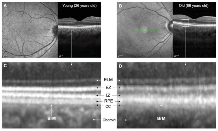
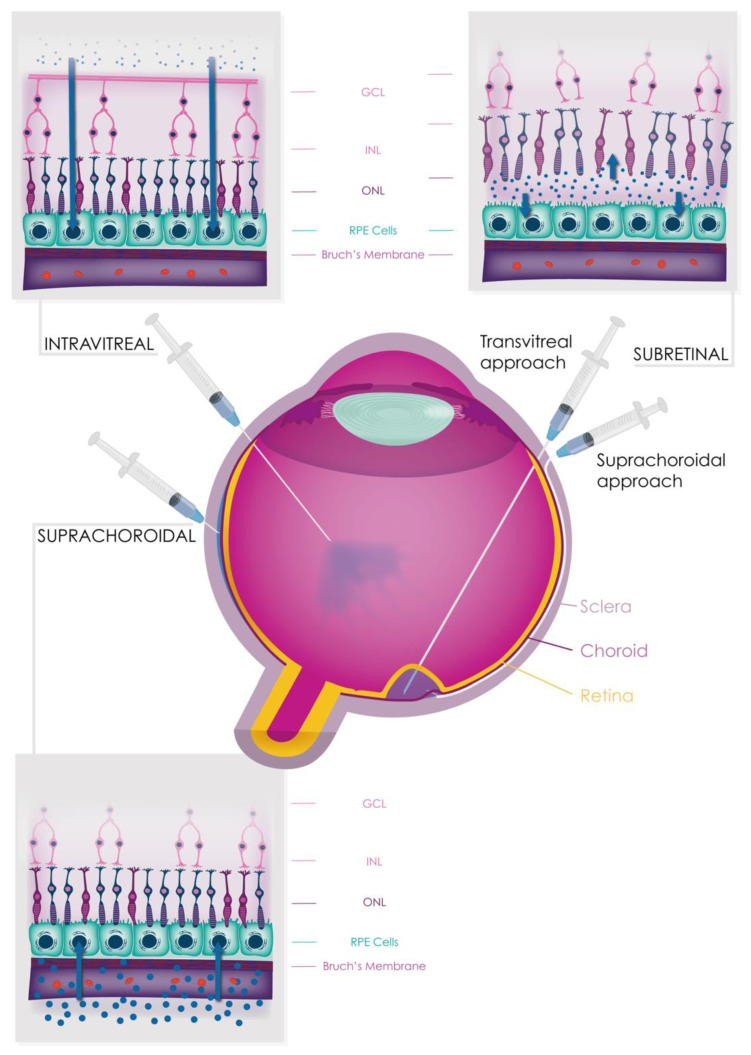
The complement system is crucial for immune surveillance, providing the body’s first line of defence against pathogens. However, an imbalance in its regulators can lead to inappropriate overactivation, resulting in diseases such as age-related macular degeneration (AMD), a leading cause of irreversible blindness globally affecting around 200 million people. Complement activation in AMD is believed to begin in the choriocapillaris, but it also plays a critical role in the subretinal and retinal pigment epithelium (RPE) spaces. Bruch’s membrane (BrM) acts as a barrier between the retina/RPE and choroid, hindering complement protein diffusion. This impediment increases with age and AMD, leading to compartmentalisation of complement activation. In this review, we comprehensively examine the structure and function of BrM, including its age-related changes visible through in vivo imaging, and the consequences of complement dysfunction on AMD pathogenesis. We also explore the potential and limitations of various delivery routes (systemic, intravitreal, subretinal, and suprachoroidal) for safe and effective delivery of conventional and gene therapy-based complement inhibitors to treat AMD. Further research is needed to understand the diffusion of complement proteins across BrM and optimise therapeutic delivery to the retina.
Overview
CoChat Summary
TL;DR
This review argues that Bruch’s membrane (BrM) is a selectively permeable, age-changing barrier that compartmentalises complement activation in age-related macular degeneration (AMD) and complicates drug delivery to the retina. It evaluates systemic, intravitreal, subretinal, and suprachoroidal routes for complement inhibitors and gene therapies, concluding that further work is needed to understand and optimise transit across BrM.
Key findings
- BrM is a 2–4 μm five-layer extracellular matrix between the RPE and choroid; its permeability declines with age, with one study reporting an almost 100% reduction in serum protein permeability from the first to ninth decades of life (from 3.5 × 10⁻⁶ to 0.2 × 10⁻⁶ cm/s), and a permeability threshold falling from >200 kDa in younger donors to 100 kDa in elderly donors.
- Complement protein passage across BrM is selective: FHL-1, Factor D, and C5a can cross, whereas C3a is not able to despite its small molecular weight; C3/C3b, FH, FB, and FI are unable to penetrate BrM even at small quantities. FHL-1 (49 kDa) has better tissue penetration than full-length glycosylated FH (155 kDa).
- AMD affects around 200 million people globally; 95% of cases are non-exudative. Genetic risk includes CFH p.Y402H, conferring approximately two-fold higher risk of late-stage AMD per allele, and GWASs have detected 52 independent and rare variants across 34 loci. Smoking is associated with an odds ratio of 1.78 (95% CI 1.52–2.09) for current versus ex-smokers.
- Intravitreal complement inhibitors pegcetacoplan and avacincaptad pegol showed anatomical benefits for geographic atrophy at one year in Phase 3 studies, but visual outcome benefit remains unknown and both have safety concerns regarding induction of exudative AMD. Systemic eculizumab completed a randomised controlled trial in AMD but failed to show significant anatomical or functional benefit.
Methods
- Narrative review synthesising literature on BrM structure and function, age-related changes, AMD pathogenesis, complement dysfunction, in vivo imaging, and therapeutic delivery routes.
- Evidence base includes human tissue studies, animal and cell models, genetic and imaging studies, and clinical trial reports; no systematic search strategy, meta-analysis, or primary data collection is described.
- Main analysis is qualitative synthesis, with tables and figures summarising age-related BrM changes and delivery approaches.
Limitations
- Stated: further research is needed to understand diffusion of complement proteins across BrM and to optimise therapeutic delivery to the retina; diffusion chamber models may not fully capture human physiology; murine models have limitations for studying AMD; the effects of complete complement inhibition in the eye remain unclear.
- Stated: the eculizumab AMD trial had limited treatment duration (6 months), a small sample size, and several methodological biases.
- Evident: as a narrative review, it does not describe a systematic literature search or formal risk-of-bias assessment, and many conclusions rely on preclinical/animal models or early-phase and interim clinical data.
Model-written summary. Check it against the paper before relying on it.
Integrity signals
Registry
Cites
Statistics
Figures



Verification
Claims the paper makes
32 Valid · 5 Not valid · 95 Not checked · 20 Own results and interpretations
Some cited sources support this claim and some don’t — read each source’s verdict below.
In the paper
“This may exacerbate the effect of the FH p.Y402H polymorphism (a major genetic risk factor for AMD) as it has been proposed that the variant protein binds less well to HSPGs than the wild-type protein [56,57].”
§ Bruch’s Membrane: A Key Consideration with Complement-Based Therapies for Age-Related Macular Degeneration > 3. Ageing Processes in BrM > 3.1. Anatomical Changes
What the sources say
- Disputedcites [56] Clark, 2006, His-384 allotypic variant of factor H associated with age-related macular degeneration has different heparin binding properties from the non-disease-associated form, J. Biol. Chem. sourceExperimental — accuracy not yet measured
In Determining Whether His-384/Tyr-384 Bind HS (page unavailable), the authors report that neither variant bound the heparan sulfate preparations tested, so they did not find weaker binding by the risk variant to HS. In Characterization of the His-384/Tyr-384-Heparin Interaction (page unavailable), the risk-associated His-384 variant bound more strongly to several unfractionated heparin preparations, although binding depended on the preparation. The citing authors’ suggestion about the consequences of reduced HSPGs is not itself assessed here. (LLM, conf=0.84)
Checked by a reasoning model — may be wrong.
- Partly supportedcites [57] Prosser, 2007, Structural basis for complement factor H linked age-related macular degeneration, J. Exp. Med. sourceExperimental — accuracy not yet measured
The Results and Discussion section ('Direct role for AMD-related polymorphic residue in glycan binding'; page unavailable) says the His402 and Tyr402 variants have different affinities and specificities for differently sulfated GAGs. It does not establish that the variant binds HSPGs less well than the wild type in general. The claim that reduced HSPGs may exacerbate the variant's effect is therefore not established by this source. (LLM, conf=0.84)
hedgedstates a possible cause- Asserted: passes — The authors put it forward as true.
- Checkable: passes — It can be checked against evidence.
- Load-bearing: unclear — It is unclear whether the paper’s conclusions depend on it.
- Based: passes — The paper gives a reason why it holds.
- Disputed
Some cited sources support this claim and some don’t — read each source’s verdict below.
In the paper
“Several experiments using mouse models of laser-induced CNV have demonstrated that all complement pathways, particularly the alternative and terminal pathways, play a significant role in angiogenesis [150,152,153,154,155,156,157,158,159,160,161,162,163].”
§ Bruch’s Membrane: A Key Consideration with Complement-Based Therapies for Age-Related Macular Degeneration > 7. Complement Dysfunction in AMD > 7.4. Complement Dysfunction in Macular Neovascularisation
What the sources say
- Partly supportedcites [150] Cashman, S.M., Ramo, K., and Kumar-Singh, R. (2011). A non membrane-targeted human soluble CD59 attenuates choroidal neovascularization in a model of age related macular degeneration. PLoS ONE, 6. sourceExperimental — accuracy not yet measured
The Abstract and Results report that inhibiting membrane attack complex (MAC) formation reduced laser-induced CNV in mice, supporting a role for the terminal complement pathway. This paper does not demonstrate that all complement pathways, including the alternative pathway, play a significant role; it tests inhibition at the terminal step. (LLM, conf=0.87)
- Partly supportedcites [152] Schnabolk, 2015, Local production of the alternative pathway component factor B is sufficient to promote laser-induced choroidal neovascularization, Investig. Ophthalmol. Vis. Sci. sourceExperimental — accuracy not yet measured
In “RPE-Derived CFB Promotes Choroidal Neovascularization” (page not available), the paper reports that factor B knockout or alternative-pathway inhibition reduced laser-induced CNV lesions in mice. It also detects the terminal-pathway marker C5b-9 in lesions, but does not establish a significant role for the terminal pathway in angiogenesis or demonstrate that all complement pathways play such a role. (LLM, conf=0.88)
- Not checked yetcites [153] Rohrer, 2011, The alternative pathway is required, but not alone sufficient, for retinal pathology in mouse laser-induced choroidal neovascularization, Mol. Immunol. source
- Not checked yetcites [154] Bora, 2006, Complement activation via alternative pathway is critical in the development of laser-induced choroidal neovascularization: Role of factor B and factor H, J. Immunol. source
- Not checked yetcites [155] Bora, 2005, Role of complement and complement membrane attack complex in laser-induced choroidal neovascularization, J. Immunol. source
- Not checked yetcites [156] Nozaki, 2006, Drusen complement components C3a and C5a promote choroidal neovascularization, Proc. Natl. Acad. Sci. USA source
- Not checked yetcites [157] Ramo, 2008, Evaluation of adenovirus-delivered human CD59 as a potential therapy for AMD in a model of human membrane attack complex formation on murine RPE, Investig. Ophthalmol. Vis. Sci. source
- Partly supportedcites [158] Gandhi, J., Cashman, S.M., and Kumar-Singh, R. (2011). Soluble CD59 expressed from an adenovirus in vivo is a potent inhibitor of complement deposition on murine liver vascular endothelium. PLoS ONE, 6. sourceExperimental — accuracy not yet measured
The Introduction and Discussion (page unavailable) report that soluble CD59 reduced membrane attack complex deposition and laser-induced choroidal neovascularization in a mouse model, supporting a role for terminal complement activity. The cited paper’s own experiments concern murine liver endothelium; it does not establish that all complement pathways, particularly the alternative pathway, contribute to angiogenesis. (LLM, conf=0.85)
- Partly supportedcites [159] Bora, 2010, Recombinant membrane-targeted form of CD59 inhibits the growth of choroidal neovascular complex in mice, J. Biol. Chem. sourceExperimental — accuracy not yet measured
The Introduction links alternative-pathway activation and membrane attack complex (MAC) formation to angiogenic growth factors in mouse CNV. The Discussion also reports that inhibiting the alternative pathway or blocking MAC reduced those factors in laser-induced CNV. These passages support the role of the alternative and terminal pathways, but do not establish that all complement pathways play a significant role or rank their relative importance. Page numbers are not available in our copy. (LLM, conf=0.87)
- Disputedcites [160] Rohrer, 2009, A targeted inhibitor of the alternative complement pathway reduces angiogenesis in a mouse model of age-related macular degeneration, Investig. Ophthalmol. Vis. Sci. sourceExperimental — accuracy not yet measured
The Discussion supports roles for the alternative and terminal pathways in mouse CNV, but directly contradicts the claim that all complement pathways play a significant role: it says deficiency or inhibition of classical and lectin pathway proteins has no impact on murine CNV. (LLM, conf=0.88)
Checked by a reasoning model — may be wrong.
boostedstates an association- Asserted: passes — The authors put it forward as true.
- Checkable: passes — It can be checked against evidence.
- Load-bearing: passes — The paper’s conclusions depend on it.
- Based: passes — The paper gives a reason why it holds.
- Partly supported
The cited sources support only part of this claim — read each source’s verdict below.
In the paper
“This observation may reflect the higher positive net charge of C3a at neutral pH [35], causing it to be trapped within the BrM, which is negatively charged due to the presence of glycosaminoglycans [35,37,38].”
§ Bruch’s Membrane: A Key Consideration with Complement-Based Therapies for Age-Related Macular Degeneration > 2. The Structure and Function of BrM
What the sources say
- Supportedcites [35] Clark, 2017, Bruch’s Membrane Compartmentalizes Complement Regulation in the Eye with Implications for Therapeutic Design in Age-Related Macular Degeneration, Front. Immunol. source
The Discussion (p. of 'Diffusion of the anaphylatoxins C3a and C5a through BrM' / Discussion section) states that although C3a and C5a are small with almost identical hydrodynamic radii, 'C3a is calculated to have a significantly greater net positive charge that C5a (Table 2)' and 'Given BrM is loaded with negatively charged glycosaminoglycans [such as heparan sulfate (22,23)], it is plausible that C3a becomes bound and collects on/within BrM.' The Results section confirms the premise that 'no detectable levels of recombinant C3a crossed the enriched macula BrM in our test system,' and Table 2 gives C3a net charge at pH 7 as 9.2 versus 5.2 for C5a. The citing sentence's hedge ('may reflect') matches the source's own hedging ('it is plausible'), so the paraphrase carries the qualifiers intact. (LLM, conf=0.92)
- Not checked yetcites [37] Day, 2015, Complementing the Sugar Code: Role of GAGs and Sialic Acid in Complement Regulation, Front. Immunol.
- Partly supportedcites [38] Clark, 2013, The proteoglycan glycomatrix: A sugar microenvironment essential for complement regulation, Front. Immunol. sourceExperimental — accuracy not yet measured
Our copy of Clark et al. (2013) supports only the matrix side of the sentence: the 'Modulation of the Complement System by Proteoglycans' section states that Bruch's membrane carries a sulfated glycomatrix ('CFH-binding sites in Bruch's membrane are comprised mainly of HS, but with dermatan sulfate also playing a minor role'; Table 1 shows 402Y binding BrM '+++' and requiring 2- and 6-O sulfation). What it does not support is the C3a-specific assertion: no passage mentions C3a, anaphylatoxins, or a net positive charge at neutral pH (searched: 'C3a anaphylatoxin positive net charge', 'anaphylatoxin C3a C5a complement fragment diffusion', 'electrostatic interactions basic residues positively charged'), and the paper never describes Bruch's membrane as 'negatively charged' — it frames GAG recognition as structure/sulfation-specific 'postcodes' rather than generic charge trapping. The charge-based trapping of C3a is therefore not this paper's point; only the GAG-rich, sulfated nature of BrM is. (LLM, conf=0.68)
hedgedstates a possible cause- Asserted: passes — The authors put it forward as true.
- Checkable: passes — It can be checked against evidence.
- Load-bearing: unclear — It is unclear whether the paper’s conclusions depend on it.
- Based: passes — The paper gives a reason why it holds.
- Supported
The cited sources support only part of this claim — read each source’s verdict below.
In the paper
“Some studies have shown that FB has a pro-angiogenic effect mediated through increased VEGF and VEGFR2 expression by endothelial cells in mouse models of laser-induced CNV and retinal hypoxia [81,153,171].”
§ Bruch’s Membrane: A Key Consideration with Complement-Based Therapies for Age-Related Macular Degeneration > 7. Complement Dysfunction in AMD > 7.4. Complement Dysfunction in Macular Neovascularisation
What the sources say
- Could not verifycites [81] Tzoumas, 2022, Rare complement factor I variants associated with reduced macular thickness and age-related macular degeneration in the UK Biobank, Hum. Mol. Genet. sourceExperimental — accuracy not yet measured
The Introduction and Discussion discuss complement Factor I variants, macular thickness, and AMD, but do not address a pro-angiogenic effect of Factor B. Searches for VEGF, VEGFR2, endothelial cells, laser-induced CNV, and retinal hypoxia found no passage supporting the attributed mechanism. (LLM, conf=0.76)
- Not checked yetcites [153] Rohrer, 2011, The alternative pathway is required, but not alone sufficient, for retinal pathology in mouse laser-induced choroidal neovascularization, Mol. Immunol. source
- Partly supportedcites [171] Murray, H., Qiu, B., Ho, S.Y., and Wang, X. (2021). Complement Factor B Mediates Ocular Angiogenesis through Regulating the VEGF Signaling Pathway. Int. J. Mol. Sci., 22. sourceExperimental — accuracy not yet measured
Results §2.1 reports increased CFB expression in mouse models of laser-induced CNV and oxygen-induced retinopathy, while Results §2.4 shows that CFB promotes angiogenesis through VEGF signaling in cultured human retinal endothelial cells. The paper does not show that increased VEGF and VEGFR2 expression by endothelial cells mediates the effect in those mouse models: it reports correlated CFB and VEGF expression in the hypoxic retina and reduced VEGFR2 expression after CFB knockdown in cultured cells. Page numbers were not supplied. (LLM, conf=0.88)
hedgedstates a cause- Asserted: passes — The authors put it forward as true.
- Checkable: passes — It can be checked against evidence.
- Load-bearing: unclear — It is unclear whether the paper’s conclusions depend on it.
- Based: passes — The paper gives a reason why it holds.
- Could not verify
Every source the paper cites for this claim was checked and supports it.
In the paper
“the common FH risk variant in AMD, CFH p.Y402H, resulted in even poorer microglial elimination [203].”
§ Bruch’s Membrane: A Key Consideration with Complement-Based Therapies for Age-Related Macular Degeneration > 7. Complement Dysfunction in AMD > 7.5. Immune Cell Regulation
What the sources say
- Supportedcites [203] Calippe, 2017, Complement Factor H Inhibits CD47-Mediated Resolution of Inflammation, Immunity source
The Results section (p. ~ 'The CFH(H402) Variant Inhibits Subretinal MC Elimination More Potently than the Common CFH(Y402)') states that the Y402H substitution accounts for a major part of AMD genetic risk and that CFH(H402) limited microglial (MC) clearance by 37% relative to CFH(Y402), and was 50% more potent at inhibiting TSP-1-driven MC elimination. The Summary (p. 2, Florian Sennlaub) likewise states the AMD-associated CFH(H402) variant markedly increased this inhibitory effect on microglial cells. Both bound the claim to the AMD risk variant and to microglial elimination, as the citing sentence does. (LLM, conf=0.92)
plainstates a cause- Asserted: passes — The authors put it forward as true.
- Checkable: passes — It can be checked against evidence.
- Load-bearing: passes — The paper’s conclusions depend on it.
- Based: passes — The paper gives a reason why it holds.
- Supported
95 more claims · 19 with their cited sources checked
Create a free account to see all 152 claims and to verify the 174 references still open.
- Probably not a substantial claim. 16
- It may state two separate claims as one. 40
Claims other papers make about this one
Each is checked against this paper’s own text: does it say what the citing paper says it does?
In the citing paper
“This could be due to the structural differences between species that result in different transduction cell types, including Bruch’s membrane (BrM) thickness, the thickness or number of cells within the retinal layers73,74.”
From An adeno-associated virus variant enabling efficient ocular-directed gene delivery across species (2024) · cites this paper as [73]
Self-check
Percentages, sums and reported differences re-computed from the paper’s own numbers.
Each contradiction between two passages listed here was confirmed by an independent check.
Rated minor: it would not touch a result — a value no result rests on, a p-value that stays on the same side of the significance level, or a conflict that is clerical, resolved elsewhere in the paper, or outside its own findings.
In the paper
§ Bruch’s Membrane: A Key Consideration with Complement-Based Therapies for Age-Related Macular Degeneration > 2. The Structure and Function of BrM
“BrM is a 2–4 μm thin layer of extracellular matrix between the retina and choroid in the eye serving as a selectively permeable membrane between the RPE and choriocapillaris (CC).”
§ Bruch’s Membrane: A Key Consideration with Complement-Based Therapies for Age-Related Macular Degeneration > 2. The Structure and Function of BrM
“The OCL’s thickness ranges from 1 to 5 μm [29]”
Citations
Cites
1 retracted
RETRACTED: Avacopan for the Treatment of ANCA-Associated Vasculitis
Jayne, D. R. W., Merkel, P. A., Schall, T. J., & Bekker, P. (2021) · New England Journal of Medicine · doi:10.1056/nejmoa2023386 · Cited by 1,021
RetractedRepertoires of Autophagy in the Pathogenesis of Ocular Diseases
Li, Y.-J., Jiang, Q., Cao, G.-F., Yao, J., & Yan, B. (2015) · Cellular Physiology and Biochemistry · doi:10.1159/000373980 · Cited by 16,304
Wong, W. L., Su, X., Li, X., Cheung, C. M. G., Klein, R., Cheng, C.-Y., & Wong, T. Y. (2014) · The Lancet Global Health · doi:10.1016/s2214-109x(13)70145-1 · Cited by 5,331
Complement factor H polymorphism in age-related macular degeneration
Klein, R. J., Zeiss, C., Chew, E. Y., Tsai, J.-Y., Sackler, R. S., Haynes, C., Henning, A. K., SanGiovanni, J. P., Mane, S. M., Mayne, S. T., et al. (2005) · Science · doi:10.1126/science.1109557 · Cited by 4,492
Non-viral vectors for gene-based therapy
Yin, H., Kanasty, R. L., Eltoukhy, A. A., Vegas, A. J., Dorkin, J. R., & Anderson, D. G. (2014) · Nature Reviews Genetics · doi:10.1038/nrg3763 · Cited by 3,123
Walport, M. J. (2001) · New England Journal of Medicine · doi:10.1056/nejm200104053441406 · Cited by 2,927
VEGF in signaling and disease: beyond discovery and development
Apte, R. S., Chen, D. S., & Ferrara, N. (2019) · Cell · doi:10.1016/j.cell.2019.01.021 · Cited by 2,721
Safety and Efficacy of Gene Transfer for Leber's Congenital Amaurosis
Maguire, A. M., Simonelli, F., Pierce, E. A., Pugh, E. N., Mingozzi, F., Bennicelli, J., Banfi, S., Marshall, K. A., Testa, F., Surace, E. M., et al. (2008) · New England Journal of Medicine · doi:10.1056/nejmoa0802315 · Cited by 2,145
Hageman, G. S., Anderson, D. H., Johnson, L. V., Hancox, L. S., Taiber, A. J., Hardisty, L. I., Hageman, J. L., Stockman, H. A., Borchardt, J. D., Gehrs, K. M., et al. (2005) · Proceedings of the National Academy of Sciences · doi:10.1073/pnas.0501536102 · Cited by 1,961
Nickla, D. L., & Wallman, J. (2010) · Progress in Retinal and Eye Research · doi:10.1016/j.preteyeres.2009.12.002 · Cited by 1,945
Research works printed but not linked: 3
- Hogan, M.J., Alvarado, A.J., and Weddell, J.E. (1971). Histology of the Human. Eye: An. Atlas and Textbook, Saunders.
- Johnson, M., and Curcio, C.A. (2012). Structure, Function, and Pathology of Bruch’s Membrane, Elsevier.
- Gamlin, P.D., Alexander, J.J., Boye, S.L., Witherspoon, C.D., and Boye, S.E. (2019). Adeno-Associated Virus Vectors. Methods in Molecular Biology, Humana Press.
Cited by
Kurzawa‐Akanbi, M., Tzoumas, N., Corral-Serrano, J. C., Guarascio, R., STEEL, D. H., Cheetham, M. E., Armstrong, L., & Lako, M. (2024) · Progress in Retinal and Eye Research · doi:10.1016/j.preteyeres.2024.101248 · Cited by 44
Self-citationAn adeno-associated virus variant enabling efficient ocular-directed gene delivery across species
Luo, S., Jiang, H., Li, Q., Qin, Y., Yang, S., Li, J., Xu, L., Gou, Y., Zhang, Y., Liu, F., et al. (2024) · Nature Communications · doi:10.1038/s41467-024-48221-4 · Cited by 39
Antioxidants in Age-Related Macular Degeneration: Lights and Shadows
Parmar, U. P. S., Surico, P. L., Mori, T., Singh, R. B., Cutrupi, F., Premkishore, P., Afflitto, G. G., Zazzo, A. D., Coassin, M., & Romano, F. (2025) · Antioxidants · doi:10.3390/antiox14020152 · Cited by 37
Therapeutic targeting of the complement system in ocular disease
Rathi, S., Hasan, R., Ueffing, M., & Clark, S. J. (2023) · Drug Discovery Today · doi:10.1016/j.drudis.2023.103757 · Cited by 24
Alhelaly, M., Soylu, C., Corradetti, G., Corvi, F., Mahmoudi, A., Abbasgholizadeh, R., Bellisario, G., He, Y., Quarta, A., Nittala, M. G., et al. (2025) · Investigative Ophthalmology & Visual Science · doi:10.1167/iovs.66.9.19 · Cited by 16
Metadata
- Authors
- Hammadi, S., Tzoumas, N., Ferrara, M., Meschede, I. P., Lo, K., Harris, C., Lako, M., & Steel, D. H.
- Affiliations
- Newcastle University (GB)
- Sunderland Eye Infirmary (GB)
- Manchester Royal Eye Hospital (GB)
- Gyroscope Therapeutics Limited, a Novartis Company, Rolling Stock Yard, 6th Floor, 188 York Way, London N7 9AS, UK
- Corresponding author
- Steel, David H.
- Venue
- Journal of Clinical Medicine
- Publisher
- MDPI AG
- Published
- 2023-04-14
- Volume
- 12
- Issue
- 8
- Pages
- 2870
- ISSN
- 2077-0383 (ISSN-L)
- PMC
- PMC10145879
- OpenAlex
- W4365514215
- Semantic Scholar
- 8936880dfad5b2eafe433ef7ed04c4ff1ddb3b9f
- Cited by
- 44
- References
- 321
- Citations by year
- 2026: 15 · 2025: 16 · 2024: 8 · 2023: 5
- FWCI
- 2.24
- Type
- journal article
- Language
- en
- Open access
- gold
- doi.org
- Licence
- Locations
- Journal of Clinical Medicine (publishedVersion, cc-by)
- PubMed Central (publishedVersion, cc-by)
- PubMed (publishedVersion)
- Institutional Research Information System (Università degli Studi di Brescia) (submittedVersion)
- Newcastle University ePrints (Newcastle Univesity) (cc-by)
- Funders
- Newcastle University (ACF-2021-01-008, #GR602, #GR601)
- Gyroscope Therapeutics, a Novartis company (ACF-2021-01-008, #GR602, #GR601)
- NIHR Academic Clinical Fellowship (ACF-2021-01-008, #GR602, #GR601)
- Retina UK (ACF-2021-01-008, #GR602, #GR601)
- Macular Society UK (ACF-2021-01-008, #GR602, #GR601)
- companies developing complement therapeutics (ACF-2021-01-008, #GR602, #GR601)
- Gyroscope Therapeutics (a Novartis company) (relevant to work under consideration), Alcon, Boehringer Ingelheim, Bayer, DORC, and Roche (ACF-2021-01-008, #GR602, #GR601)
- Topics
- Health Sciences › Medicine › Ophthalmology › Retinal Diseases and Treatments
- Life Sciences › Immunology and Microbiology › Immunology › Complement system in diseases
- Health Sciences › Medicine › Ophthalmology › Ocular Diseases and Behçet’s Syndrome
- Keywords
- Bruch's membrane, age-related macular degeneration, complement system, complement activation, complement inhibitors, retinal pigment epithelium, choriocapillaris, gene therapy, drug delivery routes, intravitreal delivery, subretinal delivery, systemic drug delivery, in vivo imaging, AMD pathogenesis
About these checks
Registry, retraction, Cites and statistics results are computed, not judged, with one exception: a printed reference that no index lists is read once by a model before it counts as a citation error. Claims are extracted by a model and kept only when their quote appears verbatim in the text; each cited source is then checked, with a model’s help, for whether it says what the claim relies on. Those verdicts can be wrong — read the claim’s quote and the cited source before relying on one.
The claim verdicts this report counts are experimental: their accuracy is not yet measured.
A language model drew the claims from the paper on 2026-10-04, under version 3 of the claim definition (model deepseek-v4.1-flash, prompt extract-v3).
A second model labelled each claim on 2026-10-04 (model jev-1.13-20260917, definition revision gold-guideline-v0.2).
Cited sources are checked with a language model’s help (gpt-6-sol and qwen3.8-omni-flash).
Statistics and arithmetic are recomputed, not judged.
Confidence bands are shown only for fields calibrated on our annotated test set. None is calibrated yet, so no band is shown.
What counts as a claim ›How we measure confidence ›How we check this ›
Are you an author, or do you see a problem with this page? Request a review
How to cite
Cite CoChat for the checks on this page, and the paper for its own findings.
- This page
- CoChat. (2026). Citation check: Bruch’s Membrane: A Key Consideration with Complement-Based Therapies for Age-Related Macular Degeneration. https://app.cochat.ai/papers/10.3390/jcm12082870
- The paper
- Hammadi, S., Tzoumas, N., Ferrara, M., Meschede, I. P., Lo, K., Harris, C., Lako, M., & Steel, D. H. (2023). Bruch’s Membrane: A Key Consideration with Complement-Based Therapies for Age-Related Macular Degeneration. Journal of Clinical Medicine. https://doi.org/10.3390/jcm12082870
Data sources
Bibliographic data: Semantic Scholar, OpenAlex, Crossref.
Contains information from the Semantic Scholar Academic Graph Datasets, made available under the ODC Attribution License (ODC-BY 1.0).
Semantic Scholar’s data is described in: Kinney, R., et al. (2023). The Semantic Scholar Open Data Platform. arXiv:2301.10140.
