

Bruch’s Membrane: A Key Consideration with Complement-Based Therapies for Age-Related Macular Degeneration
Open accessThe DOI matches its Crossref record; 315 of the 321 references it cites were found in a registry or an index; 69 of the 243 sources its claims cite were checked for support.
Abstract
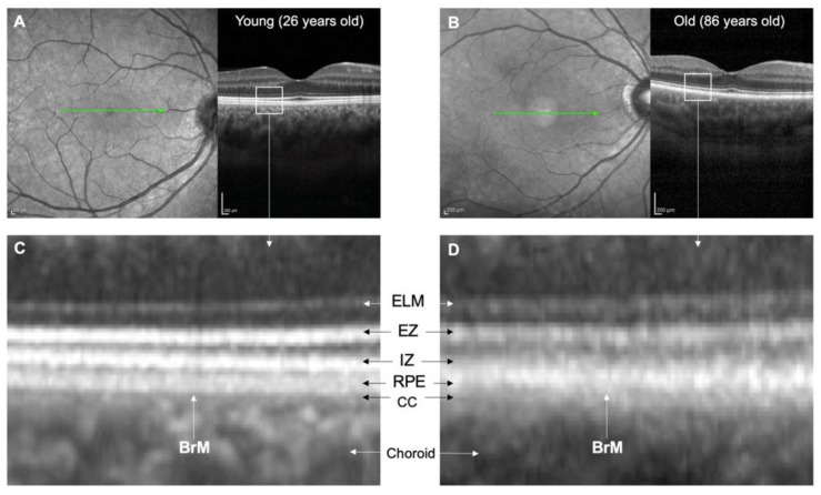
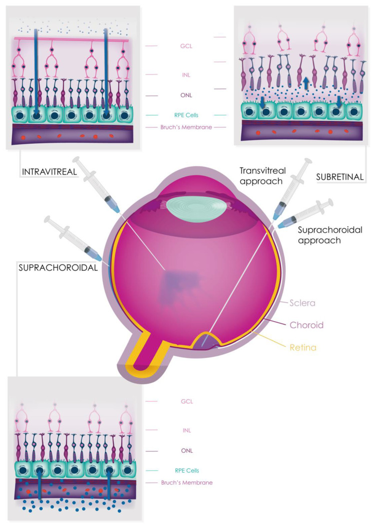
The complement system is crucial for immune surveillance, providing the body’s first line of defence against pathogens. However, an imbalance in its regulators can lead to inappropriate overactivation, resulting in diseases such as age-related macular degeneration (AMD), a leading cause of irreversible blindness globally affecting around 200 million people. Complement activation in AMD is believed to begin in the choriocapillaris, but it also plays a critical role in the subretinal and retinal pigment epithelium (RPE) spaces. Bruch’s membrane (BrM) acts as a barrier between the retina/RPE and choroid, hindering complement protein diffusion. This impediment increases with age and AMD, leading to compartmentalisation of complement activation. In this review, we comprehensively examine the structure and function of BrM, including its age-related changes visible through in vivo imaging, and the consequences of complement dysfunction on AMD pathogenesis. We also explore the potential and limitations of various delivery routes (systemic, intravitreal, subretinal, and suprachoroidal) for safe and effective delivery of conventional and gene therapy-based complement inhibitors to treat AMD. Further research is needed to understand the diffusion of complement proteins across BrM and optimise therapeutic delivery to the retina.
Overview
CoChat Summary
TL;DR
This review argues that Bruch’s membrane (BrM) is a selectively permeable, age-changing barrier that compartmentalises complement activation in age-related macular degeneration (AMD) and complicates drug delivery to the retina. It evaluates systemic, intravitreal, subretinal, and suprachoroidal routes for complement inhibitors and gene therapies, concluding that further work is needed to understand and optimise transit across BrM.
Key findings
- BrM is a 2–4 μm five-layer extracellular matrix between the RPE and choroid; its permeability declines with age, with one study reporting an almost 100% reduction in serum protein permeability from the first to ninth decades of life (from 3.5 × 10⁻⁶ to 0.2 × 10⁻⁶ cm/s), and a permeability threshold falling from >200 kDa in younger donors to 100 kDa in elderly donors.
- Complement protein passage across BrM is selective: FHL-1, Factor D, and C5a can cross, whereas C3a is not able to despite its small molecular weight; C3/C3b, FH, FB, and FI are unable to penetrate BrM even at small quantities. FHL-1 (49 kDa) has better tissue penetration than full-length glycosylated FH (155 kDa).
- AMD affects around 200 million people globally; 95% of cases are non-exudative. Genetic risk includes CFH p.Y402H, conferring approximately two-fold higher risk of late-stage AMD per allele, and GWASs have detected 52 independent and rare variants across 34 loci. Smoking is associated with an odds ratio of 1.78 (95% CI 1.52–2.09) for current versus ex-smokers.
- Intravitreal complement inhibitors pegcetacoplan and avacincaptad pegol showed anatomical benefits for geographic atrophy at one year in Phase 3 studies, but visual outcome benefit remains unknown and both have safety concerns regarding induction of exudative AMD. Systemic eculizumab completed a randomised controlled trial in AMD but failed to show significant anatomical or functional benefit.
Methods
- Narrative review synthesising literature on BrM structure and function, age-related changes, AMD pathogenesis, complement dysfunction, in vivo imaging, and therapeutic delivery routes.
- Evidence base includes human tissue studies, animal and cell models, genetic and imaging studies, and clinical trial reports; no systematic search strategy, meta-analysis, or primary data collection is described.
- Main analysis is qualitative synthesis, with tables and figures summarising age-related BrM changes and delivery approaches.
Limitations
- Stated: further research is needed to understand diffusion of complement proteins across BrM and to optimise therapeutic delivery to the retina; diffusion chamber models may not fully capture human physiology; murine models have limitations for studying AMD; the effects of complete complement inhibition in the eye remain unclear.
- Stated: the eculizumab AMD trial had limited treatment duration (6 months), a small sample size, and several methodological biases.
- Evident: as a narrative review, it does not describe a systematic literature search or formal risk-of-bias assessment, and many conclusions rely on preclinical/animal models or early-phase and interim clinical data.
Model-written summary. Check it against the paper before relying on it.
Integrity signals
Registry
Cites
Statistics
Figures



Verification
Claims the paper makes
32 Valid · 5 Not valid · 95 Not checked · 20 Own results and interpretations
Some cited sources support this claim and some don’t — read each source’s verdict below.
In the paper
“This may exacerbate the effect of the FH p.Y402H polymorphism (a major genetic risk factor for AMD) as it has been proposed that the variant protein binds less well to HSPGs than the wild-type protein [56,57].”
§ Bruch’s Membrane: A Key Consideration with Complement-Based Therapies for Age-Related Macular Degeneration > 3. Ageing Processes in BrM > 3.1. Anatomical Changes
What the sources say
- Disputedcites [56] Clark, 2006, His-384 allotypic variant of factor H associated with age-related macular degeneration has different heparin binding properties from the non-disease-associated form, J. Biol. Chem. sourceExperimental — accuracy not yet measured
In Determining Whether His-384/Tyr-384 Bind HS (page unavailable), the authors report that neither variant bound the heparan sulfate preparations tested, so they did not find weaker binding by the risk variant to HS. In Characterization of the His-384/Tyr-384-Heparin Interaction (page unavailable), the risk-associated His-384 variant bound more strongly to several unfractionated heparin preparations, although binding depended on the preparation. The citing authors’ suggestion about the consequences of reduced HSPGs is not itself assessed here. (LLM, conf=0.84)
Checked by a reasoning model — may be wrong.
- Partly supportedcites [57] Prosser, 2007, Structural basis for complement factor H linked age-related macular degeneration, J. Exp. Med. sourceExperimental — accuracy not yet measured
The Results and Discussion section ('Direct role for AMD-related polymorphic residue in glycan binding'; page unavailable) says the His402 and Tyr402 variants have different affinities and specificities for differently sulfated GAGs. It does not establish that the variant binds HSPGs less well than the wild type in general. The claim that reduced HSPGs may exacerbate the variant's effect is therefore not established by this source. (LLM, conf=0.84)
hedgedstates a possible cause- Asserted: passes — The authors put it forward as true.
- Checkable: passes — It can be checked against evidence.
- Load-bearing: unclear — It is unclear whether the paper’s conclusions depend on it.
- Based: passes — The paper gives a reason why it holds.
- Disputed
Some cited sources support this claim and some don’t — read each source’s verdict below.
In the paper
“Several experiments using mouse models of laser-induced CNV have demonstrated that all complement pathways, particularly the alternative and terminal pathways, play a significant role in angiogenesis [150,152,153,154,155,156,157,158,159,160,161,162,163].”
§ Bruch’s Membrane: A Key Consideration with Complement-Based Therapies for Age-Related Macular Degeneration > 7. Complement Dysfunction in AMD > 7.4. Complement Dysfunction in Macular Neovascularisation
What the sources say
- Partly supportedcites [150] Cashman, S.M., Ramo, K., and Kumar-Singh, R. (2011). A non membrane-targeted human soluble CD59 attenuates choroidal neovascularization in a model of age related macular degeneration. PLoS ONE, 6. sourceExperimental — accuracy not yet measured
The Abstract and Results report that inhibiting membrane attack complex (MAC) formation reduced laser-induced CNV in mice, supporting a role for the terminal complement pathway. This paper does not demonstrate that all complement pathways, including the alternative pathway, play a significant role; it tests inhibition at the terminal step. (LLM, conf=0.87)
- Partly supportedcites [152] Schnabolk, 2015, Local production of the alternative pathway component factor B is sufficient to promote laser-induced choroidal neovascularization, Investig. Ophthalmol. Vis. Sci. sourceExperimental — accuracy not yet measured
In “RPE-Derived CFB Promotes Choroidal Neovascularization” (page not available), the paper reports that factor B knockout or alternative-pathway inhibition reduced laser-induced CNV lesions in mice. It also detects the terminal-pathway marker C5b-9 in lesions, but does not establish a significant role for the terminal pathway in angiogenesis or demonstrate that all complement pathways play such a role. (LLM, conf=0.88)
- Not checked yetcites [153] Rohrer, 2011, The alternative pathway is required, but not alone sufficient, for retinal pathology in mouse laser-induced choroidal neovascularization, Mol. Immunol. source
- Not checked yetcites [154] Bora, 2006, Complement activation via alternative pathway is critical in the development of laser-induced choroidal neovascularization: Role of factor B and factor H, J. Immunol. source
- Not checked yetcites [155] Bora, 2005, Role of complement and complement membrane attack complex in laser-induced choroidal neovascularization, J. Immunol. source
- Not checked yetcites [156] Nozaki, 2006, Drusen complement components C3a and C5a promote choroidal neovascularization, Proc. Natl. Acad. Sci. USA source
- Not checked yetcites [157] Ramo, 2008, Evaluation of adenovirus-delivered human CD59 as a potential therapy for AMD in a model of human membrane attack complex formation on murine RPE, Investig. Ophthalmol. Vis. Sci. source
- Partly supportedcites [158] Gandhi, J., Cashman, S.M., and Kumar-Singh, R. (2011). Soluble CD59 expressed from an adenovirus in vivo is a potent inhibitor of complement deposition on murine liver vascular endothelium. PLoS ONE, 6. sourceExperimental — accuracy not yet measured
The Introduction and Discussion (page unavailable) report that soluble CD59 reduced membrane attack complex deposition and laser-induced choroidal neovascularization in a mouse model, supporting a role for terminal complement activity. The cited paper’s own experiments concern murine liver endothelium; it does not establish that all complement pathways, particularly the alternative pathway, contribute to angiogenesis. (LLM, conf=0.85)
- Partly supportedcites [159] Bora, 2010, Recombinant membrane-targeted form of CD59 inhibits the growth of choroidal neovascular complex in mice, J. Biol. Chem. sourceExperimental — accuracy not yet measured
The Introduction links alternative-pathway activation and membrane attack complex (MAC) formation to angiogenic growth factors in mouse CNV. The Discussion also reports that inhibiting the alternative pathway or blocking MAC reduced those factors in laser-induced CNV. These passages support the role of the alternative and terminal pathways, but do not establish that all complement pathways play a significant role or rank their relative importance. Page numbers are not available in our copy. (LLM, conf=0.87)
- Disputedcites [160] Rohrer, 2009, A targeted inhibitor of the alternative complement pathway reduces angiogenesis in a mouse model of age-related macular degeneration, Investig. Ophthalmol. Vis. Sci. sourceExperimental — accuracy not yet measured
The Discussion supports roles for the alternative and terminal pathways in mouse CNV, but directly contradicts the claim that all complement pathways play a significant role: it says deficiency or inhibition of classical and lectin pathway proteins has no impact on murine CNV. (LLM, conf=0.88)
Checked by a reasoning model — may be wrong.
boostedstates an association- Asserted: passes — The authors put it forward as true.
- Checkable: passes — It can be checked against evidence.
- Load-bearing: passes — The paper’s conclusions depend on it.
- Based: passes — The paper gives a reason why it holds.
- Partly supported
The cited sources support only part of this claim — read each source’s verdict below.
In the paper
“This observation may reflect the higher positive net charge of C3a at neutral pH [35], causing it to be trapped within the BrM, which is negatively charged due to the presence of glycosaminoglycans [35,37,38].”
§ Bruch’s Membrane: A Key Consideration with Complement-Based Therapies for Age-Related Macular Degeneration > 2. The Structure and Function of BrM
What the sources say
- Supportedcites [35] Clark, 2017, Bruch’s Membrane Compartmentalizes Complement Regulation in the Eye with Implications for Therapeutic Design in Age-Related Macular Degeneration, Front. Immunol. source
The Discussion (p. of 'Diffusion of the anaphylatoxins C3a and C5a through BrM' / Discussion section) states that although C3a and C5a are small with almost identical hydrodynamic radii, 'C3a is calculated to have a significantly greater net positive charge that C5a (Table 2)' and 'Given BrM is loaded with negatively charged glycosaminoglycans [such as heparan sulfate (22,23)], it is plausible that C3a becomes bound and collects on/within BrM.' The Results section confirms the premise that 'no detectable levels of recombinant C3a crossed the enriched macula BrM in our test system,' and Table 2 gives C3a net charge at pH 7 as 9.2 versus 5.2 for C5a. The citing sentence's hedge ('may reflect') matches the source's own hedging ('it is plausible'), so the paraphrase carries the qualifiers intact. (LLM, conf=0.92)
- Not checked yetcites [37] Day, 2015, Complementing the Sugar Code: Role of GAGs and Sialic Acid in Complement Regulation, Front. Immunol.
- Partly supportedcites [38] Clark, 2013, The proteoglycan glycomatrix: A sugar microenvironment essential for complement regulation, Front. Immunol. sourceExperimental — accuracy not yet measured
Our copy of Clark et al. (2013) supports only the matrix side of the sentence: the 'Modulation of the Complement System by Proteoglycans' section states that Bruch's membrane carries a sulfated glycomatrix ('CFH-binding sites in Bruch's membrane are comprised mainly of HS, but with dermatan sulfate also playing a minor role'; Table 1 shows 402Y binding BrM '+++' and requiring 2- and 6-O sulfation). What it does not support is the C3a-specific assertion: no passage mentions C3a, anaphylatoxins, or a net positive charge at neutral pH (searched: 'C3a anaphylatoxin positive net charge', 'anaphylatoxin C3a C5a complement fragment diffusion', 'electrostatic interactions basic residues positively charged'), and the paper never describes Bruch's membrane as 'negatively charged' — it frames GAG recognition as structure/sulfation-specific 'postcodes' rather than generic charge trapping. The charge-based trapping of C3a is therefore not this paper's point; only the GAG-rich, sulfated nature of BrM is. (LLM, conf=0.68)
hedgedstates a possible cause- Asserted: passes — The authors put it forward as true.
- Checkable: passes — It can be checked against evidence.
- Load-bearing: unclear — It is unclear whether the paper’s conclusions depend on it.
- Based: passes — The paper gives a reason why it holds.
- Supported
The cited sources support only part of this claim — read each source’s verdict below.
In the paper
“Some studies have shown that FB has a pro-angiogenic effect mediated through increased VEGF and VEGFR2 expression by endothelial cells in mouse models of laser-induced CNV and retinal hypoxia [81,153,171].”
§ Bruch’s Membrane: A Key Consideration with Complement-Based Therapies for Age-Related Macular Degeneration > 7. Complement Dysfunction in AMD > 7.4. Complement Dysfunction in Macular Neovascularisation
What the sources say
- Could not verifycites [81] Tzoumas, 2022, Rare complement factor I variants associated with reduced macular thickness and age-related macular degeneration in the UK Biobank, Hum. Mol. Genet. sourceExperimental — accuracy not yet measured
The Introduction and Discussion discuss complement Factor I variants, macular thickness, and AMD, but do not address a pro-angiogenic effect of Factor B. Searches for VEGF, VEGFR2, endothelial cells, laser-induced CNV, and retinal hypoxia found no passage supporting the attributed mechanism. (LLM, conf=0.76)
- Not checked yetcites [153] Rohrer, 2011, The alternative pathway is required, but not alone sufficient, for retinal pathology in mouse laser-induced choroidal neovascularization, Mol. Immunol. source
- Partly supportedcites [171] Murray, H., Qiu, B., Ho, S.Y., and Wang, X. (2021). Complement Factor B Mediates Ocular Angiogenesis through Regulating the VEGF Signaling Pathway. Int. J. Mol. Sci., 22. sourceExperimental — accuracy not yet measured
Results §2.1 reports increased CFB expression in mouse models of laser-induced CNV and oxygen-induced retinopathy, while Results §2.4 shows that CFB promotes angiogenesis through VEGF signaling in cultured human retinal endothelial cells. The paper does not show that increased VEGF and VEGFR2 expression by endothelial cells mediates the effect in those mouse models: it reports correlated CFB and VEGF expression in the hypoxic retina and reduced VEGFR2 expression after CFB knockdown in cultured cells. Page numbers were not supplied. (LLM, conf=0.88)
hedgedstates a cause- Asserted: passes — The authors put it forward as true.
- Checkable: passes — It can be checked against evidence.
- Load-bearing: unclear — It is unclear whether the paper’s conclusions depend on it.
- Based: passes — The paper gives a reason why it holds.
- Could not verify
Every source the paper cites for this claim was checked and supports it.
In the paper
“the common FH risk variant in AMD, CFH p.Y402H, resulted in even poorer microglial elimination [203].”
§ Bruch’s Membrane: A Key Consideration with Complement-Based Therapies for Age-Related Macular Degeneration > 7. Complement Dysfunction in AMD > 7.5. Immune Cell Regulation
What the sources say
- Supportedcites [203] Calippe, 2017, Complement Factor H Inhibits CD47-Mediated Resolution of Inflammation, Immunity source
The Results section (p. ~ 'The CFH(H402) Variant Inhibits Subretinal MC Elimination More Potently than the Common CFH(Y402)') states that the Y402H substitution accounts for a major part of AMD genetic risk and that CFH(H402) limited microglial (MC) clearance by 37% relative to CFH(Y402), and was 50% more potent at inhibiting TSP-1-driven MC elimination. The Summary (p. 2, Florian Sennlaub) likewise states the AMD-associated CFH(H402) variant markedly increased this inhibitory effect on microglial cells. Both bound the claim to the AMD risk variant and to microglial elimination, as the citing sentence does. (LLM, conf=0.92)
plainstates a cause- Asserted: passes — The authors put it forward as true.
- Checkable: passes — It can be checked against evidence.
- Load-bearing: passes — The paper’s conclusions depend on it.
- Based: passes — The paper gives a reason why it holds.
- Supported
95 more claims · 19 with their cited sources checked
Create a free account to see all 152 claims and to verify the 174 references still open.
- Probably not a substantial claim. 16
- It may state two separate claims as one. 40
Claims other papers make about this one
Each is checked against this paper’s own text: does it say what the citing paper says it does?
In the citing paper
“This could be due to the structural differences between species that result in different transduction cell types, including Bruch’s membrane (BrM) thickness, the thickness or number of cells within the retinal layers73,74.”
From An adeno-associated virus variant enabling efficient ocular-directed gene delivery across species (2024) · cites this paper as [73]
Self-check
Percentages, sums and reported differences re-computed from the paper’s own numbers.
Each contradiction between two passages listed here was confirmed by an independent check.
Rated minor: it would not touch a result — a value no result rests on, a p-value that stays on the same side of the significance level, or a conflict that is clerical, resolved elsewhere in the paper, or outside its own findings.
In the paper
§ Bruch’s Membrane: A Key Consideration with Complement-Based Therapies for Age-Related Macular Degeneration > 2. The Structure and Function of BrM
“BrM is a 2–4 μm thin layer of extracellular matrix between the retina and choroid in the eye serving as a selectively permeable membrane between the RPE and choriocapillaris (CC).”
§ Bruch’s Membrane: A Key Consideration with Complement-Based Therapies for Age-Related Macular Degeneration > 2. The Structure and Function of BrM
“The OCL’s thickness ranges from 1 to 5 μm [29]”
Citations
Cites
1 retracted
RETRACTED: Avacopan for the Treatment of ANCA-Associated Vasculitis
Jayne, D. R. W., Merkel, P. A., Schall, T. J., & Bekker, P. (2021) · New England Journal of Medicine · doi:10.1056/nejmoa2023386 · Cited by 1,021
RetractedRepertoires of Autophagy in the Pathogenesis of Ocular Diseases
Li, Y.-J., Jiang, Q., Cao, G.-F., Yao, J., & Yan, B. (2015) · Cellular Physiology and Biochemistry · doi:10.1159/000373980 · Cited by 16,304
Wong, W. L., Su, X., Li, X., Cheung, C. M. G., Klein, R., Cheng, C.-Y., & Wong, T. Y. (2014) · The Lancet Global Health · doi:10.1016/s2214-109x(13)70145-1 · Cited by 5,331
Complement factor H polymorphism in age-related macular degeneration
Klein, R. J., Zeiss, C., Chew, E. Y., Tsai, J.-Y., Sackler, R. S., Haynes, C., Henning, A. K., SanGiovanni, J. P., Mane, S. M., Mayne, S. T., et al. (2005) · Science · doi:10.1126/science.1109557 · Cited by 4,492
Non-viral vectors for gene-based therapy
Yin, H., Kanasty, R. L., Eltoukhy, A. A., Vegas, A. J., Dorkin, J. R., & Anderson, D. G. (2014) · Nature Reviews Genetics · doi:10.1038/nrg3763 · Cited by 3,123 · cited 1×
“There is growing interest in the development of non-viral vectors, such as polymeric, liposomal, lipid, and inorganic vectors, due to concerns about the immunogenicity of viral vectors [269].”
BackgroundWalport, M. J. (2001) · New England Journal of Medicine · doi:10.1056/nejm200104053441406 · Cited by 2,927 · cited 1×
“Complement represents a significant 15% of the globulin fraction of blood and is responsible for lytic activity against invading pathogens as part of the innate immune system [98].”
BackgroundVEGF in signaling and disease: beyond discovery and development
Apte, R. S., Chen, D. S., & Ferrara, N. (2019) · Cell · doi:10.1016/j.cell.2019.01.021 · Cited by 2,721 · cited 1×
“Further research into the biological interactions between complement and VEGF will be important for guiding drug development and clinical trial design [183].”
BackgroundSafety and Efficacy of Gene Transfer for Leber's Congenital Amaurosis
Maguire, A. M., Simonelli, F., Pierce, E. A., Pugh, E. N., Mingozzi, F., Bennicelli, J., Banfi, S., Marshall, K. A., Testa, F., Surace, E. M., et al. (2008) · New England Journal of Medicine · doi:10.1056/nejmoa0802315 · Cited by 2,145
Hageman, G. S., Anderson, D. H., Johnson, L. V., Hancox, L. S., Taiber, A. J., Hardisty, L. I., Hageman, J. L., Stockman, H. A., Borchardt, J. D., Gehrs, K. M., et al. (2005) · Proceedings of the National Academy of Sciences · doi:10.1073/pnas.0501536102 · Cited by 1,961
Nickla, D. L., & Wallman, J. (2010) · Progress in Retinal and Eye Research · doi:10.1016/j.preteyeres.2009.12.002 · Cited by 1,945 · cited 1×
“The choroid, a vital vascular connective tissue, regulates blood flow and temperature and secretes growth factors to maintain eye homeostasis [10].”
BackgroundFritsche, L. G., Igl, W., Bailey, J. N. C., Grassmann, F., Sengupta, S., Bragg-Gresham, J. L., Burdon, K. P., Hebbring, S. J., Wen, C., Gorski, M., et al. (2016) · Nature Genetics · doi:10.1038/ng.3448 · Cited by 1,544 · cited 1×
“Subsequent GWASs have detected 52 independent and rare variants distributed across 34 loci that account for more than half of the condition’s heritability [78].”
BackgroundAge-related macular degeneration
Mitchell, P., Liew, G., Gopinath, B., & Wong, T. Y. (2018) · The Lancet · doi:10.1016/s0140-6736(18)31550-2 · Cited by 1,511 · cited 1×
“Of over 100 proteins identified in drusen, the commonest include vitronectin and clusterin (which both participate in the control of the lytic activity of complement), with TIMP-3, serum albumin, and crystallins [67] also identified—several of which have undergone oxidative protein modifications [68].”
BackgroundHageman, G. (2001) · Progress in Retinal and Eye Research · doi:10.1016/s1350-9462(01)00010-6 · Cited by 1,265
Drusen proteome analysis: an approach to the etiology of age-related macular degeneration
Crabb, J. W., Miyagi, M., Gu, X., Shadrach, K., West, K. A., Sakaguchi, H., Kamei, M., Hasan, A., Yan, L., Rayborn, M. E., et al. (2002) · Proceedings of the National Academy of Sciences · doi:10.1073/pnas.222551899 · Cited by 1,208 · cited 1×
“Of over 100 proteins identified in drusen, the commonest include vitronectin and clusterin (which both participate in the control of the lytic activity of complement), with TIMP-3, serum albumin, and crystallins [67] also identified—several of which have undergone oxidative protein modifications [68].”
BackgroundSeven-Year Outcomes in Ranibizumab-Treated Patients in ANCHOR, MARINA, and HORIZON
Rofagha, S., Bhisitkul, R. B., Boyer, D. S., Sadda, S. R., & Zhang, K. (2013) · Ophthalmology · doi:10.1016/j.ophtha.2013.03.046 · Cited by 1,023
Hauswirth, W. W., Aleman, T. S., Kaushal, S., Cideciyan, A. V., Schwartz, S. B., Wang, L., Conlon, T. J., Boye, S. L., Flotte, T. R., Byrne, B. J., et al. (2008) · Human Gene Therapy · doi:10.1089/hum.2008.107 · Cited by 954
Morphometric analysis of Bruch's membrane, the choriocapillaris, and the choroid in aging.
Ramrattan, R. S., van der Schaft, T. L., Mooy, C. M., de Bruijn, W. C., Mulder, P. G. H., & de Jong, P. T. (1994) · PubMed · Cited by 906
Ageing and degeneration in the macular region: A clinico-pathological study
Sarks, S. H. (1976) · British Journal of Ophthalmology · doi:10.1136/bjo.60.5.324 · Cited by 891 · cited 1×
“Advanced AMD can be broadly classified into two clinical forms: neovascular AMD and geographic atrophy [45].”
BackgroundStaurenghi, G., Sadda, S., Chakravarthy, U., & Spaide, R. F. (2014) · Ophthalmology · doi:10.1016/j.ophtha.2014.02.023 · Cited by 861
Clinical risk factors for age-related macular degeneration: a systematic review and meta-analysis
Chakravarthy, U., Wong, T. Y., Fletcher, A., Piault, E., Evans, C., Zlateva, G., Buggage, R., Pleil, A., & Mitchell, P. (2010) · BMC Ophthalmology · doi:10.1186/1471-2415-10-31 · Cited by 842 · cited 1×
“Its strong genetic component, with an up to 10-fold increase in risk when a parent or sibling is affected, makes it one of the most heritable complex diseases [73,74].”
BackgroundMacLaren, R. E., Groppe, M., Barnard, A. R., Cottriall, C. L., Tolmachova, T., Seymour, L., Clark, K. R., During, M. J., Cremers, F. P. M., Black, G. C. M., et al. (2014) · The Lancet · doi:10.1016/s0140-6736(13)62117-0 · Cited by 796 · cited 1×
“Overall, while subretinal viral gene therapy has a favourable safety profile [300], it is important to carefully consider the potential risks and limitations of the procedure.”
Age-related Macular Degeneration Histopathologic Studies
Green, W. R., & Enger, C. (1993) · Ophthalmology · doi:10.1016/s0161-6420(93)31466-1 · Cited by 757
Long-Term Effect of Gene Therapy on Leber’s Congenital Amaurosis
Bainbridge, J. W. B., Mehat, M. S., Sundaram, V., Robbie, S. J., Barker, S. E., Ripamonti, C., Georgiadis, A., Mowat, F. M., Beattie, S. G., Gardner, P. J., et al. (2015) · New England Journal of Medicine · doi:10.1056/nejmoa1414221 · Cited by 743 · cited 1×
“Dose-dependent immune responses have also been observed, with high doses potentially leading to persistent inflammation and clinically significant vision loss in patients receiving high-dose AAV2 vector-based subretinal RPE65 therapy [296].”
BackgroundDalkara, D., Byrne, L. C., Klimczak, R. R., Visel, M., Yin, L., Merigan, W. H., Flannery, J. G., & Schaffer, D. V. (2013) · Science Translational Medicine · doi:10.1126/scitranslmed.3005708 · Cited by 734 · cited 2×
“This affinity allows AAVs to home to the vitreoretinal interface and facilitates retinal transduction, though it also reduces their diffusion through the retina [246].”
BackgroundAnderson, D. H., Radeke, M. J., Gallo, N. B., Chapin, E. A., Johnson, P. T., Curletti, C. R., Hancox, L. S., Hu, J., Ebright, J. N., Malek, G., et al. (2010) · Progress in Retinal and Eye Research · doi:10.1016/j.preteyeres.2009.11.003 · Cited by 727 · cited 3×
“Under normal conditions, complement expression in the RPE and neural retina is generally low compared to the choroid [114], but this may increase under conditions of stress or stimulation.”
BackgroundPharmacokinetic aspects of retinal drug delivery
del Amo, E. M., Rimpelä, A.-K., Heikkinen, E., Kari, O. K., Ramsay, E., Lajunen, T., Schmitt, M., Pelkonen, L., Bhattacharya, M., Richardson, D., et al. (2017) · Progress in Retinal and Eye Research · doi:10.1016/j.preteyeres.2016.12.001 · Cited by 693
Drusen complement components C3a and C5a promote choroidal neovascularization
Nozaki, M., Raisler, B. J., Sakurai, E., Sarma, J. V., Barnum, S. R., Lambris, J. D., Chen, Y., Zhang, K., Ambati, B. K., Baffi, J. Z., et al. (2006) · Proceedings of the National Academy of Sciences · doi:10.1073/pnas.0408835103 · Cited by 650
AAV Vector Immunogenicity in Humans: A Long Journey to Successful Gene Transfer
Costa Verdera, H., Kuranda, K., & Mingozzi, F. (2020) · Molecular Therapy · doi:10.1016/j.ymthe.2019.12.010 · Cited by 641 · cited 1×
“The high prevalence and cross-reactivity of neutralising AAV antibodies [239] indicates that methods to mitigate anti-AAV immunity will be necessary before the safety and efficacy of high-dose intravitreal viral vector-based gene therapy can be fully evaluated [267].”
BackgroundJacobson, S. G. (2012) · Archives of Ophthalmology · doi:10.1001/archophthalmol.2011.298 · Cited by 639
Risk factors associated with age-related macular degeneration
(2000) · Ophthalmology · doi:10.1016/s0161-6420(00)00409-7 · Cited by 633
Risk of geographic atrophy in the comparison of age-related macular degeneration treatments trials
Grunwald, J. E., Daniel, E., Huang, J., Ying, G.-S., Maguire, M. G., Toth, C. A., Jaffe, G. J., Fine, S. L., Blodi, B., Klein, M. L., et al. (2014) · Ophthalmology · doi:10.1016/j.ophtha.2013.08.015 · Cited by 590
The dynamic nature of Bruch's membrane
Booij, J. C., Baas, D. C., Beisekeeva, J., Gorgels, T. G. M. F., & Bergen, A. A. B. (2010) · Progress in Retinal and Eye Research · doi:10.1016/j.preteyeres.2009.08.003 · Cited by 581 · cited 3×
“Finally, solute transmission through BrM is determined by the hydrostatic pressure and the concentration gradient of molecules [14].”
BackgroundStrainic, M. G., Liu, J., Huang, D., An, F., Lalli, P. N., Muqim, N., Shapiro, V. S., Dubyak, G. R., Heeger, P. S., & Medof, M. E. (2008) · Immunity · doi:10.1016/j.immuni.2008.02.001 · Cited by 580 · cited 1×
“C5a signalling generally leads to inflammation through the protection of CD4+ T-cells against apoptosis and the upregulation of pro-inflammatory cytokines such as IL-17 and IL-22 [187].”
BackgroundComplement factor H binds malondialdehyde epitopes and protects from oxidative stress
Weismann, D., Hartvigsen, K., Lauer, N., Bennett, K. L., Scholl, H. P. N., Issa, P. C., Cano, M., Brandstätter, H., Tsimikas, S., Skerka, C., et al. (2011) · Nature · doi:10.1038/nature10449 · Cited by 533 · cited 2×
“A lack of functional FH may also impair the clearance of lipid peroxidation products [131], leading to oxidative stress [139].”
Backgroundγδ T cells in tissue physiology and surveillance
Ribot, J. C., Lopes, N., & Silva-Santos, B. (2021) · Nature Reviews Immunology · doi:10.1038/s41577-020-00452-4 · Cited by 532 · cited 1×
“Furthermore, in the choriocapillaris, C5a upregulates the expression of leukocyte adhesion molecules on endothelial cells [109] and plays a key role in recruiting γδ T-cells [194], a specialised class of tissue-resident lymphocytes that rapidly expand in response to stress induced by pathogens or endogenous stimuli an…”
BackgroundAn essential role for RPE-derived soluble VEGF in the maintenance of the choriocapillaris
Saint-Geniez, M., Kurihara, T., Sekiyama, E., Maldonado, A. E., & D'Amore, P. A. (2009) · Proceedings of the National Academy of Sciences · doi:10.1073/pnas.0905010106 · Cited by 530
Liao, D. S., Grossi, F. V., El Mehdi, D., Gerber, M. R., Brown, D. M., Heier, J. S., Wykoff, C. C., Singerman, L. J., Abraham, P., Grassmann, F., et al. (2020) · Ophthalmology · doi:10.1016/j.ophtha.2019.07.011 · Cited by 519 · cited 1×
“An interesting hypothesis relating to reduced C3 breakdown products on endothelial cell surfaces with a shift to a reparative proangiogenic macrophage phenotype has been proposed [238].”
BackgroundImmunological and Aetiological Aspects of Macular Degeneration
Penfold, P. L., Madigan, M. C., Gillies, M. C., & Provis, J. M. (2001) · Progress in Retinal and Eye Research · doi:10.1016/s1350-9462(00)00025-2 · Cited by 460 · cited 1×
“These deposits have similar lipid and protein constituents to drusen [71] and are often confluent with them [70].”
BackgroundJohnson, L. V., Leitner, W. P., Rivest, A. J., Staples, M. K., Radeke, M. J., & Anderson, D. H. (2002) · Proceedings of the National Academy of Sciences · doi:10.1073/pnas.192203399 · Cited by 447
Seddon, J. M. (2005) · Archives of Ophthalmology · doi:10.1001/archopht.123.3.321 · Cited by 446 · cited 1×
“Indeed, a large twin study has proposed heritability (additive genetic) estimates of 46% to 71% for AMD severity classification based on clinical examination and colour fundus photography [75].”
BackgroundPegcetacoplan versus eculizumab in paroxysmal nocturnal hemoglobinuria
Hillmen, P., Szer, J., Weitz, I., Röth, A., Höchsmann, B., Panse, J., Usuki, K., Griffin, M., Kiladjian, J.-J., de Castro, C., et al. (2021) · New England Journal of Medicine · doi:10.1056/nejmoa2029073 · Cited by 440 · cited 1×
“Complement inhibition, in particular, has been associated with an increased risk of encapsulated bacterial infection and the loading of host cells with complement proteins upstream of the inhibited molecule [231,232], resulting in a need for vaccination and the exclusion of patients who are immunocompromised.”
BackgroundThe cell biology of the retinal pigment epithelium
Lakkaraju, A., Umapathy, A., Tan, L. X., Daniele, L., Philp, N. J., Boesze-Battaglia, K., & Williams, D. S. (2020) · Progress in Retinal and Eye Research · doi:10.1016/j.preteyeres.2020.100846 · Cited by 422 · cited 1×
“The RPE is a crucial monolayer of epithelial cells supporting the high metabolic activity of the neural retina through various key metabolic, transport, and immunoregulatory functions [9].”
BackgroundMullins, R. F., Johnson, M. N., Faidley, E. A., Skeie, J. M., & Huang, J. (2011) · Investigative Opthalmology & Visual Science · doi:10.1167/iovs.10-6476 · Cited by 394 · cited 2×
“Early in the disease process, there is a vascular loss of CC that corresponds to the presence of drusen [103].”
BackgroundContext-dependent roles of complement in cancer
Roumenina, L. T., Daugan, M. V., Petitprez, F., Sautès-Fridman, C., & Fridman, W. H. (2019) · Nature Reviews Cancer · doi:10.1038/s41568-019-0210-0 · Cited by 389 · cited 1×
“Complement plays a vital role in directing the cellular immune response to diseases, largely explored in cancer and sepsis but also relevant for AMD [184].”
BackgroundLi, J. Q., Welchowski, T., Schmid, M., Mauschitz, M. M., Holz, F. G., & Finger, R. P. (2020) · British Journal of Ophthalmology · doi:10.1136/bjophthalmol-2019-314422 · Cited by 381 · cited 2×
“Age-related macular degeneration (AMD) is a leading cause of irreversible sight loss, affecting around 200 million globally, including 25% of those over 60 in Europe [1,2].”
BackgroundSystemic Complement Activation in Age-Related Macular Degeneration
Scholl, H. P. N., Issa, P. C., Walier, M., Janzer, S., Pollok-Kopp, B., Börncke, F., Fritsche, L. G., Chong, N. V., Fimmers, R., Wienker, T., et al. (2008) · PLoS ONE · doi:10.1371/journal.pone.0002593 · Cited by 363
Inner limiting membrane barriers to aav-mediated retinal transduction from the vitreous
Dalkara, D., Kolstad, K. D., Caporale, N., Visel, M., Klimczak, R. R., Schaffer, D. V., & Flannery, J. G. (2009) · Molecular Therapy · doi:10.1038/mt.2009.181 · Cited by 357 · cited 6×
“The inner limiting membrane (ILM) and glial processes of the neural retina act as barriers to the diffusion of naturally occurring AAV serotypes in humans [244,245].”
BackgroundInfluentialWang, A. L., Lukas, T. J., Yuan, M., Du, N., Tso, M. O., & Neufeld, A. H. (2009) · PLoS ONE · doi:10.1371/journal.pone.0004160 · Cited by 355 · cited 1×
“Exosomes secreted by stressed RPE cells have also been found to be coated with complement components [125,126].”
BackgroundClinical promise of next-generation complement therapeutics
Mastellos, D. C., Ricklin, D., & Lambris, J. D. (2019) · Nature Reviews Drug Discovery · doi:10.1038/s41573-019-0031-6 · Cited by 343 · cited 2×
“pat ay of complement has garnered significant interest as a therapeutic strategy [226].”
BackgroundEvans, J. R., & Lawrenson, J. G. (2017) · Cochrane Database of Systematic Reviews · doi:10.1002/14651858.cd000254.pub4 · Cited by 335
Menon, M., Mohammadi, S., Davila-Velderrain, J., Goods, B. A., Cadwell, T. D., Xing, Y., Stemmer-Rachamimov, A., Shalek, A. K., Love, J. C., Kellis, M., et al. (2019) · Nature Communications · doi:10.1038/s41467-019-12780-8 · Cited by 331 · cited 2×
“Müller cells are also considered a major source of complement proteins in the inner retina [115,130,277].”
BackgroundSuprachoroidal drug delivery to the back of the eye using hollow microneedles
Patel, S. R., Lin, A. S. P., Edelhauser, H. F., & Prausnitz, M. R. (2011) · Pharmaceutical Research · doi:10.1007/s11095-010-0271-y · Cited by 330 · cited 1×
“Its structural continuity and small volume are important for understanding the potential of the suprachoroidal space as a drug delivery route, which allows for rapid dissemination of a small amount of agent around most of the eye [305].”
BackgroundA rare penetrant mutation in CFH confers high risk of age-related macular degeneration
Raychaudhuri, S., Iartchouk, O., Chin, K., Tan, P. L., Tai, A. K., Ripke, S., Gowrisankar, S., Vemuri, S., Montgomery, K., Yu, Y., et al. (2011) · Nature Genetics · doi:10.1038/ng.976 · Cited by 323
HARBOE, M., ULVUND, G., VIEN, L., FUNG, M., & MOLLNES, T. E. (2004) · Clinical and Experimental Immunology · doi:10.1111/j.1365-2249.2004.02627.x · Cited by 319 · cited 1×
“The alternative pathway is responsible for the majority of C5a and MAC formation [164], which suggests that the terminal pathway may be causative in CNV formation.”
BackgroundControl of retinal and choroidal blood flow
Bill, A., & Sperber, G. O. (1990) · Eye · doi:10.1038/eye.1990.43 · Cited by 304 · cited 1×
“Additionally, the high blood flow in the choroid makes it possible for these therapies to quickly reach their target tissue [227].”
BackgroundYehoshua, Z., Alexandre de Amorim Garcia Filho, C., Nunes, R. P., Gregori, G., Penha, F. M., Moshfeghi, A. A., Zhang, K., Sadda, S., Feuer, W., & Rosenfeld, P. J. (2014) · Ophthalmology · doi:10.1016/j.ophtha.2013.09.044 · Cited by 299 · cited 1×
“To date, intravenous eculizumab (manufactured by Alexion Pharmaceuticals, New Haven, CT, USA), a monoclonal antibody against C5, is the only systemic complement inhibitor to have completed a randomised controlled trial in AMD, and it failed to show any significant benefits in terms of anatomy or function in a randomis…”
BackgroundReynolds, R., Hartnett, M. E., Atkinson, J. P., Giclas, P. C., Rosner, B., & Seddon, J. M. (2009) · Investigative Opthalmology & Visual Science · doi:10.1167/iovs.09-3928 · Cited by 294
Subretinal drusenoid deposits AKA pseudodrusen
Spaide, R. F., Ooto, S., & Curcio, C. A. (2018) · Survey of Ophthalmology · doi:10.1016/j.survophthal.2018.05.005 · Cited by 292 · cited 1×
“Collections of extracellular material in the subretinal space between the apical RPE and photoreceptors, known as subretinal drusenoid deposits or reticular pseudodrusen, have also been observed and are considered a high-risk phenotype for AMD progression [72].”
BackgroundAge-related variation in the hydraulic conductivity of Bruch's membrane.
Moore, D., Hussain, A., & Marshall, J. (1995) · PubMed · Cited by 288
Complications of intravitreal injections
Sampat, K. M., & Garg, S. J. (2010) · Current Opinion in Ophthalmology · doi:10.1097/icu.0b013e328338679a · Cited by 287 · cited 1×
“Intravitreal delivery is a relatively simple procedure that can be performed in an outpatient setting with minimal safety concerns [237].”
BackgroundKidney and eye diseases: Common risk factors, etiological mechanisms, and pathways
Wong, C. W., Wong, T. Y., Cheng, C.-Y., & Sabanayagam, C. (2014) · Kidney International · doi:10.1038/ki.2013.491 · Cited by 276 · cited 1×
“This concept may be particularly relevant in the context of the kidney and eye, both of which have structural and developmental similarities [208] and are known to be susceptible to complement dysregulation [7,209].”
BackgroundEffective delivery of large genes to the retina by dual AAV vectors
Trapani, I., Colella, P., Sommella, A., Iodice, C., Cesi, G., de Simone, S., Marrocco, E., Rossi, S., Giunti, M., Palfi, A., et al. (2014) · EMBO Molecular Medicine · doi:10.1002/emmm.201302948 · Cited by 269 · cited 1×
“While AMD is a complex genetic disease, advances in dual and triple AAV vectors with enhanced cargo capacity now allow for the delivery of larger genes [242], including anti-VEGF and complement regulatory molecules, which could potentially be used in gene therapy for AMD.”
BackgroundIncrease in the advanced glycation end product pentosidine in Bruch's membrane with age.
Handa, J. T., Verzijl, N., Matsunaga, H., Aotaki-Keen, A. E., Lutty, G. A., Koppele, J. M. T., Miyata, T., & Hjelmeland, L. M. (1999) · PubMed · Cited by 264
Permeability of Retinal Pigment Epithelium: Effects of Permeant Molecular Weight and Lipophilicity
Pitka¨nen, L., Ranta, V.-P., Moilanen, H., & Urtti, A. (2005) · Investigative Opthalmology & Visual Science · doi:10.1167/iovs.04-1051 · Cited by 261
A comparison of the elastic properties of human choroid and sclera
Friberg, T. R., & Lace, J. W. (1988) · Experimental Eye Research · doi:10.1016/0014-4835(88)90053-x · Cited by 258 · cited 6×
“Its high elasticity, with an estimated modulus of 7 to 19 MPa [40], allows it to stretch and accommodate changes in intraocular pressure [28].”
BackgroundInfluentialAnderson, D. H., Ozaki, S., Nealon, M., Neitz, J., Mullins, R. F., Hageman, G. S., & Johnson, L. V. (2001) · American Journal of Ophthalmology · doi:10.1016/s0002-9394(00)00961-2 · Cited by 255
Rosenfeld, P. J., Shapiro, H., Tuomi, L., Webster, M., Elledge, J., & Blodi, B. (2011) · Ophthalmology · doi:10.1016/j.ophtha.2010.07.011 · Cited by 252
Restoration of vision after de novo genesis of rod photoreceptors in mammalian retinas
Yao, K., Qiu, S., Wang, Y. V., Park, S. J. H., Mohns, E. J., Mehta, B., Liu, X., Chang, B., Zenisek, D., Crair, M. C., et al. (2018) · Nature · doi:10.1038/s41586-018-0425-3 · Cited by 251 · cited 1×
“Müller glia, which are the most abundant glial cells in the retina and play a multifaceted role in retinal homeostasis and response to injury, including potentially photoreceptor regeneration, may be a target for gene therapy [275,276].”
BackgroundBora, P. S., Sohn, J.-H., Cruz, J. M. C., Jha, P., Nishihori, H., Wang, Y., Kaliappan, S., Kaplan, H. J., & Bora, N. S. (2005) · The Journal of Immunology · doi:10.4049/jimmunol.174.1.491 · Cited by 249
Age-Related Macular Degeneration and Smoking: The Rotterdam Study
Vingerling, J. R. (1996) · Archives of Ophthalmology · doi:10.1001/archopht.1996.01100140393005 · Cited by 246
Sarks, S., Cherepanoff, S., Killingsworth, M., & Sarks, J. (2007) · Investigative Opthalmology & Visual Science · doi:10.1167/iovs.06-0443 · Cited by 244 · cited 2×
“These deposits have similar lipid and protein constituents to drusen [71] and are often confluent with them [70].”
BackgroundSafety evaluation of ocular drug delivery formulations: Techniques and practical considerations
Short, B. G. (2008) · Toxicologic Pathology · doi:10.1177/0192623307310955 · Cited by 244 · cited 1×
“Unfortunately, animal models may not always provide reliable data on this issue [268].”
BackgroundDosage thresholds for AAV2 and AAV8 photoreceptor gene therapy in monkey
Vandenberghe, L. H., Bell, P., Maguire, A. M., Cearley, C. N., Xiao, R., Calcedo, R., Wang, L., Castle, M. J., Maguire, A. C., Grant, R., et al. (2011) · Science Translational Medicine · doi:10.1126/scitranslmed.3002103 · Cited by 242
Kilgore, K. S., Schmid, E., Shanley, T. P., Flory, C. M., Maheswari, V., Tramontini, N. L., Cohen, H. C., Ward, P. A., Friedl, H. P., & Warren, J. S. (1997) · Europe PMC (PubMed Central) · Cited by 230
The Membrane Attack Complex in Aging Human Choriocapillaris
Mullins, R. F., Schoo, D. P., Sohn, E. H., Flamme-Wiese, M. J., Workamelahu, G., Johnston, R. M., Wang, K., Tucker, B. A., & Stone, E. M. (2014) · The American Journal of Pathology · doi:10.1016/j.ajpath.2014.07.017 · Cited by 228 · cited 1×
“Complement may play a constitutive role in the choroid and retina decades before the onset of AMD, as indicated in human tissue studies by higher MAC deposition [108,112], CD59 expression [113], and FH production relative to other retinal tissues.”
BackgroundWhitmore, S. S., Sohn, E. H., Chirco, K. R., Drack, A. V., Stone, E. M., Tucker, B. A., & Mullins, R. F. (2015) · Progress in Retinal and Eye Research · doi:10.1016/j.preteyeres.2014.11.005 · Cited by 218 · cited 1×
“Complement may play a constitutive role in the choroid and retina decades before the onset of AMD, as indicated in human tissue studies by higher MAC deposition [108,112], CD59 expression [113], and FH production relative to other retinal tissues.”
BackgroundChong, N. H. V., Keonin, J., Luthert, P. J., Frennesson, C. I., Weingeist, D. M., Wolf, R. L., Mullins, R. F., & Hageman, G. S. (2005) · The American Journal of Pathology · doi:10.1016/s0002-9440(10)62248-1 · Cited by 218 · cited 1×
“8 μm thick, discontinuous in the macular region [27], and contains elastin fibres that are contiguous with the ICL and outer collagenous layer (OCL).”
BackgroundJüttner, J., Szabo, A., Gross-Scherf, B., Morikawa, R. K., Rompani, S. B., Hantz, P., Szikra, T., Esposti, F., Cowan, C. S., Bharioke, A., et al. (2019) · Nature Neuroscience · doi:10.1038/s41593-019-0431-2 · Cited by 218
Engineering adeno-associated viral vectors to evade innate immune and inflammatory responses
Chan, Y. K., Wang, S. K., Chu, C. J., Copland, D. A., Letizia, A. J., Costa Verdera, H., Chiang, J. J., Sethi, M., Wang, M. K., Neidermyer, W. J., et al. (2021) · Science Translational Medicine · doi:10.1126/scitranslmed.abd3438 · Cited by 215 · cited 1×
“The high prevalence and cross-reactivity of neutralising AAV antibodies [239] indicates that methods to mitigate anti-AAV immunity will be necessary before the safety and efficacy of high-dose intravitreal viral vector-based gene therapy can be fully evaluated [267].”
BackgroundThe relationships of age changes in retinal pigment epithelium and Bruch's membrane.
Okubo, A., Rosa, R. H., Bunce, C., Alexander, R., Fan, J. T., Bird, A. C., & Luthert, P. J. (1999) · PubMed · Cited by 206
Hageman, G. S., Mullins, R. F., Russell, S. R., Johnson, L. V., & Anderson, D. H. (1999) · The FASEB Journal · doi:10.1096/fasebj.13.3.477 · Cited by 206
TIMP-3 in Bruch's membrane: changes during aging and in age-related macular degeneration.
Kamei, M., & Hollyfield, J. G. (1999) · PubMed · Cited by 206
The membrane attack complex as an inflammatory trigger
Morgan, B. P. (2016) · Immunobiology · doi:10.1016/j.imbio.2015.04.006 · Cited by 205 · cited 2×
“For example, sublytic levels of MAC have been shown to stimulate the release of growth factors from nucleated cells [99,100], including VEGF from RPE in cell culture studies [146,165].”
BackgroundSmoking and Age-related Macular Degeneration: The POLA Study
Delcourt, C. (1998) · Archives of Ophthalmology · doi:10.1001/archopht.116.8.1031 · Cited by 201 · cited 1×
“Large longitudinal studies have suggested that smokers remain at high risk of developing neovascular AMD up to 20 years after smoking cessation [85,86], although these analyses were not adjusted for socioeconomic status.”
BackgroundInfluence of Pre-existing Anti-capsid Neutralizing and Binding Antibodies on AAV Vector Transduction
Fitzpatrick, Z., Leborgne, C., Barbon, E., Masat, E., Ronzitti, G., van Wittenberghe, L., Vignaud, A., Collaud, F., Charles, S., Simon Sola, M., et al. (2018) · Molecular Therapy - Methods & Clinical Development · doi:10.1016/j.omtm.2018.02.003 · Cited by 192 · cited 1×
“Additionally, triggering a systemic immune response against an AAV-based vector can result in unintended side effects such as transgene expression in lymphoid organs [263,264] or systemic complement activation [265,266].”
BackgroundRetinal pigment epithelial detachments in the elderly.
Bird, A. C., & Marshall, J. (1986) · PubMed · Cited by 191
Technique of retinal gene therapy: delivery of viral vector into the subretinal space
Xue, K., Groppe, M., Salvetti, A. P., & MacLaren, R. E. (2017) · Eye · doi:10.1038/eye.2017.158 · Cited by 191
Early and late stage gene therapy interventions for inherited retinal degenerations
Botto, C., Rucli, M., Tekinsoy, M. D., Pulman, J., Sahel, J.-A., & Dalkara, D. (2022) · Progress in Retinal and Eye Research · doi:10.1016/j.preteyeres.2021.100975 · Cited by 190 · cited 1×
“Additionally, the viscosity of these formulations may limit their ability to adequately cover the subretinal area [302].”
BackgroundVitreous: A Barrier to Nonviral Ocular Gene Therapy
Peeters, L., Sanders, N. N., Braeckmans, K., Boussery, K., Van de Voorde, J., De Smedt, S. C., & Demeester, J. (2005) · Investigative Opthalmology & Visual Science · doi:10.1167/iovs.05-0165 · Cited by 189 · cited 3×
“It is unclear whether this adverse event is related to class effects of complement inhibition or PEGylation, as discussed in Section 7.4, or other clinical factors such as the presence and activity of macular neovascularisation in the fellow eye of study participants.”
BackgroundInfluentialClark, S. J., Higman, V. A., Mulloy, B., Perkins, S. J., Lea, S. M., Sim, R. B., & Day, A. J. (2006) · Journal of Biological Chemistry · doi:10.1074/jbc.m605083200 · Cited by 185 · cited 3×
“However, heparan sulphate proteoglycans (HSPGs), key binders of the complement regulator Factor H (FH), have been shown to be reduced with age [55] and may lead to increased complement activation [56,57].”
BackgroundOxidative Stress Renders Retinal Pigment Epithelial Cells Susceptible to Complement-mediated Injury
Thurman, J. M., Renner, B., Kunchithapautham, K., Ferreira, V. P., Pangburn, M. K., Ablonczy, Z., Tomlinson, S., Holers, V. M., & Rohrer, B. (2009) · Journal of Biological Chemistry · doi:10.1074/jbc.m808166200 · Cited by 185
On phagocytes and macular degeneration
Guillonneau, X., Eandi, C. M., Paques, M., Sahel, J.-A., Sapieha, P., & Sennlaub, F. (2017) · Progress in Retinal and Eye Research · doi:10.1016/j.preteyeres.2017.06.002 · Cited by 185 · cited 2×
“It has also been shown that MAC recruits and activates leukocytes by coordinating inflammasome activation and endothelial cytokine signalling [196–200], which may initially preserve tissue function by rapidly suppressing danger; however, sustained production of reactive oxygen species, proteases, and inflammatory cyto…”
BackgroundStructural basis for complement factor H linked age-related macular degeneration
Prosser, B. E., Johnson, S., Roversi, P., Herbert, A. P., Blaum, B. S., Tyrrell, J., Jowitt, T. A., Clark, S. J., Tarelli, E., Uhrín, D., et al. (2007) · The Journal of Experimental Medicine · doi:10.1084/jem.20071069 · Cited by 184 · cited 3×
“However, heparan sulphate proteoglycans (HSPGs), key binders of the complement regulator Factor H (FH), have been shown to be reduced with age [55] and may lead to increased complement activation [56,57].”
Background(2008) · PubMed Central · Cited by 183
Complement Is an Essential Component of the Immune Response to Adeno-Associated Virus Vectors
Zaiss, A. K., Cotter, M. J., White, L. R., Clark, S. A., Wong, N. C. W., Holers, V. M., Bartlett, J. S., & Muruve, D. A. (2008) · Journal of Virology · doi:10.1128/jvi.01990-07 · Cited by 177 · cited 1×
“Additionally, triggering a systemic immune response against an AAV-based vector can result in unintended side effects such as transgene expression in lymphoid organs [263,264] or systemic complement activation [265,266].”
BackgroundLanger, H. F., Chung, K.-J., Orlova, V. V., Choi, E. Y., Kaul, S., Kruhlak, M. J., Alatsatianos, M., DeAngelis, R. A., Roche, P. A., Magotti, P., et al. (2010) · Blood · doi:10.1182/blood-2010-01-261503 · Cited by 176 · cited 17×
“This method has been particularly successful in the treatment of vascular retinal disorders such as neovascular AMD through the use of anti-VEGF agents.”
BackgroundInfluentialChen, M., Forrester, J. V., & Xu, H. (2007) · Experimental Eye Research · doi:10.1016/j.exer.2006.11.015 · Cited by 175 · cited 1×
“To protect themselves from harmful complement activation, RPE cells secrete FH and express several regulators of the complement system, such as CD59 on their apical side [113,127,128] and CD46 (also known as membrane cofactor protein or MCP) on their basal side [113,129,130].”
BackgroundKim, H., Robinson, S. B., & Csaky, K. G. (2009) · Pharmaceutical Research · doi:10.1007/s11095-008-9745-6 · Cited by 174 · cited 1×
“Strategies such as PEGylation [271], charge modification [272], and coating with hyaluronate or chloroquine [273,274] have been shown to improve retinal transduction in animal models when administered intravitreally.”
BackgroundThe effect of age on the macromolecular permeability of human Bruch's membrane.
Moore, D., & Clover, G. M. (2001) · PubMed · Cited by 173
Iyengar, S. K., Song, D., Klein, B. E. K., Klein, R., Schick, J. H., Humphrey, J., Millard, C., Liptak, R., Russo, K., Jun, G., et al. (2004) · The American Journal of Human Genetics · doi:10.1086/380912 · Cited by 173 · cited 1×
“Y402H) conferring an approximately two-fold higher risk of developing late-stage AMD per allele [76] and a common haplotype affecting the age-related maculopathy susceptibility 2 (ARMS2) and HtrA serine peptidase 1 (HTRA1) gene loci [77].”
BackgroundFields, M. A., Del Priore, L. V., Adelman, R. A., & Rizzolo, L. J. (2020) · Progress in Retinal and Eye Research · doi:10.1016/j.preteyeres.2019.100803 · Cited by 173 · cited 2×
“The relative importance of the RPE and CC in the development of AMD is a subject of ongoing debate, but it is clear that both tissues play a role [101,102].”
BackgroundGarafalo, A. V., Cideciyan, A. V., Héon, E., Sheplock, R., Pearson, A., WeiYang Yu, C., Sumaroka, A., Aguirre, G. D., & Jacobson, S. G. (2020) · Progress in Retinal and Eye Research · doi:10.1016/j.preteyeres.2019.100827 · Cited by 173 · cited 3×
“Subretinal administration is a method of delivering drugs to the retina that has been commonly used in IRD gene therapy trials [281].”
BackgroundMethodInfluentialBasal deposits and drusen in eyes with age-related maculopathy: Evidence for solid lipid particles
Curcio, C. A., Presley, J. B., Millican, C. L., & Medeiros, N. E. (2005) · Experimental Eye Research · doi:10.1016/j.exer.2004.09.017 · Cited by 171 · cited 1×
“These are primarily termed drusen, focal deposits of proteins and neutral lipids such as esterified cholesterol, with apolipoproteins B and E located between the basal lamina of the RPE and the inner collagenous layer of BrM [65,66] (Figure 3).”
BackgroundHydrodynamics of ageing Bruch’s membrane: Implications for macular disease
STARITA, C., HUSSAIN, A. A., PAGLIARINI, S., & MARSHALL, J. (1996) · Experimental Eye Research · doi:10.1006/exer.1996.0066 · Cited by 170
Kotterman, M. A., Yin, L., Strazzeri, J. M., Flannery, J. G., Merigan, W. H., & Schaffer, D. V. (2015) · Gene Therapy · doi:10.1038/gt.2014.115 · Cited by 169 · cited 1×
“These higher doses may trigger a stronger immune response and increase titres of neutralising antibodies [257–259], which can spread through the blood and lymphatic tissues [260] and potentially lead to reduced transduction efficacy and a lack of transgene expression with repeated administration [261,262].”
BackgroundNewman, N. J., Yu-Wai-Man, P., Carelli, V., Moster, M. L., Biousse, V., Vignal-Clermont, C., Sergott, R. C., Klopstock, T., Sadun, A. A., Barboni, P., et al. (2021) · Ophthalmology · doi:10.1016/j.ophtha.2020.12.012 · Cited by 168 · cited 1×
“While intravitreal gene therapies have shown some efficacy in transducing inner retinal ganglion cells for the treatment of Leber hereditary optic neuropathy [243], transduction of photoreceptors in the outer retina has proven more challenging.”
BackgroundComplement Factor H Inhibits CD47-Mediated Resolution of Inflammation
Calippe, B., Augustin, S., Beguier, F., Charles-Messance, H., Poupel, L., Conart, J.-B., Hu, S. J., Lavalette, S., Fauvet, A., Rayes, J., et al. (2017) · Immunity · doi:10.1016/j.immuni.2017.01.006 · Cited by 168 · cited 2×
“Y402H, resulted in even poorer microglial elimination [203].”
BackgroundSelf-citationHISTOLOGY OF GEOGRAPHIC ATROPHY SECONDARY TO AGE-RELATED MACULAR DEGENERATION: A Multilayer Approach
Li, M., Huisingh, C., Messinger, J., Dolz-Marco, R., Ferrara, D., Freund, K. B., & Curcio, C. A. (2018) · Retina · doi:10.1097/iae.0000000000002182 · Cited by 166 · cited 1×
“However, it is unclear if Müller cell gliosis in advanced AMD could obstruct vector diffusion [278].”
BackgroundChanges in Bruch’s membrane and related structures with age
Guymer, R., Luthert, P., & Bird, A. (1999) · Progress in Retinal and Eye Research · doi:10.1016/s1350-9462(98)00012-3 · Cited by 163 · cited 1×
“15 μm thick [13,14] and contains collagen IV α1–5, collagen V, heparan sulphate, chondroitin sulphate, laminins 1, 5, 10, and 11, and nindogen-1 [15–20].”
BackgroundRetinal ultrastructure of murine models of dry age-related macular degeneration (AMD)
Ramkumar, H. L., Zhang, J., & Chan, C.-C. (2010) · Progress in Retinal and Eye Research · doi:10.1016/j.preteyeres.2010.02.002 · Cited by 163 · cited 1×
“Despite this, it is important to acknowledge the limitations of using murine models to study AMD, as has been explored elsewhere [122].”
BackgroundRegulation of age-related macular degeneration-like pathology by complement factor H
Toomey, C. B., Kelly, U., Saban, D. R., & Bowes Rickman, C. (2015) · Proceedings of the National Academy of Sciences · doi:10.1073/pnas.1424391112 · Cited by 154
Rohrer, B., Long, Q., Coughlin, B., Wilson, R. B., Huang, Y., Qiao, F., Tang, P. H., Kunchithapautham, K., Gilkeson, G. S., & Tomlinson, S. (2009) · Investigative Opthalmology & Visual Science · doi:10.1167/iovs.08-2222 · Cited by 154
VEGF regulates local inhibitory complement proteins in the eye and kidney
Keir, L. S., Firth, R., Aponik, L., Feitelberg, D., Sakimoto, S., Aguilar, E., Welsh, G. I., Richards, A., Usui, Y., Satchell, S. C., et al. (2016) · Journal of Clinical Investigation · doi:10.1172/jci86418 · Cited by 154 · cited 1×
“It is worth noting that the VEGF–complement interaction may also be reciprocal; VEGF inhibition has been shown to increase complement activation by decreasing FH expression in RPE and renal podocytes through VEGFR2- and PKC-α/CREB-dependent signalling [168].”
BackgroundYiu, G., Chung, S. H., Mollhoff, I. N., Nguyen, U. T., Thomasy, S. M., Yoo, J., Taraborelli, D., & Noronha, G. (2020) · Molecular Therapy - Methods & Clinical Development · doi:10.1016/j.omtm.2020.01.002 · Cited by 153 · cited 2×
“It is possible that the exposure of these agents to the resident macrophages and lymphatic lacunae of the choroid may enhance their antigen presentation systemically, which could lead to subretinal immune cell infiltration and the neutralization of antibody production in nonhuman primates and rats [310,312].”
BackgroundChoriocapillaris: Fundamentals and advancements
Lejoyeux, R., Benillouche, J., Ong, J., Errera, M., Rossi, E. A., Singh, S. R., Dansingani, K. K., da Silva, S., Sinha, D., Sahel, J., et al. (2021) · Progress in Retinal and Eye Research · doi:10.1016/j.preteyeres.2021.100997 · Cited by 152 · cited 2×
“The relative importance of the RPE and CC in the development of AMD is a subject of ongoing debate, but it is clear that both tissues play a role [101,102].”
BackgroundSura, A. A., Chen, L., Messinger, J. D., Swain, T. A., McGwin, G., Freund, K. B., & Curcio, C. A. (2020) · Investigative Opthalmology & Visual Science · doi:10.1167/iovs.61.13.19 · Cited by 148 · cited 4×
“This can appear as a “double-layer sign” or splitting within the RPE–BrM complex on OCT [69], which is an emerging biomarker of neovascular AMD [96].”
BackgroundInfluentialEpisomal vectors for gene expression in mammalian cells
Van Craenenbroeck, K., Vanhoenacker, P., & Haegeman, G. (2000) · European Journal of Biochemistry · doi:10.1046/j.1432-1327.2000.01645.x · Cited by 147
Evaluation of Dose and Safety of AAV7m8 and AAV8BP2 in the Non-Human Primate Retina
Ramachandran, P. S., Lee, V., Wei, Z., Song, J. Y., Casal, G., Cronin, T., Willett, K., Huckfeldt, R., Morgan, J. I. W., Aleman, T. S., et al. (2017) · Human Gene Therapy · doi:10.1089/hum.2016.111 · Cited by 147 · cited 1×
“Increased activation of glial responses and chronic choroidal inflammation following subretinal, but not intravitreal, AAV vector administration has additionally been reported in primates [257].”
BackgroundAAV8 Can Induce Innate and Adaptive Immune Response in the Primate Eye
Reichel, F. F., Dauletbekov, D. L., Klein, R., Peters, T., Ochakovski, G. A., Seitz, I. P., Wilhelm, B., Ueffing, M., Biel, M., Wissinger, B., et al. (2017) · Molecular Therapy · doi:10.1016/j.ymthe.2017.08.018 · Cited by 145
Jane-wit, D., Manes, T. D., Yi, T., Qin, L., Clark, P., Kirkiles-Smith, N. C., Abrahimi, P., Devalliere, J., Moeckel, G., Kulkarni, S., et al. (2013) · Circulation · doi:10.1161/circulationaha.113.002972 · Cited by 144
Haimovici, R., Gantz, D., Rumelt, S., Freddo, T. F., & SMALL, D. M. (2001) · PubMed · Cited by 143
Hellström, M., Ruitenberg, M. J., Pollett, M. A., Ehlert, E. M. E., Twisk, J., Verhaagen, J., & Harvey, A. R. (2009) · Gene Therapy · doi:10.1038/gt.2008.178 · Cited by 142 · cited 1×
“The inner limiting membrane (ILM) and glial processes of the neural retina act as barriers to the diffusion of naturally occurring AAV serotypes in humans [244,245].”
BackgroundQuick-Freeze/Deep-Etch Visualization of Age-Related Lipid Accumulation in Bruch’s Membrane
Ruberti, J. W., Curcio, C. A., Millican, C. L., Menco, B. P. M., Huang, J.-D., & Johnson, M. (2003) · Investigative Opthalmology & Visual Science · doi:10.1167/iovs.02-0496 · Cited by 141
AAV8-vectored suprachoroidal gene transfer produces widespread ocular transgene expression
Ding, K., Shen, J., Hafiz, Z., Hackett, S. F., Silva, R. L. E., Khan, M., Lorenc, V. E., Chen, D., Chadha, R., Zhang, M., et al. (2019) · Journal of Clinical Investigation · doi:10.1172/jci129085 · Cited by 141
Amado, D., Mingozzi, F., Hui, D., Bennicelli, J. L., Wei, Z., Chen, Y., Bote, E., Grant, R. L., Golden, J. A., Narfstrom, K., et al. (2010) · Science Translational Medicine · doi:10.1126/scitranslmed.3000659 · Cited by 140 · cited 1×
“Subretinal delivery is also less likely to result in systemic exposure and, potentially, immunogenicity compared to intravitreal administration [260], and the effectiveness of subretinal gene therapy does not seem to be affected by the presence of neutralising antibodies in the host serum [287].”
BackgroundWinkler, T. W., Grassmann, F., Brandl, C., Kiel, C., Günther, F., Strunz, T., Weidner, L., Zimmermann, M. E., Korb, C. A., Poplawski, A., et al. (2020) · BMC Medical Genomics · doi:10.1186/s12920-020-00760-7 · Cited by 138
Localization of the site of major resistance to fluid transport in Bruch's membrane.
Starita, C., Hussain, A. A., Patmore, A., & Marshall, J. (1997) · PubMed · Cited by 133
Ebrahimi, K. B., Fijalkowski, N., Cano, M., & Handa, J. T. (2013) · The Journal of Pathology · doi:10.1002/path.4128 · Cited by 133 · cited 2×
“Complement may play a constitutive role in the choroid and retina decades before the onset of AMD, as indicated in human tissue studies by higher MAC deposition [108,112], CD59 expression [113], and FH production relative to other retinal tissues.”
BackgroundGuo, L., Hussain, A. A., Limb, G. A., & Marshall, J. (1999) · PubMed · Cited by 132
Novel adeno-associated viral vectors for retinal gene therapy.
Vandenberghe, L. H., & Auricchio, A. (2012) · Gene Therapy · doi:10.1038/gt.2011.151 · Cited by 131 · cited 1×
“subretinal route [283], although transduction of other cell types may be variable [284].”
BackgroundThe Pleiotropic Effects of Natural AAV Infections on Liver-directed Gene Transfer in Macaques
Wang, L., Calcedo, R., Wang, H., Bell, P., Grant, R., Vandenberghe, L. H., Sanmiguel, J., Morizono, H., Batshaw, M. L., & Wilson, J. M. (2010) · Molecular Therapy · doi:10.1038/mt.2009.245 · Cited by 130 · cited 1×
“Additionally, triggering a systemic immune response against an AAV-based vector can result in unintended side effects such as transgene expression in lymphoid organs [263,264] or systemic complement activation [265,266].”
BackgroundGerl, V. B., Bohl, J., Pitz, S., Stoffelns, B. M., Pfeiffer, N., & Bhakdi, S. (2002) · PubMed · Cited by 129
Chen, H., Liu, B., Lukas, T. J., & Neufeld, A. H. (2008) · PLoS ONE · doi:10.1371/journal.pone.0002339 · Cited by 129 · cited 1×
“This is also seen in mice, where increased AP activation in the RPE/choroid has been identified [124].”
BackgroundBhutto, I. A., Baba, T., Merges, C., Juriasinghani, V., McLeod, D. S., & Lutty, G. A. (2011) · British Journal of Ophthalmology · doi:10.1136/bjo.2010.199216 · Cited by 126 · cited 1×
“Reduced expression of FH and CD46 by RPE cells is observed in AMD, and this coincides with increased deposition of the MAC and cell lysis [132,133].”
BackgroundBora, N. S., Kaliappan, S., Jha, P., Xu, Q., Sohn, J.-H., Dhaulakhandi, D. B., Kaplan, H. J., & Bora, P. S. (2006) · The Journal of Immunology · doi:10.4049/jimmunol.177.3.1872 · Cited by 124
Duvall-Young, J., MacDonald, M. K., & McKechnie, N. M. (1989) · British Journal of Ophthalmology · doi:10.1136/bjo.73.4.297 · Cited by 124
Kavanagh, D., Yu, Y., Schramm, E. C., Triebwasser, M., Wagner, E. K., Raychaudhuri, S., Daly, M. J., Atkinson, J. P., & Seddon, J. M. (2015) · Human Molecular Genetics · doi:10.1093/hmg/ddv091 · Cited by 124
Seeing the light after 25 Years of retinal gene therapy
Trapani, I., & Auricchio, A. (2018) · Trends in Molecular Medicine · doi:10.1016/j.molmed.2018.06.006 · Cited by 124
Clark, S. J., Schmidt, C. Q., White, A. M., Hakobyan, S., Morgan, B. P., & Bishop, P. N. (2014) · The Journal of Immunology · doi:10.4049/jimmunol.1401613 · Cited by 122 · cited 3×
“The fact that FHL-1 (49 kDa), a splice variant of the CFH gene, has better tissue penetration than the full-length, glycosylated FH protein (155 kDa) suggests that it may play a specialised role in controlling complement activation locally [35,36].”
BackgroundElevated membrane attack complex in human choroid with high risk complement factor H genotypes
Mullins, R. F., Dewald, A. D., Streb, L. M., Wang, K., Kuehn, M. H., & Stone, E. M. (2011) · Experimental Eye Research · doi:10.1016/j.exer.2011.06.015 · Cited by 122
Drug and Gene Delivery to the Back of the Eye: From Bench to Bedside
Rowe-Rendleman, C. L., Durazo, S. A., Kompella, U. B., Rittenhouse, K. D., Di Polo, A., Weiner, A. L., Grossniklaus, H. E., Naash, M. I., Lewin, A. S., Horsager, A., et al. (2014) · Investigative Opthalmology & Visual Science · doi:10.1167/iovs.13-13707 · Cited by 120
Wu, Z., Lauer, T. W., Sick, A., Hackett, S. F., & Campochiaro, P. A. (2007) · Journal of Biological Chemistry · doi:10.1074/jbc.m702321200 · Cited by 118
Cashman, S. M., Ramo, K., & Kumar-Singh, R. (2011) · PLoS ONE · doi:10.1371/journal.pone.0019078 · Cited by 118 · cited 1×
“In mice, JNJ-1887 has been shown to reduce laser-induced CNV formation beyond the site of vector delivery [150], indicating the potential superior coverage of intravitreal versus subretinal gene therapies.”
BackgroundChakravarthy, U., Bailey, C. C., Johnston, R. L., McKibbin, M., Khan, R. S., Mahmood, S., Downey, L., Dhingra, N., Brand, C., Brittain, C. J., et al. (2018) · Ophthalmology · doi:10.1016/j.ophtha.2017.11.036 · Cited by 118 · cited 2×
“AMD involves progressive degeneration of the macula, including drusen formation and tissue atrophy, leading to severe central visual loss in its late stages [4].”
BackgroundDendritic cell function in allostimulation is modulated by C5aR signaling
Peng, Q., Li, K., Wang, N., Li, Q., Asgari, E., Lu, B., Woodruff, T. M., Sacks, S. H., & Zhou, W. (2009) · The Journal of Immunology · doi:10.4049/jimmunol.0804186 · Cited by 116
Keypathophysiologic pathways in age-related macular disease
Roth, F., Bindewald-Wittich, A., & Holz, F. G. (2004) · Graefe s Archive for Clinical and Experimental Ophthalmology · doi:10.1007/s00417-004-0976-x · Cited by 116 · cited 1×
“Smoking can increase inflammation and oxidative stress in the RPE, which can be further exacerbated by changes in the choriocapillaris such as abnormal blood vessel growth and vasoconstriction [83,84].”
BackgroundDiseases of complement dysregulation-an overview
Wong, E. K. S., & Kavanagh, D. (2018) · Seminars in Immunopathology · doi:10.1007/s00281-017-0663-8 · Cited by 115
Age-related changes in human macular Bruch's membrane as seen by quick-freeze/deep-etch
Huang, J.-D., Presley, J. B., Chimento, M. F., Curcio, C. A., & Johnson, M. (2007) · Experimental Eye Research · doi:10.1016/j.exer.2007.03.011 · Cited by 114
Complement component C5a activates ICAM-1 expression on human choroidal endothelial cells
Skeie, J. M., Fingert, J. H., Russell, S. R., Stone, E. M., & Mullins, R. F. (2010) · Investigative Opthalmology & Visual Science · doi:10.1167/iovs.10-5322 · Cited by 114 · cited 1×
“Furthermore, in the choriocapillaris, C5a upregulates the expression of leukocyte adhesion molecules on endothelial cells [109] and plays a key role in recruiting γδ T-cells [194], a specialised class of tissue-resident lymphocytes that rapidly expand in response to stress induced by pathogens or endogenous stimuli an…”
BackgroundChorioretinal thinning in chronic kidney disease links to inflammation and endothelial dysfunction
Balmforth, C., van Bragt, J. J. M. H., Ruijs, T. Q., Cameron, J. R., Kimmitt, R. A., Moorhouse, R., Czopek, A., Hu, M. K., Gallacher, P. J., Dear, J. W., et al. (2016) · JCI Insight · doi:10.1172/jci.insight.89173 · Cited by 112 · cited 1×
“Systemic inflammation and endothelial dysfunction have been linked to reduced chorioretinal thickness [218], which may precede the development of AMD.”
BackgroundAutophagy, exosomes and drusen formation in age-related macular degeneration
Wang, A. L., Lukas, T. J., Yuan, M., Du, N., Tso, M. O., & Neufeld, A. H. (2009) · Autophagy · doi:10.4161/auto.5.4.8163 · Cited by 110 · cited 1×
“Exosomes secreted by stressed RPE cells have also been found to be coated with complement components [125,126].”
BackgroundRisitano, A. M., Röth, A., Soret, J., Frieri, C., de Fontbrune, F. S., Marano, L., Alashkar, F., Benajiba, L., Marotta, S., Rozenberg, I., et al. (2021) · The Lancet Haematology · doi:10.1016/s2352-3026(21)00028-4 · Cited by 110 · cited 1×
“Nevertheless, small molecule therapeutics such as the FDA-approved avacopan (an inhibitor of C5a Receptor 1, previously developed by ChemoCentryx, San Carlos, CA, USA, now by Amgen, Thousand Oaks, CA, USA) and iptacopan (also known as LNP023, an inhibitor of FB, developed by Novartis, Basel, Switzerland), which has re…”
BackgroundLipoprotein Particles of Intraocular Origin in Human Bruch Membrane: An Unusual Lipid Profile
Wang, L., Li, C.-M., Rudolf, M., Belyaeva, O. V., Chung, B. H., Messinger, J. D., Kedishvili, N. Y., & Curcio, C. A. (2009) · Investigative Opthalmology & Visual Science · doi:10.1167/iovs.08-2376 · Cited by 109
Mowat, F. M., Gornik, K. R., Dinculescu, A., Boye, S. L., Hauswirth, W. W., Petersen-Jones, S. M., & Bartoe, J. T. (2014) · Gene Therapy · doi:10.1038/gt.2013.64 · Cited by 108
Functional modulation of human monocytes derived DCs by anaphylatoxins C3a and C5a
Li, K., Fazekasova, H., Wang, N., Peng, Q., Sacks, S. H., Lombardi, G., & Zhou, W. (2012) · Immunobiology · doi:10.1016/j.imbio.2011.07.033 · Cited by 108
Seitz, I. P., Michalakis, S., Wilhelm, B., Reichel, F. F., Ochakovski, G. A., Zrenner, E., Ueffing, M., Biel, M., Wissinger, B., Bartz-Schmidt, K. U., et al. (2017) · Investigative Opthalmology & Visual Science · doi:10.1167/iovs.17-22473 · Cited by 108 · cited 3×
“Subretinal gene delivery has been shown to be more effective and have a faster onset than intravitreal delivery in animal models [260,285,286].”
BackgroundNanoparticle-based technologies for retinal gene therapy
Adijanto, J., & Naash, M. I. (2015) · European Journal of Pharmaceutics and Biopharmaceutics · doi:10.1016/j.ejpb.2014.12.028 · Cited by 107 · cited 1×
“Although non-viral vectors have been explored for subretinal administration, they have generally been associated with toxicity and lower transduction levels compared to viral vectors [301].”
BackgroundCapuano, V., Mière, A., Querques, L., Sacconi, R., Carnevali, A., Amoroso, F., Bandello, F. M., Souied, E. H., & Querques, G. (2017) · American Journal of Ophthalmology · doi:10.1016/j.ajo.2017.07.009 · Cited by 107
Xu, L., Mrejen, S., Jung, J. J., Gallego-Pinazo, R., Thompson, D., Marsiglia, M., & Freund, K. B. (2015) · Retina · doi:10.1097/iae.0000000000000374 · Cited by 106
Olsen, T. W., Feng, X., Wabner, K., Csaky, K., Pambuccian, S., & Cameron, J. D. (2011) · Investigative Opthalmology & Visual Science · doi:10.1167/iovs.10-6291 · Cited by 106
Complement 5a stimulates macrophage polarization and contributes to tumor metastases of colon cancer
Piao, C., Zhang, W.-M., Li, T.-T., Zhang, C.-C., Qiu, S., Liu, Y., Liu, S., Jin, M., Jia, L.-X., Song, W.-C., et al. (2018) · Experimental Cell Research · doi:10.1016/j.yexcr.2018.03.009 · Cited by 106
Hanna, R. M., Barsoum, M., Arman, F., Selamet, U., Hasnain, H., & Kurtz, I. (2019) · Kidney International · doi:10.1016/j.kint.2019.02.042 · Cited by 106 · cited 1×
“The complexity of this field is further compounded by limited evidence from case reports suggesting that intravitreal inhibition of VEGF may lead to disorders of complement overactivation in the kidneys [174].”
BackgroundBruch’s membrane age-related changes vary by region
Newsome, D. A., Huh, W., & Green, W. R. (1987) · Current Eye Research · doi:10.3109/02713688709025231 · Cited by 105 · cited 1×
“• The OCL’s thickness ranges from 1 to 5 μm [29] and contains collagen I, III, and V, fibronectin, fibulin-5, chondroitin sulphate, lipoproteins, dermatan sulphate, clusterin, and apoE [17,19,24,26,30–32].”
BackgroundBhutto, I. A. (2004) · Investigative Ophthalmology & Visual Science · doi:10.1167/iovs.03-0862 · Cited by 105 · cited 1×
“07 μm thick and contains collagen IV, α1, α2, V, and VI, heparan sulphate, laminin, endostatin, and chondroitin sulphate [15,19,20,33,34].”
BackgroundVogt, S. D., Curcio, C. A., Wang, L., Li, C.-M., McGwin, G., Medeiros, N. E., Philp, N. J., Kimble, J. A., & Read, R. W. (2011) · Experimental Eye Research · doi:10.1016/j.exer.2011.06.002 · Cited by 104 · cited 1×
“Reduced expression of FH and CD46 by RPE cells is observed in AMD, and this coincides with increased deposition of the MAC and cell lysis [132,133].”
BackgroundOxidized low density lipoproteins induce a pathologic response by retinal pigmented epithelial cells
Yamada, Y., Tian, J., Yang, Y., Cutler, R. G., Wu, T., Telljohann, R. S., Mattson, M. P., & Handa, J. T. (2008) · Journal of Neurochemistry · doi:10.1111/j.1471-4159.2008.05211.x · Cited by 102 · cited 1×
“07 μm thick and contains collagen IV, α1, α2, V, and VI, heparan sulphate, laminin, endostatin, and chondroitin sulphate [15,19,20,33,34].”
BackgroundEvaluation of a novel biomaterial in the suprachoroidal space of the rabbit eye.
Einmahl, S., Savoldelli, M., D'Hermies, F., Tabatabay, C., Gurny, R., & Béhar‐Cohen, F. (2002) · PubMed · Cited by 102
On the origin of proteins in human drusen: The meet, greet and stick hypothesis
Bergen, A. A., Arya, S., Koster, C., Pilgrim, M. G., Wiatrek-Moumoulidis, D., van der Spek, P. J., Hauck, S. M., Boon, C. J. F., Emri, E., Stewart, A. J., et al. (2019) · Progress in Retinal and Eye Research · doi:10.1016/j.preteyeres.2018.12.003 · Cited by 101 · cited 1×
“Additionally, drusen, a hallmark of AMD containing systemic proteins, have been suggested to result from pathological transport of visual cycle by-products across the capillary wall or BrM [219].”
BackgroundCell-Type-Specific Complement Expression in the Healthy and Diseased Retina
Pauly, D., Agarwal, D., Dana, N., Schäfer, N., Biber, J., Wunderlich, K. A., Jabri, Y., Straub, T., Zhang, N. R., Gautam, A. K., et al. (2019) · Cell Reports · doi:10.1016/j.celrep.2019.10.084 · Cited by 100 · cited 1×
“Müller cells produce most of the complement activators, retinal neurones factor I, while RPE cells express factor H and terminal complement components [8].”
BackgroundChen, M., Muckersie, E., Robertson, M., Forrester, J. V., & Xu, H. (2008) · Experimental Eye Research · doi:10.1016/j.exer.2008.09.005 · Cited by 99 · cited 1×
“In ageing and AMD, there is increased expression of the complement protein C3 within RPE cells and on their basal membranes [114,123].”
BackgroundTakahashi, K., Igarashi, T., Miyake, K., Kobayashi, M., Yaguchi, C., Iijima, O., Yamazaki, Y., Katakai, Y., Miyake, N., Kameya, S., et al. (2017) · Molecular Therapy · doi:10.1016/j.ymthe.2016.10.008 · Cited by 99 · cited 1×
“Other experimental techniques, such as ILM peeling or vitrectomy [252,253], sub-ILM injection [254,255], and ILM photo-disruption with indocyanine green [256], have also been proposed, but their safety is uncertain.”
BackgroundDas, A. (1990) · Archives of Ophthalmology · doi:10.1001/archopht.1990.01070050119045 · Cited by 98 · cited 1×
“07 μm thick and contains collagen IV, α1, α2, V, and VI, heparan sulphate, laminin, endostatin, and chondroitin sulphate [15,19,20,33,34].”
BackgroundLi, K., Sacks, S. H., & Zhou, W. (2007) · Molecular Immunology · doi:10.1016/j.molimm.2007.06.006 · Cited by 98 · cited 3×
“While most complement proteins are produced in the liver, others (such as C1q, C7, Factor D, and properdin) are produced at extrahepatic sites by various cells, including monocytes, macrophages, and glial and endothelial cells [7,204,205].”
BackgroundUgarte, M. (2006) · British Journal of Ophthalmology · doi:10.1136/bjo.2005.086579 · Cited by 97 · cited 1×
“The elasticity of BrM has been shown to decrease linearly from as early as 21 years of age at a rate of approximately 1% per year [59], although this does not appear to be exaggerated in AMD [59].”
BackgroundMüller cells as a target for retinal therapy
Devoldere, J., Peynshaert, K., Smedt, S. C. D., & Remaut, K. (2019) · Drug Discovery Today · doi:10.1016/j.drudis.2019.01.023 · Cited by 97 · cited 1×
“Müller glia, which are the most abundant glial cells in the retina and play a multifaceted role in retinal homeostasis and response to injury, including potentially photoreceptor regeneration, may be a target for gene therapy [275,276].”
BackgroundDosage thresholds and influence of transgene cassette in adeno-associated virus–related toxicity
Khabou, H., Cordeau, C., Pacot, L., Fisson, S., & Dalkara, D. (2018) · Human Gene Therapy · doi:10.1089/hum.2018.144 · Cited by 93
Complement expression in retinal pigment epithelial cells is modulated by activated macrophages
Luo, C., Zhao, J., Madden, A. F., Chen, M., & Xu, H. (2013) · Experimental Eye Research · doi:10.1016/j.exer.2013.04.016 · Cited by 92
Properdin and factor h: Opposing players on the alternative complement pathway “see-saw”
Kouser, L., Abdul-Aziz, M., Nayak, A., Stover, C. M., Sim, R. B., & Kishore, U. (2013) · Frontiers in Immunology · doi:10.3389/fimmu.2013.00093 · Cited by 91 · cited 1×
“While most complement proteins are produced in the liver, others (such as C1q, C7, Factor D, and properdin) are produced at extrahepatic sites by various cells, including monocytes, macrophages, and glial and endothelial cells [7,204,205].”
BackgroundBora, N. S., Kaliappan, S., Jha, P., Xu, Q., Sivasankar, B., Harris, C. L., Morgan, B. P., & Bora, P. S. (2007) · The Journal of Immunology · doi:10.4049/jimmunol.178.3.1783 · Cited by 90
Self-citationMathematical modelling of the elastic properties of retina: A determination of Young's modulus
Jones, I. L., Warner, M., & Stevens, J. D. (1992) · Eye · doi:10.1038/eye.1992.121 · Cited by 90
Xie, C. B., Qin, L., Li, G., Fang, C., Kirkiles-Smith, N. C., Tellides, G., Pober, J. S., & Jane-wit, D. (2019) · Circulation Research · doi:10.1161/circresaha.119.314845 · Cited by 89
Pfau, M., Möller, P. T., Künzel, S. H., von der Emde, L. A., Lindner, M., Thiele, S., Dysli, C., Nadal, J., Schmid, M. C., Schmitz-Valckenberg, S., et al. (2019) · Ophthalmology Retina · doi:10.1016/j.oret.2019.09.016 · Cited by 89
Wykoff, C. C., Rosenfeld, P. J., Waheed, N. K., Singh, R. P., Ronca, N., Slakter, J. S., Staurenghi, G., Monés, J., Baumal, C. R., Saroj, N., et al. (2021) · Ophthalmology · doi:10.1016/j.ophtha.2021.02.025 · Cited by 88 · cited 1×
“This can appear as a “double-layer sign” or splitting within the RPE–BrM complex on OCT [69], which is an emerging biomarker of neovascular AMD [96].”
BackgroundEndothelial Cell Activation by Pore-Forming Structures: Pivotal Role for Interleukin-1α
Saadi, S., Holzknecht, R. A., Patte, C. P., & Platt, J. L. (2000) · Circulation · doi:10.1161/01.cir.101.15.1867 · Cited by 87
Humoral immune response after intravitreal but not after subretinal AAV8 in primates and patients
Reichel, F. F., Peters, T., Wilhelm, B., Biel, M., Ueffing, M., Wissinger, B., Bartz-Schmidt, K. U., Klein, R., Michalakis, S., Fischer, M. D., et al. (2018) · Investigative Opthalmology & Visual Science · doi:10.1167/iovs.17-22494 · Cited by 86
Aisenbrey, S., Zhang, M., Bacher, D., Yee, J., Brunken, W. J., & Hunter, D. D. (2006) · Investigative Opthalmology & Visual Science · doi:10.1167/iovs.05-1590 · Cited by 85 · cited 3×
“Changes in BrM laminin and proteoglycans, essential for its structural properties and RPE cell attachment, also occur with age [16].”
BackgroundInfluentialIncreased retinal mtDNA damage in the CFH variant associated with age-related macular degeneration
Ferrington, D. A., Kapphahn, R. J., Leary, M. M., Atilano, S. R., Terluk, M. R., Karunadharma, P., Chen, G. K.-J., Ratnapriya, R., Swaroop, A., Montezuma, S. R., et al. (2016) · Experimental Eye Research · doi:10.1016/j.exer.2016.01.018 · Cited by 85
Huang, D., Chen, Y.-S., Green, C. R., & Rupenthal, I. D. (2018) · Biomaterials · doi:10.1016/j.biomaterials.2018.03.034 · Cited by 85 · cited 1×
“Strategies such as PEGylation [271], charge modification [272], and coating with hyaluronate or chloroquine [273,274] have been shown to improve retinal transduction in animal models when administered intravitreally.”
BackgroundAge-related macular degeneration: The importance of family history as a risk factor
Shahid, H., Khan, J. C., Cipriani, V., Sepp, T., Matharu, B. K., Bunce, C., Harding, S. P., Clayton, D. G., Moore, A. T., Yates, J. R. W., et al. (2012) · British Journal of Ophthalmology · doi:10.1136/bjophthalmol-2011-300193 · Cited by 84 · cited 1×
“Its strong genetic component, with an up to 10-fold increase in risk when a parent or sibling is affected, makes it one of the most heritable complex diseases [73,74].”
BackgroundSuprachoroidal delivery of viral and nonviral gene therapy for retinal diseases
Kansara, V., Muya, L., Wan, C.-R., & Ciulla, T. A. (2020) · Journal of Ocular Pharmacology and Therapeutics · doi:10.1089/jop.2019.0126 · Cited by 84 · cited 1×
“Non-viral-based vectors are also currently being actively explored as alternative suprachoroidally administered therapeutics [313].”
BackgroundExtrahepatic synthesis of complement proteins in inflammation
Laufer, J. (2001) · Molecular Immunology · doi:10.1016/s0161-5890(01)00044-x · Cited by 81 · cited 1×
“growth factors [206], and local biosynthesis is responsible for downstream complement effects in some tissues.”
BackgroundLueck, K., Wasmuth, S., Williams, J., Hughes, T. R., Morgan, B. P., Lommatzsch, A., Greenwood, J., Moss, S. E., & Pauleikhoff, D. (2011) · Eye · doi:10.1038/eye.2011.109 · Cited by 80 · cited 17×
“This method has been particularly successful in the treatment of vascular retinal disorders such as neovascular AMD through the use of anti-VEGF agents.”
BackgroundInfluentialComplementing the sugar code: Role of GAGs and sialic acid in complement regulation
Langford-Smith, A., Day, A. J., Bishop, P. N., & Clark, S. J. (2015) · Frontiers in Immunology · doi:10.3389/fimmu.2015.00025 · Cited by 80
Boye, S. L., Bennett, A., Scalabrino, M. L., McCullough, K. T., Van Vliet, K., Choudhury, S., Ruan, Q., Peterson, J., Agbandje-McKenna, M., & Boye, S. E. (2016) · Journal of Virology · doi:10.1128/jvi.00200-16 · Cited by 79 · cited 6×
“The inner limiting membrane (ILM) and glial processes of the neural retina act as barriers to the diffusion of naturally occurring AAV serotypes in humans [244,245].”
InfluentialYoung, M., Chui, L., Fallah, N., Or, C., Merkur, A. B., Kirker, A. W., Albiani, D. A., & Forooghian, F. (2014) · Retina · doi:10.1097/iae.0000000000000081 · Cited by 78
Highly Efficient Delivery of Adeno-Associated Viral Vectors to the Primate Retina
Boye, S. E., Alexander, J. J., Witherspoon, C. D., Boye, S. L., Peterson, J. J., Clark, M. E., Sandefer, K. J., Girkin, C. A., Hauswirth, W. W., & Gamlin, P. D. (2016) · Human Gene Therapy · doi:10.1089/hum.2016.085 · Cited by 78 · cited 1×
“Other experimental techniques, such as ILM peeling or vitrectomy [252,253], sub-ILM injection [254,255], and ILM photo-disruption with indocyanine green [256], have also been proposed, but their safety is uncertain.”
BackgroundHussain, A. A., Rowe, L., & Marshall, J. (2002) · Journal of the Optical Society of America A · doi:10.1364/josaa.19.000166 · Cited by 77 · cited 1×
“This phenomenon is likely due to the accumulation of lipids, including esterified cholesterol, fatty acids, and triglycerides [48,49], which reduces its permeability [50,51].”
BackgroundRegulated secretion of complement factor H by RPE and its role in RPE migration
Kim, Y. H., He, S., Kase, S., Kitamura, M., Ryan, S. J., & Hinton, D. R. (2009) · Graefe's Archive for Clinical and Experimental Ophthalmology · doi:10.1007/s00417-009-1049-y · Cited by 76 · cited 1×
“To protect themselves from harmful complement activation, RPE cells secrete FH and express several regulators of the complement system, such as CD59 on their apical side [113,127,128] and CD46 (also known as membrane cofactor protein or MCP) on their basal side [113,129,130].”
BackgroundDistribution of the collagen IV isoforms in human Bruch's membrane
Chen, L. (2003) · British Journal of Ophthalmology · doi:10.1136/bjo.87.2.212 · Cited by 76 · cited 1×
“07 μm thick and contains collagen IV, α1, α2, V, and VI, heparan sulphate, laminin, endostatin, and chondroitin sulphate [15,19,20,33,34].”
BackgroundComplementing the inflammasome
Triantafilou, M., Hughes, T. R., Morgan, B. P., & Triantafilou, K. (2016) · Immunology · doi:10.1111/imm.12556 · Cited by 76 · cited 1×
“While it has well-known pro-inflammatory functions mediated through its C5aR receptors and activation of the NLRP3 inflammasome (as previously reviewed [185,186]), it is also essential for the development and regulation of T-cell immunity [187–193].”
Background
Research works printed but not linked: 3
- Hogan, M.J., Alvarado, A.J., and Weddell, J.E. (1971). Histology of the Human. Eye: An. Atlas and Textbook, Saunders.
- Johnson, M., and Curcio, C.A. (2012). Structure, Function, and Pathology of Bruch’s Membrane, Elsevier.
- Gamlin, P.D., Alexander, J.J., Boye, S.L., Witherspoon, C.D., and Boye, S.E. (2019). Adeno-Associated Virus Vectors. Methods in Molecular Biology, Humana Press.
Cited by
Kurzawa‐Akanbi, M., Tzoumas, N., Corral-Serrano, J. C., Guarascio, R., STEEL, D. H., Cheetham, M. E., Armstrong, L., & Lako, M. (2024) · Progress in Retinal and Eye Research · doi:10.1016/j.preteyeres.2024.101248 · Cited by 44
Self-citationAn adeno-associated virus variant enabling efficient ocular-directed gene delivery across species
Luo, S., Jiang, H., Li, Q., Qin, Y., Yang, S., Li, J., Xu, L., Gou, Y., Zhang, Y., Liu, F., et al. (2024) · Nature Communications · doi:10.1038/s41467-024-48221-4 · Cited by 39
“This could be due to the structural differences between species that result in different transduction cell types, including Bruch ’ s membrane (BrM) thickness, the thickness or number of cells within the retinal layers 73,74 .”
Antioxidants in Age-Related Macular Degeneration: Lights and Shadows
Parmar, U. P. S., Surico, P. L., Mori, T., Singh, R. B., Cutrupi, F., Premkishore, P., Afflitto, G. G., Zazzo, A. D., Coassin, M., & Romano, F. (2025) · Antioxidants · doi:10.3390/antiox14020152 · Cited by 37
“A clinical trial (NCT03144999) [165] demonstrated that a single intravitreal injection of JNJ-81201887 was well tolerated and led to a sustained reduction in GA lesion growth, with the high-dose cohort showing a decline in lesion progression over a 24-month period [164].”
Therapeutic targeting of the complement system in ocular disease
Rathi, S., Hasan, R., Ueffing, M., & Clark, S. J. (2023) · Drug Discovery Today · doi:10.1016/j.drudis.2023.103757 · Cited by 24
Alhelaly, M., Soylu, C., Corradetti, G., Corvi, F., Mahmoudi, A., Abbasgholizadeh, R., Bellisario, G., He, Y., Quarta, A., Nittala, M. G., et al. (2025) · Investigative Ophthalmology & Visual Science · doi:10.1167/iovs.66.9.19 · Cited by 17
“11, 13 Furthermore, BM calcification may predispose the Postmortem histological studies have revealed multiple breaks and fragmentation of BM in AMD, with significantly higher occurrences of BM breaks in exudative compared to non-exudative AMD.”
Metadata
- Authors
- Hammadi, S., Tzoumas, N., Ferrara, M., Meschede, I. P., Lo, K., Harris, C., Lako, M., & Steel, D. H.
- Affiliations
- Newcastle University (GB)
- Sunderland Eye Infirmary (GB)
- Manchester Royal Eye Hospital (GB)
- Gyroscope Therapeutics Limited, a Novartis Company, Rolling Stock Yard, 6th Floor, 188 York Way, London N7 9AS, UK
- Corresponding author
- Steel, David H.
- Venue
- Journal of Clinical Medicine
- Publisher
- MDPI AG
- Published
- 2023-04-14
- Volume
- 12
- Issue
- 8
- Pages
- 2870
- ISSN
- 2077-0383 (ISSN-L)
- PMC
- PMC10145879
- OpenAlex
- W4365514215
- Semantic Scholar
- 8936880dfad5b2eafe433ef7ed04c4ff1ddb3b9f
- Cited by
- 44
- References
- 321
- Citations by year
- 2026: 15 · 2025: 16 · 2024: 8 · 2023: 5
- FWCI
- 2.24
- Type
- journal article
- Language
- en
- Open access
- gold
- doi.org
- Licence
- Locations
- Journal of Clinical Medicine (publishedVersion, cc-by)
- PubMed Central (publishedVersion, cc-by)
- PubMed (publishedVersion)
- Institutional Research Information System (Università degli Studi di Brescia) (submittedVersion)
- Newcastle University ePrints (Newcastle Univesity) (cc-by)
- Funders
- Newcastle University (ACF-2021-01-008, #GR602, #GR601)
- Gyroscope Therapeutics, a Novartis company (ACF-2021-01-008, #GR602, #GR601)
- NIHR Academic Clinical Fellowship (ACF-2021-01-008, #GR602, #GR601)
- Retina UK (ACF-2021-01-008, #GR602, #GR601)
- Macular Society UK (ACF-2021-01-008, #GR602, #GR601)
- companies developing complement therapeutics (ACF-2021-01-008, #GR602, #GR601)
- Gyroscope Therapeutics (a Novartis company) (relevant to work under consideration), Alcon, Boehringer Ingelheim, Bayer, DORC, and Roche (ACF-2021-01-008, #GR602, #GR601)
- Topics
- Health Sciences › Medicine › Ophthalmology › Retinal Diseases and Treatments
- Life Sciences › Immunology and Microbiology › Immunology › Complement system in diseases
- Health Sciences › Medicine › Ophthalmology › Ocular Diseases and Behçet’s Syndrome
- Keywords
- Bruch's membrane, age-related macular degeneration, complement system, complement activation, complement inhibitors, retinal pigment epithelium, choriocapillaris, gene therapy, drug delivery routes, intravitreal delivery, subretinal delivery, systemic drug delivery, in vivo imaging, AMD pathogenesis
About these checks
Registry, retraction, Cites and statistics results are computed, not judged, with one exception: a printed reference that no index lists is read once by a model before it counts as a citation error. Claims are extracted by a model and kept only when their quote appears verbatim in the text; each cited source is then checked, with a model’s help, for whether it says what the claim relies on. Those verdicts can be wrong — read the claim’s quote and the cited source before relying on one.
The claim verdicts this report counts are experimental: their accuracy is not yet measured.
A language model drew the claims from the paper on 2026-10-04, under version 3 of the claim definition (model deepseek-v4.1-flash, prompt extract-v3).
A second model labelled each claim on 2026-10-04 (model jev-1.13-20260917, definition revision gold-guideline-v0.2).
Cited sources are checked with a language model’s help (gpt-6-sol and qwen3.8-omni-flash).
Statistics and arithmetic are recomputed, not judged.
Confidence bands are shown only for fields calibrated on our annotated test set. None is calibrated yet, so no band is shown.
What counts as a claim ›How we measure confidence ›How we check this ›
Are you an author, or do you see a problem with this page? Request a review
Data sources
Bibliographic data: Semantic Scholar, OpenAlex, Crossref.
Contains information from the Semantic Scholar Academic Graph Datasets, made available under the ODC Attribution License (ODC-BY 1.0).
Semantic Scholar’s data is described in: Kinney, R., et al. (2023). The Semantic Scholar Open Data Platform. arXiv:2301.10140.
