

The DOI matches its Crossref record; 7 of the 7 references it cites were found in a registry or an index.
Controlled Drug Delivery Systems: Current Status and Future Directions
Abstract
The drug delivery system enables the release of the active pharmaceutical ingredient to achieve a desired therapeutic response. Conventional drug delivery systems (tablets, capsules, syrups, ointments, etc.) suffer from poor bioavailability and fluctuations in plasma drug level and are unable to achieve sustained release. Without an efficient delivery mechanism, the whole therapeutic process can be rendered useless. Moreover, the drug has to be delivered at a specified controlled rate and at the target site as precisely as possible to achieve maximum efficacy and safety. Controlled drug delivery systems are developed to combat the problems associated with conventional drug delivery. There has been a tremendous evolution in controlled drug delivery systems from the past two decades ranging from macro scale and nano scale to intelligent targeted delivery. The initial part of this review provides a basic understanding of drug delivery systems with an emphasis on the pharmacokinetics of the drug. It also discusses the conventional drug delivery systems and their limitations. Further, controlled drug delivery systems are discussed in detail with the design considerations, classifications and drawings. In addition, nano-drug delivery, targeted and smart drug delivery using stimuli-responsive and intelligent biomaterials is discussed with recent key findings. The paper concludes with the challenges faced and future directions in controlled drug delivery.
Overview
The claims and statistics of this paper haven’t been checked yet.
Run the check — free, no account needed. It reads the open-access text and checks the sources it cites.
Integrity signals
Registry
Cites
Statistics
Figures
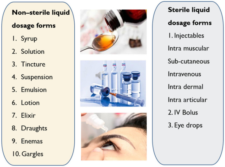
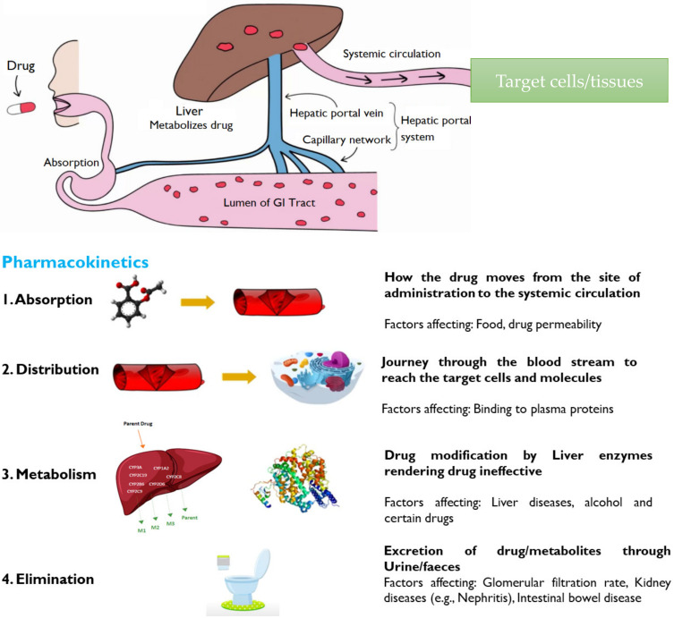
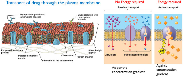
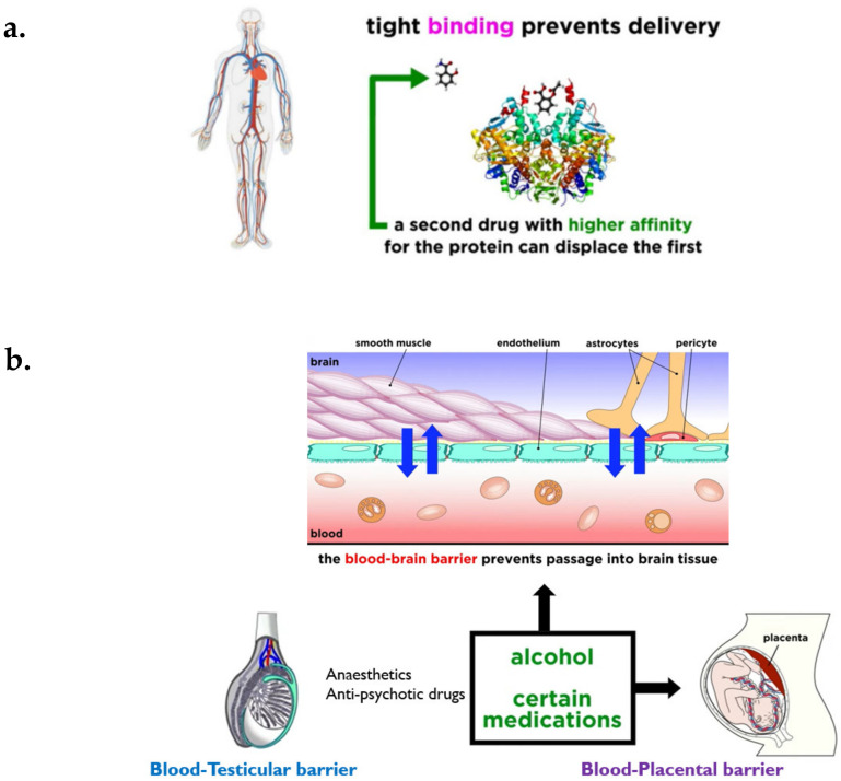
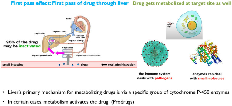
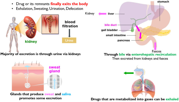
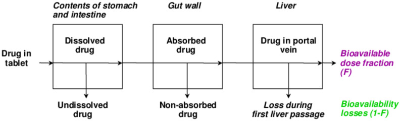
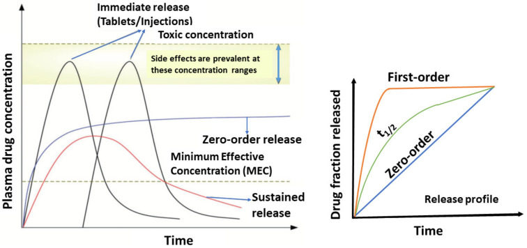
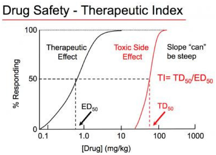
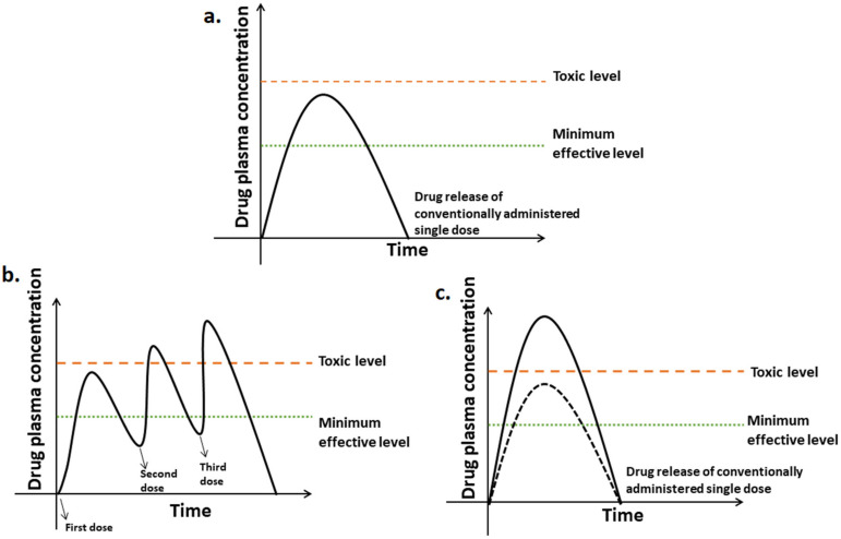
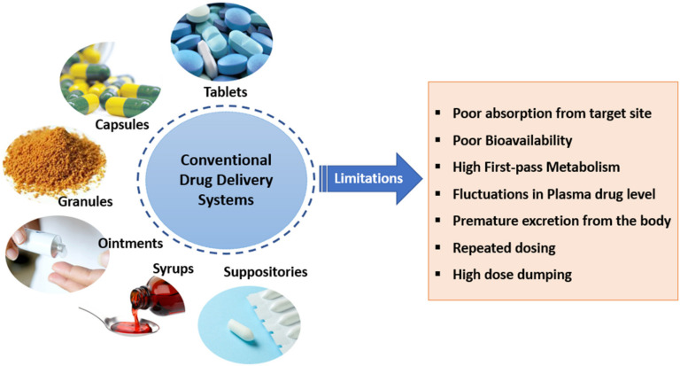
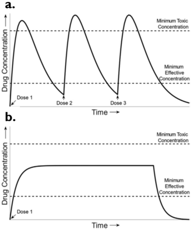
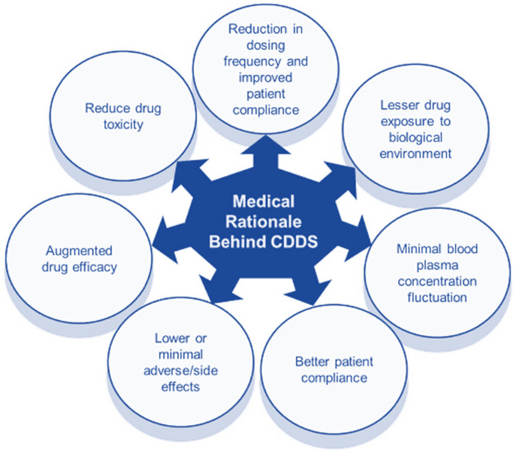
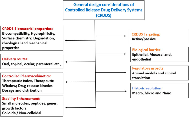
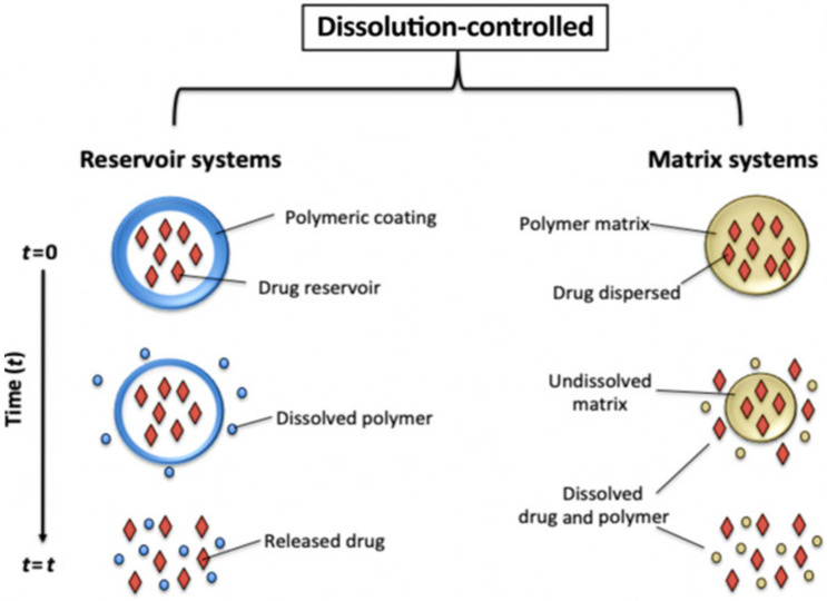
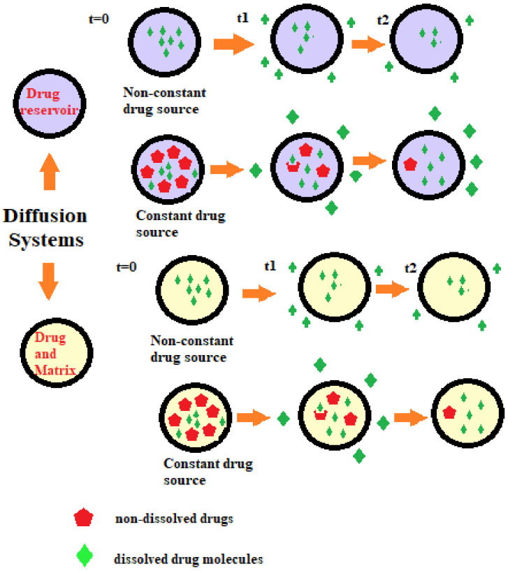
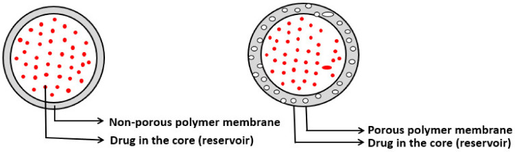
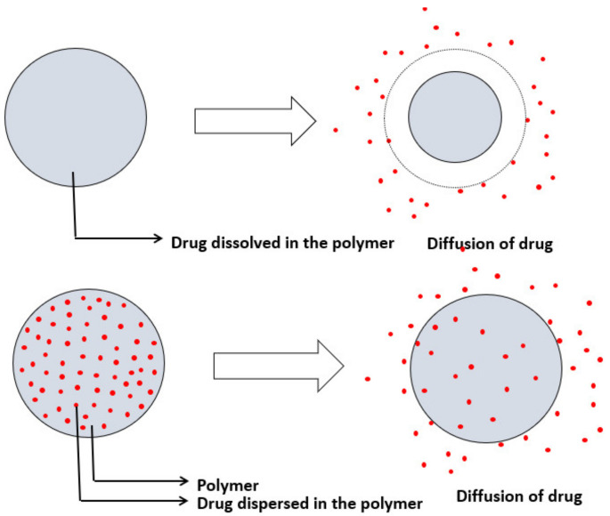
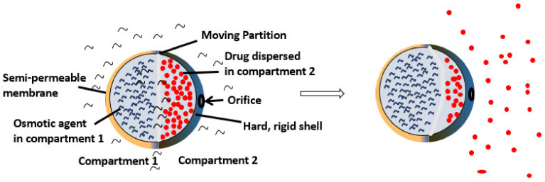
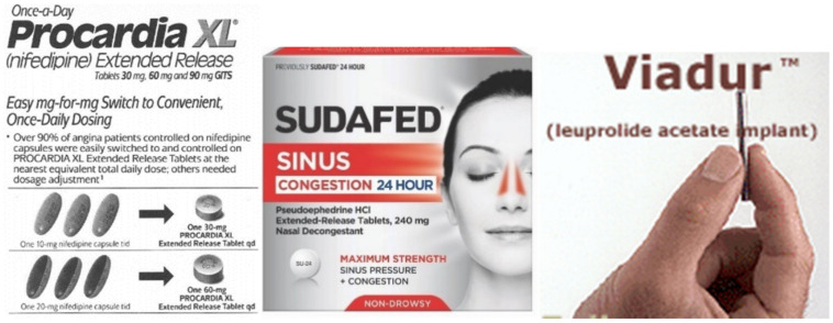
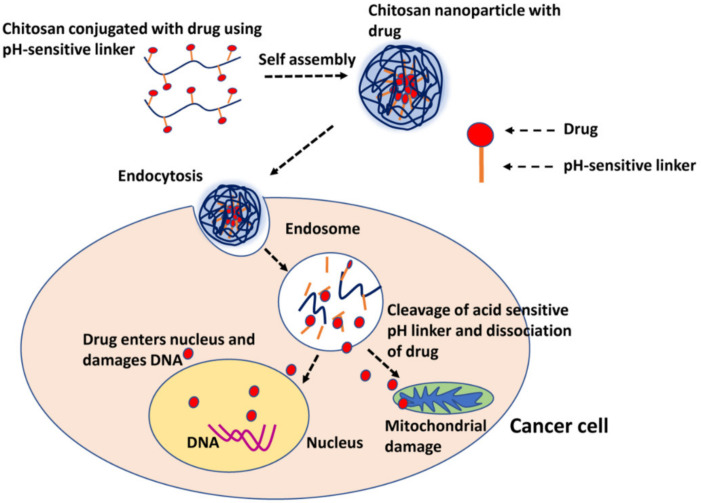
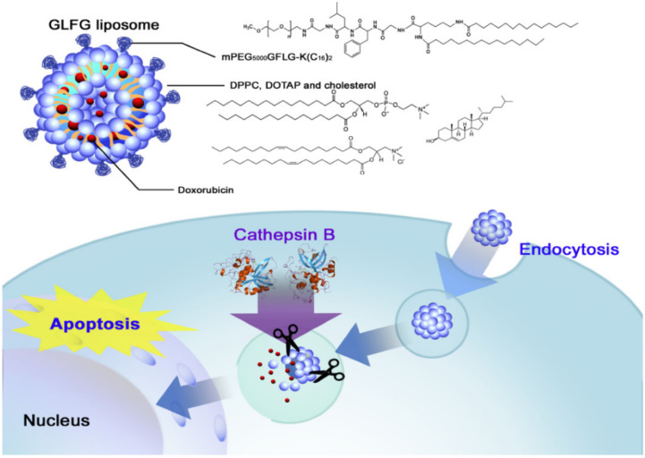
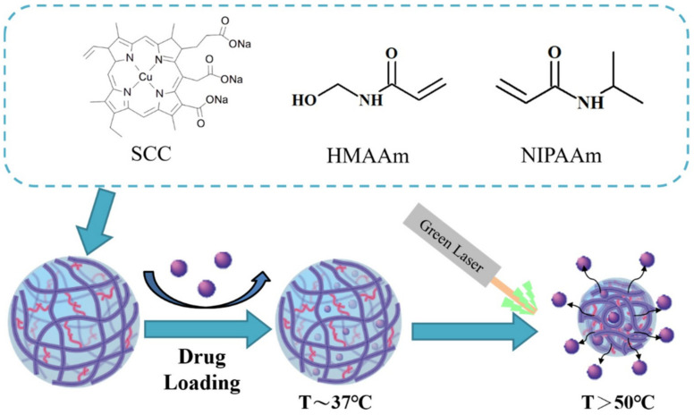
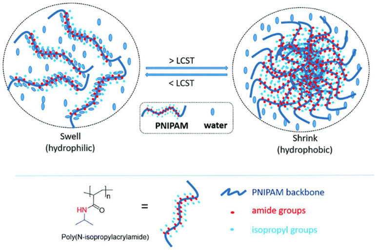
Verification
Claims the paper makes
The claims and statistics of this paper haven’t been checked yet.
Run the check — free, no account needed. It reads the open-access text and checks the sources it cites.
Claims other papers make about this one
Each is checked against this paper’s own text: does it say what the citing paper says it does?
In the citing paper
“With MNP, it can be achieved by optimizing temperature, pH or enzyme-sensitive coating that releases the substance in response to specific stimuli found in tumor microelements. This ensures that the substance remains preserved during circulation and is released only when it reaches cancerous tissue [31].”
From Advances in green-synthesized magnetic nanoparticles for targeted cancer therapy: mechanisms, applications, and future perspectives (2026) · cites this paper as [31]
Self-check
Percentages, sums and reported differences re-computed from the paper’s own numbers.
The self-check needs the paper’s full text.
Citations
Cites
Liposomal drug delivery systems: From concept to clinical applications
Allen, T. M., & Cullis, P. R. (2013) · Advanced Drug Delivery Reviews · doi:10.1016/j.addr.2012.09.037 · Cited by 4,839
Langer, R. (1990) · Science · doi:10.1126/science.2218494 · Cited by 1,778
Overcoming the challenges in administering biopharmaceuticals: Formulation and delivery strategies
Mitragotri, S., Burke, P. A., & Langer, R. (2014) · Nature Reviews Drug Discovery · doi:10.1038/nrd4363 · Cited by 1,702
Engineering exosomes for targeted drug delivery
Liang, Y., Duan, L., Lu, J., & Xia, J. (2021) · Theranostics · doi:10.7150/thno.52570 · Cited by 1,547
Quantum dots in imaging, drug delivery and sensor applications
Matea, C., Mocan, T., Tabaran, F., Pop, T., Mosteanu, O., Puia, C., Iancu, C., & Mocan, L. (2017) · International Journal of Nanomedicine · doi:10.2147/ijn.s138624 · Cited by 666
Targeted therapy in chronic diseases using nanomaterial-based drug delivery vehicles
Singh, A. P., Biswas, A., Shukla, A., & Maiti, P. (2019) · Signal Transduction and Targeted Therapy · doi:10.1038/s41392-019-0068-3 · Cited by 645
A brief review on solid lipid nanoparticles: part and parcel of contemporary drug delivery systems
Duan, Y., Dhar, A., Patel, C., Khimani, M., Neogi, S., Sharma, P., Siva Kumar, N., & Vekariya, R. L. (2020) · RSC Advances · doi:10.1039/d0ra03491f · Cited by 614
Research works printed but not linked: 163
- Langer, 1998, Drug delivery and targeting, Nature, 392, 5
- Benoit, D.S., Overby, C.T., Sims, K.R., and Ackun-Farmmer, M.A. (2020). Drug delivery systems. Biomaterials Science, Elsevier. · DOI
- Chaudhari, 2012, Pharmaceutical excipients: A review, IJAPBC, 1, 21
- Jain, 2020, An overview of drug delivery systems, Drug Deliv. Syst., 2059, 1, 10.1007/978-1-4939-9798-5_1 · DOI
- Patel, 2011, New pharmaceutical excipients in solid dosage forms-A review, Int. J. Pharm. Life Sci., 2, 1006
- Kalasz, 2006, Drug excipients, Curr. Med. Chem., 13, 2535, 10.2174/092986706778201648 · DOI
- Ku, 2008, Use of the biopharmaceutical classification system in early drug development, AAPS J., 10, 208, 10.1208/s12248-008-9020-0 · DOI
- Verma, 2010, Research. Routes of drug administration, Int. J. Pharm. Stud. Res., 1, 54
- Augsburger, L.L., and Hoag, S.W. (2016). Pharmaceutical Dosage Forms-Tablets, CRC Press. · DOI
- Qiu, Y., Chen, Y., Zhang, G.G., Yu, L., and Mantri, R.V. (2016). Developing Solid Oral Dosage Forms: Pharmaceutical Theory and Practice, Academic Press.
- Mahato, R.I., and Narang, A.S. (2017). Pharmaceutical Dosage Forms and Drug Delivery: Revised and Expanded, CRC Press.
- Bora, 2014, Recent advances in semisolid dosage form, Int. J. Pharm. Sci. Res., 5, 3596
- Niazi, S.K. (2019). Handbook of Pharmaceutical Manufacturing Formulations: Volume Four, Semisolid Products, CRC Press. · DOI
- Mahalingam, 2008, Semisolid dosages: Ointments, creams, and gels, Pharm. Manuf. Handb., 1, 267, 10.1002/9780470259818.ch9 · DOI
- Allen, 2014, Basics of compounding: Tips and hints, Part 3: Compounding with ointments, creams, pastes, gels, and gel-creams, Int. J. Pharm. Compd., 18, 228
- Prausnitz, 2008, Transdermal drug delivery, Nat. Biotechnol., 26, 1261, 10.1038/nbt.1504 · DOI
- Prausnitz, 2004, Current status and future potential of transdermal drug delivery, Nat. Rev. Drug Discov., 3, 115, 10.1038/nrd1304 · DOI
- Khan, 2019, Transdermal patches: Design and current approaches to painless drug delivery, Acta Pharm., 69, 197, 10.2478/acph-2019-0016 · DOI
- Dhiman, 2011, Transdermal patches: A recent approach to new drug delivery system, Int. J. Pharm. Pharm. Sci., 3, 26
- Gad, 2008, Semisolid dosages: Ointments, creams and gels, Pharmaceutical Manufacturing Handbook: Production and Processes, Volume 5, 267
- Londono, 2008, Liquid dosage forms, Pharmaceutical Manufacturing Handbook: Production and Processes, Volume 5, 313
- Kumar, 2016, Pharmaceutical suspensions: Patient compliance oral dosage forms, World J. Pharm. Pharm. Sci., 5, 1471
- Kalantzi, 2006, Biowaiver monographs for immediate release solid oral dosage forms: Acetaminophen (paracetamol), J. Pharm. Sci., 95, 4, 10.1002/jps.20477 · DOI
- Sears, 1951, Linctuses, Practitioner, 166, 91
- Payne, 2002, Pharmacokinetics of oral tramadol drops for postoperative pain relief in children aged 4 to 7 years—A pilot study, Anesth. Prog., 49, 109
- 2011, Using gargles and mouthwashes: Medicine cupboard, SA Pharm. Assist., 11, 26
- Jambhekar, S.S., and Breen, P.J. (2009). Basic Pharmacokinetics, Pharmaceutical Press.
- Fleisher, 1999, Drug, meal and formulation interactions influencing drug absorption after oral administration, Clin. Pharmacokinet., 36, 233, 10.2165/00003088-199936030-00004 · DOI
- Obata, 2005, Prediction of oral drug absorption in humans by theoretical passive absorption model, Int. J. Pharm., 293, 183, 10.1016/j.ijpharm.2005.01.005 · DOI
- Hubatsch, 2007, Determination of drug permeability and prediction of drug absorption in Caco-2 monolayers, Nat. Protoc., 2, 2111, 10.1038/nprot.2007.303 · DOI
- Seydel, J.K., and Wiese, M. (2009). Drug-Membrane Interactions: Analysis, Drug Distribution, Modeling, John Wiley & Sons.
- Gillette, 1971, Factors affecting drug metabolism, Ann. N. Y. Acad. Sci., 179, 43, 10.1111/j.1749-6632.1971.tb46890.x · DOI
- Ekins, 2000, Present and future in vitro approaches for drug metabolism, J. Pharmacol. Toxicol. Methods, 44, 313, 10.1016/S1056-8719(00)00110-6 · DOI
- Taft, D.R. (2009). Drug excretion. Pharmacology, Elsevier. · DOI
- Reinberg, 1991, Concepts of circadian chronopharmacology, Ann. N. Y. Acad. Sci., 618, 102, 10.1111/j.1749-6632.1991.tb27239.x · DOI
- Prabu, S.L., Suriyaprakash, T., Ruckmani, K., and Thirumurugan, R. (2015). Biopharmaceutics and Pharmacokinetics. Basic Pharmacokinetic Concepts and Some Clinical Applications, IntechOpen. · DOI · Paper page
- Hardenia, A., Maheshwari, N., Hardenia, S.S., Dwivedi, S.K., Maheshwari, R., and Tekade, R.K. (2019). Scientific rationale for designing controlled drug delivery systems. Basic Fundamentals of Drug Delivery, Elsevier. · DOI
- Paarakh, 2018, Release kinetics–concepts and applications, Int. J. Pharm. Res. Technol., 8, 12
- Habet, 2021, Narrow Therapeutic Index drugs: Clinical pharmacology perspective, J. Pharm. Pharmacol., 73, 1285, 10.1093/jpp/rgab102 · DOI
- Lowe, E.S., and BALIS, F.M. (2007). Dose-effect and concentration-effect analysis. Principles of Clinical Pharmacology, Elsevier. · DOI
- Park, 2014, Controlled drug delivery systems: Past forward and future back, J. Control. Release, 190, 3, 10.1016/j.jconrel.2014.03.054 · DOI
- Fenton, 2018, Advances in biomaterials for drug delivery, Adv. Mater., 30, 1705328, 10.1002/adma.201705328 · DOI
- Gupta, 2010, Osmotically controlled drug delivery system with associated drugs, J. Pharm. Pharm. Sci., 13, 571, 10.18433/J38W25 · DOI
- Siepmann, J., Siegel, R.A., and Siepmann, F. (2012). Diffusion controlled drug delivery systems. Fundamentals and Applications of Controlled Release Drug Delivery, Springer. · DOI
- Siepmann, 2012, Modeling of diffusion controlled drug delivery, J. Control. Release, 161, 351, 10.1016/j.jconrel.2011.10.006 · DOI
- Srikonda, 2006, Osmotic controlled drug delivery systems, Des. Control. Release Drug Deliv. Syst., 1, 203
- Patil, 2018, A review: Osmotic drug delivery system, Pharma Sci. Monit., 9, 2
- Kumar, 2007, An overview of recent patents on oral osmotic drug delivery systems, Recent Pat. Drug Deliv. Formul., 1, 236, 10.2174/187221107782331638 · DOI
- Siepmann, J., and Siepmann, F. (2012). Swelling controlled drug delivery systems. Fundamentals and Applications of Controlled Release Drug Delivery, Springer. · DOI
- 2013, Polymer–drug conjugates: Origins, progress to date and future directions, Adv. Drug Deliv. Rev., 65, 49, 10.1016/j.addr.2012.10.014 · DOI
- Yun, 2015, Controlled drug delivery: Historical perspective for the next generation, J. Control. Release, 219, 2, 10.1016/j.jconrel.2015.10.005 · DOI
- Guo, M.X. (2010). Dissolution Testing: In Vitro Characterization of Oral Controlled Release Dosage Forms, Wiley. · DOI
- Adeosun, S.O., Ilomuanya, M.O., Gbenebor, O.P., Dada, M.O., and Odili, C.C. (2020). Biomaterials for Drug Delivery: Sources, Classification, Synthesis, Processing, and Applications. Advanced Functional Materials, IntechOpen. · DOI
- Neuse, 2008, Synthetic polymers as drug-delivery vehicles in medicine, Met. Based Drugs, 2008, 469531, 10.1155/2008/469531 · DOI
- Srivastava, 2015, Polymers in drug delivery, J. Biosci. Med., 4, 69
- Grumezescu, A.M. (2016). Chapter 8—Natural and synthetic polymers for drug delivery and targeting. Nanobiomaterials in Drug Delivery, William Andrew Publishing.
- Bawa, 2009, Stimuli-responsive polymers and their applications in drug delivery, Biomed. Mater., 4, 022001, 10.1088/1748-6041/4/2/022001 · DOI
- Deng, 2020, Application of the Nano-Drug Delivery System in Treatment of Cardiovascular Diseases, Front. Bioeng. Biotechnol., 7, 489, 10.3389/fbioe.2019.00489 · DOI
- Natarajan, 2014, Sustained-release from nanocarriers: A review, J. Control. Release, 193, 122, 10.1016/j.jconrel.2014.05.029 · DOI
- Chamundeeswari, 2019, Nanocarriers for drug delivery applications, Environ. Chem. Lett., 17, 849, 10.1007/s10311-018-00841-1 · DOI
- Tekade, R.K. (2019). Chapter 7—Levels of Drug Targeting. Basic Fundamentals of Drug Delivery, Academic Press.
- Naz, 2021, Nanotechnology: Its application in treating neurodegenerative diseases, CNS Neurol. Disord. Drug Targets, 20, 34, 10.2174/1871527319666200916121515 · DOI
- Attia, 2019, An overview of active and passive targeting strategies to improve the nanocarriers efficiency to tumour sites, J. Pharm. Pharmacol., 71, 1185, 10.1111/jphp.13098 · DOI
- Shah, 2021, Nanocarriers for targeted drug delivery, J. Drug Deliv. Sci. Technol., 13, 102426, 10.1016/j.jddst.2021.102426 · DOI · Paper page
- Sercombe, 2015, Advances and Challenges of Liposome Assisted Drug Delivery, Front. Pharmacol., 6, 286, 10.3389/fphar.2015.00286 · DOI
- Dimov, 2017, Formation and purification of tailored liposomes for drug delivery using a module-based micro continuous-flow system, Sci. Rep., 7, 12045, 10.1038/s41598-017-11533-1 · DOI
- Yadav, 2017, Liposomes for drug delivery, Drugs, 7, 15
- Caminade, 2014, Dendrimers for drug delivery, J. Mater. Chem. B, 2, 4055, 10.1039/C4TB00171K · DOI
- Chauhan, A.S. (2018). Dendrimers for Drug Delivery. Molecules, 23. · DOI
- Le, N.T.T., Nguyen, T.N.Q., Cao, V.D., Hoang, D.T., Ngo, V.C., and Hoang Thi, T.T. (2019). Recent progress and advances of multi-stimuli-responsive dendrimers in drug delivery for cancer treatment. Pharmaceutics, 11. · DOI
- Bunggulawa, 2018, Recent advancements in the use of exosomes as drug delivery systems, J. Nanobiotechnology, 16, 1, 10.1186/s12951-018-0403-9 · DOI
- Patra, 2018, Nano based drug delivery systems: Recent developments and future prospects, J. Nanobiotechnology, 16, 71, 10.1186/s12951-018-0392-8 · DOI
- Adepu, S., Luo, H., and Ramakrishna, S. (2021). Heparin-Tagged PLA-PEG Copolymer-Encapsulated Biochanin A-Loaded (Mg/Al) LDH Nanoparticles Recommended for Non-Thrombogenic and Anti-Proliferative Stent Coating. Int. J. Mol. Sci., 22. · DOI
- Adepu, 2018, Broad-spectrum antimicrobial activity of bacterial cellulose silver nanocomposites with sustained release, Int. J. Mol. Sci., 53, 1596
- Adepu, 2020, Bacterial cellulose with microencapsulated antifungal essential oils: A novel double barrier release system, Materialia, 9, 100585, 10.1016/j.mtla.2020.100585 · DOI
- Muraca, 2020, Solid Lipid Nanoparticles for Drug Delivery: Pharmacological and Biopharmaceutical Aspects, Front. Mol. Biosci., 7, 319
- Kocbek, 2019, Electrospun nanofibers for customized drug-delivery systems, J. Drug Deliv. Sci. Technol., 51, 672, 10.1016/j.jddst.2019.03.038 · DOI
- Zare, 2020, Current Progress of Electrospun Nanocarriers for Drug Delivery Applications, Proceedings, 4, 8790
- Adepu, 2017, Effect of micropatterning induced surface hydrophobicity on drug release from electrospun cellulose acetate nanofibers, Appl. Surf. Sci., 426, 755, 10.1016/j.apsusc.2017.07.197 · DOI
- Adepu, 2021, Drug release behaviour and mechanism from unmodified and in situ modified bacterial cellulose, Proc. Indian Natl. Sci. Acad., 87, 110, 10.1007/s43538-021-00012-x · DOI
- Adepu, 2021, Bacterial Cellulose-Based Drug Delivery System for Dual Mode Drug Release, Trans. Indian Natl. Acad. Eng., 6, 265, 10.1007/s41403-020-00192-w · DOI
- Adepu, 2020, Ex-situ modification of bacterial cellulose for immediate and sustained drug release with insights into release mechanism, Carbohydr. Polym., 249, 116816, 10.1016/j.carbpol.2020.116816 · DOI
- Farokhi, 2020, Functionalized silk fibroin nanofibers as drug carriers: Advantages and challenges, J. Control. Release, 321, 324, 10.1016/j.jconrel.2020.02.022 · DOI
- 2018, A Summary of Electrospun Nanofibers as Drug Delivery System: Drugs Loaded and Biopolymers Used as Matrices, Curr. Drug Deliv., 15, 1360, 10.2174/1567201815666180723114326 · DOI
- Mortensen, 1998, Structural properties of self-assembled polymeric micelles, Curr. Opin. Colloid Interface Sci., 3, 12, 10.1016/S1359-0294(98)80036-8 · DOI
- Alex, 2017, Self assembled dual responsive micelles stabilized with protein for co-delivery of drug and siRNA in cancer therapy, Biomaterials, 133, 94, 10.1016/j.biomaterials.2017.04.022 · DOI
- Saleemi, 2020, An overview of recent development in therapeutic drug carrier system using carbon nanotubes, J. Drug Deliv. Sci. Technol., 59, 101855, 10.1016/j.jddst.2020.101855 · DOI
- Zare, 2021, Carbon Nanotubes: Smart Drug/Gene Delivery Carriers, Int. J. Nanomed., 16, 1681, 10.2147/IJN.S299448 · DOI
- Madani, 2011, A new era of cancer treatment: Carbon nanotubes as drug delivery tools, Int. J. Nanomed., 6, 2963
- Kaur, J., Gill, G.S., and Jeet, K. (2019). Chapter 5 - Applications of Carbon Nanotubes in Drug Delivery A Comprehensive Review. Characterization and Biology of Nanomaterials for Drug Delivery, Elsevier. · DOI
- Li, 2017, Functionalized single-walled carbon nanotubes: Cellular uptake, biodistribution and applications in drug delivery, Int. J. Pharm., 524, 41, 10.1016/j.ijpharm.2017.03.017 · DOI
- Tayeb, 2018, Nanoemulsions in drug delivery: Formulation to medical application, Nanomedicine, 13, 2507, 10.2217/nnm-2018-0088 · DOI
- Li, 2016, Designing hydrogels for controlled drug delivery, Nat. Rev. Mater., 1, 16071, 10.1038/natrevmats.2016.71 · DOI
- Mi, 2000, The study of gelation kinetics and chain-relaxation properties of glutaraldehyde-cross-linked chitosan gel and their effects on microspheres preparation and drug release, Carbohydr. Polym., 41, 389, 10.1016/S0144-8617(99)00104-6 · DOI
- Hoare, 2008, Hydrogels in drug delivery: Progress and challenges, Polymer, 49, 1993, 10.1016/j.polymer.2008.01.027 · DOI
- Wei, 2017, Synthesis and characterization of a multi-sensitive polysaccharide hydrogel for drug delivery, Carbohydr. Polym., 177, 275, 10.1016/j.carbpol.2017.08.133 · DOI
- Li, 2020, Supramolecular hydrogels: Mechanical strengthening with dynamics, Polymer, 210, 122993, 10.1016/j.polymer.2020.122993 · DOI
- Jacob, S., Nair, A.B., Shah, J., Sreeharsha, N., Gupta, S., and Shinu, P. (2021). Emerging role of hydrogels in drug delivery systems, tissue engineering and wound management. Pharmaceutics, 13. · DOI
- Zou, 2020, A sodium alginate-based sustained-release IPN hydrogel and its applications, RSC Adv., 10, 39722, 10.1039/D0RA04316H · DOI
- Shubhika, 2012, Nanotechnology and medicine-The upside and the downside, Int. J. Drug Dev. Res., 5, 1
- Manzoor, 2012, Overcoming limitations in nanoparticle drug delivery: Triggered, intravascular release to improve drug penetration into tumors, Cancer Res., 72, 5566, 10.1158/0008-5472.CAN-12-1683 · DOI
- Barenholz, 2001, Liposome application: Problems and prospects, Curr. Opin. Colloid Interface Sci., 6, 66, 10.1016/S1359-0294(00)00090-X · DOI · Paper page
- Lin, M., Wang, R., and Qi, X.-R. (2021). Quality Evaluation of Drug-Loaded Liposomes. Liposome-Based Drug Deliv. Syst., 123–140. · DOI
- Chis, A.A., Dobrea, C., Morgovan, C., Arseniu, A.M., Rus, L.L., Butuca, A., Juncan, A.M., Totan, M., Vonica-Tincu, A.L., and Cormos, G. (2020). Applications and limitations of dendrimers in biomedicine. Molecules, 25. · DOI
- Vaidya, 2018, Dendrimers: Nanosized multifunctional platform for drug delivery, Drug Deliv. Lett., 8, 3
- Antimisiaris, S.G., Mourtas, S., and Marazioti, A. (2018). Exosomes and exosome-inspired vesicles for targeted drug delivery. Pharmaceutics, 10. · DOI · Paper page
- Agarwal, 2020, Exosomes: Structure, biogenesis, types and application in diagnosis and gene and Drug delivery, Curr. Gene Ther., 20, 195, 10.2174/1566523220999200731011702 · DOI
- Desai, 2021, Metallic nanoparticles as drug delivery system for the treatment of cancer, Expert Opin. Drug Deliv., 18, 1
- Alyassin, 2020, Application of mesoporous silica nanoparticles as drug delivery carriers for chemotherapeutic agents, Drug Discov. Today, 25, 1513, 10.1016/j.drudis.2020.06.006 · DOI
- Bharti, 2015, Mesoporous silica nanoparticles in target drug delivery system: A review, Int. J. Pharm. Investig., 5, 124, 10.4103/2230-973X.160844 · DOI
- Timur, 2020, Recent pros and cons of nanomaterials in drug delivery systems, Int. J. Polym. Mater. Polym. Biomater., 69, 1090, 10.1080/00914037.2019.1655753 · DOI
- Bilensoy, 2018, Nanocapsules for drug delivery: An updated review of the last decade, Recent Pat. Drug Deliv. Formul., 12, 252
- Thassu, D., Pathak, Y., and Deleers, M. (2007). Nanoparticulate Drug-Delivery Systems: An Overview, CRC Press. · DOI
- Ghaderi, 2011, Fluorescence nanoparticles “quantum dots” as drug delivery system and their toxicity: A review, J. Drug Target., 19, 475, 10.3109/1061186X.2010.526227 · DOI
- Barroso, 2011, Quantum dots in cell biology, J. Histochem. Cytochem., 59, 237, 10.1369/0022155411398487 · DOI
- Sahu, D.K., Ghosh, G., and Rath, G. (2021). Nanofibers in Drug Delivery. Nanopharmaceutical Adv. Deliv. Syst., 99–123. · DOI
- Zare, M., Dziemidowicz, K., Williams, G.R., and Ramakrishna, S. (2021). Encapsulation of pharmaceutical and nutraceutical active ingredients using electrospinning processes. Nanomaterials, 11. · DOI
- Torchilin, V.P. (2018). CHAPTER 1 Fundamentals of Stimuli-responsive Drug and Gene Delivery Systems. Stimuli-Responsive Drug Delivery Systems, The Royal Society of Chemistry. · DOI
- Mohapatra, S.S., Ranjan, S., Dasgupta, N., Mishra, R.K., and Thomas, S. (2019). Chapter 6—Thermoresponsive Drug Delivery Systems, Characterization and Application. Applications of Targeted Nano Drugs and Delivery Systems, Elsevier.
- Palanikumar, 2020, pH-responsive high stability polymeric nanoparticles for targeted delivery of anticancer therapeutics, Commun. Biol., 3, 95, 10.1038/s42003-020-0817-4 · DOI
- Datta, 2018, Unravelling the Excellent Chemical Stability and Bioavailability of Solvent Responsive Curcumin-Loaded 2-Ethyl-2-oxazoline-grad-2-(4-dodecyloxyphenyl)-2-oxazoline Copolymer Nanoparticles for Drug Delivery, Biomacromolecules, 19, 2459, 10.1021/acs.biomac.8b00057 · DOI
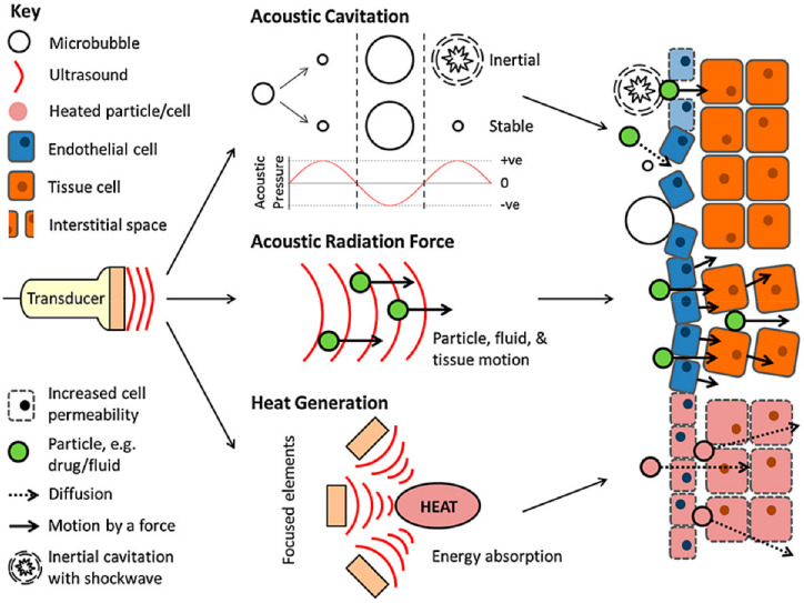
- Cai, 2020, Ultrasound-Responsive Materials for Drug/Gene Delivery, Front. Pharmacol., 10, 1650, 10.3389/fphar.2019.01650 · DOI
- Gibot, 2019, Electric field-responsive nanoparticles and electric fields: Physical, chemical, biological mechanisms and therapeutic prospects, Adv. Drug Deliv. Rev., 138, 56, 10.1016/j.addr.2018.10.017 · DOI
- Price, 2018, Magnetic Drug Delivery: Where the Field Is Going, Front. Chem., 6, 609, 10.3389/fchem.2018.00619 · DOI
- Amukarimi, 2021, Smart biomaterials-A proposed definition and overview of the field, Curr. Opin. Biomed. Eng., 19, 100311, 10.1016/j.cobme.2021.100311 · DOI
- Zhuo, S., Zhang, F., Yu, J., Zhang, X., Yang, G., and Liu, X. (2020). pH-Sensitive Biomaterials for Drug Delivery. Molecules, 25. · DOI · Paper page
- Rai, 2016, Eudragit-coated dextran microspheres of 5-fluorouracil for site-specific delivery to colon, Drug Deliv., 23, 328, 10.3109/10717544.2014.913733 · DOI
- Deirram, 2019, pH-Responsive Polymer Nanoparticles for Drug Delivery, Macromol. Rapid Commun., 40, 1800917, 10.1002/marc.201800917 · DOI · Paper page
- Yang, 2019, A Smart pH-Sensitive Delivery System for Enhanced Anticancer Efficacy via Paclitaxel Endosomal Escape, Front. Pharmacol., 10, 10, 10.3389/fphar.2019.00010 · DOI
- Vivek, 2013, pH-responsive drug delivery of chitosan nanoparticles as Tamoxifen carriers for effective anti-tumor activity in breast cancer cells, Colloids Surf. B Biointerfaces, 111, 117, 10.1016/j.colsurfb.2013.05.018 · DOI
- Babu, A., and Ramesh, R. (2017). Multifaceted applications of chitosan in cancer drug delivery and therapy. Mar. Drugs, 15. · DOI
- Monteiro, 2021, Reduction-responsive polymers for drug delivery in cancer therapy—Is there anything new to discover?, Wiley Interdiscip. Rev. Nanomed. Nanobiotechnology, 13, e1678, 10.1002/wnan.1678 · DOI
- Geiselhart, 2021, Degradable Redox-Responsive Polyolefins, Macromolecules, 54, 1775, 10.1021/acs.macromol.1c00010 · DOI
- Shahriari, 2019, Enzyme responsive drug delivery systems in cancer treatment, J. Control. Release, 308, 172, 10.1016/j.jconrel.2019.07.004 · DOI
- Lee, S., Song, S.J., Lee, J., Ha, T.H., and Choi, J.S. (2020). Cathepsin B-Responsive Liposomes for Controlled Anticancer Drug Delivery in Hep G2 Cells. Pharmaceutics, 12. · DOI
- Linsley, 2017, Recent advances in light-responsive on-demand drug-delivery systems, Ther. Deliv., 8, 89, 10.4155/tde-2016-0060 · DOI
- Zhao, 2019, Remote Light-Responsive Nanocarriers for Controlled Drug Delivery: Advances and Perspectives, Small, 15, 1903060, 10.1002/smll.201903060 · DOI
- Chang, R., and Tsai, W.-B. (2016). Photo-induced thermal-responsive nanogels for controlled drug release. Front. Bioeng. Biotechnol., 4. · DOI
- Singh, 2018, NIR triggered liposome gold nanoparticles entrapping curcumin as in situ adjuvant for photothermal treatment of skin cancer, Int. J. Biol. Macromol., 110, 375, 10.1016/j.ijbiomac.2017.11.163 · DOI
- Chang, R., and Tsai, W.-B. (2018). Fabrication of photothermo-responsive drug-loaded nanogel for synergetic cancer therapy. Polymers, 10. · DOI
- Jain, 2015, Tunable LCST behavior of poly(N-isopropylacrylamide/ionic liquid) copolymers, Polym. Chem., 6, 6819, 10.1039/C5PY00998G · DOI
- Cao, 2019, Reversible thermoresponsive peptide–PNIPAM hydrogels for controlled drug delivery, Biomacromolecules, 20, 3601, 10.1021/acs.biomac.9b01009 · DOI
- Tu, 2019, Advances in injectable self-healing biomedical hydrogels, Acta Biomater., 90, 1, 10.1016/j.actbio.2019.03.057 · DOI
- Doberenz, 2020, Thermoresponsive polymers and their biomedical application in tissue engineering–A review, J. Mater. Chem. B, 8, 607, 10.1039/C9TB02052G · DOI
- Ge, 2012, Drug release from electric-field-responsive nanoparticles, ACS Nano, 6, 227, 10.1021/nn203430m · DOI
- Qu, 2018, Injectable antibacterial conductive hydrogels with dual response to an electric field and pH for localized “smart” drug release, Acta Biomater., 72, 55, 10.1016/j.actbio.2018.03.018 · DOI · Paper page
- Zhao, 2016, Nano-engineered electro-responsive drug delivery systems, J. Mater. Chem. B, 4, 3019, 10.1039/C6TB00049E · DOI
- Patil, T.V., Patel, D.K., Dutta, S.D., Ganguly, K., and Lim, K.-T. (2021). Graphene Oxide-Based Stimuli-Responsive Platforms for Biomedical Applications. Molecules, 26. · DOI
- Hou, 2020, Electrochemically controlled cleavage of imine bonds on a graphene platform: Towards new electro-responsive hybrids for drug release, Nanoscale, 12, 23824, 10.1039/D0NR04102E · DOI
- Zhao, 2013, Potential and problems in ultrasound-responsive drug delivery systems, Int. J. Nanomed., 8, 1621
- Browning, R.J., and Stride, E. (2018). Microbubble-mediated delivery for cancer therapy. Fluids, 3. · DOI
- Bartucci, 2020, Comparative study of nanoparticle uptake and impact in murine lung, liver and kidney tissue slices, Nanotoxicology, 14, 847, 10.1080/17435390.2020.1771785 · DOI
- Lu, 2020, Targeted immunomodulation of inflammatory monocytes across the blood-brain barrier by curcumin-loaded nanoparticles delays the progression of experimental autoimmune encephalomyelitis, Biomaterials, 245, 119987, 10.1016/j.biomaterials.2020.119987 · DOI
- Vallet-Regí, M., Colilla, M., Izquierdo-Barba, I., and Manzano, M. (2018). Mesoporous silica nanoparticles for drug delivery: Current insights. Molecules, 23. · DOI
- Santos, H.A., Liu, D., and Zhang, H. (2019). Chapter 10—Microfluidic technologies for local drug delivery. Microfluidics for Pharmaceutical Applications, William Andrew Publishing.
- Santos, H.A., Liu, D., and Zhang, H. (2019). Chapter 12—Antibody discovery using microfluidic systems. Microfluidics for Pharmaceutical Applications, William Andrew Publishing.
- Santos, H.A., Liu, D., and Zhang, H. (2019). Chapter 11—Droplet-based microfluidics for cell encapsulation and delivery. Microfluidics for Pharmaceutical Applications, William Andrew Publishing.
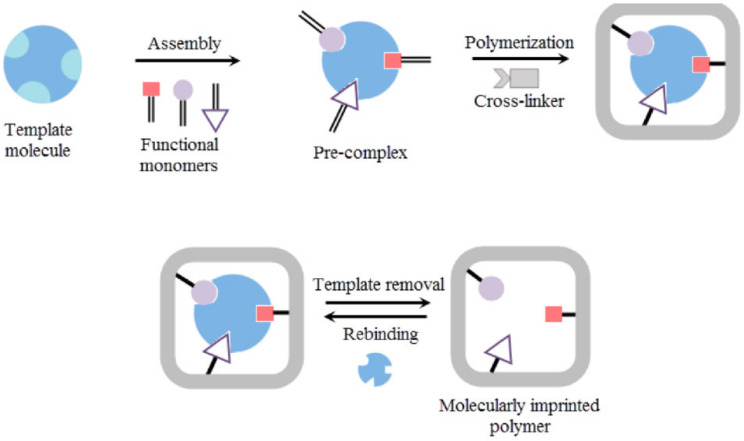
- Zaidi, 2020, Molecular imprinting: A useful approach for drug delivery, Mater. Sci. Energy Technol., 3, 72
- Saylan, Y., Akgönüllü, S., Yavuz, H., Ünal, S., and Denizli, A. (2019). Molecularly imprinted polymer based sensors for medical applications. Sensors, 19. · DOI
- Wei, 2020, Systemic nanoparticle delivery of CRISPR-Cas9 ribonucleoproteins for effective tissue specific genome editing, Nat. Commun., 11, 3232, 10.1038/s41467-020-17029-3 · DOI
- Preeti, 2021, Application of Quantum Dots in Drug Delivery, Nanosci. Nanotechnol. Asia, 11, 1
- Beg, 2020, 3D printing for drug delivery and biomedical applications, Drug Discov. Today, 25, 1668, 10.1016/j.drudis.2020.07.007 · DOI
- Trenfield, 2019, Shaping the future: Recent advances of 3D printing in drug delivery and healthcare, Expert Opin. Drug Deliv., 16, 1081, 10.1080/17425247.2019.1660318 · DOI
Cited by
Sorted by citations
Yanar, F., Carugo, D., & Zhang, X. (2023) · Molecules · doi:10.3390/molecules28155694 · Cited by 80
Gulati, S., Ansari, N., Moriya, Y., Joshi, K., Prasad, D., Sajwan, G., Shukla, S., Kumar, S., & Varma, R. S. (2024) · Journal of Materials Chemistry B · doi:10.1039/d4tb00599f · Cited by 52
PLGA-Based Co-Delivery Nanoformulations: Overview, Strategies, and Recent Advances
Stevanović, M. M., Qian, K., Huang, L., & Vukomanović, M. (2025) · Pharmaceutics · doi:10.3390/pharmaceutics17121613 · Cited by 42
Zhuo, Q., Liang, Y., Hu, Y., Shi, M., Zhao, C., & Zhang, S. (2023) · Carbon Research · doi:10.1007/s44246-023-00066-0 · Cited by 39
Pramoda, G., Verma, R. K., Kesharwani, P., & Shukla, R. (2025) · Journal of Molecular Liquids · doi:10.1016/j.molliq.2025.127938 · Cited by 15
Metadata
- Authors
- Adepu, S., & Ramakrishna, S.
- Venue
- Molecules
- Publisher
- MDPI AG
- Published
- 2021-09-29
- Volume
- 26
- Issue
- 19
- Pages
- 5905
- ISSN
- 1420-3049
- PMC
- PMC8512302
- Cited by
- 1,691
- References
- 7
- Language
- en
About these checks
Registry, retraction, Cites and statistics results are computed, not judged, with one exception: a printed reference that no index lists is read once by a model before it counts as a citation error. Claims are extracted by a model and kept only when their quote appears verbatim in the text; each cited source is then checked, with a model’s help, for whether it says what the claim relies on. Those verdicts can be wrong — read the claim’s quote and the cited source before relying on one.
Statistics and arithmetic are recomputed, not judged.
Confidence bands are shown only for fields calibrated on our annotated test set. None is calibrated yet, so no band is shown.
What counts as a claim ›How we measure confidence ›How we check this ›
Are you an author, or do you see a problem with this page? Request a review
