

The DOI matches its Crossref record; 14 of the 14 references it cites were found in a registry or an index.
Recent Studies and Progress in the Intratumoral Administration of Nano-Sized Drug Delivery Systems
Abstract
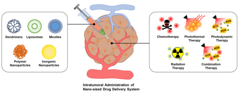
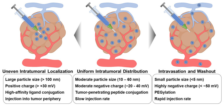
Over the last 30 years, diverse types of nano-sized drug delivery systems (nanoDDSs) have been intensively explored for cancer therapy, exploiting their passive tumor targetability with an enhanced permeability and retention effect. However, their systemic administration has aroused some unavoidable complications, including insufficient tumor-targeting efficiency, side effects due to their undesirable biodistribution, and carrier-associated toxicity. In this review, the recent studies and advancements in intratumoral nanoDDS administration are generally summarized. After identifying the factors to be considered to enhance the therapeutic efficacy of intratumoral nanoDDS administration, the experimental results on the application of intratumoral nanoDDS administration to various types of cancer therapies are discussed. Subsequently, the reports on clinical studies of intratumoral nanoDDS administration are addressed in short. Intratumoral nanoDDS administration is proven with its versatility to enhance the tumor-specific accumulation and retention of therapeutic agents for various therapeutic modalities. Specifically, it can improve the efficacy of therapeutic agents with poor bioavailability by increasing their intratumoral concentration, while minimizing the side effect of highly toxic agents by restricting their delivery to normal tissues. Intratumoral administration of nanoDDS is considered to expand its application area due to its potent ability to improve therapeutic effects and relieve the systemic toxicities of nanoDDSs.
Overview
The claims and statistics of this paper haven’t been checked yet.
Run the check — free, no account needed. It reads the open-access text and checks the sources it cites.
Integrity signals
Registry
Cites
Statistics
Figures
Verification
Claims the paper makes
The claims and statistics of this paper haven’t been checked yet.
Run the check — free, no account needed. It reads the open-access text and checks the sources it cites.
Claims other papers make about this one
Each is checked against this paper’s own text: does it say what the citing paper says it does?
The source backs part of the claim, not all of it — or backs it for a different population, condition or comparison.
In the citing paper
“Systemically administered drugs are subject to rapid clearance, limited tumor accumulation, and most critically, poor tissue penetration.”
From Overcoming tumor microenvironment barriers: transformable and bioinspired nanomedicine strategies for deep tumor penetration (2026) · cites this paper as [2]
Experimental — accuracy not yet measuredIn the citing paper
“Solid tumors pose a spatial “delivery-at-depth” bottleneck: therapeutics that reach tumors often remain sequestered near vessels and fail to distribute uniformly into tumor cores.”
From Overcoming tumor microenvironment barriers: transformable and bioinspired nanomedicine strategies for deep tumor penetration (2026) · cites this paper as [2]
Self-check
Percentages, sums and reported differences re-computed from the paper’s own numbers.
The self-check needs the paper’s full text.
Citations
Cites
Doxil® — The first FDA-approved nano-drug: Lessons learned
Barenholz, Y. (. (2012) · Journal of Controlled Release · doi:10.1016/j.jconrel.2012.03.020 · Cited by 4,792
A framework for advancing our understanding of cancer-associated fibroblasts
Sahai, E., Astsaturov, I., Cukierman, E., DeNardo, D. G., Egeblad, M., Evans, R. M., Fearon, D., Greten, F. R., Hingorani, S. R., Hunter, T., et al. (2020) · Nature Reviews Cancer · doi:10.1038/s41568-019-0238-1 · Cited by 3,863
Accumulation of sub-100 nm polymeric micelles in poorly permeable tumours depends on size
Cabral, H., Matsumoto, Y., Mizuno, K., Chen, Q., Murakami, M., Kimura, M., Terada, Y., Kano, M. R., Miyazono, K., Uesaka, M., et al. (2011) · Nature Nanotechnology · doi:10.1038/nnano.2011.166 · Cited by 2,429
High Interstitial Fluid Pressure - an Obstacle in Cancer Therapy
Heldin, C.-H., Rubin, K., Pietras, K., & Östman, A. (2004) · Nature reviews. Cancer · doi:10.1038/nrc1456 · Cited by 2,222
Dobrovolskaia, M. A., Aggarwal, P., Hall, J. B., & McNeil, S. E. (2008) · Molecular Pharmaceutics · doi:10.1021/mp800032f · Cited by 1,022
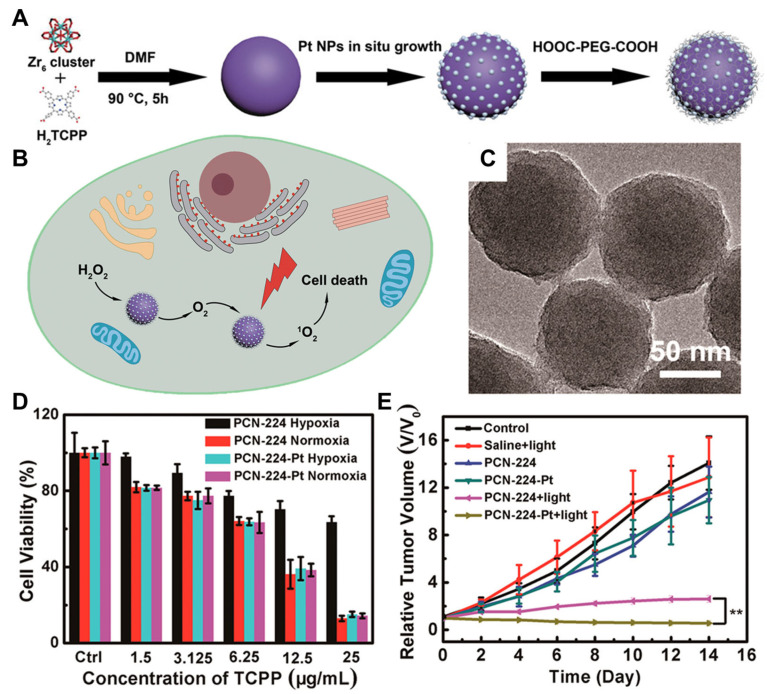
Nanozyme Decorated Metal–Organic Frameworks for Enhanced Photodynamic Therapy
Zhang, Y., Wang, F., Liu, C., Wang, Z., Kang, L., Huang, Y., Dong, K., Ren, J., & Qu, X. (2018) · ACS Nano · doi:10.1021/acsnano.7b07746 · Cited by 854
Stromal cells in tumor microenvironment and breast cancer
Mao, Y., Keller, E. T., Garfield, D. H., Shen, K., & Wang, J. (2013) · Cancer and Metastasis Reviews · doi:10.1007/s10555-012-9415-3 · Cited by 671
The extracellular matrix in tumor progression and metastasis
Eble, J. A., & Niland, S. (2019) · Clinical & Experimental Metastasis · doi:10.1007/s10585-019-09966-1 · Cited by 613
Multifunctional and stimuli-sensitive pharmaceutical nanocarriers
Torchilin, V. (2009) · European Journal of Pharmaceutics and Biopharmaceutics · doi:10.1016/j.ejpb.2008.09.026 · Cited by 583
Lee, H., Fonge, H., Hoang, B., Reilly, R. M., & Allen, C. (2010) · Molecular Pharmaceutics · doi:10.1021/mp100038h · Cited by 349
Research works printed but not linked: 181
- Wakaskar, 2018, Promising effects of nanomedicine in cancer drug delivery, J. Drug Target., 26, 319, 10.1080/1061186X.2017.1377207 · DOI
- Stylianopoulos, 2013, EPR-effect: Utilizing size-dependent nanoparticle delivery to solid tumors, Ther. Deliv., 4, 421, 10.4155/tde.13.8 · DOI
- Ngoune, 2016, Accumulating nanoparticles by EPR: A route of no return, J. Control. Release, 238, 58, 10.1016/j.jconrel.2016.07.028 · DOI
- Greish, 2010, Enhanced permeability and retention (EPR) effect for anticancer nanomedicine drug targeting, Cancer Nanotechnol. Methods Protoc., 624, 25, 10.1007/978-1-60761-609-2_3 · DOI · Paper page
- Li, 2021, Recent advances in development of dendritic polymer-based nanomedicines for cancer diagnosis, Wiley Interdiscip. Rev. Nanomed. Nanobiotechnol., 13, e1670, 10.1002/wnan.1670 · DOI
- Davis, 2008, Nanoparticle therapeutics: An emerging treatment modality for cancer, Nat. Rev. Drug Discov., 7, 771, 10.1038/nrd2614 · DOI · Paper page
- Blanchette, 2004, Nanoparticle and targeted systems for cancer therapy, Adv. Drug Deliv. Rev., 56, 1649, 10.1016/j.addr.2004.02.014 · DOI
- Hu, 2010, Nanoparticle-assisted combination therapies for effective cancer treatment, Ther. Deliv., 1, 323, 10.4155/tde.10.13 · DOI
- Wang, 2022, Strategies to improve the EPR effect: A mechanistic perspective and clinical translation, J. Control. Release, 345, 512, 10.1016/j.jconrel.2022.03.043 · DOI · Paper page
- Egusquiaguirre, 2012, Nanoparticle delivery systems for cancer therapy: Advances in clinical and preclinical research, Clin. Transl. Oncol., 14, 83, 10.1007/s12094-012-0766-6 · DOI
- Nichols, 2014, EPR: Evidence and fallacy, J. Control. Release, 190, 451, 10.1016/j.jconrel.2014.03.057 · DOI · Paper page
- He, 2019, Survey of clinical translation of cancer nanomedicines—Lessons learned from successes and failures, Acc. Chem. Res., 52, 2445, 10.1021/acs.accounts.9b00228 · DOI
- Danhier, 2016, To exploit the tumor microenvironment: Since the EPR effect fails in the clinic, what is the future of nanomedicine?, J. Control. Release, 244, 108, 10.1016/j.jconrel.2016.11.015 · DOI · Paper page
- Kreuter, 1991, Nanoparticle-based dmg delivery systems, J. Control. Release, 16, 169, 10.1016/0168-3659(91)90040-K · DOI
- Takemoto, 2008, Clinical toxicities of nanocarrier systems, Adv. Drug Deliv. Rev., 60, 929, 10.1016/j.addr.2007.11.007 · DOI · Paper page
- Byrne, 2008, Active targeting schemes for nanoparticle systems in cancer therapeutics, Adv. Drug Deliv. Rev., 60, 1615, 10.1016/j.addr.2008.08.005 · DOI
- Yoo, J., Park, C., Yi, G., Lee, D., and Koo, H. (2019). Active targeting strategies using biological ligands for nanoparticle drug delivery systems. Cancers, 11. · DOI
- Chu, 2019, Improving antitumor outcomes for palliative intratumoral injection therapy through lecithin–chitosan nanoparticles loading paclitaxel–cholesterol complex, Int. J. Nanomed., 14, 689, 10.2147/IJN.S188667 · DOI
- Moon, 2015, Nanoparticle approaches to combating drug resistance, Future Med. Chem., 7, 1503, 10.4155/fmc.15.82 · DOI
- Jia, 2012, Co-encapsulation of magnetic Fe3O4 nanoparticles and doxorubicin into biodegradable PLGA nanocarriers for intratumoral drug delivery, Int. J. Nanomed., 7, 1697
- Powell, 2010, Colloidal gold: A novel nanoparticle for targeted cancer therapeutics, Cancer Nanotechnol. Methods Protoc., 64, 375, 10.1007/978-1-60761-609-2_25 · DOI
- Brachi, 2020, Intratumoral injection of hydrogel-embedded nanoparticles enhances retention in glioblastoma, Nanoscale, 12, 23838, 10.1039/D0NR05053A · DOI
- Santini, 2017, Intravenous and intratumoral injection of Pluronic P94: The effect of administration route on biodistribution and tumor retention, Nanomed. Nanotechnol. Biol. Med., 13, 2179, 10.1016/j.nano.2017.04.015 · DOI
- Xie, 2012, Effect of intratumoral administration on biodistribution of 64Cu-labeled nanoshells, Int. J. Nanomed., 7, 2227, 10.2147/IJN.S30699 · DOI
- Kaffashi, 2017, Effects of curcumin-loaded PLGA nanoparticles on the RG2 rat glioma model, Mater. Sci. Eng. C, 78, 32, 10.1016/j.msec.2017.03.292 · DOI
- Jin, 2014, Encapsulating tantalum oxide into polypyrrole nanoparticles for X-ray CT/photoacoustic bimodal imaging-guided photothermal ablation of cancer, Biomaterials, 35, 5795, 10.1016/j.biomaterials.2014.03.086 · DOI
- Fukumura, 2007, Tumor microenvironment abnormalities: Causes, consequences, and strategies to normalize, J. Cell. Biochem., 101, 937, 10.1002/jcb.21187 · DOI · Paper page
- Danquah, 2011, Extravasation of polymeric nanomedicines across tumor vasculature, Adv. Drug Deliv. Rev., 63, 623, 10.1016/j.addr.2010.11.005 · DOI
- Jiang, 2020, Stroma-targeting therapy in pancreatic cancer: One coin with two sides?, Front. Oncol., 10, 576399, 10.3389/fonc.2020.576399 · DOI
- Rofstad, 2010, Associations between Radiocurability and Interstitial Fluid Pressure in Human Tumor Xenografts without Hypoxic TissueRadiocurability and Interstitial Fluid Pressure, Clin. Cancer Res., 16, 936, 10.1158/1078-0432.CCR-09-2718 · DOI
- Raju, 2008, High interstitial fluid pressure in rat tongue cancer is related to increased lymph vessel area, tumor size, invasiveness and decreased body weight, J. Oral Pathol. Med., 37, 137, 10.1111/j.1600-0714.2007.00602.x · DOI
- Gerstner, 2009, VEGF inhibitors in the treatment of cerebral edema in patients with brain cancer, Nat. Rev. Clin. Oncol., 6, 229, 10.1038/nrclinonc.2009.14 · DOI
- Sen, 2011, Mild Elevation of Body Temperature Reduces Tumor Interstitial Fluid Pressure and Hypoxia and Enhances Efficacy of Radiotherapy in Murine Tumor ModelsSystemic Heating Lowers Tumor IFP, Cancer Res., 71, 3872, 10.1158/0008-5472.CAN-10-4482 · DOI
- Pietras, 2003, PDGF receptors as cancer drug targets, Cancer Cell, 3, 439, 10.1016/S1535-6108(03)00089-8 · DOI
- Jain, 2002, Tumor angiogenesis and accessibility: Role of vascular endothelial growth factor, Semin. Oncol., 29, 3, 10.1053/sonc.2002.37265 · DOI
- Rippe, 1994, Transport of macromolecules across microvascular walls: The two-pore theory, Physiol. Rev., 74, 163, 10.1152/physrev.1994.74.1.163 · DOI
- Ernsting, 2013, Factors controlling the pharmacokinetics, biodistribution and intratumoral penetration of nanoparticles, J. Control. Release, 172, 782, 10.1016/j.jconrel.2013.09.013 · DOI
- Najafi, 2019, Extracellular matrix (ECM) stiffness and degradation as cancer drivers, J. Cell. Biochem., 120, 2782, 10.1002/jcb.27681 · DOI
- Miroshnikova, 2017, α5β1-Integrin promotes tension-dependent mammary epithelial cell invasion by engaging the fibronectin synergy site, Mol. Biol. Cell, 28, 2958, 10.1091/mbc.e17-02-0126 · DOI
- Leef, 2013, Molecular communication between tumor-associated fibroblasts and head and neck squamous cell carcinoma, Oral Oncol., 49, 381, 10.1016/j.oraloncology.2012.12.014 · DOI
- Cox, 2013, LOX-Mediated Collagen Crosslinking Is Responsible for Fibrosis-Enhanced MetastasisLOX, Fibrosis, and Metastasis, Cancer Res., 73, 1721, 10.1158/0008-5472.CAN-12-2233 · DOI · Paper page
- Zhang, 2011, Ovarian cancer-associated fibroblasts contribute to epithelial ovarian carcinoma metastasis by promoting angiogenesis, lymphangiogenesis and tumor cell invasion, Cancer Lett., 303, 47, 10.1016/j.canlet.2011.01.011 · DOI
- Denton, 2018, Stromal cells in the tumor microenvironment, Stromal Immunol., 1060, 99
- Tanaka, 2018, Stromal barriers to nanomedicine penetration in the pancreatic tumor microenvironment, Cancer Sci., 109, 2085, 10.1111/cas.13630 · DOI
- Jackson, 2007, Magnetic resonance imaging of therapy-induced necrosis using gadolinium-chelated polyglutamic acids, Int. J. Radiat. Oncol. Biol. Phys., 68, 830, 10.1016/j.ijrobp.2007.01.011 · DOI
- Li, 2013, Deep penetration of nanoparticulate drug delivery systems into tumors: Challenges and solutions, Curr. Med. Chem., 20, 2881, 10.2174/09298673113209990004 · DOI
- Roy, 2015, A highly tumor-targeted nanoparticle of podophyllotoxin penetrated tumor core and regressed multidrug resistant tumors, Biomaterials, 52, 335, 10.1016/j.biomaterials.2015.02.041 · DOI
- Zhang, 2019, Strategies to improve tumor penetration of nanomedicines through nanoparticle design, Wiley Interdiscip. Rev. Nanomed. Nanobiotechnol., 11, e1519, 10.1002/wnan.1519 · DOI
- Hu, 2021, The progress and perspective of strategies to improve tumor penetration of nanomedicines, Chin. Chem. Lett., 32, 1341, 10.1016/j.cclet.2020.11.006 · DOI
- Gaumet, 2008, Nanoparticles for drug delivery: The need for precision in reporting particle size parameters, Eur. J. Pharm. Biopharm., 69, 1, 10.1016/j.ejpb.2007.08.001 · DOI
- Blanco, 2015, Principles of nanoparticle design for overcoming biological barriers to drug delivery, Nat. Biotechnol., 33, 941, 10.1038/nbt.3330 · DOI · Paper page
- Wang, 1998, Intratumoral injection of rhenium-188 microspheres into an animal model of hepatoma, J. Nucl. Med., 39, 1752
- Bakker, 2018, Intratumoral injection of radioactive holmium (166Ho) microspheres for treatment of oral squamous cell carcinoma in cats, Vet. Comp. Oncol., 16, 114, 10.1111/vco.12319 · DOI
- Bakker, 2018, Intratumoral injection of radioactive holmium-166 microspheres in recurrent head and neck squamous cell carcinoma: Preliminary results of first use, Nucl. Med. Commun., 39, 213, 10.1097/MNM.0000000000000792 · DOI
- Dreher, 2006, Tumor vascular permeability, accumulation, and penetration of macromolecular drug carriers, J. Natl. Cancer Inst., 98, 335, 10.1093/jnci/djj070 · DOI
- Liu, 2010, A nanoparticle size series for in vivo fluorescence imaging, Angew. Chem., 122, 8831, 10.1002/ange.201003142 · DOI
- Goodman, 2008, Spatio-temporal modeling of nanoparticle delivery to multicellular tumor spheroids, Biotechnol. Bioeng., 101, 388, 10.1002/bit.21910 · DOI
- Tanaka, 2005, Intratumoral injection of immature dendritic cells enhances antitumor effect of hyperthermia using magnetic nanoparticles, Int. J. Cancer, 116, 624, 10.1002/ijc.21061 · DOI
- Cho, 2010, The effects of size, shape, and surface functional group of gold nanostructures on their adsorption and internalization by cells, Small, 6, 517, 10.1002/smll.200901622 · DOI
- Sohrabi Kashani, A., and Packirisamy, M. (2021). Cancer-Nano-Interaction: From Cellular Uptake to Mechanobiological Responses. Int. J. Mol. Sci., 22. · DOI
- Durymanov, 2015, Current approaches for improving intratumoral accumulation and distribution of nanomedicines, Theranostics, 5, 1007, 10.7150/thno.11742 · DOI
- Chauhan, 2011, Fluorescent nanorods and nanospheres for real-time in vivo probing of nanoparticle shape-dependent tumor penetration, Angew. Chem. Int. Ed., 50, 11417, 10.1002/anie.201104449 · DOI
- Black, 2014, Radioactive 198Au-doped nanostructures with different shapes for in vivo analyses of their biodistribution, tumor uptake, and intratumoral distribution, ACS Nano, 8, 4385, 10.1021/nn406258m · DOI
- Debotton, 2007, Targeting of nanoparticles to the clathrin-mediated endocytic pathway, Biochem. Biophys. Res. Commun., 353, 26, 10.1016/j.bbrc.2006.11.135 · DOI
- Salatin, 2015, Effect of the surface modification, size, and shape on cellular uptake of nanoparticles, Cell Biol. Int., 39, 881, 10.1002/cbin.10459 · DOI
- Campbell, 2002, Cationic charge determines the distribution of liposomes between the vascular and extravascular compartments of tumors, Cancer Res., 62, 6831
- Kim, 2010, Tuning payload delivery in tumour cylindroids using gold nanoparticles, Nat. Nanotechnol., 5, 465, 10.1038/nnano.2010.58 · DOI
- Priwitaningrum, 2016, Tumor stroma-containing 3D spheroid arrays: A tool to study nanoparticle penetration, J. Control. Release, 244, 257, 10.1016/j.jconrel.2016.09.004 · DOI
- Jokerst, 2011, Nanoparticle PEGylation for imaging and therapy, Nanomedicine, 6, 715, 10.2217/nnm.11.19 · DOI
- Suk, 2016, PEGylation as a strategy for improving nanoparticle-based drug and gene delivery, Adv. Drug Deliv. Rev., 99, 28, 10.1016/j.addr.2015.09.012 · DOI · Paper page
- Mishra, 2016, PEGylation in anti-cancer therapy: An overview, Asian J. Pharm. Sci., 11, 337, 10.1016/j.ajps.2015.08.011 · DOI
- Bevilacqua, P., Nuzzo, S., Torino, E., Condorelli, G., Salvatore, M., and Grimaldi, A.M. (2021). Antifouling strategies of nanoparticles for diagnostic and therapeutic application: A systematic review of the literature. Nanomaterials, 11. · DOI
- Lieleg, 2009, Selective filtering of particles by the extracellular matrix: An electrostatic bandpass, Biophys. J., 97, 1569, 10.1016/j.bpj.2009.07.009 · DOI
- Soenen, 2014, The cellular interactions of PEGylated gold nanoparticles: Effect of PEGylation on cellular uptake and cytotoxicity, Part. Part. Syst. Charact., 31, 794, 10.1002/ppsc.201300357 · DOI
- Ferrari, 2014, Investigation of size, surface charge, PEGylation degree and concentration on the cellular uptake of polymer nanoparticles, Colloids Surf. B Biointerfaces, 123, 639, 10.1016/j.colsurfb.2014.10.003 · DOI
- Chattopadhyay, 2012, Role of antibody-mediated tumor targeting and route of administration in nanoparticle tumor accumulation in vivo, Mol. Pharm., 9, 2168, 10.1021/mp300016p · DOI
- Lin, 2015, Self-targeted, bacillus-shaped, and controlled-release methotrexate prodrug polymeric nanoparticles for intratumoral administration with improved therapeutic efficacy in tumor-bearing mice, J. Mater. Chem. B, 3, 7707, 10.1039/C5TB00724K · DOI
- Ruoslahti, 2017, Tumor penetrating peptides for improved drug delivery, Adv. Drug Deliv. Rev., 110, 3, 10.1016/j.addr.2016.03.008 · DOI
- Teesalu, 2013, Tumor-penetrating peptides, Front. Oncol., 3, 216, 10.3389/fonc.2013.00216 · DOI
- Chen, 2013, Application of a Proapoptotic Peptide to Intratumorally Spreading Cancer TherapyIntratumorally Spreading Tumor Therapy, Cancer Res., 73, 1352, 10.1158/0008-5472.CAN-12-1979 · DOI
- Feron, 2010, Tumor-penetrating peptides: A shift from magic bullets to magic guns, Sci. Transl. Med., 2, 34ps26, 10.1126/scitranslmed.3001174 · DOI
- Dahaghin, 2021, A comparative study on the effects of increase in injection sites on the magnetic nanoparticles hyperthermia, J. Drug Deliv. Sci. Technol., 63, 102542, 10.1016/j.jddst.2021.102542 · DOI
- Subbotin, 2019, Modeling multi-needle injection into solid tumor, Am. J. Cancer Res., 9, 2209
- Willink, 2023, Intratumoral injection therapies for locally advanced pancreatic cancer: Systematic review, BJS Open, 7, zrad052, 10.1093/bjsopen/zrad052 · DOI
- Bult, W., Kroeze, S.G., Elschot, M., Seevinck, P.R., Beekman, F.J., de Jong, H.W., Uges, D.R., Kosterink, J.G., Luijten, P.R., and Hennink, W.E. (2013). Intratumoral administration of holmium-166 acetylacetonate microspheres: Antitumor efficacy and feasibility of multimodality imaging in renal cancer. PLoS ONE, 8. · DOI
- Shevtsov, 2014, Pilot study of intratumoral injection of recombinant heat shock protein 70 in the treatment of malignant brain tumors in children, OncoTargets Ther., 7, 1071, 10.2147/OTT.S62764 · DOI
- Chen, 2006, A prospective randomized trial comparing percutaneous local ablative therapy and partial hepatectomy for small hepatocellular carcinoma, Ann. Surg., 243, 321, 10.1097/01.sla.0000201480.65519.b8 · DOI
- Tselikas, 2021, Interventional radiology for local immunotherapy in oncology, Clin. Cancer Res., 27, 2698, 10.1158/1078-0432.CCR-19-4073 · DOI
- Lau, 2000, Local ablative therapy for hepatocellular carcinoma, Endoscopy, 32, 644, 10.1055/s-2000-4664 · DOI
- Li, 2014, Interventional therapy for human breast cancer in nude mice with 131I gelatin microspheres (131I-GMSs) following intratumoral injection, Radiat. Oncol., 9, 144, 10.1186/1748-717X-9-144 · DOI
- Pacheco, 2023, Recent advances in long-acting drug delivery systems for anticancer drug, Adv. Drug Deliv. Rev., 194, 114724, 10.1016/j.addr.2023.114724 · DOI
- Kim, J., Choi, Y., Yang, S., Lee, J., Choi, J., Moon, Y., Kim, J., Shim, N., Cho, H., and Shim, M.K. (2022). Sustained and Long-Term Release of Doxorubicin from PLGA Nanoparticles for Eliciting Anti-Tumor Immune Responses. Pharmaceutics, 14. · DOI
- Zhao, 2019, Thermosensitive nanogels with cross-linked Pd (II) ions for improving therapeutic effects on platinum-resistant cancers via intratumoral formation of hydrogels, Chem. Mater., 31, 5089, 10.1021/acs.chemmater.9b00986 · DOI
- Lan, 2018, Microneedle-mediated delivery of lipid-coated cisplatin nanoparticles for efficient and safe cancer therapy, ACS Appl. Mater. Interfaces, 10, 33060, 10.1021/acsami.8b12926 · DOI
- Yi, 2010, Matrix metalloproteinase sensitive gold nanorod for simultaneous bioimaging and photothermal therapy of cancer, Bioconjugate Chem., 21, 2173, 10.1021/bc100308p · DOI
- Zhang, 2019, Acid-triggered in situ aggregation of gold nanoparticles for multimodal tumor imaging and photothermal therapy, ACS Biomater. Sci. Eng., 5, 1589, 10.1021/acsbiomaterials.8b01623 · DOI
- Wang, 2020, Designing immunogenic nanotherapeutics for photothermal-triggered immunotherapy involving reprogramming immunosuppression and activating systemic antitumor responses, Biomaterials, 255, 120153, 10.1016/j.biomaterials.2020.120153 · DOI
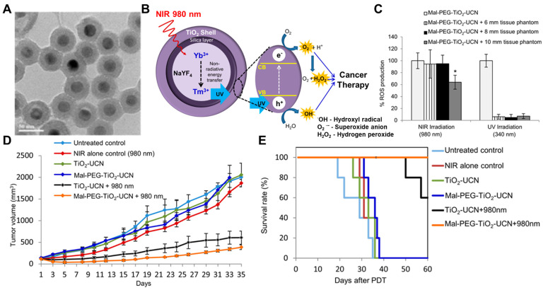
- Lucky, 2015, Titania coated upconversion nanoparticles for near-infrared light triggered photodynamic therapy, ACS Nano, 9, 191, 10.1021/nn503450t · DOI
- Wang, 2011, Near-infrared light induced in vivo photodynamic therapy of cancer based on upconversion nanoparticles, Biomaterials, 32, 6145, 10.1016/j.biomaterials.2011.05.007 · DOI
- Petriev, 2019, Nuclear nanomedicine using Si nanoparticles as safe and effective carriers of 188Re radionuclide for cancer therapy, Sci. Rep., 9, 2017, 10.1038/s41598-018-38474-7 · DOI
- Gong, 2018, Core–shell TaOx@MnO2 nanoparticles as a nano-radiosensitizer for effective cancer radiotherapy, J. Mater. Chem. B, 6, 2250, 10.1039/C8TB00070K · DOI
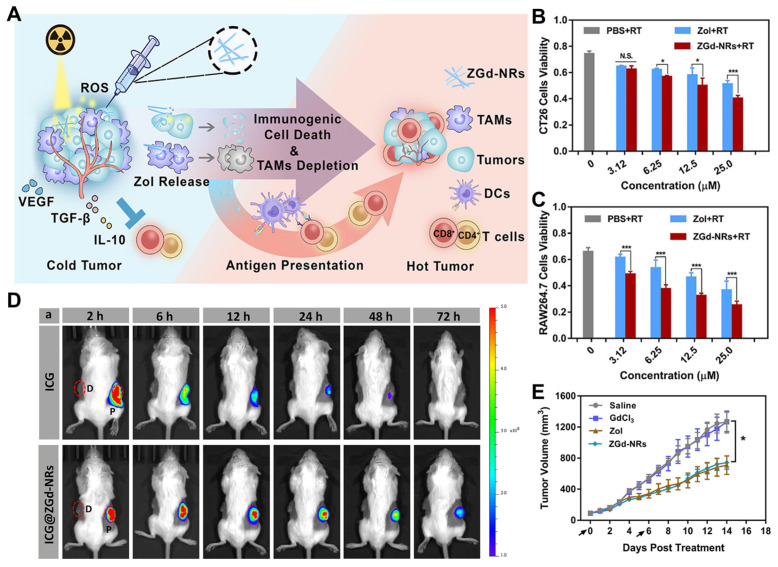
- Huang, 2021, Zoledronic Acid–Gadolinium Coordination Polymer Nanorods for Improved Tumor Radioimmunotherapy by Synergetically Inducing Immunogenic Cell Death and Reprogramming the Immunosuppressive Microenvironment, ACS Nano, 15, 8450, 10.1021/acsnano.0c10764 · DOI
- Guo, 2014, Combinatorial photothermal and immuno cancer therapy using chitosan-coated hollow copper sulfide nanoparticles, ACS Nano, 8, 5670, 10.1021/nn5002112 · DOI
- Noh, 2010, Pegylated poly-l-arginine derivatives of chitosan for effective delivery of siRNA, J. Control. Release, 145, 159, 10.1016/j.jconrel.2010.04.005 · DOI
- Ngamcherdtrakul, 2021, In situ tumor vaccination with nanoparticle co-delivering CpG and STAT3 siRNA to effectively induce whole-body antitumor immune response, Adv. Mater., 33, 2100628, 10.1002/adma.202100628 · DOI
- He, Q., Zhou, L., Yu, D., Zhu, R., Chen, Y., Song, M., Liu, X., Liao, Y., Ding, T., and Fan, W. (2023). Near-Infrared-Activatable PROTAC Nanocages for Controllable Target Protein Degradation and On-Demand Antitumor Therapy. J. Med. Chem. · DOI
- Li, 2023, c-Myc-Targeting PROTAC Based on a TNA-DNA Bivalent Binder for Combination Therapy of Triple-Negative Breast Cancer, J. Am. Chem. Soc., 145, 9334, 10.1021/jacs.3c02619 · DOI
- Deng, 2013, Layer-by-layer nanoparticles for systemic codelivery of an anticancer drug and siRNA for potential triple-negative breast cancer treatment, ACS Nano, 7, 9571, 10.1021/nn4047925 · DOI
- Gao, 2021, Intratumoral administration of thermosensitive hydrogel co-loaded with norcantharidin nanoparticles and doxorubicin for the treatment of hepatocellular carcinoma, Int. J. Nanomed., 16, 4073, 10.2147/IJN.S308057 · DOI
- Kwon, 2016, Synergistic anti-tumor activity through combinational intratumoral injection of an in-situ injectable drug depot, Biomaterials, 85, 232, 10.1016/j.biomaterials.2016.02.001 · DOI
- Ma, 2015, Drug coated microneedles for minimally-invasive treatment of oral carcinomas: Development and in vitro evaluation, Biomed. Microdevices, 17, 44, 10.1007/s10544-015-9944-y · DOI
- Bhatnagar, 2019, Dissolvable microneedle patch containing doxorubicin and docetaxel is effective in 4T1 xenografted breast cancer mouse model, Int. J. Pharm., 556, 263, 10.1016/j.ijpharm.2018.12.022 · DOI
- Li, 2016, Designing hydrogels for controlled drug delivery, Nat. Rev. Mater., 1, 16071, 10.1038/natrevmats.2016.71 · DOI
- Tiwari, 2018, pH/NIR-responsive polypyrrole-functionalized fibrous localized drug-delivery platform for synergistic cancer therapy, ACS Appl. Mater. Interfaces, 10, 20256, 10.1021/acsami.7b17664 · DOI
- Prausnitz, 2004, Microneedles for transdermal drug delivery, Adv. Drug Deliv. Rev., 56, 581, 10.1016/j.addr.2003.10.023 · DOI
- Indermun, 2014, Current advances in the fabrication of microneedles for transdermal delivery, J. Control. Release, 185, 130, 10.1016/j.jconrel.2014.04.052 · DOI
- Chen, 2018, In vitro and in vivo assessment of polymer microneedles for controlled transdermal drug delivery, J. Drug Target., 26, 720, 10.1080/1061186X.2018.1424859 · DOI
- Chen, 2018, Controlled Delivery of Insulin Using Rapidly Separating Microneedles Fabricated from Genipin-Crosslinked Gelatin, Macromol. Rapid Commun., 39, 1800075, 10.1002/marc.201800075 · DOI
- Puolakkainen, 1991, Contact Nd: YAG laser potentiates the tumor cell killing effect of hyperthermia, Lasers Surg. Med., 11, 595, 10.1002/lsm.1900110615 · DOI
- Waldow, 1988, Nd: YAG laser-induced hyperthermia in a mouse tumor model, Lasers Surg. Med., 8, 510, 10.1002/lsm.1900080510 · DOI
- Yun, W.S., Shim, M.K., Lim, S., Song, S., Kim, J., Yang, S., Hwang, H.S., Kim, M.R., Yoon, H.Y., and Lim, D.-K. (2022). Mesenchymal Stem Cell-Mediated Deep Tumor Delivery of Gold Nanorod for Photothermal Therapy. Nanomaterials, 12. · DOI
- Marmor, 1979, Treatment of superficial human neoplasms by local hyperthermia induced by ultrasound, Cancer, 43, 188, 10.1002/1097-0142(197901)43:1<188::AID-CNCR2820430128>3.0.CO;2-7 · DOI
- Seki, 1999, Percutaneous microwave coagulation therapy for patients with small hepatocellular carcinoma: Comparison with percutaneous ethanol injection therapy, Cancer Interdiscip. Int. J. Am. Cancer Soc., 85, 1694
- Ash, 2017, Effect of wavelength and beam width on penetration in light-tissue interaction using computational methods, Lasers Med. Sci., 32, 1909, 10.1007/s10103-017-2317-4 · DOI · Paper page
- Link, 1999, Laser photothermal melting and fragmentation of gold nanorods: Energy and laser pulse-width dependence, J. Phys. Chem. A, 103, 1165, 10.1021/jp983141k · DOI
- Huang, 2006, Selective laser photo-thermal therapy of epithelial carcinoma using anti-EGFR antibody conjugated gold nanoparticles, Cancer Lett., 239, 129, 10.1016/j.canlet.2005.07.035 · DOI
- Ke, 2014, Gold nanoshelled liquid perfluorocarbon nanocapsules for combined dual modal ultrasound/CT imaging and photothermal therapy of cancer, Small, 10, 1220, 10.1002/smll.201302252 · DOI
- Liu, 2015, A plasmonic gold nanostar theranostic probe for in vivo tumor imaging and photothermal therapy, Theranostics, 5, 946, 10.7150/thno.11974 · DOI
- Link, 2005, Simulation of the optical absorption spectra of gold nanorods as a function of their aspect ratio and the effect of the medium dielectric constant, J. Phys. Chem. B, 109, 10531, 10.1021/jp058091f · DOI
- Xu, 2018, PEGylated hydrazided gold nanorods for pH-triggered chemo/photodynamic/photothermal triple therapy of breast cancer, Acta Biomater., 82, 171, 10.1016/j.actbio.2018.10.019 · DOI
- Hwang, 2022, Escherichia coli mimetic gold nanorod-mediated photo-and immunotherapy for treating cancer and its metastasis, ACS Nano, 16, 8472, 10.1021/acsnano.2c03379 · DOI
- Rengan, 2014, Multifunctional gold coated thermo-sensitive liposomes for multimodal imaging and photo-thermal therapy of breast cancer cells, Nanoscale, 6, 916, 10.1039/C3NR04448C · DOI
- Melamed, 2015, Elucidating the fundamental mechanisms of cell death triggered by photothermal therapy, ACS Nano, 9, 6, 10.1021/acsnano.5b00021 · DOI
- Pelaz, 2012, Tailoring the synthesis and heating ability of gold nanoprisms for bioapplications, Langmuir, 28, 8965, 10.1021/la204712u · DOI
- Mitchell, 2015, Dissecting the molecular mechanism of apoptosis during photothermal therapy using gold nanoprisms, ACS Nano, 9, 52, 10.1021/nn505468v · DOI
- Nam, 2018, Chemo-photothermal therapy combination elicits anti-tumor immunity against advanced metastatic cancer, Nat. Commun., 9, 1074, 10.1038/s41467-018-03473-9 · DOI · Paper page
- Ma, 2019, Near-infrared II phototherapy induces deep tissue immunogenic cell death and potentiates cancer immunotherapy, ACS Nano, 13, 11967, 10.1021/acsnano.9b06040 · DOI
- Feng, 2021, A combination strategy based on an Au nanorod/doxorubicin gel via mild photothermal therapy combined with antigen-capturing liposomes and anti-PD-L1 agent promote a positive shift in the cancer-immunity cycle, Acta Biomater., 136, 495, 10.1016/j.actbio.2021.09.052 · DOI
- Zhao, 2021, Recent advances in selective photothermal therapy of tumor, J. Nanobiotechnol., 19, 335, 10.1186/s12951-021-01080-3 · DOI
- Allison, 2013, Photodynamic therapy (PDT): PDT mechanisms, Clin. Endosc., 46, 24, 10.5946/ce.2013.46.1.24 · DOI
- Baptista, 2017, Type I and type II photosensitized oxidation reactions: Guidelines and mechanistic pathways, Photochem. Photobiol., 93, 912, 10.1111/php.12716 · DOI
- Li, 2022, Type I AIE photosensitizers: Mechanism and application, View, 3, 20200121, 10.1002/VIW.20200121 · DOI
- Cheng, 2014, Functional nanomaterials for phototherapies of cancer, Chem. Rev., 114, 10869, 10.1021/cr400532z · DOI
- Huang, 2008, Photodynamic therapy for treatment of solid tumors—Potential and technical challenges, Technol. Cancer Res. Treat., 7, 309, 10.1177/153303460800700405 · DOI
- Chen, 2008, Nanoparticle self-lighting photodynamic therapy for cancer treatment, J. Biomed. Nanotechnol., 4, 369, 10.1166/jbn.2008.001 · DOI
- Deng, 2017, Recent progress in near infrared light triggered photodynamic therapy, Small, 13, 1702299, 10.1002/smll.201702299 · DOI
- Yuan, 2013, Application of near-infrared dyes for tumor imaging, photothermal, and photodynamic therapies, J. Pharm. Sci., 102, 6, 10.1002/jps.23356 · DOI
- Breskey, 2013, Photodynamic therapy: Occupational hazards and preventative recommendations for clinical administration by healthcare providers, Photomed. Laser Surg., 31, 398, 10.1089/pho.2013.3496 · DOI
- Paszko, 2011, Nanodrug applications in photodynamic therapy, Photodiagn. Photodyn. Ther., 8, 14, 10.1016/j.pdpdt.2010.12.001 · DOI
- Gu, 2023, Enzyme-triggered deep tumor penetration of a dual-drug nanomedicine enables an enhanced cancer combination therapy, Bioact. Mater., 26, 102, 10.1016/j.bioactmat.2023.02.015 · DOI
- Song, 2017, Emerging nanotechnology and advanced materials for cancer radiation therapy, Adv. Mater., 29, 1700996, 10.1002/adma.201700996 · DOI
- Martins, 2018, Radioimmunotherapy for delivery of cytotoxic radioisotopes: Current status and challenges, Expert Opin. Drug Deliv., 15, 185, 10.1080/17425247.2018.1378180 · DOI
- Pei, 2021, Biomaterial-mediated internal radioisotope therapy, Mater. Horiz., 8, 1348, 10.1039/D0MH01761B · DOI
- Clement, 2020, Mechanisms for tuning engineered nanomaterials to enhance radiation therapy of cancer, Adv. Sci., 7, 2003584, 10.1002/advs.202003584 · DOI
- Nigro, 1974, Combined therapy for cancer of the anal canal: A preliminary report, Dis. Colon Rectum, 17, 354, 10.1007/BF02586980 · DOI
- Carrick, 2009, Single agent versus combination chemotherapy for metastatic breast cancer, Cochrane Database Syst. Rev., 2, 1
- Mokhtari, 2017, Combination therapy in combating cancer, Oncotarget, 8, 38022, 10.18632/oncotarget.16723 · DOI · Paper page
- Vanneman, 2012, Combining immunotherapy and targeted therapies in cancer treatment, Nat. Rev. Cancer, 12, 237, 10.1038/nrc3237 · DOI
- Esfahani, 2020, A review of cancer immunotherapy: From the past, to the present, to the future, Curr. Oncol., 27, 87, 10.3747/co.27.5223 · DOI
- Wang, 2018, Combining immunotherapy and radiotherapy for cancer treatment: Current challenges and future directions, Front. Pharmacol., 9, 185, 10.3389/fphar.2018.00185 · DOI
- Nam, 2019, Cancer nanomedicine for combination cancer immunotherapy, Nat. Rev. Mater., 4, 398, 10.1038/s41578-019-0108-1 · DOI
- Apetoh, 2015, Combining immunotherapy and anticancer agents: The right path to achieve cancer cure?, Ann. Oncol., 26, 1813, 10.1093/annonc/mdv209 · DOI
- Hu, 2012, Nanoparticle-based combination therapy toward overcoming drug resistance in cancer, Biochem. Pharmacol., 83, 1104, 10.1016/j.bcp.2012.01.008 · DOI
- Xu, 2015, Cancer nanomedicine: From targeted delivery to combination therapy, Trends Mol. Med., 21, 223, 10.1016/j.molmed.2015.01.001 · DOI · Paper page
- Lee, 2016, Delivery strategies and potential targets for siRNA in major cancer types, Adv. Drug Deliv. Rev., 104, 2, 10.1016/j.addr.2016.05.010 · DOI
- Gewirtz, 2007, On future’s doorstep: RNA interference and the pharmacopeia of tomorrow, J. Clin. Investig., 117, 3612, 10.1172/JCI34274 · DOI
- Huang, 2016, Pharmacokinetic behaviors of intravenously administered siRNA in glandular tissues, Theranostics, 6, 1528, 10.7150/thno.15246 · DOI
- Gavrilov, 2012, Therapeutic siRNA: Principles, challenges, and strategies, Yale J. Biol. Med., 85, 187
- Tatiparti, K., Sau, S., Kashaw, S.K., and Iyer, A.K. (2017). siRNA delivery strategies: A comprehensive review of recent developments. Nanomaterials, 7. · DOI
- Oh, 2009, siRNA delivery systems for cancer treatment, Adv. Drug Deliv. Rev., 61, 850, 10.1016/j.addr.2009.04.018 · DOI
- Pan, 2015, New paradigms on siRNA local application, BMB Rep., 48, 147, 10.5483/BMBRep.2015.48.3.089 · DOI
- Zou, 2019, The PROTAC technology in drug development, Cell Biochem. Funct., 37, 21, 10.1002/cbf.3369 · DOI
- Paiva, 2019, Targeted protein degradation: Elements of PROTAC design, Curr. Opin. Chem. Biol., 50, 111, 10.1016/j.cbpa.2019.02.022 · DOI
- Chen, 2022, Proteolysis-targeting chimera (PROTAC) delivery system: Advancing protein degraders towards clinical translation, Chem. Soc. Rev., 51, 5330, 10.1039/D1CS00762A · DOI
- Moon, Y., Jeon, S.I., Shim, M.K., and Kim, K. (2023). Cancer-specific delivery of proteolysis-targeting chimeras (PROTACs) and their application to cancer immunotherapy. Pharmaceutics, 15. · DOI
- Thapa, 2023, Nanomedicine-based commercial formulations: Current developments and future prospects, J. Pharm. Investig., 53, 19, 10.1007/s40005-022-00607-6 · DOI
- Wilson, 2021, Facile bioinspired synthesis of iron oxide encapsulating silica nanocapsules, J. Colloid Interface Sci., 601, 78, 10.1016/j.jcis.2021.05.021 · DOI
- Rodríguez, F., Caruana, P., De la Fuente, N., Español, P., Gámez, M., Balart, J., Llurba, E., Rovira, R., Ruiz, R., and Martín-Lorente, C. (2022). Nano-based approved pharmaceuticals for cancer treatment: Present and future challenges. Biomolecules, 12. · DOI
- Anselmo, 2019, Nanoparticles in the clinic: An update, Bioeng. Transl. Med., 4, e10143, 10.1002/btm2.10143 · DOI · Paper page
- Bonvalot, 2019, NBTXR3, a first-in-class radioenhancer hafnium oxide nanoparticle, plus radiotherapy versus radiotherapy alone in patients with locally advanced soft-tissue sarcoma (Act. In. Sarc): A multicentre, phase 2–3, randomised, controlled trial, Lancet Oncol., 20, 1148, 10.1016/S1470-2045(19)30326-2 · DOI
- Bonvalot, 2017, First-in-human study testing a new radioenhancer using nanoparticles (NBTXR3) activated by radiation therapy in patients with locally advanced soft tissue sarcomas, Clin. Cancer Res., 23, 908, 10.1158/1078-0432.CCR-16-1297 · DOI · Paper page
Cited by
Sorted by citations
Sheng, J., Yuan, W., Zhang, M., Chen, Z., Qi, Y., Zhao, Y., & Zhang, S. (2026) · Journal of Nanobiotechnology · doi:10.1186/s12951-026-04028-7 · Cited by 35
Metadata
- Authors
- Yun, W. S., Kim, J., Lim, D.-K., Kim, D.-H., Jeon, S. I., & Kim, K.
- Venue
- Nanomaterials
- Publisher
- MDPI AG
- Published
- 2023-07-31
- Volume
- 13
- Issue
- 15
- Pages
- 2225
- ISSN
- 2079-4991
- PMC
- PMC10421122
- Cited by
- 76
- References
- 14
- Language
- en
- Funders
- National Research Foundation (NRF) of Korea (NRF-2022M3H4A1A03067401)
About these checks
Registry, retraction, Cites and statistics results are computed, not judged, with one exception: a printed reference that no index lists is read once by a model before it counts as a citation error. Claims are extracted by a model and kept only when their quote appears verbatim in the text; each cited source is then checked, with a model’s help, for whether it says what the claim relies on. Those verdicts can be wrong — read the claim’s quote and the cited source before relying on one.
Statistics and arithmetic are recomputed, not judged.
Confidence bands are shown only for fields calibrated on our annotated test set. None is calibrated yet, so no band is shown.
What counts as a claim ›How we measure confidence ›How we check this ›
Are you an author, or do you see a problem with this page? Request a review
