

The DOI matches its Crossref record; 114 of the 114 references it cites were found in a registry or an index.
The Eye, Oxidative Damage and Polyunsaturated Fatty Acids
Open accessAbstract
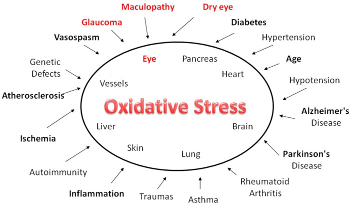
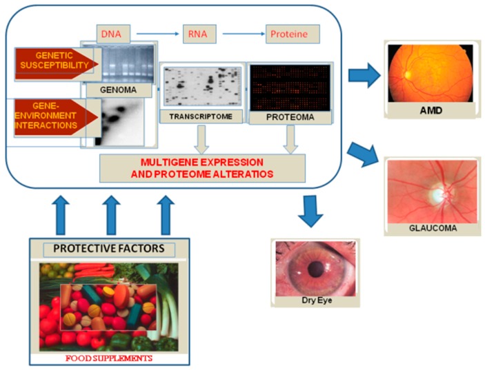
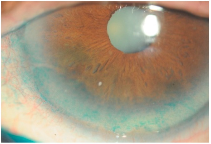
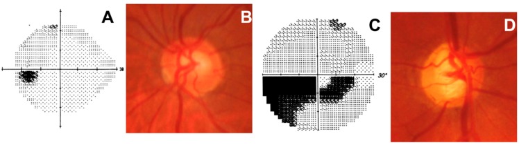
Polyunsaturated fatty acids (PUFA) are known to have numerous beneficial effects, owing to their anti-inflammatory and antioxidant properties. From a metabolic standpoint, the mitochondria play a fundamental role in cellular homeostasis, and oxidative stress can affect their functioning. Indeed, the mitochondria are the main source of ROS, and an imbalance between ROS and antioxidant defenses leads to oxidative stress. In addition, aging, the decline of cellular functions, and continual exposure to light underlie many diseases, particularly those of the eye. Long-term exposure to insults, such as UV light, visible light, ionizing radiation, chemotherapeutics, and environmental toxins, contribute to oxidative damage in ocular tissues and expose the aging eye to considerable risk of pathological consequences of oxidative stress. Ample antioxidant defenses responsible for scavenging free radicals are essential for redox homeostasis in the eye, indeed, eye tissues, starting from the tear film, which normally are exposed to high oxygen levels, have strong antioxidant defenses that are efficient for protecting against ROS-related injuries. On the contrary, instead, the trabecular meshwork is not directly exposed to light and its endothelial cells are poorly equipped with antioxidant defenses. All this makes the eye a target organ of oxidative damage. This review focuses on the role of the polyunsaturated fatty acids in the human eye, particularly in such pathologies as dry eye, glaucoma, and macular degeneration, in which dietary PUFA supplementation can be a valid therapeutic aid.
Overview
The claims and statistics of this paper haven’t been checked yet.
Run the check — free, no account needed. It reads the open-access text and checks the sources it cites.
Integrity signals
Registry
Cites
Statistics
Figures
Verification
Claims the paper makes
The claims and statistics of this paper haven’t been checked yet.
Run the check — free, no account needed. It reads the open-access text and checks the sources it cites.
Claims other papers make about this one
Each is checked against this paper’s own text: does it say what the citing paper says it does?
The check wasn’t confident either way, so it gives no verdict — never disputed.
In the citing paper
“MGF has demonstrated therapeutic benefits in preclinical models of retinal and lens injury through antioxidant and antiangiogenic mechanisms (127).”
From Mangiferin in human disease: Multifaceted mechanisms and applications (Review) (2026) · cites this paper as [127]
Experimental — accuracy not yet measured
Self-check
Percentages, sums and reported differences re-computed from the paper’s own numbers.
The self-check needs the paper’s full text.
Citations
Cites
Free Radicals in the Physiological Control of Cell Function
Dröge, W. (2002) · Physiological Reviews · doi:10.1152/physrev.00018.2001 · Cited by 9,855
Oxidants, oxidative stress and the biology of ageing
Finkel, T., & Holbrook, N. J. (2000) · Nature · doi:10.1038/35041687 · Cited by 9,413
Aging: a theory based on free radical and radiation chemistry
Harman, D. (1956) · Journal of Gerontology · doi:10.1093/geronj/11.3.298 · Cited by 8,567
Cillà, S. D., Farruggio, S., Vujosevic, S., Raina, G., Filippini, D. A., Gatti, V., Clemente, N., Mary, D., Vezzola, D., Casini, G., et al. (2017) · Cellular Physiology and Biochemistry · doi:10.1159/000479441 · Cited by 5,154
Oxyl radicals, redox-sensitive signalling cascades and antioxidants
Genestra, M. (2007) · Cellular Signalling · doi:10.1016/j.cellsig.2007.04.009 · Cited by 4,930
(2007) · The Ocular Surface · doi:10.1016/s1542-0124(12)70081-2 · Cited by 3,299
Baeuerle, P. A., & Baltimore, D. (1996) · Cell · doi:10.1016/s0092-8674(00)81318-5 · Cited by 3,002
Senescent cells, tumor suppression, and organismal aging: good citizens, bad neighbors
Campisi, J. (2005) · Cell · doi:10.1016/j.cell.2005.02.003 · Cited by 2,379
Oxidative stress in Parkinson's disease
Jenner, P. (2003) · Annals of Neurology · doi:10.1002/ana.10483 · Cited by 2,053
The Biologic Clock: The Mitochondria?
Harman, D. (1972) · Journal of the American Geriatrics Society · doi:10.1111/j.1532-5415.1972.tb00787.x · Cited by 2,026
Functional Roles of Fatty Acids and Their Effects on Human Health
Calder, P. C. (2015) · Journal of Parenteral and Enteral Nutrition · doi:10.1177/0148607115595980 · Cited by 1,276
Prevalence of and Risk Factors for Dry Eye Syndrome
Moss, S. E. (2000) · Archives of Ophthalmology · doi:10.1001/archopht.118.9.1264 · Cited by 1,223
Resolvin E1 and protectin D1 activate inflammation-resolution programmes
Schwab, J. M., Chiang, N., Arita, M., & Serhan, C. N. (2007) · Nature · doi:10.1038/nature05877 · Cited by 1,219
Wall, R., Ross, R. P., Fitzgerald, G. F., & Stanton, C. (2010) · Nutrition Reviews · doi:10.1111/j.1753-4887.2010.00287.x · Cited by 1,101
Mechanisms of Action of (n-3) Fatty Acids,
Calder, P. C. (2012) · Journal of Nutrition · doi:10.3945/jn.111.155259 · Cited by 920
Mechanisms of action of docosahexaenoic acid in the nervous system
Salem, N., Litman, B., Kim, H., & Gawrisch, K. (2001) · Lipids · doi:10.1007/s11745-001-0805-6 · Cited by 919 · cited 1×
“Omega-3 docosahexaenoic acid (DHA) is essential for the proper functioning [1] and development of the brain [2].”
BackgroundDietary (n-3) Fatty Acids and Brain Development1
Innis, S. M. (2007) · The Journal of Nutrition · doi:10.1093/jn/137.4.855 · Cited by 906
The role of omega-3 long-chain polyunsaturated fatty acids in health and disease of the retina
SanGiovanni, J. P., & Chew, E. Y. (2004) · Progress in Retinal and Eye Research · doi:10.1016/j.preteyeres.2004.06.002 · Cited by 905
Oxidative stress‐induced cataract: mechanism of action
Spector, A. (1995) · The FASEB Journal · doi:10.1096/fasebj.9.12.7672510 · Cited by 896
Expanding insights of mitochondrial dysfunction in Parkinson's disease
Abou-Sleiman, P. M., Muqit, M. M. K., & Wood, N. W. (2006) · Nature Reviews Neuroscience · doi:10.1038/nrn1868 · Cited by 869
Mitochondrial dysfunction accounts for the stochastic heterogeneity in telomere-dependent senescence
Passos, J. F., Saretzki, G., Ahmed, S., Nelson, G., Richter, T., Peters, H., Wappler, I., Birket, M. J., Harold, G., Schaeuble, K., et al. (2007) · PLoS Biology · doi:10.1371/journal.pbio.0050110 · Cited by 796 · cited 1×
“Senescent cells are associated to a high level of intracellular ROS and display oxidative damage to DNA and proteins [88].”
BackgroundFatty acid composition of brain, retina, and erythrocytes in breast- and formula-fed infants
Makrides, M., Neumann, M., Byard, R. W., Simmer, K. N., & Gibson, R. A. (1994) · American Journal of Clinical Nutrition · doi:10.1093/ajcn/60.2.189 · Cited by 787
Oxidative damage to mitochondrial DNA shows marked age-dependent increases in human brain
Mecocci, P., MacGarvey, U., Kaufman, A. E., Koontz, D., Shoffner, J. M., Wallace, D. C., & Beal, M. F. (1993) · Annals of Neurology · doi:10.1002/ana.410340416 · Cited by 757 · cited 2×
“Moreover, high levels of 8-OHdG have been found in nuclear DNA (nDNA) and in mtDNA in post mortem brains of elderly subjects [42], and in trabecular mtDNA this damage has been seen to be greater in glaucoma patients than in healthy subjects.”
BackgroundThies, F., Garry, J. M., Yaqoob, P., Rerkasem, K., Williams, J., Shearman, C. P., Gallagher, P. J., Calder, P. C., & Grimble, R. F. (2003) · The Lancet · doi:10.1016/s0140-6736(03)12468-3 · Cited by 739 · cited 2×
“Supplementation with polyunsaturated fatty acids, in addition to reducing inflammation [53], can improve the lipid layer of the lacri al film and normalize the function of the meibomian glands and lacrimal gland [54].”
BackgroundOxidative DNA damage and senescence of human diploid fibroblast cells.
Chen, Q., Fischer, A., Reagan, J. D., Yan, L. J., & Ames, B. N. (1995) · Proceedings of the National Academy of Sciences · doi:10.1073/pnas.92.10.4337 · Cited by 720
Barja, G. (2004) · Trends in Neurosciences · doi:10.1016/j.tins.2004.07.005 · Cited by 653
Consequences of oxidative stress in age-related macular degeneration
Jarrett, S. G., & Boulton, M. E. (2012) · Molecular Aspects of Medicine · doi:10.1016/j.mam.2012.03.009 · Cited by 549
The Mechanisms of UV Mutagenesis
Ikehata, H., & Ono, T. (2011) · Journal of Radiation Research · doi:10.1269/jrr.10175 · Cited by 542
Oxidative stress and protein aggregation during biological aging
Squier, T. C. (2001) · Experimental Gerontology · doi:10.1016/s0531-5565(01)00139-5 · Cited by 519
Hastürk, H., Kantarcı, A., Goguet, E., Blackwood, A., Andry, C., Serhan, C. N., & Dyke, T. E. V. (2007) · The Journal of Immunology · doi:10.4049/jimmunol.179.10.7021 · Cited by 470
Resolvins and protectins in the termination program of acute inflammation
Ariel, A., & Serhan, C. N. (2007) · Trends in Immunology · doi:10.1016/j.it.2007.02.007 · Cited by 386
Oxidative DNA Damage in the Human Trabecular Meshwork
Saccà, S. C. (2005) · Archives of Ophthalmology · doi:10.1001/archopht.123.4.458 · Cited by 377
Self-citationCell survival matters: docosahexaenoic acid signaling, neuroprotection and photoreceptors
Bazan, N. G. (2006) · Trends in Neurosciences · doi:10.1016/j.tins.2006.03.005 · Cited by 353
The cAMP Pathway as Therapeutic Target in Autoimmune and Inflammatory Diseases
Raker, V., Becker, C., & Steinbrink, K. (2016) · Frontiers in Immunology · doi:10.3389/fimmu.2016.00123 · Cited by 330
Evidence of Oxidative Stress in the Pathogenesis of Fuchs Endothelial Corneal Dystrophy
Jurkunas, U. V., Bitar, M. S., Funaki, T., & Azizi, B. (2010) · American Journal Of Pathology · doi:10.2353/ajpath.2010.100279 · Cited by 329
Frippiat, C. C., Chen, Q. M., Zdanov, S., Magalhaes, J.-P., Remacle, J., & Toussaint, O. (2001) · Journal of Biological Chemistry · doi:10.1074/jbc.m006809200 · Cited by 316
Oxidative deoxyribonucleic acid damage in the eyes of glaucoma patients
Izzotti, A., Saccà, S. C., Cartiglia, C., & Flora, S. D. (2003) · The American Journal of Medicine · doi:10.1016/s0002-9343(03)00114-1 · Cited by 304
Self-citationChong, E. W.-T. (2008) · Archives of Ophthalmology · doi:10.1001/archopht.126.6.826 · Cited by 279
n−3 Fatty Acid Supplementation for the Treatment of Dry Eye Disease
The Dry Eye Assessment and Management Study Research Group (2018) · New England Journal of Medicine · doi:10.1056/nejmoa1709691 · Cited by 274
Glaucomatous outflow pathway and oxidative stress
Saccà, S. C., Izzotti, A., Rossi, P., & Traverso, C. E. (2006) · Experimental Eye Research · doi:10.1016/j.exer.2006.10.008 · Cited by 269
Self-citationIs docosahexaenoic acid synthesis from α-linolenic acid sufficient to supply the adult brain?
Domenichiello, A. F., Kitson, A. P., & Bazinet, R. P. (2015) · Progress in Lipid Research · doi:10.1016/j.plipres.2015.04.002 · Cited by 268
Wang, N., Chintala, S. K., Fini, M. E., & Schuman, J. S. (2001) · Nature Medicine · doi:10.1038/85446 · Cited by 238
Vidgren, H. M., Ågren, J. J., Schwab, U., Rissanen, T., Hänninen, O., & Uusitupa, M. I. J. (1997) · Lipids · doi:10.1007/s11745-997-0089-x · Cited by 236 · cited 1×
“They are preferentially incorporated as structural components into membrane phospholipids [6].”
BackgroundLepretti, M., Martucciello, S., Burgos Aceves, M., Putti, R., & Lionetti, L. (2018) · Nutrients · doi:10.3390/nu10030350 · Cited by 232
Alterations in microRNA expression in stress-induced cellular senescence
Li, G., Luna, C., Qiu, J., Epstein, D. L., & González, P. (2009) · Mechanisms of Ageing and Development · doi:10.1016/j.mad.2009.09.002 · Cited by 218
Endothelial dysfunction in glaucoma
Resch, H., Garhöfer, G., Fuchsjäger‐Mayrl, G., Hommer, A., & Schmetterer, L. F. (2008) · Acta Ophthalmologica · doi:10.1111/j.1755-3768.2007.01167.x · Cited by 206
Cellular senescence in the glaucomatous outflow pathway
Liton, P. B., Challa, P., Stinnett, S. S., Luna, C., Epstein, D. L., & González, P. (2005) · Experimental Gerontology · doi:10.1016/j.exger.2005.06.005 · Cited by 198
Diet impact on mitochondrial bioenergetics and dynamics
Putti, R., Sica, R., Migliaccio, V., & Lionetti, L. (2015) · Frontiers in Physiology · doi:10.3389/fphys.2015.00109 · Cited by 192
Gibson, N. J., & Brown, M. F. (1993) · Biochemistry · doi:10.1021/bi00060a040 · Cited by 178
Borow, K. M., Nelson, J. R., & Mason, R. P. (2015) · Atherosclerosis · doi:10.1016/j.atherosclerosis.2015.07.035 · Cited by 175
Involvement of Oxidative Stress on Corneal Epithelial Alterations in a Blink-Suppressed Dry Eye
Nakamura, S., Shibuya, M., Nakashima, H., Hisamura, R., Masuda, N., Imagawa, T., Uehara, M., & Tsubota, K. (2007) · Investigative Ophthalmology & Visual Science · doi:10.1167/iovs.06-1027 · Cited by 174
Unesterified docosahexaenoic acid is protective in neuroinflammation
Orr, S. K., Palumbo, S., Bosetti, F., Mount, H. T. J., Kang, J. X., Greenwood, C. E., David, W. L., Serhan, C. N., & Bazinet, R. P. (2013) · Journal of Neurochemistry · doi:10.1111/jnc.12392 · Cited by 173
Deinema, L. A., Vingrys, A. J., Wong, C. Y., Jackson, D. C., Chinnery, H. R., & Downie, L. E. (2017) · Ophthalmology · doi:10.1016/j.ophtha.2016.09.023 · Cited by 160
Protein oxidation and degradation during proliferative senescence of human MRC-5 fibroblasts
Sitte, N., Merker, K., von Zglinicki, T., & Grune, T. (2000) · Free Radical Biology and Medicine · doi:10.1016/s0891-5849(99)00279-8 · Cited by 153
From DNA damage to functional changes of the trabecular meshwork in aging and glaucoma
Saccà, S. C., Gandolfi, S., Bagnis, A., Manni, G., Damonte, G., Traverso, C. E., & Izzotti, A. (2016) · Ageing Research Reviews · doi:10.1016/j.arr.2016.05.012 · Cited by 141
Self-citationThe Dysfunction of the Trabecular Meshwork During Glaucoma Course
Saccà, S. C., Pulliero, A., & Izzotti, A. (2014) · Journal of Cellular Physiology · doi:10.1002/jcp.24826 · Cited by 130
Self-citationRoles and mechanisms of cellular senescence in regulation of tissue homeostasis
Ohtani, N., & Hara, E. (2013) · Cancer Science · doi:10.1111/cas.12118 · Cited by 122
Glutathione in calf trabecular meshwork and its relation to aqueous humor outflow facility.
Kahn, M. G., Giblin, F. J., & Epstein, D. L. (1983) · PubMed · Cited by 121
Pro- and antiarrhythmic properties of a diet rich in fish oil
den Ruijter, H. M., Berecki, G., Opthof, T., Verkerk, A. O., Zock, P. L., & Coronel, R. (2006) · Cardiovascular Research · doi:10.1016/j.cardiores.2006.06.014 · Cited by 118
Peripheral endothelial dysfunction in normal pressure glaucoma.
Henry, E., Newby, D. E., Webb, D. J., & O'Brien, C. J. (1999) · PubMed · Cited by 116
Aging and protein oxidative damage
Agarwal, S., & Sohal, R. S. (1994) · Mechanisms of Ageing and Development · doi:10.1016/0047-6374(94)90024-8 · Cited by 116
Plausible explanations for effects of long chain polyunsaturated fatty acids (LCPUFA) on neonates
Kurlak, L. O., & Stephenson, T. J. (1999) · Archives of Disease in Childhood Fetal & Neonatal · doi:10.1136/fn.80.2.f148 · Cited by 116
Izzotti, A., Longobardi, M., Cartiglia, C., & Saccà, S. C. (2011) · PLoS ONE · doi:10.1371/journal.pone.0014567 · Cited by 113
Self-citationN-Docosahexaenoylethanolamine: A neurotrophic and neuroprotective metabolite of docosahexaenoic acid
Kim, H., & Spector, A. A. (2018) · Molecular Aspects of Medicine · doi:10.1016/j.mam.2018.03.004 · Cited by 112
TXNIP shuttling: missing link between oxidative stress and inflammasome activation
Lane, T., Flam, B. R., Lockey, R. F., & Kolliputi, N. (2013) · Frontiers in Physiology · doi:10.3389/fphys.2013.00050 · Cited by 110
Ding, G., Franki, N., Kapasi, A. A., Reddy, K., Gibbons, N., & Singhal, P. C. (2001) · Experimental and Molecular Pathology · doi:10.1006/exmp.2000.2346 · Cited by 103
Oral omega-3 fatty acids treatment in computer vision syndrome related dry eye
Bhargava, R., Kumar, P., Phogat, H., Kaur, A., & Kumar, M. (2015) · Contact Lens and Anterior Eye · doi:10.1016/j.clae.2015.01.007 · Cited by 103
Saccà, S. C., Roszkowska, A. M., & Izzotti, A. (2013) · Mutation Research/Reviews in Mutation Research · doi:10.1016/j.mrrev.2013.01.001 · Cited by 100
Self-citationProteasome inhibition by chronic oxidative stress in human trabecular meshwork cells
Caballero, M., Liton, P. B., Epstein, D. L., & González, P. (2003) · Biochemical and Biophysical Research Communications · doi:10.1016/s0006-291x(03)01385-8 · Cited by 98
Mediterranean diet reduces senescence-associated stress in endothelial cells
Marin, C., Delgado-Lista, J., Ramirez, R., Carracedo, J., Caballero, J., Perez-Martinez, P., Gutierrez-Mariscal, F. M., Garcia-Rios, A., Delgado-Casado, N., Cruz-Teno, C., et al. (2012) · AGE · doi:10.1007/s11357-011-9305-6 · Cited by 96
Dyall, S. C. (2017) · Lipids · doi:10.1007/s11745-017-4292-8 · Cited by 95
Effect of age on superoxide dismutase activity of human trabecular meshwork.
Paz, M. A. D. L., & Epstein, D. L. (1996) · PubMed · Cited by 94
New Proteins as Vascular Biomarkers in Primary Open Angle Glaucomatous Aqueous Humor
Saccà, S. C., Centofanti, M., & Izzotti, A. (2012) · Investigative Ophthalmology & Visual Science · doi:10.1167/iovs.11-8902 · Cited by 87
Self-citationThe Outflow Pathway: A Tissue With Morphological and Functional Unity
Saccà, S. C., Gandolfi, S., Bagnis, A., Manni, G., Damonte, G., Traverso, C. E., & Izzotti, A. (2016) · Journal of Cellular Physiology · doi:10.1002/jcp.25305 · Cited by 83
Self-citationJi, J. (2014) · Medical Science Monitor · doi:10.12659/msm.891364 · Cited by 83
Choi, S., Kim, T., Kim, K. S., Kim, B.-Y., Ahn, S.-Y., Cho, H.-J., Lee, H. K., Cho, H., & Kim, E. K. (2009) · American Journal Of Pathology · doi:10.2353/ajpath.2009.081001 · Cited by 82
Kim, H., & Spector, A. A. (2012) · Prostaglandins Leukotrienes and Essential Fatty Acids · doi:10.1016/j.plefa.2012.08.002 · Cited by 81
Nutritional influences on visual development and function
Lien, E. L., & Hammond, B. R. (2011) · Progress in Retinal and Eye Research · doi:10.1016/j.preteyeres.2011.01.001 · Cited by 80
Cejkova, J., Ardan, T., Šimonová, Z., Čejka, Č., Malec, J., Dotřelová, D., & Brùnová, B. (2008) · PubMed · doi:10.14670/hh-23.1477 · Cited by 79
Nutrigenomics of ω-3 fatty acids: Regulators of the master transcription factors
Rodríguez‐Cruz, M., & Serna, D. S. (2017) · Nutrition · doi:10.1016/j.nut.2017.04.012 · Cited by 78
Lipid peroxidation in open‐angle glaucoma
Babizhayev, M. A., & Bunin, A. Y. (1989) · Acta Ophthalmologica · doi:10.1111/j.1755-3768.1989.tb01617.x · Cited by 75
Rieusset, J. (2015) · Diabetes & Metabolism · doi:10.1016/j.diabet.2015.02.006 · Cited by 69
Effects of Dietary Polyunsaturated Fatty Acids on Mitochondria
Rohrbach, S. (2009) · Current Pharmaceutical Design · doi:10.2174/138161209789909692 · Cited by 69
A Randomized Controlled Trial of Omega 3 Fatty Acids in Rosacea Patients with Dry Eye Symptoms
Bhargava, R., Chandra, M., Bansal, U., Singh, D., Ranjan, S., & Sharma, S. (2016) · Current Eye Research · doi:10.3109/02713683.2015.1122810 · Cited by 67
Black, A. T., Gordon, M. K., Heck, D. E., Gallo, M. A., Laskin, D. L., & Laskin, J. D. (2011) · Biochemical Pharmacology · doi:10.1016/j.bcp.2011.01.014 · Cited by 64
Focus on molecular events in the anterior chamber leading to glaucoma
Saccà, S. C., & Izzotti, A. (2013) · Cellular and Molecular Life Sciences · doi:10.1007/s00018-013-1493-z · Cited by 62
Self-citationTGF-β2 Induces Senescence-Associated Changes in Human Trabecular Meshwork Cells
Yu, A. L., Birke, K., Moriniere, J., & Welge‐Lüßen, U. (2010) · Investigative Ophthalmology & Visual Science · doi:10.1167/iovs.10-5679 · Cited by 60
Investigation of oxidative stress in pterygium tissue.
Balci, M., Şahin, Ş., Mutlu, F. M., Yağcı, R., Karanci, P., & Yildiz, M. (2011) · PubMed Central · Cited by 58
McCann, P., Hogg, R. E., Fallis, R., & Azuara‐Blanco, A. (2016) · Investigative Ophthalmology & Visual Science · doi:10.1167/iovs.15-18595 · Cited by 57
Schnebelen, C., Pasquis, B., Salinas‐Navarro, M., Joffre, C., Creuzot-Garcher, C. P., Vidal‐Sanz, M., Bron, A. M., Brétillon, L., & Acar, N. (2009) · Graefe s Archive for Clinical and Experimental Ophthalmology · doi:10.1007/s00417-009-1094-6 · Cited by 56
Cohen, E., Kramer, M., Shochat, T., Goldberg, E., Garty, M., & Krause, I. (2016) · Journal of Glaucoma · doi:10.1097/ijg.0000000000000374 · Cited by 56
Yog, R., Barhoumi, R., McMurray, D. N., & Chapkin, R. S. (2010) · The Journal of Immunology · doi:10.4049/jimmunol.0904102 · Cited by 55
Mitochondria in Aging and Alzheimer's Disease
Crouch, P. J., Cimdins, K., Duce, J. A., Bush, A. I., & Trounce, I. A. (2007) · Rejuvenation Research · doi:10.1089/rej.2007.0592 · Cited by 54
Sidhu, V. K., Huang, B. X., & Kim, H. (2011) · Journal of Proteome Research · doi:10.1021/pr2007285 · Cited by 53
Cell Senescence in Human Aging and Disease
Fossel, M. (2002) · Annals of the New York Academy of Sciences · doi:10.1111/j.1749-6632.2002.tb02078.x · Cited by 49
Docosahexaenoic acid, protectins and dry eye
Cortina, M. S., & Bazán, H. E. P. (2010) · Current Opinion in Clinical Nutrition & Metabolic Care · doi:10.1097/mco.0b013e328342bb1a · Cited by 48
Kaarniranta, K., & Salminen, A. (2009) · Experimental Gerontology · doi:10.1016/j.exger.2009.09.002 · Cited by 45
The Role of Diet in Glaucoma: A Review of the Current Evidence
Owaifeer, A. M. A., & Taisan, A. A. A. (2018) · Ophthalmology and Therapy · doi:10.1007/s40123-018-0120-3 · Cited by 43
Endothelial senescence after high-cholesterol, high-fat diet challenge in baboons
Shi, Q., Hubbard, G. B., Kushwaha, R. S., Rainwater, D., Thomas, C. A., Leland, M. M., VandeBerg, J. L., & Wang, X. L. (2007) · American Journal of Physiology-Heart and Circulatory Physiology · doi:10.1152/ajpheart.01405.2006 · Cited by 43
Senescent Phenotype of Trabecular Meshwork Cells Displays Biomarkers in Primary Open-Angle Glaucoma
Babizhayev, M. A., & Yegorov, Y. E. (2011) · Current Molecular Medicine · doi:10.2174/156652411800615126 · Cited by 42
Zhu, W. J., Wu, Y. T., Li, G., Wang, J., & Li, X. (2014) · Nutrition Reviews · doi:10.1111/nure.12145 · Cited by 40
Wang, Y. X., Xu, L., Zhang, X. H., You, Q. S., Zhao, L., & Jonas, J. B. (2013) · PLoS ONE · doi:10.1371/journal.pone.0077180 · Cited by 40
Tourtas, T., Birke, M. T., Kruse, F. E., Welge-Lüßen, U.-C., & Birke, K. (2012) · PLoS ONE · doi:10.1371/journal.pone.0031340 · Cited by 40
Borlon, C., Debacq-Chainiaux, F., Hinrichs, C., Scharffetter-Kochanek, K., Toussaint, O., & Wlaschek, M. (2007) · Experimental Gerontology · doi:10.1016/j.exger.2007.04.009 · Cited by 35
Russell, P., & Johnson, D. H. (1996) · Journal of Glaucoma · doi:10.1097/00061198-199610000-00006 · Cited by 33
Light, lipids and photoreceptor survival: live or let die?
German, O. L., Agnolazza, D. L., Politi, L. E., & Rotstein, N. P. (2015) · Photochemical & Photobiological Sciences · doi:10.1039/c5pp00194c · Cited by 31
Impact of nutrition on pollutant toxicity: an update with new insights into epigenetic regulation
Hoffman, J. B., Petriello, M. C., & Hennig, B. (2017) · Reviews on Environmental Health · doi:10.1515/reveh-2016-0041 · Cited by 30
Age-related macular degeneration
Holz, F. G., Pauleikhoff, D., Spaide, R. F., & Bird, A. C. (2004) · Springer eBooks · doi:10.1007/978-3-662-05199-3 · Cited by 29
Othman, K. R. B., Cercy, C., Amri, M., Doly, M., & Ranchon‐Cole, I. (2015) · PLoS ONE · doi:10.1371/journal.pone.0128395 · Cited by 28
Kinetics of Proton Binding to the Sarcoplasmic Reticulum Ca-ATPase in the E1 State
Fibich, A., Janko, K., & Apell, H. (2007) · Biophysical Journal · doi:10.1529/biophysj.107.110791 · Cited by 22
Gene–environment interactions in non-cancer degenerative diseases
Izzotti, A. (2009) · Mutation research. Fundamental and molecular mechanisms of mutagenesis · doi:10.1016/j.mrfmmm.2009.06.001 · Cited by 20
Effects of atorvastatin on porcine aqueous humour outflow and trabecular meshwork cells
Cong, L., Fu, S., Zhang, J., Zhao, J., & Zhang, Y. (2017) · Experimental and Therapeutic Medicine · doi:10.3892/etm.2017.5353 · Cited by 17
Phase 2 Protein Inducers in the Diet Promote Healthier Aging
Noyan-Ashraf, M. H., Sadeghinejad, Z., Davies, G. F., Ross, A. R. S., Saucier, D. M., Harkness, T. A. A., & Juurlink, B. H. J. (2008) · The Journals of Gerontology Series A · doi:10.1093/gerona/63.11.1168 · Cited by 7
Cornelli, U., Berra, B., & Frega, N. G. (2010) · Progress in nutrition · Cited by 1
Research works printed but not linked: 1
- Cornelli, 2010, I Modulatori Fisiologici, Prog. Nutr., 12, 99
Cited by
Sorted by citations
Oxidative stress in the eye and its role in the pathophysiology of ocular diseases
Böhm, E. W., Buonfiglio, F., Voigt, A. M., Bachmann, P., Safi, T., Pfeiffer, N., & Gericke, A. (2023) · Redox Biology · doi:10.1016/j.redox.2023.102967 · Cited by 334
The Pathomechanism, Antioxidant Biomarkers, and Treatment of Oxidative Stress-Related Eye Diseases
Hsueh, Y., Chen, Y., Tsao, Y., Cheng, C., Wu, W., & Chen, H. (2022) · International Journal of Molecular Sciences · doi:10.3390/ijms23031255 · Cited by 199
Zhao, T., Guo, X., & Sun, Y. (2021) · Aging and Disease · doi:10.14336/ad.2020.0912 · Cited by 180
Anti-oxidative and anti-inflammatory micelles: break the dry eye vicious cycle
Li, S., Lu, Z., Huang, Y., Wang, Y., Jin, Q., Shentu, X., Ye, J., Ji, J., Yao, K., & Han, H. (2022) · Advanced Science · doi:10.1002/advs.202200435 · Cited by 174
Mechanistic aspects of carotenoid health benefits – where are we now?
Bohn, T., Bonet, M. L., Borel, P., Keijer, J., Landrier, J., Milisav, I., Ribot, J., Riso, P., Winklhofer‐Roob, B. M., Sharoni, Y., et al. (2021) · Nutrition Research Reviews · doi:10.1017/s0954422421000147 · Cited by 155
Metadata
- Authors
- Saccà, S. C., Cutolo, C. A., Ferrari, D., Corazza, P., & Traverso, C. E.
- Affiliations
- Ospedale Policlinico San Martino (IT)
- Corresponding author
- Saccà, Sergio Claudio
- Venue
- Nutrients
- Publisher
- MDPI AG
- Published
- 2018-05-24
- Volume
- 10
- Issue
- 6
- Pages
- 668
- ISSN
- 2072-6643 (ISSN-L)
- PMC
- PMC6024720
- OpenAlex
- W2804346130
- Cited by
- 150
- References
- 114
- Citations by year
- 2026: 18 · 2025: 15 · 2024: 26 · 2023: 20 · 2022: 18 · 2021: 24 · 2020: 17 · 2019: 10 · 2018: 2
- FWCI
- 1.70
- Type
- journal article
- Language
- en
- Open access
- gold
- doi.org
- Licence
- Locations
- Nutrients (publishedVersion, cc-by)
- Europe PMC (PubMed Central) (publishedVersion, cc-by)
- PubMed Central (publishedVersion, cc-by)
- PubMed (publishedVersion)
- DOAJ (DOAJ: Directory of Open Access Journals) (submittedVersion)
- CINECA IRIS Institutial Research Information System (University of Genoa) (submittedVersion, other-oa)
- Topics
- Health Sciences › Nursing › Nutrition and Dietetics › Fatty Acid Research and Health
- Health Sciences › Medicine › Ophthalmology › Retinal Diseases and Treatments
- Health Sciences › Medicine › Biochemistry (medical) › Antioxidant Activity and Oxidative Stress
- Keywords
- polyunsaturated fatty acids, oxidative stress, reactive oxygen species, eye, antioxidants, mitochondrial dysfunction, dry eye, glaucoma, macular degeneration, dietary supplementation, ocular aging, light exposure, trabecular meshwork, redox homeostasis
- MeSH terms
- Animals, Antioxidants, Eye, Eye Diseases, Fatty Acids, Unsaturated, Humans, Risk Factors, Reactive Oxygen Species, Oxidative Stress, Dietary Supplements, Protective Factors
About these checks
Registry, retraction, Cites and statistics results are computed, not judged, with one exception: a printed reference that no index lists is read once by a model before it counts as a citation error. Claims are extracted by a model and kept only when their quote appears verbatim in the text; each cited source is then checked, with a model’s help, for whether it says what the claim relies on. Those verdicts can be wrong — read the claim’s quote and the cited source before relying on one.
Statistics and arithmetic are recomputed, not judged.
Confidence bands are shown only for fields calibrated on our annotated test set. None is calibrated yet, so no band is shown.
What counts as a claim ›How we measure confidence ›How we check this ›
Are you an author, or do you see a problem with this page? Request a review
