

The DOI matches its Crossref record; 271 of the 271 references it cites were found in a registry or an index.
Three-Dimensional Spheroids as In Vitro Preclinical Models for Cancer Research
Open accessAbstract
Most cancer biologists still rely on conventional two-dimensional (2D) monolayer culture techniques to test in vitro anti-tumor drugs prior to in vivo testing. However, the vast majority of promising preclinical drugs have no or weak efficacy in real patients with tumors, thereby delaying the discovery of successful therapeutics. This is because 2D culture lacks cell–cell contacts and natural tumor microenvironment, important in tumor signaling and drug response, thereby resulting in a reduced malignant phenotype compared to the real tumor. In this sense, three-dimensional (3D) cultures of cancer cells that better recapitulate in vivo cell environments emerged as scientifically accurate and low cost cancer models for preclinical screening and testing of new drug candidates before moving to expensive and time-consuming animal models. Here, we provide a comprehensive overview of 3D tumor systems and highlight the strategies for spheroid construction and evaluation tools of targeted therapies, focusing on their applicability in cancer research. Examples of the applicability of 3D culture for the evaluation of the therapeutic efficacy of nanomedicines are discussed.
Overview
The claims and statistics of this paper haven’t been checked yet.
Run the check — free, no account needed. It reads the open-access text and checks the sources it cites.
Integrity signals
Registry
Cites
Statistics
Figures
Verification
Claims the paper makes
The claims and statistics of this paper haven’t been checked yet.
Run the check — free, no account needed. It reads the open-access text and checks the sources it cites.
Claims other papers make about this one
Each is checked against this paper’s own text: does it say what the citing paper says it does?
The cited source says what the claim attributes to it.
In the citing paper
“Despite their widespread use, conventional two-dimensional monolayer cultures fail to reproduce these complexities, contributing to the poor translational predictability of many preclinical candidates.”
From Modeling Tumor Microenvironment Complexity In Vitro: Spheroids as Physiologically Relevant Tumor Models and Strategies for Their Analysis (2025) · cites this paper as [6]
Self-check
Percentages, sums and reported differences re-computed from the paper’s own numbers.
The self-check needs the paper’s full text.
Citations
Cites
1 retracted
Jin, H., Gui, R., Sun, J., et al. (2017) · Talanta · doi:10.1016/j.talanta.2017.08.043 · Cited by 24
RetractedHallmarks of Cancer: The Next Generation
Hanahan, D., & Weinberg, R. A. (2011) · Cell · doi:10.1016/j.cell.2011.02.013 · Cited by 66,590
Microenvironmental regulation of tumor progression and metastasis
Quail, D. F., & Joyce, J. A. (2013) · Nature Medicine · doi:10.1038/nm.3394 · Cited by 7,994
A framework for advancing our understanding of cancer-associated fibroblasts
Sahai, E., Astsaturov, I., Cukierman, E., DeNardo, D. G., Egeblad, M., Evans, R. M., Fearon, D., Greten, F. R., Hingorani, S. R., Hunter, T., et al. (2020) · Nature Reviews Cancer · doi:10.1038/s41568-019-0238-1 · Cited by 3,863 · cited 2×
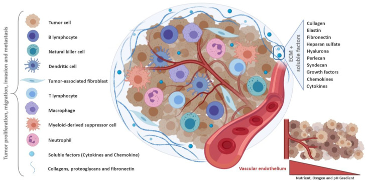
“In this context, CAFs contribute to tumor cell invasion, as well as to changes in tumor growth and immune microenvironment, through ECM remodeling and production of soluble factors [21,22].”
BackgroundThe Tumor Microenvironment Innately Modulates Cancer Progression
Hinshaw, D. C., & Shevde, L. A. (2019) · Cancer Research · doi:10.1158/0008-5472.can-18-3962 · Cited by 3,270
Regulation of transport pathways in tumor vessels: Role of tumor type and microenvironment
Hobbs, S. K., Monsky, W. L., Yuan, F., Roberts, W. G., Griffith, L., Torchilin, V. P., & Jain, R. K. (1998) · Proceedings of the National Academy of Sciences · doi:10.1073/pnas.95.8.4607 · Cited by 2,438
Optical Sectioning Deep Inside Live Embryos by Selective Plane Illumination Microscopy
Huisken, J., Swoger, J., Bene, F. D., Wittbrodt, J., & Stelzer, E. H. K. (2004) · Science · doi:10.1126/science.1100035 · Cited by 2,370
Roles of the immune system in cancer: From tumor initiation to metastatic progression
Gonzalez, H., Hagerling, C., & Werb, Z. (2018) · Genes & Development · doi:10.1101/gad.314617.118 · Cited by 2,111
Role of hypoxia in cancer therapy by regulating the tumor microenvironment
Jing, X., Yang, F., Shao, C., Wei, K., Xie, M., Shen, H., & Shu, Y. (2019) · Molecular Cancer · doi:10.1186/s12943-019-1089-9 · Cited by 2,054
Cancer immunoediting and resistance to T cell-based immunotherapy
O’Donnell, J. S., Teng, M. W. L., & Smyth, M. J. (2018) · Nature Reviews Clinical Oncology · doi:10.1038/s41571-018-0142-8 · Cited by 1,826
Tumor microenvironment and therapeutic response
Wu, T., & Dai, Y. (2017) · Cancer Letters · doi:10.1016/j.canlet.2016.01.043 · Cited by 1,802
Deconstructing the third dimension – how 3D culture microenvironments alter cellular cues
Baker, B. M., & Chen, C. S. (2012) · Journal of Cell Science · doi:10.1242/jcs.079509 · Cited by 1,760
Modeling Tissue Morphogenesis and Cancer in 3D
Yamada, K. M., & Cukierman, E. (2007) · Cell · doi:10.1016/j.cell.2007.08.006 · Cited by 1,751
Spheroid-based drug screen: Considerations and practical approach
Friedrich, J., Seidel, C., Ebner, R., & Kunz-Schughart, L. A. (2009) · Nature Protocols · doi:10.1038/nprot.2008.226 · Cited by 1,742
Multicellular tumor spheroids: An underestimated tool is catching up again
Hirschhaeuser, F., Menne, H., Dittfeld, C., West, J., Mueller-Klieser, W., & Kunz-Schughart, L. A. (2010) · Journal of Biotechnology · doi:10.1016/j.jbiotec.2010.01.012 · Cited by 1,674
Prabhakar, U., Maeda, H., Jain, R. K., Sevick-Muraca, E. M., Zamboni, W. C., Farokhzad, O. C., Barry, S. T., Gabizón, A., Grodzinski, P. A., & Blakey, D. C. (2013) · Cancer Research · doi:10.1158/0008-5472.can-12-4561 · Cited by 1,493
Lin, Y., Xu, J., & Lan, H. (2019) · Journal of Hematology & Oncology · doi:10.1186/s13045-019-0760-3 · Cited by 1,398
Pitt, J. M., Marabelle, A., Eggermont, A. M. M., Soria, J., Kroemer, G., & Zitvogel, L. (2016) · Annals of Oncology · doi:10.1093/annonc/mdw168 · Cited by 1,355
Can Animal Models of Disease Reliably Inform Human Studies?
van der Worp, H. B., Howells, D. W., Sena, E. S., Porritt, M. J., Rewell, S., O'Collins, V., & Macleod, M. R. (2010) · PLoS Medicine · doi:10.1371/journal.pmed.1000245 · Cited by 1,354
3D Cell Culture Systems: Advantages and Applications
Ravi, M., Paramesh, V., Kaviya, S. R., Anuradha, E., & Solomon, F. D. P. (2015) · Journal of Cellular Physiology · doi:10.1002/jcp.24683 · Cited by 1,206
Spherical Cancer Models in Tumor Biology
Weiswald, L., Bellet, D. H., & Dangles‐Marie, V. (2015) · Neoplasia · doi:10.1016/j.neo.2014.12.004 · Cited by 1,194
Targeting Tumor Microenvironment for Cancer Therapy
Roma-Rodrigues, C., Mendes, R., Baptista, P. V., & Fernandes, A. R. (2019) · International Journal of Molecular Sciences · doi:10.3390/ijms20040840 · Cited by 1,187
Opportunities and challenges for use of tumor spheroids as models to test drug delivery and efficacy
Mehta, G., Hsiao, A. Y., Ingram, M., Luker, G. D., & Takayama, S. (2012) · Journal of Controlled Release · doi:10.1016/j.jconrel.2012.04.045 · Cited by 1,172
Zanoni, M., Piccinini, F., Arienti, C., Zamagni, A., Santi, S., Polico, R., Bevilacqua, A., & Tesei, A. (2016) · Scientific Reports · doi:10.1038/srep19103 · Cited by 1,144
Three-Dimensional Cell Culture: A Breakthrough in Vivo
Antoni, D., Burckel, H., Josset, E., & Noël, G. (2015) · International Journal of Molecular Sciences · doi:10.3390/ijms16035517 · Cited by 1,049
Ernsting, M. J., Murakami, M., Roy, A., & Li, S. (2013) · Journal of Controlled Release · doi:10.1016/j.jconrel.2013.09.013 · Cited by 1,036
Vinci, M., Gowan, S., Boxall, F., Patterson, L., Zimmermann, M., Court, W., Lomas, C., Mendiola, M., Hardisson, D., & Eccles, S. A. (2012) · BMC Biology · doi:10.1186/1741-7007-10-29 · Cited by 1,024
3D tumor spheroids: an overview on the tools and techniques used for their analysis
Costa, E. C., Moreira, A. F., de Melo-Diogo, D., Gaspar, V. M., Carvalho, M. P., & Correia, I. J. (2016) · Biotechnology Advances · doi:10.1016/j.biotechadv.2016.11.002 · Cited by 929
Rubinstein, L., Shoemaker, R. H., Paull, K. D., Simon, R. M., Tosini, S., Skehan, P., Scudiero, D. A., Monks, A. P., & Boyd, M. R. (1990) · JNCI Journal of the National Cancer Institute · doi:10.1093/jnci/82.13.1113 · Cited by 927
Three-dimensional culture systems in cancer research: Focus on tumor spheroid model
Nath, S., & Devi, G. R. (2016) · Pharmacology & Therapeutics · doi:10.1016/j.pharmthera.2016.03.013 · Cited by 919
Huang, K., Ma, H., Liu, J., Huo, S., Kumar, A., Wei, T., Zhang, X., Jin, S., Gan, Y., Wang, P. C., et al. (2012) · ACS Nano · doi:10.1021/nn301282m · Cited by 859
Three-Dimensional Cell Cultures in Drug Discovery and Development
Fang, Y., & Eglen, R. M. (2017) · SLAS Discovery · doi:10.1177/1087057117696795 · Cited by 849
Comparison of 2D- and 3D-culture models as drug-testing platforms in breast cancer
IMAMURA, Y., MUKOHARA, T., SHIMONO, Y., FUNAKOSHI, Y., CHAYAHARA, N., TOYODA, M., KIYOTA, N., TAKAO, S., KONO, S., NAKATSURA, T., et al. (2015) · Oncology Reports · doi:10.3892/or.2015.3767 · Cited by 845
Engineering tumors with 3D scaffolds
Fischbach, C., Chen, R., Matsumoto, T., Schmelzle, T., Brugge, J. S., Polverini, P. J., & Mooney, D. J. (2007) · Nature Methods · doi:10.1038/nmeth1085 · Cited by 834
Wei, C., Yang, C., Wang, S., Shi, D., Zhang, C., Lin, X., Liu, Q., Dou, R., & Xiong, B. (2019) · Molecular Cancer · doi:10.1186/s12943-019-0976-4 · Cited by 833
A Single-Cell Atlas of the Tumor and Immune Ecosystem of Human Breast Cancer
Wagner, J., Rapsomaniki, M., Chevrier, S., Anzeneder, T., Langwieder, C., Dykgers, A., Rees, M., Ramaswamy, A., Muenst, S. E., Soysal, S. D., et al. (2019) · Cell · doi:10.1016/j.cell.2019.03.005 · Cited by 807
Sethi, T., Rintoul, R. C., Moore, S. M., MacKinnon, A. C., Salter, D. M., Choo, C., Chilvers, E. R., Dransfield, I., Donnelly, S. C., Strieter, R. M., et al. (1999) · Nature Medicine · doi:10.1038/9511 · Cited by 739
Nunes, A. S., Barros, A. S., Costa, E. C., Moreira, A. F., & Correia, I. J. (2019) · Biotechnology and Bioengineering · doi:10.1002/bit.26845 · Cited by 721
Extracellular matrix (ECM) stiffness and degradation as cancer drivers
Najafi, M., Farhood, B., & Mortezaee, K. (2018) · Journal of Cellular Biochemistry · doi:10.1002/jcb.27681 · Cited by 711
Sacks, D., Baxter, B., Campbell, B. C. V., Carpenter, J., Cognard, C., Dippel, D. W. J., Eesa, M., Fischer, U., Hausegger, K. A., Hirsch, J. A., et al. (2018) · Journal of Vascular and Interventional Radiology · doi:10.1016/j.jvir.2017.11.026 · Cited by 678
Tumor-Associated Macrophages: Recent Insights and Therapies
Zhou, J., Tang, Z., Gao, S., Li, C., Feng, Y., & Zhou, X. (2020) · Frontiers in Oncology · doi:10.3389/fonc.2020.00188 · Cited by 619
Sacks, D., Baxter, B., Campbell, B. C. V., Carpenter, J., Cognard, C., Dippel, D. W. J., Eesa, M., Fischer, U., Hausegger, K. A., Hirsch, J. A., et al. (2018) · American Journal of Neuroradiology · doi:10.3174/ajnr.a5638 · Cited by 605
Advances in the formation, use and understanding of multi-cellular spheroids
Achilli, T.-M., Meyer, J., & Morgan, J. R. (2012) · Expert Opinion on Biological Therapy · doi:10.1517/14712598.2012.707181 · Cited by 600
Gene expression perturbation in vitro—A growing case for three-dimensional (3D) culture systems
Birgersdotter, A., Sandberg, R., & Ernberg, I. (2005) · Seminars in Cancer Biology · doi:10.1016/j.semcancer.2005.06.009 · Cited by 591
Spheroid Culture System Methods and Applications for Mesenchymal Stem Cells
Ryu, N.-E., Lee, S., & Park, H. (2019) · Cells · doi:10.3390/cells8121620 · Cited by 563
Tumor Microenvironment as A “Game Changer” in Cancer Radiotherapy
Jarosz–Biej, M., Smolarczyk, R., Cichoń, T., & Kułach, N. (2019) · International Journal of Molecular Sciences · doi:10.3390/ijms20133212 · Cited by 549
Lefranc, F., Brotchi, J., & Robert Kiss, A. (2005) · Journal of Clinical Oncology · doi:10.1200/jco.2005.03.089 · Cited by 532
Targeting the microenvironment in solid tumors
Belli, C., Trapani, D., Viale, G., D’Amico, P., Duso, B. A., Vigna, P. D., Orsi, F., & Curigliano, G. (2018) · Cancer Treatment Reviews · doi:10.1016/j.ctrv.2018.02.004 · Cited by 518
High drug attrition rates—where are we going wrong?
Hutchinson, L., & Kirk, R. (2011) · Nature Reviews Clinical Oncology · doi:10.1038/nrclinonc.2011.34 · Cited by 517
Alessandri, K., Sarangi, B. R., Gurchenkov, V., Sinha, B., Kießling, T. R., Fetler, L., Rico, F., Scheuring, S., Lamaze, C., Simon, A., et al. (2013) · Proceedings of the National Academy of Sciences · doi:10.1073/pnas.1309482110 · Cited by 495
Tuning payload delivery in tumour cylindroids using gold nanoparticles
Kim, B., Han, G., Toley, B. J., Kim, C., Rotello, V. M., & Forbes, N. S. (2010) · Nature Nanotechnology · doi:10.1038/nnano.2010.58 · Cited by 480
Chauhan, V. P., Popović, Z., Chen, O., Cui, J., Fukumura, D., Bawendi, M. G., & Jain, R. K. (2011) · Angewandte Chemie International Edition · doi:10.1002/anie.201104449 · Cited by 474
Tumor microenvironment: recent advances in various cancer treatments.
Jj, W., Kf, L., & Han, F. (2018) · PubMed · doi:10.26355/eurrev_201806_15270 · Cited by 418
Białkowska, K., Komorowski, P., Bryszewska, M. J., & Miłowska, K. (2020) · International Journal of Molecular Sciences · doi:10.3390/ijms21176225 · Cited by 416
The production of 3D tumor spheroids for cancer drug discovery
Sant, S., & Johnston, P. A. (2017) · Drug Discovery Today: Technologies · doi:10.1016/j.ddtec.2017.03.002 · Cited by 404
Longati, P., Jia, X., Eimer, J., Wagman, A., Witt, M.-R., Rehnmark, S., Verbeke, C., Toftgård, R., Löhr, M., & Heuchel, R. L. (2013) · BMC Cancer · doi:10.1186/1471-2407-13-95 · Cited by 394
Live-cell 3D super-resolution imaging in thick biological samples
Zanacchi, F. C., Lavagnino, Z., Donnorso, M. P., et al. (2011) · Nature Methods · doi:10.1038/nmeth.1744 · Cited by 385
Generation of Multicellular Tumor Spheroids with Microwell-Based Agarose Scaffolds for Drug Testing
Gong, X., Lin, C., Cheng, J., Su, J., Zhao, H., Liu, T., Wen, X., & Zhao, P. (2015) · PLOS ONE · doi:10.1371/journal.pone.0130348 · Cited by 373
Liao, J., Qian, F., Tchabo, N. E., Mhawech-Fauceglia, P. Y., Beck, A. F., Zikun, Q., Wang, X., Huss, W. J., Lele, S., Morrison, C. D., et al. (2014) · PLoS ONE · doi:10.1371/journal.pone.0084941 · Cited by 365
Spatial Heterogeneity in the Tumor Microenvironment
Yuan, Y. (2016) · Cold Spring Harbor Perspectives in Medicine · doi:10.1101/cshperspect.a026583 · Cited by 342
Integrin signaling inhibits paclitaxel-induced apoptosis in breast cancer cells
Aoudjit, F., & Vuori, K. (2001) · Oncogene · doi:10.1038/sj.onc.1204554 · Cited by 336
Weigelt, B., Lo, A. T., Park, C. C., Gray, J. W., & Bissell, M. J. (2009) · Breast Cancer Research and Treatment · doi:10.1007/s10549-009-0502-2 · Cited by 334
Cancer cell behaviors mediated by dysregulated pH dynamics at a glance
White, K. A., Grillo‐Hill, B. K., & Barber, D. L. (2017) · Journal of Cell Science · doi:10.1242/jcs.195297 · Cited by 330
Three-dimensional context regulation of metastasis
Erler, J. T., & Weaver, V. M. (2008) · Clinical & Experimental Metastasis · doi:10.1007/s10585-008-9209-8 · Cited by 326
Xin, H., Sha, X., Jiang, X., Zhang, W., Chen, L., & Fang, X. (2012) · Biomaterials · doi:10.1016/j.biomaterials.2012.07.046 · Cited by 316
Immunity, Hypoxia, and Metabolism–the Ménage à Trois of Cancer: Implications for Immunotherapy
Riera-Domingo, C., Audigé, A., Granja, S., Cheng, W.-C., Ho, P.-C., Baltazar, F., Stockmann, C., & Mazzone, M. (2020) · Physiological Reviews · doi:10.1152/physrev.00018.2019 · Cited by 310
Increased nanoparticle penetration in collagenase-treated multicellular spheroids.
Goodman, T. T., Olive, P. L., & Pun, S. H. (2007) · PubMed Central · Cited by 298
The immune system in cancer metastasis: friend or foe?
Janssen, L. M. E., Ramsay, E. E., Logsdon, C. D., & Overwijk, W. W. (2017) · Journal for ImmunoTherapy of Cancer · doi:10.1186/s40425-017-0283-9 · Cited by 297
Metabolism shapes the tumor microenvironment
Reina‐Campos, M., Moscat, J., & Díaz-Meco, M. T. (2017) · Current Opinion in Cell Biology · doi:10.1016/j.ceb.2017.05.006 · Cited by 292
Jeong, S.-Y., Lee, J.-H., Shin, Y., Chung, S., & Kuh, H.-J. (2016) · PLOS ONE · doi:10.1371/journal.pone.0159013 · Cited by 287
Targeting 3D Bladder Cancer Spheroids with Urease-Powered Nanomotors
Hortelão, A. C., Carrascosa, R., Murillo-Cremaes, N., Patiño, T., & Sánchez, S. (2019) · ACS Nano · doi:10.1021/acsnano.8b06610 · Cited by 287
Zhang, J., Zhang, Q., Lou, Y., Fu, Q., Chen, Q., Wei, T., Yang, J., Tang, J., Wang, J., Chen, Y., et al. (2018) · Hepatology · doi:10.1002/hep.29681 · Cited by 286
Drug and radiation resistance in spheroids: cell contact and kinetics
Olive, P. L., & Durand, R. E. (1994) · Cancer and Metastasis Reviews · doi:10.1007/bf00689632 · Cited by 271
Lazzari, G., Nicolas, V., Matsusaki, M., Akashi, M., Couvreur, P., & Mura, S. (2018) · Acta Biomaterialia · doi:10.1016/j.actbio.2018.08.008 · Cited by 271
Nanoparticles for modulating tumor microenvironment to improve drug delivery and tumor therapy
Yang, S., & Gao, H. (2017) · Pharmacological Research · doi:10.1016/j.phrs.2017.05.004 · Cited by 256
Three-Dimensional (3D) Tumor Spheroid Invasion Assay
Vinci, M., Box, C., & Eccles, S. A. (2015) · Journal of Visualized Experiments · doi:10.3791/52686 · Cited by 252
Design of spherically structured 3D in vitro tumor models -Advances and prospects
Ferreira, L. P., Gaspar, V. M., & Mano, J. F. (2018) · Acta Biomaterialia · doi:10.1016/j.actbio.2018.05.034 · Cited by 249
Ivanov, D. P., Parker, T. L., Walker, D. A., Alexander, C., Ashford, M. B., Gellert, P. R., & Garnett, M. C. (2014) · PLoS ONE · doi:10.1371/journal.pone.0103817 · Cited by 244
Characterization of Triple-Negative Breast Cancer MDA-MB-231 Cell Spheroid Model
Huang, Z., Yu, P., & Tang, J. (2020) · OncoTargets and Therapy · doi:10.2147/ott.s249756 · Cited by 242
Metabolic Reprogramming in Breast Cancer and Its Therapeutic Implications
Gandhi, N. S., & Das, G. M. (2019) · Cells · doi:10.3390/cells8020089 · Cited by 237
Heterotypic CAF-tumor spheroids promote early peritoneal metastasis of ovarian cancer
Gao, Q., Yang, Z., Xu, S., Li, X., Yang, X., Jin, P., Liu, Y., Zhou, X., Zhang, T., Gong, C., et al. (2019) · The Journal of Experimental Medicine · doi:10.1084/jem.20180765 · Cited by 236
Multicellular Tumor Spheroids (MCTS) as a 3D In Vitro Evaluation Tool of Nanoparticles
Lu, H., & Stenzel, M. H. (2018) · Small · doi:10.1002/smll.201702858 · Cited by 234
Three-Dimensional In Vitro Co-Culture Model of Breast Tumor using Magnetic Levitation
Jaganathan, H., Gage, J., Leonard, F., Srinivasan, S., Souza, G. R., Dave, B., & Godin, B. (2014) · Scientific Reports · doi:10.1038/srep06468 · Cited by 231
Anticancer drug development: the grand challenges
Hait, W. N. (2010) · Nature Reviews Drug Discovery · doi:10.1038/nrd3144 · Cited by 231
A reliable tool to determine cell viability in complex 3-d culture: the acid phosphatase assay
Friedrich, J., Eder, W., Castaneda, J., Doss, M., Huber, E., Ebner, R., & Kunz-Schughart, L. A. (2007) · SLAS DISCOVERY · doi:10.1177/1087057107306839 · Cited by 227
Wang, X., Xu, Z. C., Wang, J., Zhang, J., Wu, W., & Jiang, X. (2013) · Biomaterials · doi:10.1016/j.biomaterials.2013.03.008 · Cited by 223
Patra, B., Peng, C.-C., Liao, W.-H., Lee, C.-H., & Tung, Y.-C. (2016) · Scientific Reports · doi:10.1038/srep21061 · Cited by 215
Porous chitosan-hyaluronic acid scaffolds as a mimic of glioblastoma microenvironment ECM
Florczyk, S. J., Wang, K., Jana, S., Wood, D. L., Sytsma, S. K., Sham, J. G., Kievit, F. M., & Zhang, M. (2013) · Biomaterials · doi:10.1016/j.biomaterials.2013.09.034 · Cited by 213
Cancer-associated fibroblasts as key regulators of the breast cancer tumor microenvironment
Houthuijzen, J. M., & Jonkers, J. (2018) · Cancer and Metastasis Reviews · doi:10.1007/s10555-018-9768-3 · Cited by 213
Three‐dimensional cell culture models for anticancer drug screening: Worth the effort?
Verjans, E., Doijen, J., Luyten, W., Landuyt, B., & Schoofs, L. (2018) · Journal of Cellular Physiology · doi:10.1002/jcp.26052 · Cited by 212
Generation of Multicellular Breast Cancer Tumor Spheroids: Comparison of Different Protocols
Fröhlich, K., Häeger, J.-D., Héger, J., Pastuschek, J., Photini, S. M., Yan, Y., Lupp, A., Pfarrer, C. D., Mrowka, R., Schleußner, E., et al. (2016) · Journal of Mammary Gland Biology and Neoplasia · doi:10.1007/s10911-016-9359-2 · Cited by 203
Beauchamp, P., Jackson, C. B., Ozhathil, L. C., Agarkova, I., Galindo, C. L., Sawyer, D. B., Suter, T. M., & Zuppinger, C. (2020) · Frontiers in Molecular Biosciences · doi:10.3389/fmolb.2020.00014 · Cited by 202
Frankel, A., Man, S., Elliott, P. J., Adams, J., & Kerbel, R. S. (2000) · PubMed · Cited by 201
Khaitan, D., Chandna, S., Arya, M., & Dwarakanath, B. (2006) · Journal of Translational Medicine · doi:10.1186/1479-5876-4-12 · Cited by 196
Santos, J. M., Camões, S. P., Filipe, E., Cipriano, M., Barcia, R. N., Filipe, M., Teixeira, M., Simões, S., Gaspar, M., Mosqueira, D., et al. (2015) · Stem Cell Research & Therapy · doi:10.1186/s13287-015-0082-5 · Cited by 196
Tumor pH and metastasis: a malignant process beyond hypoxia
Thews, O., & Riemann, A. (2019) · Cancer and Metastasis Reviews · doi:10.1007/s10555-018-09777-y · Cited by 195
Tumor models for efficacy determination
Teicher, B. A. (2006) · Molecular Cancer Therapeutics · doi:10.1158/1535-7163.mct-06-0391 · Cited by 194
A mechanistic investigation of the oxygen fixation hypothesis and oxygen enhancement ratio
Grimes, D. R., & Partridge, M. (2015) · Biomedical Physics & Engineering Express · doi:10.1088/2057-1976/1/4/045209 · Cited by 191
The pro-tumorigenic host response to cancer therapies
Shaked, Y. (2019) · Nature reviews. Cancer · doi:10.1038/s41568-019-0209-6 · Cited by 185
Heo, D. N., Hospodiuk, M., & Ozbolat, I. T. (2019) · Acta Biomaterialia · doi:10.1016/j.actbio.2019.02.046 · Cited by 184
Ma, H.-L., Jiang, Q., Han, S., Wu, Y., Tomshine, J. C., Wang, D., Gan, Y., Zou, G., & Liang, X.-J. (2012) · Molecular Imaging · doi:10.2310/7290.2012.00012 · Cited by 182
de Souza, A. G., Silva, I. B. B., Campos‐Fernández, E., Barcelos, L. S., de Souza, J. B., Marangoni, K., Goulart, L. R., & Alonso‐Goulart, V. (2018) · Current Pharmaceutical Design · doi:10.2174/1381612824666180404152304 · Cited by 176
Ho, W. Y., Yeap, S. K., Ho, C. L., Rahim, R. A., & Alitheen, N. B. (2012) · PLoS ONE · doi:10.1371/journal.pone.0044640 · Cited by 173
Ekert, J. E., Johnson, K., Strake, B., Pardinas, J. R., Jarantow, S. W., Perkinson, R. A., & Colter, D. C. (2014) · PLoS ONE · doi:10.1371/journal.pone.0092248 · Cited by 172
Agarwal, R., Jurney, P., Raythatha, M., Singh, V., Sreenivasan, S. V., Shi, L., & Roy, K. (2015) · Advanced Healthcare Materials · doi:10.1002/adhm.201500441 · Cited by 169
Three-Dimensional Spheroid Culture Increases Exosome Secretion from Mesenchymal Stem Cells
Kim, M., Yun, H., Park, D. Y., Choi, B. H., & Min, B. (2018) · Tissue Engineering and Regenerative Medicine · doi:10.1007/s13770-018-0139-5 · Cited by 166
Preclinical development of molecular-targeted agents for cancer
Ocaña, A., Pandiella, A., Siu, L. L., & Tannock, I. F. (2010) · Nature Reviews Clinical Oncology · doi:10.1038/nrclinonc.2010.194 · Cited by 161
Wu, H., Zhiyong, L., Xiaohua, S., Xinyu, R., Wang, K., & Tonghua, L. (2009) · Molecular Cancer · doi:10.1186/1476-4598-8-125 · Cited by 157
Optimization of liquid overlay technique to formulate heterogenic 3D co-cultures models
Costa, E. C., Gaspar, V. M., Coutinho, P., & Correia, I. J. (2014) · Biotechnology and Bioengineering · doi:10.1002/bit.25210 · Cited by 156
Tumor Models for Efficacy Determination
Teicher, B. A. (2008) · AACR Education book · doi:10.1158/aacr.edb-mct-06-0391 · Cited by 155
A versatile 3D tissue matrix scaffold system for tumor modeling and drug screening
Rijal, G., & Li, W. (2017) · Science Advances · doi:10.1126/sciadv.1700764 · Cited by 154
Yao, H.-J., Ju, R.-J., Wang, X., Zhang, Y., Li, R., Yu, Y., Zhang, L., & Lü, W.-L. (2011) · Biomaterials · doi:10.1016/j.biomaterials.2011.01.038 · Cited by 151
Barbone, D., Yang, T., Morgan, J. R., Gaudino, G., & Broaddus, V. C. (2008) · Journal of Biological Chemistry · doi:10.1074/jbc.m709698200 · Cited by 149
Sarısözen, C., Abouzeid, A. H., & Torchilin, V. P. (2014) · European Journal of Pharmaceutics and Biopharmaceutics · doi:10.1016/j.ejpb.2014.07.001 · Cited by 146
Aung, A., Kumar, V., Theprungsirikul, J., Davey, S. K., & Varghese, S. (2020) · Cancer Research · doi:10.1158/0008-5472.can-19-0342 · Cited by 146
Millard, M., Yakavets, I., Zorin, V. P., Kulmukhamedova, A., Marchal, S., & Bezdetnaya, L. (2017) · International Journal of Nanomedicine · doi:10.2147/ijn.s146927 · Cited by 144
Cell Culture Based in vitro Test Systems for Anticancer Drug Screening
Kitaeva, K. V., Rutland, C. S., Rizvanov, A. A., & Solovyeva, V. V. (2020) · Frontiers in Bioengineering and Biotechnology · doi:10.3389/fbioe.2020.00322 · Cited by 142
Targeting Tumor Microenvironment by Small-Molecule Inhibitors
Zhong, S., Jeong, J.-H., Chen, Z., Chen, Z., & Luo, J. (2019) · Translational Oncology · doi:10.1016/j.tranon.2019.10.001 · Cited by 139
Microfluidic Technology for the Generation of Cell Spheroids and Their Applications
Vadivelu, R. K., Kamble, H., Shiddiky, M. J. A., & Nguyen, N. (2017) · Micromachines · doi:10.3390/mi8040094 · Cited by 138
Correa de Sampaio, P., Auslaender, D., Krubasik, D., Failla, A. V., Skepper, J. N., Murphy, G., & English, W. R. (2012) · PLoS ONE · doi:10.1371/journal.pone.0030753 · Cited by 127
Impact of the spheroid model complexity on drug response
Hoffmann, O. I., Ilmberger, C., Magosch, S., Joka, M., Jauch, K.-W., & Mayer, B. (2015) · Journal of Biotechnology · doi:10.1016/j.jbiotec.2015.02.029 · Cited by 125
Influence of refractive-index mismatch in high-resolution three-dimensional confocal microscopy
Diaspro, A., Federici, F., & Robello, M. (2002) · Applied Optics · doi:10.1364/ao.41.000685 · Cited by 125
Davies, C. D. L., Berk, D. A., Pluen, A., & Jain, R. K. (2002) · British Journal of Cancer · doi:10.1038/sj.bjc.6600270 · Cited by 121
Use of Hoechst 33342 for cell selection from multicell systems.
Durand, R. E. (1982) · Journal of Histochemistry & Cytochemistry · doi:10.1177/30.2.6174559 · Cited by 118
Anticancer Drug Delivery: An Update on Clinically Applied Nanotherapeutics
Marchal, S., Hor, A. E., Millard, M., Gillon, V., & Bezdetnaya, L. (2015) · Drugs · doi:10.1007/s40265-015-0453-3 · Cited by 117
Anticancer drug discovery using multicellular tumor spheroid models
Zanoni, M., Pignatta, S., Arienti, C., Bonafè, M., & Tesei, A. (2019) · Expert Opinion on Drug Discovery · doi:10.1080/17460441.2019.1570129 · Cited by 114
Microenvironment of alginate-based microcapsules for cell culture and tissue engineering
Huang, X., Zhang, X., Wang, X., Wang, C., & Tang, B. H. (2012) · Journal of Bioscience and Bioengineering · doi:10.1016/j.jbiosc.2012.02.024 · Cited by 113
A Cancer Cell Spheroid Assay to Assess Invasion in a 3D Setting
Berens, E. B., Holy, J. M., Riegel, A. T., & Wellstein, A. (2015) · Journal of Visualized Experiments · doi:10.3791/53409 · Cited by 112
Sarısözen, C., Dhokai, S., Tsikudo, E. G., Luther, E., Rachman, I. M., & Torchilin, V. P. (2016) · European Journal of Pharmaceutics and Biopharmaceutics · doi:10.1016/j.ejpb.2016.08.013 · Cited by 109
Embedded Spheroids as Models of the Cancer Microenvironment
Tevis, K. M., Colson, Y. L., & Grinstaff, M. W. (2017) · Advanced Biosystems · doi:10.1002/adbi.201700083 · Cited by 109
Khawar, I. A., Park, J. K., Jung, E. S., Lee, M. A., Chang, S., & Kuh, H. (2018) · Neoplasia · doi:10.1016/j.neo.2018.05.008 · Cited by 109
Tumor Spheroid-Based Migration Assays for Evaluation of Therapeutic Agents
Vinci, M., Box, C., Zimmermann, M., & Eccles, S. A. (2013) · Methods in molecular biology · doi:10.1007/978-1-62703-311-4_16 · Cited by 108
Bugno, J., Hsu, H.-J., Pearson, R. M., Noh, H., & Hong, S. (2016) · Molecular Pharmaceutics · doi:10.1021/acs.molpharmaceut.5b00946 · Cited by 107
Light-sheet microscopy with attenuation-compensated propagation-invariant beams
Nylk, J., McCluskey, K., Preciado, M. A., Mazilu, M., Yang, Z., Gunn‐Moore, F. J., Aggarwal, S., Tello, J. A., Ferrier, D. E. K., & Dholakia, K. (2018) · Science Advances · doi:10.1126/sciadv.aar4817 · Cited by 103
A 3D Printed Hanging Drop Dripper for Tumor Spheroids Analysis Without Recovery
Zhao, L., Xiu, J., Liu, Y., Zhang, T., Pan, W., Zheng, X. S., & Zhang, X. (2019) · Scientific Reports · doi:10.1038/s41598-019-56241-0 · Cited by 101
Interleukin-22 promotes tumor angiogenesis
Protopsaltis, N. J., Liang, W. P., Nudleman, E., & Ferrara, N. (2018) · Angiogenesis · doi:10.1007/s10456-018-9658-x · Cited by 99
Scaffold-Free 3-D Cell Sheet Technique Bridges the Gap between 2-D Cell Culture and Animal Models
Alghuwainem, A., Alshareeda, A. T., & Al-Sowayan, B. S. (2019) · International Journal of Molecular Sciences · doi:10.3390/ijms20194926 · Cited by 97
Michy, T., Massias, T., Bernard, C., Vanwonterghem, L., Henry, M., Guidetti, M., Royal, G., Coll, J., Texier, I., Josserand, V., et al. (2019) · Cancers · doi:10.3390/cancers11111760 · Cited by 97
Biopolymer-Based Microcarriers for Three-Dimensional Cell Culture and Engineered Tissue Formation
Huang, L., Abdalla, A. M. E., Xiao, L. J., & Yang, G. (2020) · International Journal of Molecular Sciences · doi:10.3390/ijms21051895 · Cited by 97
Hypoxia Mediates Tumor Malignancy and Therapy Resistance
Luo, W., & Wang, Y. (2019) · Advances in experimental medicine and biology · doi:10.1007/978-3-030-12734-3_1 · Cited by 94
Choi, H., & Moon, A. (2018) · Archives of Pharmacal Research · doi:10.1007/s12272-018-1051-1 · Cited by 93
Lazzari, G., Vinciguerra, D., Balasso, A., Nicolas, V., Goudin, N., Garfa‐Traoré, M., Fehér, A., Dinnyés, A., Nicolas, J., Couvreur, P., et al. (2019) · European Journal of Pharmaceutics and Biopharmaceutics · doi:10.1016/j.ejpb.2019.06.019 · Cited by 93
Routine Optical Clearing of 3D-Cell Cultures: Simplicity Forward
Nürnberg, E., Vitacolonna, M., Klicks, J., von Molitor, E., Cesetti, T., Keller, F., Bruch, R., Ertongur-Fauth, T., Riedel, K., Scholz, P., et al. (2020) · Frontiers in Molecular Biosciences · doi:10.3389/fmolb.2020.00020 · Cited by 93
Optimized antiangiogenic reprogramming of the tumor microenvironment potentiates CD40 immunotherapy
Kashyap, A. S., Schmittnaegel, M., Rigamonti, N., Ferreira, D. P., Mueller, P., Buchi, M., Ooi, C.-H., Kreuzaler, M., Hirschmann, P., Guichard, A., et al. (2019) · Proceedings of the National Academy of Sciences · doi:10.1073/pnas.1902145116 · Cited by 91
Utama, R. H., Atapattu, L., O’Mahony, A. P., Fife, C. M., Baek, J., Allard, T., O'Mahony, K. J., Ribeiro, J. C. C., Gaus, K., Kavallaris, M., et al. (2020) · iScience · doi:10.1016/j.isci.2020.101621 · Cited by 89
Light exposure and cell viability in fluorescence microscopy
Schneckenburger, H., Weber, P., Wagner, M., et al. (2011) · Journal of Microscopy · doi:10.1111/j.1365-2818.2011.03576.x · Cited by 87
Mirab, F., Kang, Y. J., & Majd, S. (2019) · PLoS ONE · doi:10.1371/journal.pone.0211078 · Cited by 87
Wartenberg, M., Hoffmann, E., Schwindt, H., Grünheck, F., Petros, J. A., Arnold, J., Hescheler, J., & Sauer, H. (2005) · FEBS Letters · doi:10.1016/j.febslet.2005.06.078 · Cited by 86
Mori, Y., Yamawaki, K., Ishiguro, T., Yoshihara, K., Ueda, H., Sato, A., Ohata, H., Yoshida, Y., Minamino, T., Okamoto, K., et al. (2019) · Stem Cell Reports · doi:10.1016/j.stemcr.2019.08.015 · Cited by 86
Cancer multicellular spheroids: Volume assessment from a single 2D projection
Piccinini, F., Tesei, A., Arienti, C., & Bevilacqua, A. (2014) · Computer Methods and Programs in Biomedicine · doi:10.1016/j.cmpb.2014.12.003 · Cited by 85
Zhao, J., Lu, H., Xiao, P., & Stenzel, M. H. (2016) · ACS Applied Materials & Interfaces · doi:10.1021/acsami.6b04805 · Cited by 85
Ivanov, D. P., & Grabowska, A. M. (2017) · Scientific Reports · doi:10.1038/srep41160 · Cited by 80
Gao, W., Wu, D., Wang, Y., Wang, Z., Zou, C., Dai, Y., Ng, C., Teoh, J. Y., & Chan, F. L. (2018) · Stem Cell Research & Therapy · doi:10.1186/s13287-018-0987-x · Cited by 80
A reproducible scaffold-free 3D organoid model to study neoplastic progression in breast cancer
Djomehri, S. I., Burman, B., González, M. E., Takayama, S., & Kleer, C. G. (2018) · Journal of Cell Communication and Signaling · doi:10.1007/s12079-018-0498-7 · Cited by 79
Bauleth-Ramos, T., Feijão, T., Gonçalves, A., Shahbazi, M.-A., Liu, Z., Barrias, C., Oliveira, M. J., Granja, P., Santos, H. A., & Sarmento, B. (2020) · Journal of Controlled Release · doi:10.1016/j.jconrel.2020.04.025 · Cited by 79
PDMS and its Suitability for Analytical Microfluidic Devices
Kuncová-Kallio, J., & Kallio, P. J. (2006) · doi:10.1109/iembs.2006.260465 · Cited by 77
Rigidity of polymer micelles affects interactions with tumor cells
Stern, T., Kaner, I., Zer, N. L., Shoval, H., Dror, D., Manevitch, Z., Chai, L., Brill‐Karniely, Y., & Benny, O. (2016) · Journal of Controlled Release · doi:10.1016/j.jconrel.2016.12.013 · Cited by 75
Zhang, J., Wehrle, E., Vetsch, J. R., Paul, G. R., Rubert, M., & Müller, R. (2019) · Biomedical Materials · doi:10.1088/1748-605x/ab3c74 · Cited by 75
Tevis, K. M., Cecchi, R. J., Colson, Y. L., & Grinstaff, M. W. (2016) · Acta Biomaterialia · doi:10.1016/j.actbio.2016.12.037 · Cited by 74
Hypoxia and Its Acid–Base Consequences: From Mountains to Malignancy
Swenson, E. R. (2016) · Advances in experimental medicine and biology · doi:10.1007/978-1-4899-7678-9_21 · Cited by 73
Wang, K., Kievit, F. M., Florczyk, S. J., Stephen, Z. R., & Zhang, M. (2015) · Biomacromolecules · doi:10.1021/acs.biomac.5b01032 · Cited by 72
Multi-size spheroid formation using microfluidic funnels
Marimuthu, M., Rousset, N., St‐Georges‐Robillard, A., Lateef, M. A., Ferland, M., Mes‐Masson, A., & Gervais, T. (2017) · Lab on a Chip · doi:10.1039/c7lc00970d · Cited by 71
Zielińska, A., Ferreira, N. R., Durazzo, A., Lucarini, M., Cicero, N., Mamouni, S. E., Silva, A. M., Nowak, I., Santini, A., & Souto, E. B. (2019) · Molecules · doi:10.3390/molecules24152683 · Cited by 70
Facile Tumor Spheroids Formation in Large Quantity with Controllable Size and High Uniformity
Shi, W., Kwon, J., Huang, Y., Tan, J., Uhl, C. G., He, R., Zhou, C., & Liu, Y. (2018) · Scientific Reports · doi:10.1038/s41598-018-25203-3 · Cited by 69
Neonatal rat heart cells cultured in simulated microgravity
Akins, R. E., Schroedl, N. A., Gonda, S. R., et al. (1997) · In Vitro Cellular & Developmental Biology - Animal · doi:10.1007/s11626-997-0003-8 · Cited by 68
Characterization of organoid cultured human breast cancer
Goldhammer, N., Kim, J., Wielenga, V. T., & PETERSEN, O. W. (2019) · Breast Cancer Research · doi:10.1186/s13058-019-1233-x · Cited by 68
Smyrek, I., & Stelzer, E. H. K. (2017) · Biomedical Optics Express · doi:10.1364/boe.8.000484 · Cited by 66
Ni, D., Ding, H., Liu, S., Yue, H., Bao, Y., Wang, Z., Su, Z. G., Wei, W., & Ma, G. (2015) · Small · doi:10.1002/smll.201403632 · Cited by 62
Imaging and Analysis of 3D Tumor Spheroids Enriched for a Cancer Stem Cell Phenotype
Robertson, F. M., Ogasawara, M. A., Ye, Z., Chu, K., Pickei, R., Debeb, B. G., Woodward, W. A., Hittelman, W. N., Cristofanilli, M., & Barsky, S. H. (2010) · SLAS DISCOVERY · doi:10.1177/1087057110376541 · Cited by 61
Loss of DLG5 promotes breast cancer malignancy by inhibiting the Hippo signaling pathway
Liu, J., Li, J., Li, P., Wang, Y., Liang, Z., Jiang, Y., Li, J., Feng, C., Wang, R., Chen, H., et al. (2017) · Scientific Reports · doi:10.1038/srep42125 · Cited by 61
Flampouri, E., Imar, S., OConnell, K., & Singh, B. (2019) · ACS Sensors · doi:10.1021/acssensors.8b01490 · Cited by 61
Stroma-Rich Co-Culture Multicellular Tumor Spheroids as a Tool for Photoactive Drugs Screening
Yakavets, I., Jenard, S., François, A., Maklygina, Y., Loschenov, V. B., Lassalle, H., Dolivet, G., & Bezdetnaya, L. (2019) · Journal of Clinical Medicine · doi:10.3390/jcm8101686 · Cited by 60
A novel pancreatic tumour and stellate cell 3D co-culture spheroid model
Norberg, K. J., Liu, X., Moro, C. F., Strell, C., Nania, S., Blümel, M., Balboni, A., Bozóky, B., Heuchel, R. L., & Löhr, M. J. (2020) · BMC Cancer · doi:10.1186/s12885-020-06867-5 · Cited by 60
Urbanczyk, M., Zbinden, A., Layland, S. L., Duffy, G. P., & Schenke‐Layland, K. (2019) · Tissue Engineering Part A · doi:10.1089/ten.tea.2019.0158 · Cited by 58
Quantitative Live-Cell Confocal Imaging of 3D Spheroids in a High-Throughput Format
Leary, E., Rhee, C., Wilks, B. T., & Morgan, J. R. (2018) · SLAS TECHNOLOGY · doi:10.1177/2472630318756058 · Cited by 57
Kochanek, S. J., Close, D. A., & Johnston, P. A. (2018) · Assay and Drug Development Technologies · doi:10.1089/adt.2018.896 · Cited by 56
Beaumont, K. A., Anfosso, A., Ahmed, F., Weninger, W., & Haass, N. K. (2015) · Journal of Visualized Experiments · doi:10.3791/53486 · Cited by 55
Viable cell culture in PDMS-based microfluidic devices
Tanyeri, M., & Tay, S. (2018) · Methods in cell biology · doi:10.1016/bs.mcb.2018.09.007 · Cited by 54
Salehi, F., Jamali, T., Kavoosi, G., Ardestani, S. K., & Vahdati, S. N. (2020) · International Journal of Biological Macromolecules · doi:10.1016/j.ijbiomac.2020.08.084 · Cited by 53
Quantitative 3D Cell-Based Assay Performed with Cellular Spheroids and Fluorescence Microscopy
Ansari, N., Müller, S., Stelzer, E. H. K., & Pampaloni, F. (2013) · Methods in cell biology · doi:10.1016/b978-0-12-407239-8.00013-6 · Cited by 52
Insert-based microfluidics for 3D cell culture with analysis
Chen, C., Townsend, A. D., Hayter, E. A., Birk, H. M., Sell, S. A., & Martin, R. S. (2018) · Analytical and Bioanalytical Chemistry · doi:10.1007/s00216-018-0985-y · Cited by 52
Magnetic molding of tumor spheroids: emerging model for cancer screening
Perez, J. E., Nagle, I., & Wilhelm, C. (2020) · Biofabrication · doi:10.1088/1758-5090/abc670 · Cited by 52
Xiang, X., Phung, Y., Feng, M., Nagashima, K., Zhang, J., Broaddus, V. C., Hassan, R., FitzGerald, D., & Ho, M. (2011) · PLoS ONE · doi:10.1371/journal.pone.0014640 · Cited by 51
Citral induced apoptosis in MDA-MB-231 spheroid cells
Nigjeh, S. E., Yeap, S. K., Nordin, N., Kamalideghan, B., Kỳ, H., & Rosli, R. (2018) · BMC Complementary and Alternative Medicine · doi:10.1186/s12906-018-2115-y · Cited by 51
Sano, K., Usui, M., Moritani, Y., Nakazawa, K., Hanatani, T., Kondo, H., Nakatomi, M., Onizuka, S., Iwata, T., Sato, T., et al. (2020) · Regenerative Therapy · doi:10.1016/j.reth.2019.12.008 · Cited by 51
Assessing Cell Viability and Death in 3D Spheroid Cultures of Cancer Cells
Rolver, M. G., Elingaard-Larsen, L. O., & Pedersen, S. F. (2019) · Journal of Visualized Experiments · doi:10.3791/59714 · Cited by 50
Aldawsari, H. M., & Singh, S. (2020) · Nanomaterials · doi:10.3390/nano10030510 · Cited by 50
Characterization and reproducibility of HepG2 hanging drop spheroids toxicology in vitro
Hurrell, T., Ellero, A. A., Masso, Z. F., & Cromarty, D. (2018) · Toxicology in Vitro · doi:10.1016/j.tiv.2018.02.013 · Cited by 49
Askari, E., Naghib, S. M., Seyfoori, A., et al. (2019) · Ultrasonics Sonochemistry · doi:10.1016/j.ultsonch.2019.104615 · Cited by 49
Cancer Cell Spheroids for Screening of Chemotherapeutics and drug-delivery Systems
Patel, N. R., Aryasomayajula, B., Abouzeid, A. H., & Torchilin, V. P. (2015) · Therapeutic Delivery · doi:10.4155/tde.15.1 · Cited by 48
Bell, H. S., Wharton, S. B., LEAVER, H. A., & Whittle, I. R. (1999) · Journal of neurosurgery · doi:10.3171/jns.1999.91.6.0989 · Cited by 48
Development of a Novel Hanging Drop Platform for Engineering Controllable 3D Microenvironments
Cho, C.-Y., Chiang, T.-H., Hsieh, L., Yang, W., Hsu, H., Yeh, C., Huang, C., & Huang, J. (2020) · Frontiers in Cell and Developmental Biology · doi:10.3389/fcell.2020.00327 · Cited by 46
Fully Automated One-Step Production of Functional 3D Tumor Spheroids for High-Content Screening
Monjaret, F., Fernandes, M., Duchemin-Pelletier, E., Argento, A., Degot, S., & Young, J. (2015) · SLAS TECHNOLOGY · doi:10.1177/2211068215607058 · Cited by 45
Wang, X., Dai, X., Zhang, X., Li, X., Xu, T., & Lan, Q. (2018) · Biochemical and Biophysical Research Communications · doi:10.1016/j.bbrc.2018.03.114 · Cited by 45
Salehi, F., Behboudi, H., Kavoosi, G., et al. (2017) · Scientific Reports · doi:10.1038/s41598-017-02633-z · Cited by 44
The good and bad of targeting cancer-associated extracellular matrix
Sangaletti, S., Chiodoni, C., Tripodo, C., & Colombo, M. P. (2017) · Current Opinion in Pharmacology · doi:10.1016/j.coph.2017.06.003 · Cited by 43
Joshi, J., Mahajan, G., & Kothapalli, C. R. (2018) · Biotechnology and Bioengineering · doi:10.1002/bit.26714 · Cited by 43
Characterization of Uveal Melanoma Cell Lines and Primary Tumor Samples in 3D Culture
Aughton, K., Shahidipour, H., Djirackor, L., Coupland, S. E., & Kalirai, H. (2020) · Translational Vision Science & Technology · doi:10.1167/tvst.9.7.39 · Cited by 43
Induction of carcinoembryonic antigen expression in a three-dimensional culture system
Jessup, J. M., Brown, D., Fitzgerald, W., et al. (1997) · In Vitro Cellular & Developmental Biology - Animal · doi:10.1007/s11626-997-0005-6 · Cited by 42
Three-dimensional perfused tumour spheroid model for anti-cancer drug screening
Wan, X., Li, Z., Ye, H., & Cui, Z. (2016) · Biotechnology Letters · doi:10.1007/s10529-016-2035-1 · Cited by 42
Cited by
Sorted by citations
Shah, S., & D’Souza, G. G. M. (2025) · Cells · doi:10.3390/cells14100732 · Cited by 83
“While these models are convenient and scalable, they fail to capture essential characteristics such as spatial cell–cell interactions, oxygen and nutrient gradients, and ECM-mediated diffusion resistance—factors that critically influence drug behavior in solid tumors [6,7].”
Metadata
- Authors
- Pinto, B., Henriques, A. C., Silva, P. M. A., & Bousbaa, H.
- Affiliations
- Cooperativa de Ensino Superior Politécnico e Universitário (PT)
- Universidade do Porto (PT)
- Corresponding author
- Bousbaa, Hassan
- Venue
- Pharmaceutics
- Publisher
- MDPI AG
- Published
- 2020-12-06
- Volume
- 12
- Issue
- 12
- Pages
- 1186
- ISSN
- 1999-4923 (ISSN-L)
- PMC
- PMC7762220
- OpenAlex
- W3111112091
- Cited by
- 440
- References
- 271
- Citations by year
- 2026: 64 · 2025: 97 · 2024: 103 · 2023: 89 · 2022: 56 · 2021: 31
- FWCI
- 4.72
- Type
- journal article
- Language
- en
- Open access
- gold
- doi.org
- Licence
- Locations
- Pharmaceutics (publishedVersion, cc-by)
- PubMed Central (publishedVersion, cc-by)
- PubMed (publishedVersion)
- DOAJ (DOAJ: Directory of Open Access Journals) (submittedVersion, cc-by-sa)
- Funders
- Cooperativa de Ensino Superior Politécnico e Universitário (IMPLDEBRIS-PL-3RL-IINFACTS-2019, BD/CBAS/CESPU/01/2020)
- Fundação para a Ciência e a Tecnologia (SFRH/BD/116167/2016)
- Topics
- Health Sciences › Medicine › Oncology › Cancer Cells and Metastasis
- Physical Sciences › Engineering › Biomedical Engineering › 3D Printing in Biomedical Research
- Physical Sciences › Mathematics › Modeling and Simulation › Mathematical Biology Tumor Growth
- Keywords
- multicellular spheroids, 3D tumor models, tumor cell culture, tumor microenvironment, preclinical drug screening, targeted therapy
About these checks
Registry, retraction, Cites and statistics results are computed, not judged, with one exception: a printed reference that no index lists is read once by a model before it counts as a citation error. Claims are extracted by a model and kept only when their quote appears verbatim in the text; each cited source is then checked, with a model’s help, for whether it says what the claim relies on. Those verdicts can be wrong — read the claim’s quote and the cited source before relying on one.
Statistics and arithmetic are recomputed, not judged.
Confidence bands are shown only for fields calibrated on our annotated test set. None is calibrated yet, so no band is shown.
What counts as a claim ›How we measure confidence ›How we check this ›
Are you an author, or do you see a problem with this page? Request a review
