

The DOI matches its Crossref record; 92 of the 93 references it cites were found in a registry or an index.
2D and 3D Cell Cultures - a Comparison of Different Types of Cancer Cell Cultures
Open accessAbstract
Cell culture is a widely used in vitro tool for improving our understanding of cell biology, tissue morphology, and mechanisms of diseases, drug action, protein production and the development of tissue engineering.Most research regarding cancer biology is based on experiments using two-dimensional (2D) cell cultures in vitro.However, 2D cultures have many limitations, such as the disturbance of interactions between the cellular and extracellular environments, changes in cell morphology, polarity, and method of division.These disadvantages led to the creation of models which are more closely able to mimic conditions in vivo.One such method is three-dimensional culture (3D).Optimisation of the culture conditions may allow for a better understanding of cancer biology and facilitate the study of biomarkers and targeting therapies.In this review, we compare 2D and 3D cultures in vitro as well as different versions of 3D cultures.
Overview
The claims and statistics of this paper haven’t been checked yet.
Run the check — free, no account needed. It reads the open-access text and checks the sources it cites.
Integrity signals
Registry
Cites
Statistics
Figures
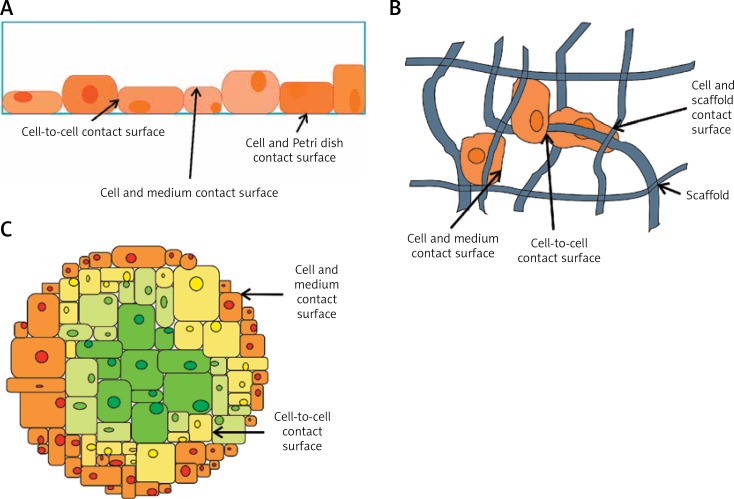
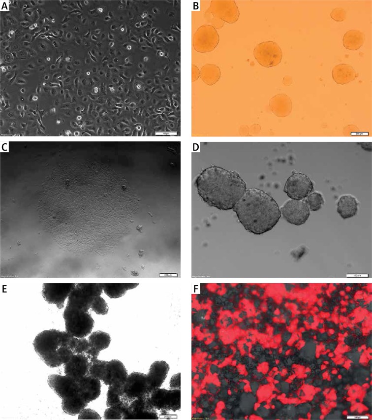
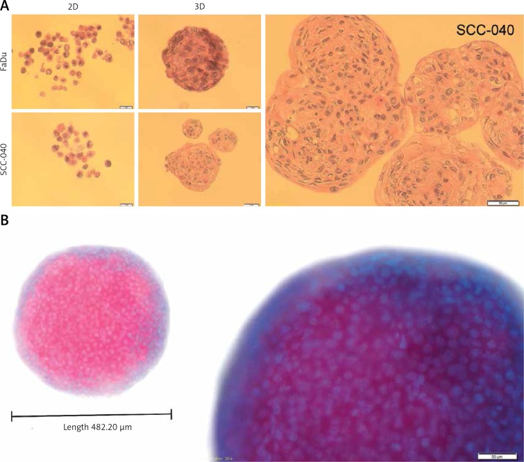
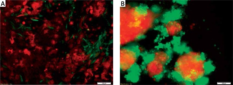
Verification
Claims the paper makes
The claims and statistics of this paper haven’t been checked yet.
Run the check — free, no account needed. It reads the open-access text and checks the sources it cites.
Claims other papers make about this one
Each is checked against this paper’s own text: does it say what the citing paper says it does?
In the citing paper
“While primary patient-derived samples are the gold standard for prediction of treatment response, long-term culture of these samples in 2D often leads to phenotypic changes and differential responses to drug treatments (Kapałczyńska et al., 2018).”
From Three-dimensional modelling of ovarian cancer: from cell lines to organoids for discovery and personalized medicine (2022) · cites this paper as Kapałczyńska et al., 2018
Self-check
Percentages, sums and reported differences re-computed from the paper’s own numbers.
The self-check needs the paper’s full text.
Citations
Cites
Matrix elasticity directs stem cell lineage specification
Engler, A. J., Sen, S., Sweeney, H. L., & Discher, D. E. (2006) · Cell · doi:10.1016/j.cell.2006.06.044 · Cited by 13,915
The third dimension bridges the gap between cell culture and live tissue
Pampaloni, F., Reynaud, E. G., & Stelzer, E. H. K. (2007) · Nature Reviews Molecular Cell Biology · doi:10.1038/nrm2236 · Cited by 2,817
Exploring and Engineering the Cell Surface Interface
Stevens, M. M., & George, J. H. (2005) · Science · doi:10.1126/science.1106587 · Cited by 2,607
Capturing complex 3D tissue physiology in vitro
Griffith, L. G., & Swartz, M. A. (2006) · Nature Reviews Molecular Cell Biology · doi:10.1038/nrm1858 · Cited by 2,347
Scudiero, D. A., Shoemaker, R. H., Paull, K. D., Monks, A. P., Tierney, S., Nofziger, T. H., Currens, M. J., Seniff, D. A., & Boyd, M. R. (1988) · PubMed · Cited by 2,342
Benya, P. D., & Shaffer, J. D. (1982) · Cell · doi:10.1016/0092-8674(82)90027-7 · Cited by 2,284
Geometric cues for directing the differentiation of mesenchymal stem cells
Kilian, K. A., Bugarija, B., Lahn, B. T., & Mrksich, M. (2010) · Proceedings of the National Academy of Sciences · doi:10.1073/pnas.0903269107 · Cited by 1,841
Deconstructing the third dimension – how 3D culture microenvironments alter cellular cues
Baker, B. M., & Chen, C. S. (2012) · Journal of Cell Science · doi:10.1242/jcs.079509 · Cited by 1,760
Modeling Tissue Morphogenesis and Cancer in 3D
Yamada, K. M., & Cukierman, E. (2007) · Cell · doi:10.1016/j.cell.2007.08.006 · Cited by 1,751
Spheroid-based drug screen: Considerations and practical approach
Friedrich, J., Seidel, C., Ebner, R., & Kunz-Schughart, L. A. (2009) · Nature Protocols · doi:10.1038/nprot.2008.226 · Cited by 1,742
Research works printed but not linked: 1
- [5]Jacoby W Pasten I Methods in Enzymology: Cell Culture 1979 58 New York Academic Press
Cited by
Sorted by citations
Items 901–1,000 of 1,001
Gupta, V. K., Vaishnavi, V. V., Arrieta‐Ortiz, M. L., Abhirami, P. S., Jyothsna, K. M., Jeyasankar, S., Raghunathan, V., Baliga, N. S., & Agarwal, R. (2024) · Advanced Healthcare Materials · doi:10.1002/adhm.202304299 · Cited by 5
Ribeiro, M. L., Castro, V. I. B., de Castro, J. V., Pires, R. A., Reis, R. L., Costa, B. M., Ferreira, H., & Neves, N. M. (2024) · International Journal of Molecular Sciences · doi:10.3390/ijms25094910 · Cited by 6
Establishing Three-Dimensional Explant Culture of Human Dental Pulp Tissue
Seo, E. J., Park, S., Lee, E., Huh, Y. H., Ha, Y. E., Tigyi, G., Jeong, T.-S., Jang, I. H., & Shin, J. (2024) · International Journal of Stem Cells · doi:10.15283/ijsc23105 · Cited by 5
Emerging Landscape of In Vitro Models for Assessing Rheumatoid Arthritis Management
Mishra, A. P., Kumar, R., Harilal, S., Nigam, M., Datta, D., & Singh, S. K. (2024) · ACS Pharmacology & Translational Science · doi:10.1021/acsptsci.4c00260 · Cited by 5
Luo, H., Zhou, H., Chen, Y., Sun, X., Li, Y., Li, G., Long, S., Wang, S., Liang, G., & Chen, S. (2024) · Journal of Cellular and Molecular Medicine · doi:10.1111/jcmm.18545 · Cited by 5
Sreekumar, S., & Kiran, M. S. (2024) · BioFactors · doi:10.1002/biof.2111 · Cited by 5
Nandi, S., Ghosh, S., Garg, S., & Ghosh, S. (2024) · ACS Chemical Neuroscience · doi:10.1021/acschemneuro.4c00388 · Cited by 5
Photodynamic therapy in glioma cell culture
Aebisher, D., Rogóż, K., Yakub, Z. A., Dynarowicz, K., Myśliwiec, A., Mytych, W., Komosińska‐Vassev, K., Misiołek, M., Kawczyk‐Krupka, A., & Bartusik‐Aebisher, D. (2024) · ONCOLOGIE · doi:10.1515/oncologie-2024-0407 · Cited by 5
Host-microbe-cancer interactions on-a-chip
Sousa, M. G. C., Brasino, D. S. K., Krieger, M., Dindar, D. A., Duhen, R., Zhang, Z., França, C. M., & Bertassoni, L. E. (2025) · Frontiers in Bioengineering and Biotechnology · doi:10.3389/fbioe.2025.1505963 · Cited by 5
Johnston, A. P., Burton, T. P., & Callanan, A. (2025) · Journal of Applied Polymer Science · doi:10.1002/app.57062 · Cited by 5
Evaluation of the effects of estrogens on endometrial cancer cells of different grades
Pavlič, R., Pušić, M. N., & Rižner, T. L. (2025) · The Journal of Steroid Biochemistry and Molecular Biology · doi:10.1016/j.jsbmb.2025.106762 · Cited by 5
Zhang, X., Guo, Q., Fang, J., Cheng, Q., Zhu, Z., Yu, Q., Wang, H., Hong, Y., Liu, C., Yang, H., et al. (2025) · Journal of Nanobiotechnology · doi:10.1186/s12951-025-03401-2 · Cited by 5
Parodi, I., Palamà, M. E. F., Lisa, D. D., Pastorino, L., Lagazzo, A., Falleroni, F., Aiello, M., Fato, M., & Scaglione, S. (2025) · Gels · doi:10.3390/gels11050356 · Cited by 5
Panatli, O., Gürcan, C., Arı, F., Ünal, M. A., Yüksekkaya, M. E., & Yılmazer, A. (2025) · Micro · doi:10.3390/micro5020025 · Cited by 5
Bridging the Gap: How Organ‐on‐a‐Chip Technology Facilitates the Battle against Glioma
Liu, S., Gong, Z., Zhou, D., Ayala‐Nunez, V., Xu, T., & Wick, P. (2025) · Small Science · doi:10.1002/smsc.202500154 · Cited by 5
Omran, M. M., Vafaei, S., Alkhrait, S., Yang, Q., & Al-Hendy, A. A. (2025) · Journal of Cellular and Molecular Medicine · doi:10.1111/jcmm.70626 · Cited by 5
Stasiak, P., Sopel, J., Płóciennik, A., Musielak, O., Lipowicz, J. M., Rawłuszko-Wieczorek, A. A., Sterzyńska, K., Korbecki, J., & Januchowski, R. (2025) · International Journal of Molecular Sciences · doi:10.3390/ijms26125800 · Cited by 5
Wu, S., Hu, H., Wang, X., Hua, Z., & Zhou, J. (2025) · Molecular Carcinogenesis · doi:10.1002/mc.70009 · Cited by 5
Cultivating human tissues and organs over lab-on-a-chip models: Recent progress and applications
Bhagat, S., & Singh, S. (2022) · Progress in molecular biology and translational science · doi:10.1016/bs.pmbts.2021.07.023 · Cited by 5
Optimization of in vitro culture of honeybee nervous tissue for pesticide risk assessment
Azevedo, P., Butolo, N. P., de Alencar, L. D., Lima, H. M. S., Sales, V. R., Malaspina, O., & Nocelli, R. C. F. (2022) · Toxicology in Vitro · doi:10.1016/j.tiv.2022.105437 · Cited by 5
Direct lysis of 3D cell cultures for RT-qPCR gene expression quantification
Gysens, F., Ostyn, L., Goeteyn, E., Blondeel, E., Nuytens, J., Wever, O. D., de Bony, E. J., Crabbé, A., & Mestdagh, P. (2023) · Scientific Reports · doi:10.1038/s41598-023-28844-1 · Cited by 5
Phon, B. W. S., Bhuvanendran, S., Ayub, Q., Radhakrishnan, A. K., & Kamarudin, M. N. A. (2023) · Biology · doi:10.3390/biology12050648 · Cited by 5
Blood-brain-barrier modeling with tissue chips for research applications in space and on Earth
Yau, A., Jogdand, A. H., & Chen, Y. (2023) · Frontiers in Space Technologies · doi:10.3389/frspt.2023.1176943 · Cited by 5
Suryavanshi, P., Kudtarkar, Y., Chaudhari, M. B., & Bodas, D. (2023) · Nanoscale Advances · doi:10.1039/d3na00420a · Cited by 5
The mannose‐binding protein from Agaricus bisporus inhibits the growth of MDA‐MB‐231 spheroids
Azzahra, F., Amalia, R., Karsono, A. H., Tjandrawinata, R. R., Ismaya, W. T., & Rachmawati, H. (2023) · Chemical Biology & Drug Design · doi:10.1111/cbdd.14365 · Cited by 5
The polymer and materials science of the bacterial fimbriae Caf1
Fulton, D. A., Durá, G., & Peters, D. T. (2023) · Biomaterials Science · doi:10.1039/d3bm01075a · Cited by 5
de Morais, É. F., da Rocha, L. D. O. S., Santos, J. L. D. S., Freitas, R. D., Souza, B. S. D. F., Coletta, R. D., & Rocha, C. A. G. (2023) · Journal of Personalized Medicine · doi:10.3390/jpm13111618 · Cited by 5
Bulygina, I. N., Karshieva, S. S., Permyakova, E. S., Korol, A. A., Kolesnikov, E. A., Choudhary, R., Senatov, F. S., & Koudan, E. V. (2024) · Toxicology in Vitro · doi:10.1016/j.tiv.2024.105830 · Cited by 5
The morphology of cell spheroids in simple shear flow
Ferraro, R., Franco, J. D., Caserta, S., & Guido, S. (2024) · Frontiers in Physics · doi:10.3389/fphy.2024.1347934 · Cited by 5
Christfort, J. F., Ortis, M., Nguyen, H. V., Marsault, R., & Doglio, A. (2024) · Biosensors · doi:10.3390/bios14080401 · Cited by 6
Tahara, S., Rentsch, S., de Faria, F. C. C., Sarchet, P., Karna, R., Pollock, R. E., & Calore, F. (2024) · Oncology Research Featuring Preclinical and Clinical Cancer Therapeutics · doi:10.32604/or.2024.053635 · Cited by 5
PLLA Porous Scaffold as a 3D Breast Cancer Model to Investigate Drug Resistance
Carbone, C., Rigogliuso, S., Brucato, V., Cusimano, A., Labbozzetta, M., Carrubba, V. L., Poma, P., Notarbartolo, M., & Pavia, F. C. (2024) · Journal of Biomedical Materials Research Part A · doi:10.1002/jbm.a.37836 · Cited by 5
A standardized in vitro bioengineered skin for penetrating wound modeling
Silva, C. S. S., Petzold, W., Hirsch, U., Schmelzer, C. E. H., & Friedmann, A. (2025) · In vitro models · doi:10.1007/s44164-025-00082-x · Cited by 5
Rafik, S. T., Bakkalci, D., MacRobert, A. J., & Cheema, U. (2025) · FEBS Open Bio · doi:10.1002/2211-5463.70018 · Cited by 5
Development and application of cell-based bioassay in aquatic toxicity assessment
Xia, Y., & Wang, W. (2025) · Aquatic Toxicology · doi:10.1016/j.aquatox.2025.107427 · Cited by 5
Gene expression profiling of tumor stroma interactions in retinoblastoma
Raguraman, R., Parameswaran, S., Kanwar, J. R., Vasudevan, M., Chitipothu, S., Kanwar, R. K., & Krishnakumar, S. (2020) · Experimental Eye Research · doi:10.1016/j.exer.2020.108067 · Cited by 5
ShujaaEdin, H. Y., Al-Haj, N. A. A., Abdullah, R., Alitheen, N. B., Kadir, A. A., How, C. W., Rahman, H. S., & Abdullah, A.-S. H. (2021) · Saudi Journal of Biological Sciences · doi:10.1016/j.sjbs.2021.01.059 · Cited by 6
Baru, A., Sharma, S., Purakayastha, B. P. D., Khan, S. M., Mazumder, S., Gupta, R., Kundu, P. K., & Arora, N. M. (2021) · Assay and Drug Development Technologies · doi:10.1089/adt.2021.031 · Cited by 5
Model Selection for the Preclinical Development of New Drug–Radiotherapy Combinations
Singh, J., Hatcher, S. V., Ku, A. A., Ding, Z., Feng, F. Y.-C., Sharma, R. A., & Pfister, S. (2021) · Clinical Oncology · doi:10.1016/j.clon.2021.08.008 · Cited by 5
Thermo-Chemical Resistance to Combination Therapy of Glioma Depends on Cellular Energy Level
Qiu, J., Li, Z., An, K., Niu, L., Huang, H., & Xu, F. (2023) · ACS Applied Materials & Interfaces · doi:10.1021/acsami.3c05683 · Cited by 5
Preclinical models in oncological pharmacology: limits and advantages
Pagano, E., Bergamo, A., Carpi, S., Donnini, S., Villarosa, M. N. D., Serpe, L., & Lisi, L. (2021) · doi:10.36118/pharmadvances.2021.05 · Cited by 5
Hung, S.-T., Rosell, A. L., Jurriens, D., Siemons, M. E., Soloviev, O., Kapitein, L. C., Grußmayer, K. S., Neukomm, L. J., Verhaegen, M., & Smith, C. (2022) · Frontiers in Neuroscience · doi:10.3389/fnins.2022.954949 · Cited by 5
Advancements in cancer research: 3D bioprinting as an optimal oral tumor model
Mohanprasanth, A., & Saravanan, M. (2024) · Oral Oncology Reports · doi:10.1016/j.oor.2024.100174 · Cited by 5
Upadhyay, V., Das, S., Nehru, A., Swetha, U. S., Vatsa, P. P., Chamoli, A., & Mandoli, A. (2025) · 3 Biotech · doi:10.1007/s13205-025-04472-6 · Cited by 4
Cell Viability Assays for 3D Cellular Constructs
Congress, Z., Brovold, M., & Söker, S. (2023) · Methods in molecular biology · doi:10.1007/978-1-0716-3052-5_25 · Cited by 4
Effects of growth factor deprivation on MKN-45 spheroid cells
TÜRKSOY, Ö., & Terzioğlu, G. (2023) · TURKISH JOURNAL OF BIOLOGY · doi:10.55730/1300-0152.2646 · Cited by 4
Zhang, Y., Liu, K., He, H., Xiao, H., Fang, Z., Chen, X., & Li, H. (2024) · Environmental Science and Pollution Research · doi:10.1007/s11356-024-32256-7 · Cited by 6
A promising breakthrough in pancreatic cancer research: The potential of spheroids as 3D models
Jamshidi, N., Jamshidi, N., Chahardehi, A. M., Shams, E., & Chaleshi, V. (2024) · Bioimpacts · doi:10.34172/bi.30241 · Cited by 4
Çakır, M., Akar, S., Çakır, Ş., Akkoç, S., Özkol, H., & Tülüce, Y. (2024) · Eastern Journal Of Medicine · doi:10.5505/ejm.2024.03708 · Cited by 4
Mimicking the immunosuppressive impact of fibroblasts in a 3D multicellular spheroid model
Grotz, M., van Gijzel, L., Bitsch, P., Carrara, S. C., Kolmar, H., & Garg, S. (2024) · Frontiers in Drug Discovery · doi:10.3389/fddsv.2024.1427407 · Cited by 4
Unveiling the effects of micro and nano plastics in embryonic development
Nair, S. R., Nihad, M., Sudheer Shenoy, P., Gupta, S., & Bose, B. (2025) · Toxicology Reports · doi:10.1016/j.toxrep.2025.101954 · Cited by 5
Recent trends in the development of in vitro 3D kidney models
Gaddam, K., Chameettachal, S., Yeleswarapu, S., Duin, S., Lode, A., Gelinsky, M., Akkineni, A. R., & Pati, F. (2025) · Biofabrication · doi:10.1088/1758-5090/adb999 · Cited by 4
Establishment and characterization study of ovine mammary organoids
Feng, R., Ma, S., Bai, R., Zhu, Y., Sarengele, Ning, J., Xu, Q., Wang, C., Wang, L., Bian, C., et al. (2025) · BMC Veterinary Research · doi:10.1186/s12917-025-04657-4 · Cited by 4
Petrauskas, V., Damaseviciute, R., & Gulla, A. (2025) · Medicina · doi:10.3390/medicina61040623 · Cited by 4
Small molecules direct the generation of ameloblast-like cells from human embryonic stem cells
Zhu, X., Zhao, Y., Bai, X., Dong, Q., Tian, C., Sun, R., Yan, C., Ruan, J., Liu, Z., & Gao, J. (2025) · Stem Cell Research & Therapy · doi:10.1186/s13287-025-04294-6 · Cited by 4
Al-Masoody, A. H., Asadi, N., Mohammadpour, H., Hodjat, M., & Kashi, T. S. J. (2025) · BMC Oral Health · doi:10.1186/s12903-025-06236-3 · Cited by 4
Weng, J., Li, S., Weng, J., Wang, Y., Deng, B., Yao, M., Hao, H., Xia, H., Gan, L., Chen, B., et al. (2025) · Materials Today Bio · doi:10.1016/j.mtbio.2025.102061 · Cited by 4
Kremmydas, S., Gourdoupi, C., Piperigkou, Z., & Karamanos, N. K. (2025) · International Journal of Molecular Sciences · doi:10.3390/ijms26178665 · Cited by 4
Khan, M., Kollenz, P., Fritzenschaft, M., Taheri, F., Colombo, F., Blumberg, J. W., Schlotterose, L., Schwarz, U. S., Leal‐Egaña, A., & Selhuber‐Unkel, C. (2025) · Bioactive Materials · doi:10.1016/j.bioactmat.2025.09.025 · Cited by 4
Grattarola, M., Pons, N., Cannet, F., Kaya, M., Kaoutari, A. E., Morel, C., Dobric, A., Borg, J., Sequera, C., & Maina, F. (2025) · Cell Communication and Signaling · doi:10.1186/s12964-025-02391-w · Cited by 4
Al-Ansari, D. E., Hu, Y., Negrini, N. C., Jones, D., Birdsey, G. M., & Celiz, A. D. (2025) · Materials Today Bio · doi:10.1016/j.mtbio.2025.102367 · Cited by 4
Perfluoroalkyl Chain-Modified Artificial Viral Capsid for Enhanced Intracellular Delivery of mRNA
Ghosh, A., Yamamoto, Y., Wada, M., Inaba, H., Aikawa, K., Ota, Y., Okazoe, T., & Matsuura, K. (2025) · Bioconjugate Chemistry · doi:10.1021/acs.bioconjchem.5c00477 · Cited by 4
Three‐Dimensional Influence of Photobiomodulation on Adipose‐Derived Stem Cell Behavior
Mulaudzi, P. E., Abrahamse, H., & Crous, A. (2025) · Journal of Biophotonics · doi:10.1002/jbio.202500414 · Cited by 4
Trautmann, R. K., Dennison, N. R., McCortney, K., Klier, S., Coşacak, M. İ., Werner, C., Akyoldaş, G., Horbinski, C., Freudenberg, U., & Kızıl, Ç. (2026) · Macromolecular Bioscience · doi:10.1002/mabi.202500394 · Cited by 4
Microfluidic Detection of Adenylate Kinase as Cell Damage Biomarker and Integration with a Cell Chip
Domingues, C., Fontes, P. M., Condelipes, P. G. M., Marques, V., Afonso, M. B., Chu, V., Rodrigues, C. M. P., & Conde, J. P. (2022) · Chemosensors · doi:10.3390/chemosensors11040220 · Cited by 4
Research and therapeutic applications of silk proteins in cancer
Kumari, N., Pullaguri, N., Sahu, V., & Ealla, K. K. R. (2023) · Journal of Biomaterials Applications · doi:10.1177/08853282231184572 · Cited by 4
Establishment of a 3D Model to Characterize the Radioresponse of Patient-Derived Glioblastoma Cells
Strand, Z., Schrickel, F., Dobiasch, S., Thomsen, A. R., Steiger, K., Gempt, J., Meyer, B., Combs, S. E., & Schilling, D. (2023) · Cancers · doi:10.3390/cancers15164051 · Cited by 4
Applications of Tumor Cells in an In Vitro 3D Environment
Hasterok, S., Gustafsson, A., & Wingren, A. G. (2023) · Applied Sciences · doi:10.3390/app131810349 · Cited by 4
3D Breast Cancer Model on Silk Fibroin–Integrated Microfluidic Chips
Yilmaz, E. G., & İnci, F. (2024) · Methods in molecular biology · doi:10.1007/978-1-0716-3674-9_16 · Cited by 4
Cells in the 3D biomatrix on-chip: better mimicking the real micro-physiological system
D’Orazio, M., Filippi, J., Antonelli, G., Curci, G., Casti, P., Mencattini, A., Cidonio, G., & Martinelli, E. (2024) · Next Materials · doi:10.1016/j.nxmate.2024.100229 · Cited by 4
Antimetastatic activity of (arene)ruthenium(II) complex of 4-aryl-4H-naphthopyran
Prachařová, J., Cyrikova, T., Berecka, M., Biersack, B., Kašpárková, J., & Brabec, V. (2024) · Chemico-Biological Interactions · doi:10.1016/j.cbi.2024.111180 · Cited by 4
Huang, J., Wang, X., Ge, S., Lü, X., & Sun, C. (2024) · Cellular and Molecular Bioengineering · doi:10.1007/s12195-024-00825-y · Cited by 4
Psarra, E., & Apostolou, D. (2024) · International Conference on Information, Intelligence, Systems & Applications (IISA) · doi:10.1109/iisa62523.2024.10786721 · Cited by 4
Developing a 3D bone model of osteosarcoma to investigate cancer mechanisms and evaluate treatments
Smith, H. L., Beers, S. A., Kanczler, J. M., & Gray, J. C. (2024) · The FASEB Journal · doi:10.1096/fj.202402011r · Cited by 4
Caratelli, S., Paolis, F. D., Silvestris, D. A., Baldari, S., Salvatori, I., Tullo, A., Lanzilli, G., Gurtner, A., Ferri, A., Valle, C., et al. (2025) · Experimental Hematology and Oncology · doi:10.1186/s40164-025-00601-2 · Cited by 4
Prakash, N., Cha, Y., Koh, W.-G., Park, H., Bello, A. B., & Lee, S. (2025) · Biomaterials Research · doi:10.34133/bmr.0184 · Cited by 4
Organoids with a Type 1 Collagen Scaffold to Model Bacterial Cancer Therapy
Farrell, L., Bonnet, C. S., Tang, A., Peneva, S., Williams, N. G., Dolwani, S., Parry, L., & Dyson, P. J. (2025) · Cells · doi:10.3390/cells14070524 · Cited by 4
The Role of HER2 in Cancer and Its Influence on the Tumor Microenvironment
Choi, S., Kim, M., & Kim, M. (2025) · IntechOpen eBooks · doi:10.5772/intechopen.1009458 · Cited by 4
Wawrowicz, K., Durak-Kozica, M., Wierzbicki, M., & Stępień, E. Ł. (2025) · ACS Omega · doi:10.1021/acsomega.5c02682 · Cited by 5
Zahmatkesh, E., & Vosough, M. (2018) · Modern Medical Laboratory Journal · doi:10.30699/mmlj17.1.3.110 · Cited by 4
Hernandes, C., de Oliveira, R. N., Santos, A. H. D. S., Malvezzi, H., de Azevedo, B. C., Gueuvoghlanian‐Silva, B. Y., Pereira, A. M. S., & Podgaec, S. (2020) · Molecules · doi:10.3390/molecules25061325 · Cited by 4
Seyfoori, A., Barough, M. S., Amereh, M., Jush, B. K., Lum, J. J., & Akbari, M. (2020) · Drug Discovery Today · doi:10.1016/j.drudis.2020.11.028 · Cited by 4
Longitudinal evaluation of five nasopharyngeal carcinoma animal models on the microPET/MR platform
Shi, J.-J., Xue, Z.-C., Tan, K., Yuan, H., Tsang, A., Tsao, S.-W., & Khong, P. (2021) · European Journal of Nuclear Medicine and Molecular Imaging · doi:10.1007/s00259-021-05633-4 · Cited by 3
Zmrhal, V., Svoradová, A., Bátik, A., & Sláma, P. (2022) · Frontiers in Cell and Developmental Biology · doi:10.3389/fcell.2021.730804 · Cited by 4
Cell Line Culture in Pharmaceutical Development and Application: A Review
Jassim, Z. (2023) · Al-Mağallaẗ al-ʻirāqiyyaẗ li-l-ṣaydalaẗ · doi:10.33899/iphr.2023.138155.01023 · Cited by 4
SMYD3 Modulates the HGF/MET Signaling Pathway in Gastric Cancer
Marco, K. D., Signorile, M. L., Nicola, E. D., Sanese, P., Fasano, C., Forte, G., Disciglio, V., Pantaleo, A., Varchi, G., Río, A. D., et al. (2023) · Cells · doi:10.3390/cells12202481 · Cited by 4
Fujiike, A. Y., de Oliveira, L. C. B., Ribeiro, D. L., Pereira, É. R., Okuyama, N. C. M., dos Santos, A. G. P., Cólus, I. M. D. S., & Serpeloni, J. M. (2023) · Journal of Toxicology and Environmental Health · doi:10.1080/15287394.2023.2293218 · Cited by 4
Meyer, B.-O., Stella, M. P. J., Holst, B., Nielsen, B. S., Holmstrøm, K., Andersen, P. E., & Marti, D. (2020) · Journal of Biomedical Optics · doi:10.1117/1.jbo.25.7.071206 · Cited by 4
Physical Cues in the Microenvironment Regulate Stemness-Dependent Homing of Breast Cancer Cells
Chu, H.-Y., Chen, Y., Hsu, C.-J., Liu, Y.-W., Chiou, J.-F., Lu, L., & Tseng, F. (2020) · Cancers · doi:10.3390/cancers12082176 · Cited by 4
Human lung-cancer-cell radioresistance investigated through 2D network topology
Tirinato, L., Onesto, V., García-Calderón, D., Pagliari, F., Spadea, M. F., Seco, J. C., & Gentile, F. (2022) · Scientific Reports · doi:10.1038/s41598-022-17018-0 · Cited by 4
Christowitz, C., Olivier, D. W., Schneider, J. W., Kotze, M. J., & Engelbrecht, A. (2024) · Mutation Research/Reviews in Mutation Research · doi:10.1016/j.mrrev.2024.108492 · Cited by 4
Bioimprinting: Bringing Together 2D and 3D in Dissecting Cancer Biology
Sarwar, M., & Evans, J. J. (2021) · BioTechniques · doi:10.2144/btn-2021-0058 · Cited by 4
Kim, Y.-R., Bae, K., Lee, J., Jeong, S., Yoon, H., Han, H., Hyun, J., Nam, A., Park, J.-H., Yoon, K., et al. (2025) · Animals · doi:10.3390/ani15081146 · Cited by 3
Yang, L., Wu, Z., Zhu, H., Ding, S., & Zhou, G. (2025) · Food Research International · doi:10.1016/j.foodres.2025.116324 · Cited by 3
Three-Dimensional Culture Systems in Neuroblastoma Research
Jung, P., & Wolpaw, A. J. (2025) · Organoids · doi:10.3390/organoids4020010 · Cited by 3
Modeling Synucleinopathy Using hESC-Derived Cerebral Organoids
Kim, S. J., Jung, W. H., Choe, M. S., Jeon, Y., & Lee, M. Y. (2025) · Cells · doi:10.3390/cells14181436 · Cited by 3
Etiologies of endometriosis and model systems: is there a risk of a tunnel vision?
Manavalan, M. A., Babtain, M., Weessies, M., Nap, A. W., Verdurmen, W. P. R., Taurin, S., Sater, M. S., & Brock, R. E. (2025) · Frontiers in Medicine · doi:10.3389/fmed.2025.1675051 · Cited by 3
Chen, X., Peng, Y., Zhao, Y., Liu, H., Lin, Q., Fu, X., Chen, L., Peng, Z., Huang, J., Luo, Y., et al. (2025) · Bioactive Materials · doi:10.1016/j.bioactmat.2025.09.041 · Cited by 3
Caliskan, A., Crouch, S. A. W., Pereira, J. G. N., Dandekar, T., Dandekar, G., & Breitenbach, T. (2025) · Computational and Structural Biotechnology Journal · doi:10.1016/j.csbj.2025.10.032 · Cited by 3
Aranaz, P., Clavel-Millan, M., Gil‐Cardoso, K., González-Arceo, M., Hernández-González, D., Les, F., Salles, J., Amri, E., Arbonés-Mainar, J. M., Atgié, C., et al. (2025) · Journal of Physiology and Biochemistry · doi:10.1007/s13105-025-01130-6 · Cited by 3
Metadata
- Authors
- Kapałczyńska, M., Kolenda, T., Przybyła, W., Zajączkowska, M., Teresiak, A., Filas, V., Ibbs, M., Bliźniak, R., Łuczewski, Ł., & Lamperska, K.
- Affiliations
- Greater Poland Cancer Center (PL)
- Poznan University of Medical Sciences (PL)
- Corresponding author
- Kolenda, Tomasz
- ORCID
- Venue
- Archives of Medical Science
- Publisher
- Termedia Sp. z.o.o.
- Published
- 2016
- Volume
- 14
- Issue
- 4
- Pages
- 910-919
- ISSN
- 1734-1922 (ISSN-L)
- PMC
- PMC6040128
- OpenAlex
- W2587788572
- Cited by
- 1,499
- References
- 93
- Citations by year
- 2026: 114 · 2025: 264 · 2024: 299 · 2023: 256 · 2022: 196 · 2021: 209 · 2020: 111 · 2019: 43 · 2018: 5
- FWCI
- 7.71
- Type
- journal article
- Language
- en
- Open access
- gold
- doi.org
- Licence
- cc-by-nc-sa
- Locations
- Archives of Medical Science (publishedVersion, cc-by-nc-sa)
- Europe PMC (PubMed Central) (publishedVersion, cc-by-nc-sa)
- PubMed Central (publishedVersion, cc-by-nc-sa)
- PubMed (publishedVersion)
- Topics
- Physical Sciences › Engineering › Biomedical Engineering › 3D Printing in Biomedical Research
- Health Sciences › Medicine › Oncology › Cancer Cells and Metastasis
- Life Sciences › Biochemistry, Genetics and Molecular Biology › Cell Biology › Cellular Mechanics and Interactions
- Keywords
- 2D cell culture, 3D cell culture, tumor cell culture, in vitro model, tumor biology, extracellular matrix, cell morphology, biomarkers, targeted therapy, tissue engineering
About these checks
Registry, retraction, Cites and statistics results are computed, not judged, with one exception: a printed reference that no index lists is read once by a model before it counts as a citation error. Claims are extracted by a model and kept only when their quote appears verbatim in the text; each cited source is then checked, with a model’s help, for whether it says what the claim relies on. Those verdicts can be wrong — read the claim’s quote and the cited source before relying on one.
Statistics and arithmetic are recomputed, not judged.
Confidence bands are shown only for fields calibrated on our annotated test set. None is calibrated yet, so no band is shown.
What counts as a claim ›How we measure confidence ›How we check this ›
Are you an author, or do you see a problem with this page? Request a review
