

The DOI matches its Crossref record; 92 of the 93 references it cites were found in a registry or an index.
2D and 3D Cell Cultures - a Comparison of Different Types of Cancer Cell Cultures
Open accessAbstract
Cell culture is a widely used in vitro tool for improving our understanding of cell biology, tissue morphology, and mechanisms of diseases, drug action, protein production and the development of tissue engineering.Most research regarding cancer biology is based on experiments using two-dimensional (2D) cell cultures in vitro.However, 2D cultures have many limitations, such as the disturbance of interactions between the cellular and extracellular environments, changes in cell morphology, polarity, and method of division.These disadvantages led to the creation of models which are more closely able to mimic conditions in vivo.One such method is three-dimensional culture (3D).Optimisation of the culture conditions may allow for a better understanding of cancer biology and facilitate the study of biomarkers and targeting therapies.In this review, we compare 2D and 3D cultures in vitro as well as different versions of 3D cultures.
Overview
The claims and statistics of this paper haven’t been checked yet.
Run the check — free, no account needed. It reads the open-access text and checks the sources it cites.
Integrity signals
Registry
Cites
Statistics
Figures
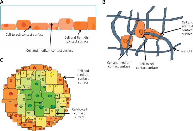
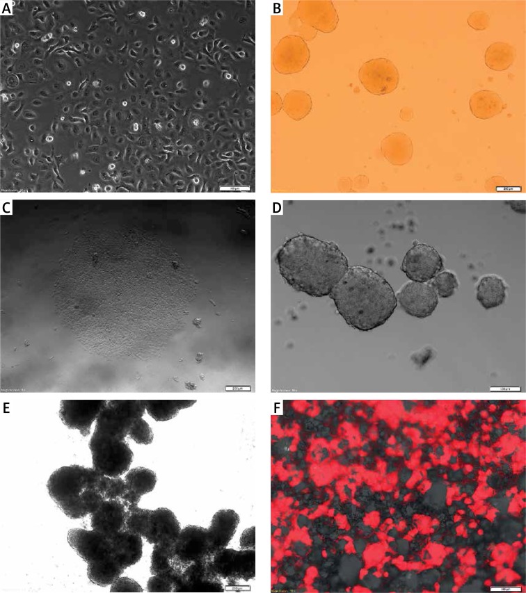
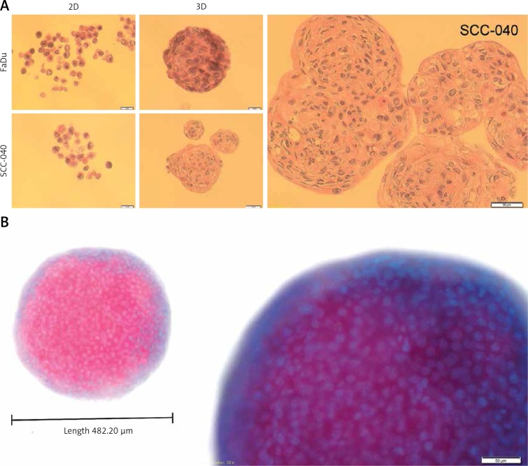
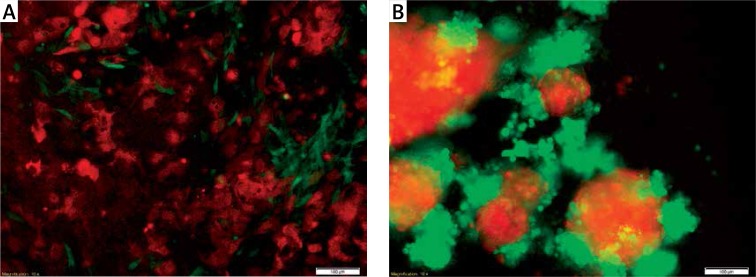
Verification
Claims the paper makes
The claims and statistics of this paper haven’t been checked yet.
Run the check — free, no account needed. It reads the open-access text and checks the sources it cites.
Claims other papers make about this one
Each is checked against this paper’s own text: does it say what the citing paper says it does?
In the citing paper
“While primary patient-derived samples are the gold standard for prediction of treatment response, long-term culture of these samples in 2D often leads to phenotypic changes and differential responses to drug treatments (Kapałczyńska et al., 2018).”
From Three-dimensional modelling of ovarian cancer: from cell lines to organoids for discovery and personalized medicine (2022) · cites this paper as Kapałczyńska et al., 2018
Self-check
Percentages, sums and reported differences re-computed from the paper’s own numbers.
The self-check needs the paper’s full text.
Citations
Cites
Matrix elasticity directs stem cell lineage specification
Engler, A. J., Sen, S., Sweeney, H. L., & Discher, D. E. (2006) · Cell · doi:10.1016/j.cell.2006.06.044 · Cited by 13,915
The third dimension bridges the gap between cell culture and live tissue
Pampaloni, F., Reynaud, E. G., & Stelzer, E. H. K. (2007) · Nature Reviews Molecular Cell Biology · doi:10.1038/nrm2236 · Cited by 2,817
Exploring and Engineering the Cell Surface Interface
Stevens, M. M., & George, J. H. (2005) · Science · doi:10.1126/science.1106587 · Cited by 2,607
Capturing complex 3D tissue physiology in vitro
Griffith, L. G., & Swartz, M. A. (2006) · Nature Reviews Molecular Cell Biology · doi:10.1038/nrm1858 · Cited by 2,347
Scudiero, D. A., Shoemaker, R. H., Paull, K. D., Monks, A. P., Tierney, S., Nofziger, T. H., Currens, M. J., Seniff, D. A., & Boyd, M. R. (1988) · PubMed · Cited by 2,342
Benya, P. D., & Shaffer, J. D. (1982) · Cell · doi:10.1016/0092-8674(82)90027-7 · Cited by 2,284
Geometric cues for directing the differentiation of mesenchymal stem cells
Kilian, K. A., Bugarija, B., Lahn, B. T., & Mrksich, M. (2010) · Proceedings of the National Academy of Sciences · doi:10.1073/pnas.0903269107 · Cited by 1,841
Deconstructing the third dimension – how 3D culture microenvironments alter cellular cues
Baker, B. M., & Chen, C. S. (2012) · Journal of Cell Science · doi:10.1242/jcs.079509 · Cited by 1,760
Modeling Tissue Morphogenesis and Cancer in 3D
Yamada, K. M., & Cukierman, E. (2007) · Cell · doi:10.1016/j.cell.2007.08.006 · Cited by 1,751
Spheroid-based drug screen: Considerations and practical approach
Friedrich, J., Seidel, C., Ebner, R., & Kunz-Schughart, L. A. (2009) · Nature Protocols · doi:10.1038/nprot.2008.226 · Cited by 1,742
Research works printed but not linked: 1
- [5]Jacoby W Pasten I Methods in Enzymology: Cell Culture 1979 58 New York Academic Press
Cited by
Sorted by citations
Items 501–600 of 1,001
Phon, B. W. S., Kamarudin, M. N. A., Bhuvanendran, S., & Radhakrishnan, A. K. (2021) · Biomedicine & Pharmacotherapy · doi:10.1016/j.biopha.2021.112396 · Cited by 16
Non-destructive monitoring of 3D cell cultures: new technologies and applications
Cortesi, M., & Giordano, E. (2022) · PeerJ · doi:10.7717/peerj.13338 · Cited by 16
Dozzo, A., Chullipalliyalil, K., McAuliffe, M. A. P., O’Driscoll, C. M., & Ryan, K. B. (2023) · Pharmaceutics · doi:10.3390/pharmaceutics15010242 · Cited by 16
Abdelmoneim, D., Porter, G. C., Duncan, W., Lim, K. S., Easingwood, R. A., Woodfield, T. B. F., & Coates, D. E. (2023) · Nanomaterials · doi:10.3390/nano13040705 · Cited by 16
Vascularized Lower Respiratory-Physiology-On-A-Chip
Nam, H., Choi, Y., & Jang, J. (2020) · Applied Sciences · doi:10.3390/app10030900 · Cited by 16
Fan, T. W. ‐., Higashi, R. M., Chernayavskaya, Y., & Lane, A. N. (2020) · Metabolites · doi:10.3390/metabo10060249 · Cited by 16
Antiproliferative and antibacterial potential of tetrahexylammonium bromide‑based ionic liquids
Popescu, R., Filimon, M. N., Vlad, D. C., Verdeş, D., Moatar, A. E., Moise, G. V., Guran, K., Carabă, I. V., Ciochină, L. P., Pînzaru, I., et al. (2021) · Experimental and Therapeutic Medicine · doi:10.3892/etm.2021.10104 · Cited by 16
Artificial tissue creation under microgravity conditions: Considerations and future applications
Swaminathan, V., Bechtel, G. N., & Tchantchaleishvili, V. (2021) · Artificial Organs · doi:10.1111/aor.14017 · Cited by 16
In vitro 3D skin culture and its sustainability in toxicology: a narrative review
Masri, S., Fauzi, M. B., Rajab, N. F., Lee, W.-H., Zainal Abidin, D. A., & Siew, E. L. (2024) · Artificial Cells, Nanomedicine, and Biotechnology · doi:10.1080/21691401.2024.2407617 · Cited by 15
Omran, M. M., Vafaei, S., Alkhrait, S., Ali, F. L., Bariani, M. V., Bai, T., Thompson, W. E., Yang, Q., Ali, M. R., & Al-Hendy, A. A. (2025) · Cell Proliferation · doi:10.1111/cpr.70025 · Cited by 15
Two-Dimensional and Three-Dimensional Cell Culture and Their Applications
Ballav, S., Deshmukh, A. J., Siddiqui, S., Aich, J., & Basu, S. (2021) · Biochemistry · doi:10.5772/intechopen.100382 · Cited by 15
Brumskill, S., Barrera, L. N., Calcraft, P. J., Phillips, C., & Costello, E. (2021) · Journal of Physiology and Biochemistry · doi:10.1007/s13105-021-00857-2 · Cited by 16
Borah, A., & Kumar, D. S. (2022) · Elsevier eBooks · doi:10.1016/b978-0-12-823966-7.00003-7 · Cited by 16
Bhargava, A., Sandoval-Castellanos, A. M., Shah, S. S., & Ning, K. (2022) · Interface Focus · doi:10.1098/rsfs.2022.0040 · Cited by 15
Rerouting the drug response: Overcoming metabolic adaptation in KRAS-mutant cancers
Moss, D., McCann, C. K., & Kerr, E. M. (2022) · Science Signaling · doi:10.1126/scisignal.abj3490 · Cited by 16
Ovarian Cancer Patient-Derived Organoid Models for Pre-Clinical Drug Testing
Fashemi, B., van Biljon, L., Rodriguez, J., Graham, O., Mullen, M., & Khabele, D. (2023) · Journal of Visualized Experiments · doi:10.3791/65068 · Cited by 15
Spheroids as a 3D in vitro model to study bone and bone mineralization
Boscaro, D., & Sikorski, P. (2023) · Biomaterials Advances · doi:10.1016/j.bioadv.2023.213727 · Cited by 15
Use of iPSC-Derived Smooth Muscle Cells to Model Physiology and Pathology
Kwartler, C. S., & Pinelo, J. E. E. (2024) · Arteriosclerosis Thrombosis and Vascular Biology · doi:10.1161/atvbaha.123.319703 · Cited by 15
Tumoroids, a valid preclinical screening platform for monitoring cancer angiogenesis
Abbasi‐Malati, Z., Khanicheragh, P., Narmi, M. T., Mardi, N., Khosrowshahi, N. D., Hiradfar, A., Rezabakhsh, A., Sadeghsoltani, F., Rashidi, S., Chegeni, S. A., et al. (2024) · Stem Cell Research & Therapy · doi:10.1186/s13287-024-03880-4 · Cited by 15
Channel-assembling tumor microenvironment on-chip for evaluating anticancer drug efficacy
Lee, J. H., Kim, Y., Jung, H., Lim, J., & Kwak, B. (2024) · Journal of Controlled Release · doi:10.1016/j.jconrel.2024.11.030 · Cited by 15
Gene Expression Signature of BRAF Inhibitor Resistant Melanoma Spheroids
Koroknai, V., Patel, V., Szász, I., Ádány, R., & Balázs, M. (2020) · Pathology & Oncology Research · doi:10.1007/s12253-020-00837-9 · Cited by 16
Feky, S. E. E., Megahed, M., Moneim, N. A. A. E., Zaher, E. R., Khamis, S. A., & Ali, L. (2021) · Experimental and Therapeutic Medicine · doi:10.3892/etm.2021.9937 · Cited by 15
Sargenti, A., Musmeci, F., Cavallo, C., Mazzeschi, M., Bonetti, S., Pasqua, S., Bacchi, F., Filardo, G., Gazzola, D., Lauriola, M., et al. (2021) · PLoS ONE · doi:10.1371/journal.pone.0252907 · Cited by 15
Spinelli, F. M., Rosales, P., Pluda, S., Vitale, D. L., Icardi, A., Guarise, C., Reszegi, A., Kovalszky, I., García, M. G., Sevic, I., et al. (2022) · IUBMB Life · doi:10.1002/iub.2604 · Cited by 15
Dynamic Three-Dimensional Cell-Culture Systems for Enhanced in Vitro Applications
Aishwarya, P., Agrawal, G., Samson, J. S., & Ravi, M. (2022) · Current Science · doi:10.18520/cs/v122/i2/149-160 · Cited by 15
Unsung versatility of elastin-like polypeptide inspired spheroid fabrication: A review
Goel, R., Gulwani, D., Upadhyay, P., Sarangthem, V., & Singh, T. D. (2023) · International Journal of Biological Macromolecules · doi:10.1016/j.ijbiomac.2023.123664 · Cited by 15
Brown, M.-J., Bahsoun, S., Morris, M. A., & Akam, E. C. (2019) · Bioengineering · doi:10.3390/bioengineering6040101 · Cited by 15
Tumor spheroid-based microtumor models for preclinical evaluation of anticancer nanomedicines
Khawar, I. A., Ghosh, T., Park, J. K., & Kuh, H. (2021) · Journal of Pharmaceutical Investigation · doi:10.1007/s40005-021-00534-y · Cited by 15
Seabra, R., Malhão, F., Correia, A., Costa, C., Kijjoa, A., & Rocha, E. (2023) · Marine Drugs · doi:10.3390/md21030166 · Cited by 15
den Bossche, S. V., Ostyn, L., Vandendriessche, V., Rigauts, C., Keersmaecker, H. D., Nickerson, C. A., & Crabbé, A. (2023) · European Journal of Pharmaceutical Sciences · doi:10.1016/j.ejps.2023.106567 · Cited by 14
Engineering models of head and neck and oral cancers on-a-chip
Sousa, M. G. D. C., Vignolo, S. M., França, C. M., Mereness, J. A., Fraga, M. A. A., Silva‐Sousa, A. C., Benoit, D. S. W., & Bertassoni, L. E. (2024) · Biomicrofluidics · doi:10.1063/5.0186722 · Cited by 14
Validating Well-Functioning Hepatic Organoids for Toxicity Evaluation
Choi, S. Y., Kim, T. H., Kim, M. J., Mun, S. J., Kim, T., Jung, K. K., Oh, I. U., Oh, J. H., Son, M. J., & Lee, J. H. (2024) · Toxics · doi:10.3390/toxics12050371 · Cited by 14
Surface-Enhanced Raman Scattering Monitoring of Tryptophan Dynamics in 3D Pancreatic Tumor Models
Valera, P. S., Henriques‐Pereira, M., Wagner, M., Gaspar, V. M., Mano, J. F., & Liz‐Marzán, L. M. (2024) · ACS Sensors · doi:10.1021/acssensors.4c01210 · Cited by 14
Kumar, R., Iden, M., Tsaih, S., Schmidt, R., Ojesina, A. I., & Rader, J. S. (2024) · Frontiers in Cell and Developmental Biology · doi:10.3389/fcell.2024.1413882 · Cited by 14
Injectable, in-situ forming, tunable, biocompatible gelatin hydrogels for biomedical applications
Chauhan, M., Roopmani, P., Rajendran, J., Narayan, K. P., & Giri, J. (2024) · International Journal of Biological Macromolecules · doi:10.1016/j.ijbiomac.2024.138200 · Cited by 14
The application of three-dimensional cell culture in clinical medicine
Chen, Q., & Wang, Y. (2020) · Biotechnology Letters · doi:10.1007/s10529-020-03003-y · Cited by 14
Winkelman, M. A., Koppes, A. N., Koppes, R. A., & Dai, G. (2021) · APL Bioengineering · doi:10.1063/5.0027211 · Cited by 14
Chakraborty, S., DePalma, T. J., & Skardal, A. (2021) · Advanced NanoBiomed Research · doi:10.1002/anbr.202100061 · Cited by 14
Hashemzadeh, N., Aghanejad, A., Abdolahinia, E. D., Dolatkhah, M., Barzegar-Jalali, M., Omidi, Y., Barar, J., & Adibkia, K. (2021) · Journal of Microencapsulation · doi:10.1080/02652048.2021.1979672 · Cited by 14
Transcriptional Factor Repertoire of Breast Cancer in 3D Cell Culture Models
Özkan, H., Öztürk, D. G., & Korkmaz, G. (2022) · Cancers · doi:10.3390/cancers14041023 · Cited by 14
Recent Advances in 3D-Cultured Brain Tissue Models Derived from Human iPSCs
Choi, N. Y., Lee, M., & Jeong, S. (2022) · BioChip Journal · doi:10.1007/s13206-022-00075-y · Cited by 14
Organoids: A New Chapter in Sarcoma Diagnosis and Treatment
Psilopatis, I., Kokkali, S., Palamaris, K., Digklia, A., Vrettou, K., & Theocharis, S. E. (2022) · International Journal of Molecular Sciences · doi:10.3390/ijms231911271 · Cited by 14
Physical and biological advances in endothelial cell-based engineered co-culture model systems
Mierke, C. T. (2023) · Seminars in Cell and Developmental Biology · doi:10.1016/j.semcdb.2023.01.012 · Cited by 14
Leo, V. D., Marras, E., Maurelli, A. M., Catucci, L., Milano, F., & Gariboldi, M. B. (2024) · International Journal of Molecular Sciences · doi:10.3390/ijms25063392 · Cited by 14
Progress and application of lung-on-a-chip for lung cancer
Li, L., Bo, W., Wang, G., Juan, X., Xue, H., & Zhang, H. (2024) · Frontiers in Bioengineering and Biotechnology · doi:10.3389/fbioe.2024.1378299 · Cited by 14
Tumor in 3D: In Vitro Complex Cellular Models to Improve Nanodrugs Cancer Therapy
Fernandes, S., Cassani, M., Pagliari, S., Filipenský, P., Cavalieri, F., & Forte, G. (2020) · Current Medicinal Chemistry · doi:10.2174/0929867327666200625151134 · Cited by 15
Sebők, C., Tráj, P., Vörösházi, J., Mackei, M., Papp, M., Gálfi, P., Neogrády, Z., & Mátis, G. (2021) · Cells · doi:10.3390/cells10081910 · Cited by 14
Navigating Cancer Treatment: A Journey from 2D to 3D Cancer Models and Nanoscale Therapies
Kaur, N., Savitsky, M. J., Scutte, A., Mehta, R., Dridi, N., Sogbesan, T., Savannah, A., Lopez, D., Yang, D., & Ali, J. (2025) · ACS Pharmacology & Translational Science · doi:10.1021/acsptsci.5c00530 · Cited by 15
Oxygen in Metabolic Dysfunction and Its Therapeutic Relevance
López-Pascual, A., Trayhurn, P., Martínez, J. A., & González‐Muniesa, P. (2021) · Antioxidants and Redox Signaling · doi:10.1089/ars.2019.7901 · Cited by 14
Extra Cellular Matrix Deposition and Assembly in Dermis Spheroids
Rescigno, F., Ceriotti, L., & Méloni, M. (2021) · Clinical Cosmetic and Investigational Dermatology · doi:10.2147/ccid.s316707 · Cited by 13
Protocol for the generation and automated confocal imaging of whole multi-cellular tumor spheroids
Genenger, B., McAlary, L., Perry, J. R., Ashford, B., & Ranson, M. (2023) · STAR Protocols · doi:10.1016/j.xpro.2023.102331 · Cited by 13
Collodet, C., Blust, K., Gkouma, S., Ståhl, E., Chen, X., Hartman, J., & Hedhammar, M. (2023) · Bioengineering & Translational Medicine · doi:10.1002/btm2.10537 · Cited by 13
Li, J., Liu, J., Xia, W., Yang, H., Sha, W., & Chen, H. H. (2024) · Technology in Cancer Research & Treatment · doi:10.1177/15330338231221856 · Cited by 13
Uniform sized cancer spheroids production using hydrogel-based droplet microfluidics: a review
Kim, S., Lam, P. Y., Jayaraman, A., & Han, A. (2024) · Biomedical Microdevices · doi:10.1007/s10544-024-00712-3 · Cited by 14
Dynamic cell culture modulates colon cancer cell migration in a novel 3D cell culture system
Namaqi, M. M., Moll, F., Wiedemeier, S., Grodrian, A., & Lemke, K. (2024) · Scientific Reports · doi:10.1038/s41598-024-69261-2 · Cited by 13
In Vitro Modeling of Idiopathic Pulmonary Fibrosis: Lung-on-a-Chip Systems and Other 3D Cultures
Corona, C., Man, K., Newton, C. A., Nguyen, K. T., & Yang, Y. S. (2024) · International Journal of Molecular Sciences · doi:10.3390/ijms252111751 · Cited by 13
Research Progress on Cyclic-Peptide Functionalized Nanoparticles for Tumor-Penetrating Delivery
Wang, C., Shen, Z., Chen, Y., Wang, Y., Zhou, X., Chen, X., Li, Y., Zhang, P., & Zhang, Q. (2024) · International Journal of Nanomedicine · doi:10.2147/ijn.s487303 · Cited by 13
Environmental Exposure to Bisphenol A Enhances Invasiveness in Papillary Thyroid Cancer
Huang, C., Xie, R., Li, P.-H., Chen, C., You, B.-H., Sun, Y.-C., Chou, C., Chang, Y., Lin, W., & Chen, G. (2025) · International Journal of Molecular Sciences · doi:10.3390/ijms26020814 · Cited by 13
Wawrzak-Pienkowska, K., Pienkowski, T., Tankiewicz‐Kwedlo, A., Ciborowski, M., Kurek, K., & Pawlak, D. (2025) · European Journal of Pharmacology · doi:10.1016/j.ejphar.2025.177309 · Cited by 14
Stasiak, P., Sopel, J., Lipowicz, J. M., Rawłuszko-Wieczorek, A. A., Korbecki, J., & Januchowski, R. (2025) · International Journal of Molecular Sciences · doi:10.3390/ijms26031124 · Cited by 13
Bokhout, L., Campeiro, J. D., & Dalm, S. U. (2025) · European Journal of Nuclear Medicine and Molecular Imaging · doi:10.1007/s00259-025-07123-3 · Cited by 13
Can Caenorhabditis elegans Serve as a Reliable Model for Drug and Nutraceutical Discovery?
Deji-Oloruntoba, O. O., Elufioye, T. O., Adefegha, S. A., & Jang, M. (2025) · Applied Biosciences · doi:10.3390/applbiosci4020023 · Cited by 13
Three-dimensional preclinical models for Osteosarcoma: Advances and translational prospects
Puce, A., Ferraresi, V., Biagini, R., Soddu, S., & Loria, R. (2025) · Biomedicine & Pharmacotherapy · doi:10.1016/j.biopha.2025.118471 · Cited by 14
Valyi‐Nagy, K., Betsou, F., Susma, A., & Vályi-Nagy, T. (2020) · Biopreservation and Biobanking · doi:10.1089/bio.2020.0050 · Cited by 13
Adeshakin, F. O., Adeshakin, A. O., Liu, Z., Cheng, J. G., Zhang, P., Yan, D., Zhang, G., & Wan, X. (2021) · Life · doi:10.3390/life12010042 · Cited by 13
Why Can Organoids Improve Current Organ-on-Chip Platforms?
Sokołowska, P., Żuchowska, A., & Brzózka, Z. (2022) · Organoids · doi:10.3390/organoids1010007 · Cited by 15
Gregory, E., Baek, I., Ala-Kokko, N., Dugan, R., Pinzon‐Herrera, L., Almodóvar, J., & Song, Y. H. (2022) · ACS Biomaterials Science & Engineering · doi:10.1021/acsbiomaterials.2c00034 · Cited by 13
Baka, Z., Stiefel, M., Figarol, A., Godier, C., Mallick, A., Joubert, O., Ashammakhi, N. A., Gaffet, É., & Alem, H. (2022) · Progress in Biomedical Engineering · doi:10.1088/2516-1091/ac8259 · Cited by 13
Advances in 3D Vascularized Tumor-on-a-Chip Technology
Jung, S., Jo, H., Hyung, S., & Jeon, N. L. (2022) · Advances in experimental medicine and biology · doi:10.1007/978-3-031-04039-9_9 · Cited by 13
3D engineered tissue models for studying human-specific infectious viral diseases
Hwang, K. S., Seo, E. U., Choi, N., Kim, J., & Kim, H. N. (2022) · Bioactive Materials · doi:10.1016/j.bioactmat.2022.09.010 · Cited by 13
Okuyama, N. C. M., Ribeiro, D. L., da Rocha, C. Q., Pereira, É. R., Cólus, I. M. D. S., & Serpeloni, J. M. (2023) · Toxicology and Applied Pharmacology · doi:10.1016/j.taap.2023.116376 · Cited by 13
Hong, S., & Song, J. M. (2023) · ACS Applied Materials & Interfaces · doi:10.1021/acsami.2c17877 · Cited by 13
Bovine Enteroids as an In Vitro Model for Infection with Bovine Coronavirus
Shakya, R., Jiménez‐Meléndez, A., Robertson, L. J., & Myrmel, M. (2023) · Viruses · doi:10.3390/v15030635 · Cited by 13
Immunomodulatory Effect of Hypericin-Mediated Photodynamic Therapy on Oral Cancer Cells
Olek, M., Machorowska-Pieniążęk, A., Czuba, Z. P., Cieślar, G., & Kawczyk‐Krupka, A. (2023) · Pharmaceutics · doi:10.3390/pharmaceutics16010042 · Cited by 13
Viale, F., Ciprandi, M., Leoni, L., Sierri, G., Renda, A., Barbugian, F., Koch, M., Sesana, S., Salvioni, L., Colombo, M., et al. (2024) · Journal of Drug Delivery Science and Technology · doi:10.1016/j.jddst.2024.105672 · Cited by 13
In Vitro and In Vivo Tumor Models for the Evaluation of Anticancer Nanoparticles
Abreu, T. R., Biscaia-Caleiras, M., Gonçalves, N., Fonseca, N. A., & Moreira, J. N. (2021) · Advances in experimental medicine and biology · doi:10.1007/978-3-030-58174-9_12 · Cited by 14
Experimental Models for SARS-CoV-2 Infection
Kim, T., Lee, J. S., & Ju, Y. S. (2021) · Molecules and Cells · doi:10.14348/molcells.2021.0094 · Cited by 13
Recent Insights into Organoid-Derived Extracellular Vesicles and Their Biomedical Applications
Dayem, A. A., Kwak, Y., Jeun, H., & Cho, S. (2025) · Journal of Personalized Medicine · doi:10.3390/jpm15100492 · Cited by 12
3D Cell Culture Technology – A New Insight Into in Vitro Research – A Review
Sośniak, J., & Opiela, J. (2021) · Annals of Animal Science · doi:10.2478/aoas-2021-0039 · Cited by 13
Kupke, P., Adenugba, A., Schemmerer, M., Bitterer, F., Schlitt, H., Geissler, E., Wenzel, J., & Werner, J. M. (2023) · Cells · doi:10.3390/cells12030453 · Cited by 10
Marques, J. R. O. F., González‐Alva, P., Lin, R. Y., Fernandes, B. F., Chaurasia, A., & Dubey, N. (2023) · SLAS TECHNOLOGY · doi:10.1016/j.slast.2023.03.005 · Cited by 12
Park, M., Kim, D., Kim, I., Im, S., & Kim, S. (2023) · EBioMedicine · doi:10.1016/j.ebiom.2023.104705 · Cited by 12
Ahirwar, K., Kumar, A., Srivastava, N., Saraf, S. A., & Shukla, R. (2024) · International Journal of Biological Macromolecules · doi:10.1016/j.ijbiomac.2024.131048 · Cited by 12
i-Rheo-optical assay: Measuring the viscoelastic properties of multicellular spheroids
Ferraro, R., Guido, S., Caserta, S., & Tassieri, M. (2024) · Materials Today Bio · doi:10.1016/j.mtbio.2024.101066 · Cited by 13
Jeong, S., Fuwad, A., Yoon, S., Jeon, T., & Kim, S. M. (2024) · Biomimetics · doi:10.3390/biomimetics9050289 · Cited by 12
Angelopoulou, A. (2024) · International Journal of Molecular Sciences · doi:10.3390/ijms25105414 · Cited by 14
A comprehensive review on organ-on-chips as powerful preclinical models to study tissue barriers
Filiz, Y., Esposito, A., Maria, C. D., Vozzi, G., & Yeşil-Çeliktaş, Ö. (2024) · Progress in Biomedical Engineering · doi:10.1088/2516-1091/ad776c · Cited by 12
Taghizadeh-Tabarsi, R., Akbari‐Birgani, S., Amjadi, M., Mohammadi, S., Nikfarjam, N., & Kusamori, K. (2024) · Scientific Reports · doi:10.1038/s41598-024-74312-9 · Cited by 12
Human brain organoids for understanding substance use disorders
Li, K., Gu, L., Cai, H., Lu, H., Mackie, K., & Guo, F. (2024) · Drug Metabolism and Pharmacokinetics · doi:10.1016/j.dmpk.2024.101036 · Cited by 14
Deep-supercooling preservation of stem cell spheroids for chondral defect repairment
Qiu, J., Yu, B., Ren, C., Wang, T., Zhang, G., Jian, Z., Ding, J. N., Xu, F., & Huang, H. (2024) · Stem Cell Reports · doi:10.1016/j.stemcr.2024.10.008 · Cited by 12
Lanskikh, D., Kuziakova, O., Baklanov, I., Penkova, A. O., Doroshenko, V., Buriak, I., Zhmenia, V., & Kumeiko, V. V. (2024) · Cells · doi:10.3390/cells13242085 · Cited by 12
New frontiers in anti-cancer drug testing: The need for a relevant In vitro testing model
M, A. V. A., Usha Nandhini, S., Kumar, V., Thangam, T., & Parthasarathy, K. (2024) · NAM journal. · doi:10.1016/j.namjnl.2024.100003 · Cited by 13
Coxon, J., Linder, E., Sweet, C. W., Magness, S. T., & Green, L. N. (2025) · Annual Review of Biomedical Engineering · doi:10.1146/annurev-bioeng-110122-122343 · Cited by 12
A two-compartment bone tumor model to investigate interactions between healthy and tumor cells
Kömez, A., Büyüksungur, A., Antmen, E., Święszkowski, W., Hasırcı, N., & Hasırcı, V. (2020) · Biomedical Materials · doi:10.1088/1748-605x/ab6b31 · Cited by 12
Akbari, Z., Saadati, F., Mahdikia, H., Freund, E., Abbasvandi, F., Shokri, B., Zali, H., & Bekeschus, S. (2021) · Applied Sciences · doi:10.3390/app11104527 · Cited by 12
Zhang, M., Yang, K.-L., Cui, Y., Zhou, Y., Zhang, H., Wang, Q., Ye, Y., Wang, S., & Jiang, K. (2021) · Frontiers in Cell and Developmental Biology · doi:10.3389/fcell.2021.609452 · Cited by 12
Hashemzadeh, H., Kelkawi, A. H. A., Allahverdi, A., Rothbauer, M., Ertl, P., & Naderi‐Manesh, H. (2022) · Cells · doi:10.3390/cells11030478 · Cited by 12
Diffusion‐induced anisotropic cancer invasion: A novel experimental method based on tumor spheroids
Ferraro, R., Ascione, F., Dogra, P., Vittorio Cristini, C., Guido, S., & Caserta, S. (2022) · AIChE Journal · doi:10.1002/aic.17678 · Cited by 12
In Vivo Models for Prostate Cancer Research
Adamiecki, R., Hryniewicz‐Jankowska, A., Ortiz, M. A., Li, X., Porter, B. A., Nsouli, I. S., Bratslavsky, G., & Kotula, L. (2022) · Cancers · doi:10.3390/cancers14215321 · Cited by 13
Dienemann, S., Schmidt, V., Fleischhammer, T. M., Mueller, J. H., & Lavrentieva, A. V. (2023) · Journal of Cellular Physiology · doi:10.1002/jcp.31002 · Cited by 12
Metadata
- Authors
- Kapałczyńska, M., Kolenda, T., Przybyła, W., Zajączkowska, M., Teresiak, A., Filas, V., Ibbs, M., Bliźniak, R., Łuczewski, Ł., & Lamperska, K.
- Affiliations
- Greater Poland Cancer Center (PL)
- Poznan University of Medical Sciences (PL)
- Corresponding author
- Kolenda, Tomasz
- ORCID
- Venue
- Archives of Medical Science
- Publisher
- Termedia Sp. z.o.o.
- Published
- 2016
- Volume
- 14
- Issue
- 4
- Pages
- 910-919
- ISSN
- 1734-1922 (ISSN-L)
- PMC
- PMC6040128
- OpenAlex
- W2587788572
- Cited by
- 1,499
- References
- 93
- Citations by year
- 2026: 114 · 2025: 264 · 2024: 299 · 2023: 256 · 2022: 196 · 2021: 209 · 2020: 111 · 2019: 43 · 2018: 5
- FWCI
- 7.71
- Type
- journal article
- Language
- en
- Open access
- gold
- doi.org
- Licence
- cc-by-nc-sa
- Locations
- Archives of Medical Science (publishedVersion, cc-by-nc-sa)
- Europe PMC (PubMed Central) (publishedVersion, cc-by-nc-sa)
- PubMed Central (publishedVersion, cc-by-nc-sa)
- PubMed (publishedVersion)
- Topics
- Physical Sciences › Engineering › Biomedical Engineering › 3D Printing in Biomedical Research
- Health Sciences › Medicine › Oncology › Cancer Cells and Metastasis
- Life Sciences › Biochemistry, Genetics and Molecular Biology › Cell Biology › Cellular Mechanics and Interactions
- Keywords
- 2D cell culture, 3D cell culture, tumor cell culture, in vitro model, tumor biology, extracellular matrix, cell morphology, biomarkers, targeted therapy, tissue engineering
About these checks
Registry, retraction, Cites and statistics results are computed, not judged, with one exception: a printed reference that no index lists is read once by a model before it counts as a citation error. Claims are extracted by a model and kept only when their quote appears verbatim in the text; each cited source is then checked, with a model’s help, for whether it says what the claim relies on. Those verdicts can be wrong — read the claim’s quote and the cited source before relying on one.
Statistics and arithmetic are recomputed, not judged.
Confidence bands are shown only for fields calibrated on our annotated test set. None is calibrated yet, so no band is shown.
What counts as a claim ›How we measure confidence ›How we check this ›
Are you an author, or do you see a problem with this page? Request a review
