

The DOI matches its Crossref record; 21 of the 21 references it cites were found in a registry or an index.
The von Economo neurons in frontoinsular and anterior cingulate cortex in great apes and humans
Overview
The claims and statistics of this paper haven’t been checked yet.
Run the check — free, no account needed. It reads the open-access text and checks the sources it cites.
Integrity signals
Registry
Cites
Statistics
Figures
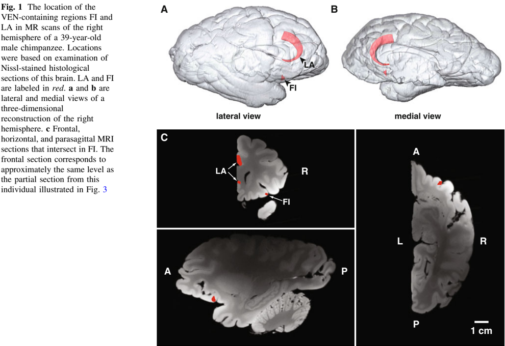
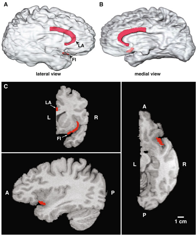
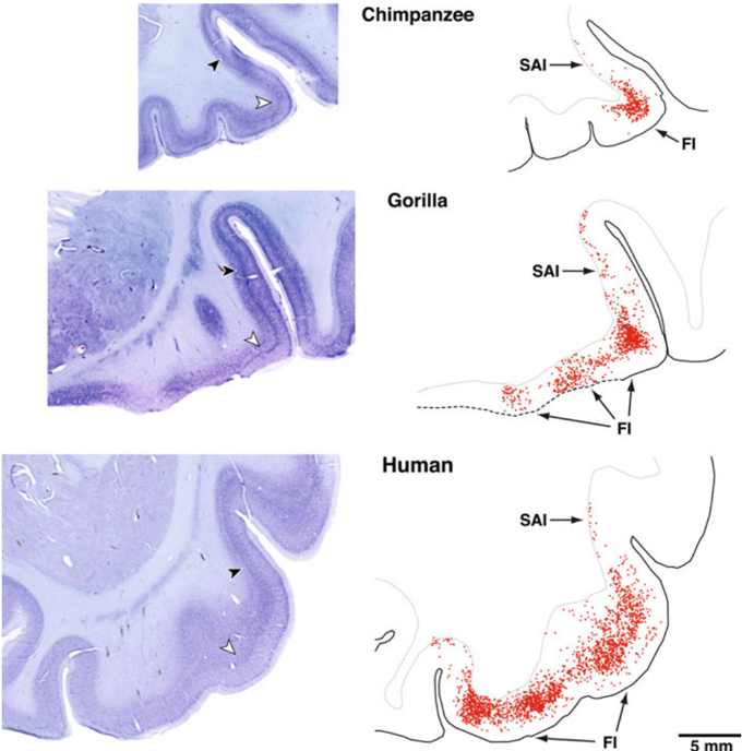
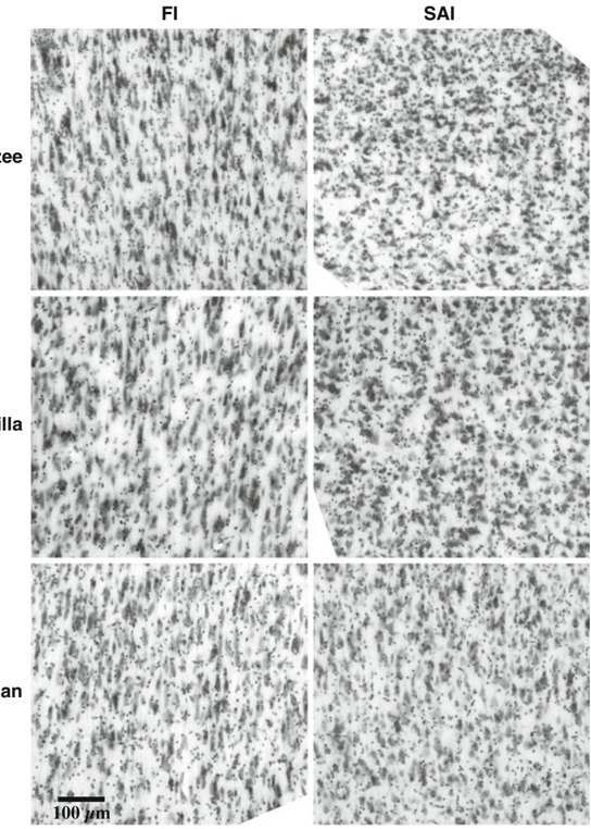
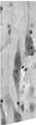
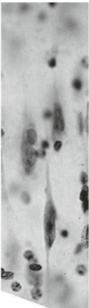
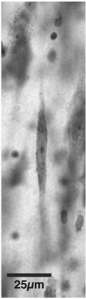
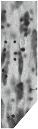
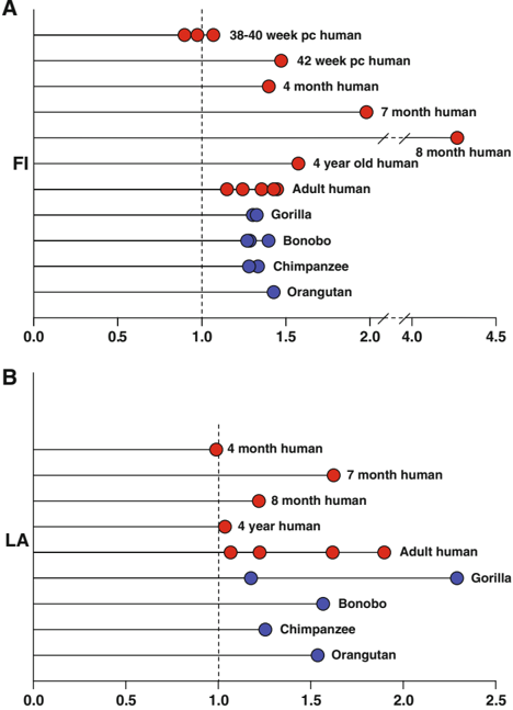
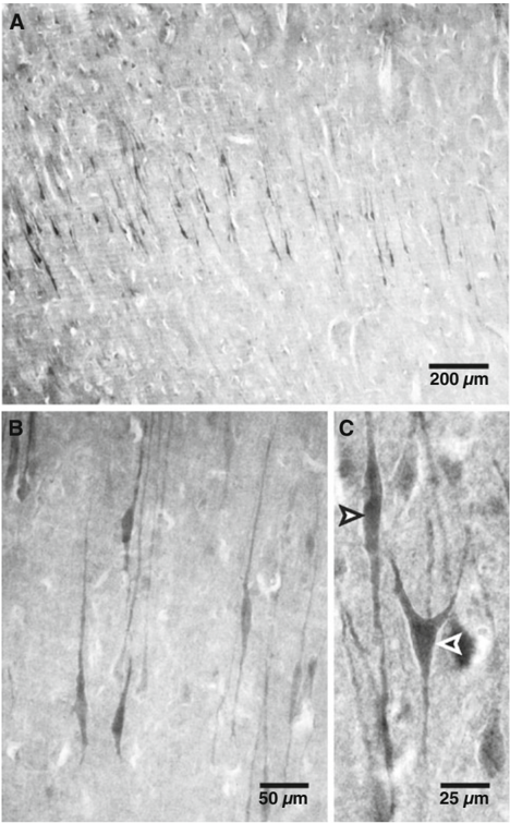
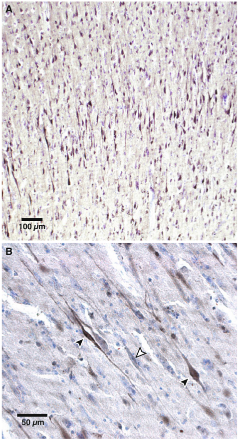
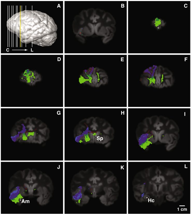
Verification
Claims the paper makes
The claims and statistics of this paper haven’t been checked yet.
Run the check — free, no account needed. It reads the open-access text and checks the sources it cites.
Claims other papers make about this one
Each is checked against this paper’s own text: does it say what the citing paper says it does?
In the citing paper
“However, both regions do correspond to the distribution of VENs. 36”
From A human brain network derived from coma-causing brainstem lesions (2016) · cites this paper as [36]
In the citing paper
“These spindle-shaped neurons are found in the AI and ACC of particular mammals, such as humans, other great apes, whales, dolphins, and elephants. 28,36”
From A human brain network derived from coma-causing brainstem lesions (2016) · cites this paper as [36]
In the citing paper
“Based on their large size and dendrites that span all cortical layers, VENs have been proposed to rapidly integrate and transmit information across large brains. 36”
From A human brain network derived from coma-causing brainstem lesions (2016) · cites this paper as [36]
Self-check
Percentages, sums and reported differences re-computed from the paper’s own numbers.
The self-check needs the paper’s full text.
Citations
Cites
How do you feel — now? The anterior insula and human awareness
(Bud) Craig, A. D. (2009) · Nature Reviews Neuroscience · doi:10.1038/nrn2555 · Cited by 6,657 · cited 2×
“The posterior insula and anterior cingulate cortex in anthropoid primates receive differentiated inputs for pain, itch, warmth, cooling, and sensual touch, which are central components of highly evolved mechanisms for physiological homeostasis (Craig 2003, 2009).”
BackgroundEmpathy for Pain Involves the Affective but not Sensory Components of Pain
Singer, T., Seymour, B., O'Doherty, J., Kaube, H., Dolan, R. J., & Frith, C. D. (2004) · Science · doi:10.1126/science.1093535 · Cited by 3,929
Sridharan, D., Levitin, D. J., & Menon, V. (2008) · Proceedings of the National Academy of Sciences · doi:10.1073/pnas.0800005105 · Cited by 2,892
The neural correlates of maternal and romantic love
Bartels, A., & Zeki, S. (2004) · NeuroImage · doi:10.1016/j.neuroimage.2003.11.003 · Cited by 1,497 · cited 1×
“Anterior insula and anterior cingulate cortex are also activated by pro-social signals, such as love and trust (Bartels and Zeki 2004; Singer et al. 2004b), which suggests that these structures register both negative and positive aspects of the states of social networks.”
BackgroundCardiovascular effects of human insular cortex stimulation
Oppenheimer, S. M., Gelb, A., Girvin, J. P., & Hachinski, V. C. (1992) · Neurology · doi:10.1212/wnl.42.9.1727 · Cited by 1,272
The hidden island of addiction: the insula
Naqvi, N. H., & Bechara, A. (2009) · Trends in Neurosciences · doi:10.1016/j.tins.2008.09.009 · Cited by 833 · cited 1×
“Disruption of this feedback could also underlie the recently recognized role of the insula in the feelings of craving associated with addiction to food or drugs (Naqvi and Bechara 2009).”
BackgroundAgenesis of the corpus callosum: Genetic, developmental and functional aspects of connectivity
Paul, L. K., Brown, W. S., Adolphs, R., Tyszka, J. M., Richards, L. J., Mukherjee, P., & Sherr, E. H. (2007) · Nature Reviews Neuroscience · doi:10.1038/nrn2107 · Cited by 830
The role of anterior insular cortex in social emotions
Lamm, C., & Singer, T. (2010) · Brain Structure and Function · doi:10.1007/s00429-010-0251-3 · Cited by 566
Structural anatomy of empathy in neurodegenerative disease
Rankin, K. P., Gorno-Tempini, M. L., Allison, S. C., Stanley, C. M., Glenn, S., Weiner, M. W., & Miller, B. L. (2006) · Brain · doi:10.1093/brain/awl254 · Cited by 504 · cited 2×
“Rankin et al. (2006) systematically investigated the capacity for empathic concern in a large population of neurological patients and found that this capacity was greatly reduced in FTD patients; while this capacity was variably expressed in Alzheimer's patients it was not significantly reduced…”
BackgroundRole of the anterior insula in task-level control and focal attention
Nelson, S. M., Dosenbach, N. U. F., Cohen, A. L., Wheeler, M. E., Schlaggar, B. L., & Petersen, S. E. (2010) · Brain Structure and Function · doi:10.1007/s00429-010-0260-2 · Cited by 435
Croxson, P. L., Johansen-Berg, H., Behrens, T. E. J., Robson, M. D., Pinsk, M. A., Gross, C. G., Richter, W., Richter, M. C., Kastner, S., & Rushworth, M. F. S. (2005) · The Journal of Neuroscience · doi:10.1523/jneurosci.1311-05.2005 · Cited by 410
Jabbi, M., Bastiaansen, J., & Keysers, C. (2008) · PLoS ONE · doi:10.1371/journal.pone.0002939 · Cited by 362
Ploran, E. J., Nelson, S. M., Velanova, K., Donaldson, D. I., Petersen, S. E., & Wheeler, M. E. (2007) · The Journal of Neuroscience · doi:10.1523/jneurosci.3522-07.2007 · Cited by 282 · cited 2×
“More recently Ploran et al. (2007) found in an experiment employing a behavioral paradigm in which objects gradually emerge from noise that the activity of anterior insula was strongly linked to the moment when the subjects became aware of the identity of the object (allowing for the delays…”
BackgroundActivation of anterior paralimbic structures during guilt-related script-driven imagery
Shin, L. M., Dougherty, D. D., Orr, S. P., Pitman, R. K., Lasko, M., Macklin, M. L., Alpert, N. M., Fischman, A. J., & Rauch, S. L. (2000) · Biological Psychiatry · doi:10.1016/s0006-3223(00)00251-1 · Cited by 252
Shaw, P., Lalonde, F., Lepage, C., Rabin, C., Eckstrand, K., Sharp, W., Greenstein, D., Evans, A., Giedd, J. N., & Rapoport, J. (2009) · Archives of General Psychiatry · doi:10.1001/archgenpsychiatry.2009.103 · Cited by 249 · cited 2×
“3 mm on the right side than the left (Shaw et al. 2009).”
BackgroundInfluentialSpindle neurons of the human anterior cingul. Ate cortex
Nimchinsky, E. A., Vogt, B. A., Morrison, J. H., & Hof, P. R. (1995) · Journal of Comparative Neurology · doi:10.1002/cne.903550106 · Cited by 220
Self-citationBook Review: Two Phylogenetic Specializations in the Human Brain
Allman, J., Hakeem, A., & Watson, K. (2002) · The Neuroscientist · doi:10.1177/107385840200800409 · Cited by 178
Self-citationSeeley, W. W., Allman, J. M., Carlin, D. A., Crawford, R. K., Macedo, M. N., Greicius, M. D., DeArmond, S. J., & Miller, B. L. (2007) · Alzheimer Disease & Associated Disorders · doi:10.1097/wad.0b013e31815c0f14 · Cited by 165
Self-citationNeural Correlates for Feeling-of-Knowing
Kikyo, H., Ohki, K., & Miyashita, Y. (2002) · Neuron · doi:10.1016/s0896-6273(02)00939-x · Cited by 161
Barger, N., Stefanacci, L., & Semendeferi, K. (2007) · American Journal of Physical Anthropology · doi:10.1002/ajpa.20684 · Cited by 79
Self-citationSelective reduction of Von Economo neuron number in agenesis of the corpus callosum
Kaufman, J. A., Paul, L. K., Manaye, K. F., Granstedt, A. E., Hof, P. R., Hakeem, A. Y., & Allman, J. M. (2008) · Acta Neuropathologica · doi:10.1007/s00401-008-0434-7 · Cited by 69
Self-citation
Research works printed but not linked: 87
- Allman JM (2000) Evolving brains. Scientific American Library-W.H.Freeman, New York
- Allman J, McLaughlin T, Hakeem A (1993) Brain weight and life-span in primate species. Proc Natl Acad Sci USA 90:118–122 · DOI
- Allman JM, Hakeem A, Erwin JM, Nimchinsky E, Hof P (2001) Anterior cingulate cortex: the evolution of an interface between emotion and cognition. Ann NY Acad Sci 935:107–117 · DOI
- Allman J, Watson K, Tetreault N, Hakeem A (2005) Intuition and autism: a possible role for von Economo neurons. Trends Cogn Sci 9:367–373 · DOI · Paper page
- Aziz-Zadeh L, Kaplan J, Iocoboni M (2009) “Aha”: the neural correlates of verbal insight solutions. Hum Brain Mapp 30:908–916 · DOI
- Baron-Cohen S, Ring HA, Wheelwright S, Bullmore ET, Brammer MJ, Simmons A, Williams SC (1999) Social intelligence in the normal and autistic brain: an fMRI study. Eur J Neurosci 11:1891–1898 · DOI · Paper page
- Bass AH, Gilland EH, Baker R (2008) Evolutionary origins for social vocalization in a vertebrate hindbrain-spinal compartment. Science 321:417–421 · DOI
- Berthoz S, Armony JL, Blair RJR, Dolan RJ (2002) An fMRI study of intentional and unintentional (embarrassing) violations of social norms. Brain 125:1696–1708 · DOI
- Brauer K, Schober W (1970) Catalogue of mammalian brains. VEB Gustav Fischer Verlag, Jena
- Butti C, Sherwood CC, Hakeem AY, Allman JM, Hof PR (2009) Total number and volume of Von Economo neurons in the cerebral cortex of cetaceans. J Comp Neurol 515:243–259 · DOI · Paper page
- Chubb JE, Bradshaw NJ, Soares DC, Porteous DJ, Millar JK (2008) The DISC locus in psychiatric illness. Mol Psychiatry 13:36–64 · DOI · Paper page
- Craig AD (2003) A new view of pain as a homeostasis emotion. Trends Neurosci 26:303–307 · DOI
- Craig AD (2005) Forebrain emotional asymmetry: a neuroanatomical basis? Trends Cogn Sci 912:566–571 · DOI · Paper page
- Craig AD (2010) The sentient self. Brain Struct Funct. doi: 10.1007/s00429-010-0248-y · DOI · Paper page
- Crespi B, Summers K, Dorus S (2007) Adaptive evolution of genes underlying schizophrenia. Proc Roy Soc B 274:2801–2810 · DOI
- Critchley HD, Elliott R, Mathias CJ, Dolan RJ (2000) Neural activity relating to generation and representation of galvanic skin conductance responses. J Neurosci 20:3033–3040 · DOI · Paper page
- Critchley HD, Mathias C, Dolan R (2001a) Neural activity in the human brain relating to uncertainty and arousal during anticipation. Neuron 29:537–545 · DOI · Paper page
- Critchley HD, Melmed RN, Featherstone E, Mathias CJ, Dolan RJ (2001b) Brain activity during biofeedback relaxation: a functional neuroimaging investigation. Brain 124:1003–1012 · DOI
- Dehaene S, Posner MI, Tucker DM (1994) Localization of a neural system for error detection and compensation. Psychol Sci 5:303–305 · DOI · Paper page
- Di Martino A, Ross K, Uddin L, Sklar A, Castellanos F, Milham M (2009) Processes in autism spectrum disorders: an activation likelihood estimation meta-analysis. Biol Psychiatry 65:63–74 · DOI
- Dosenbach NU, Fair DA, Miezin FM, Cohen AL, Wenger KK, Dosenbach RA, Fox MD, Snyder AZ, Vincent JL, Raichle ME, Schlaggar BL, Petersen SE (2007) Distinct brain networks for adaptive and stable task control in humans. Proc Natl Acad Sci USA 104:11073–11078 · DOI · Paper page
- Duan X, Chang JH, Ge S, Faulkner RL, Kim JY, Kitabatake Y, Liu XB, Yang CH, Jordan JD, Ma DK, Liu CY, Ganesan S, Cheng HJ, Ming GL, Lu B, Song H (2007) Disrupted-in-schizophrenia 1 regulates integration of newly generated neurons in the adult brain. Cell 130:1146–1158 · DOI · Paper page
- Galdikas BMF (1985) Orangutan sociality at Tanjung Puting. Am J Primatol 9:101–119 · DOI
- Gallyas F (1971) A principle for silver staining of tissue elements by physical development. Acta Morphol Acad Sci 19:57–71
- Gehring WJ, Goss B, Coles MGH, Meyer DE, Donchin E (1993) A neural system of error detection and compensation. Psychol Sci 4:385–390 · DOI · Paper page
- Gilbert S, Spengler S, Simons J, Steele J, Lawrie S, Frith C, Burgess P (2006) Functional specialization within rostral prefrontal cortex (Area 10): a meta-analysis. J Cogn Neurosci 18:932–948 · DOI
- Gilles FH, Leviton A, Dooling EC (1983) Developing human brain, growth and epidemiologic neuropathology. John Wright - PSG Inc, Boston
- Gündel H, López-Sala A, Ceballos-Baumann AO, Deus J, Cardoner N, Marten-Mittag B, Soriano-Mas C, Pujol J (2004) Alexithymia correlates with the size of right anterior cingulate. Psychosom Med 66:132–140 · DOI
- Hakeem AY, Sherwood CC, Bonar CJ, Butti C, Hof PR, Allman JM (2009) Von Economo neurons in the elephant brain. Anat Rec (Hoboken) 292:242–248 · DOI
- Hammarberg C (1895) Studien über Klinik und Pathologie der Idiotie nebst Untersuchungen über die normale Anatomie des Hirnrinde. Berling, Uppsala
- Harrison NA, Gray MA, Critchley HD (2009) Dynamic pupillary exchange engages brain regions encoding social salience. Soc Neurosci 4:233–243 · DOI
- Hashimoto R, Numakawa T, Ohnishi T, Kumamaru E, Yagasaki Y, Ishimoto T, Mori T, Nemoto K, Adachi N, Izumi A, Chiba S, Noguchi H, Suzuki T, Iwata N, Ozaki N, Taguchi T, Kamiya A, Kosuga A, Tatsumi M, Kamijima K, Weinberger DR, Sawa A, Kunugi H (2006) Impact of the DISC1 Ser704Cys polymorphism on risk for major depression, brain morphology and ERK signaling. Hum Mol Genet 15:3024–3033 · DOI
- Hayashi M, Ito M, Shimizu K (2001) The spindle neurons are present in the cingulate cortex of chimpanzee fetus. Neurosci Lett 309:97–100 · DOI
- Hof PR, Van der Gucht E (2007) Structure of the cerebral cortex of the humpback whale, Megaptera novaeangliae (Cetacea, Mysticeti, Balaenopteridae). Anat Rec 290:1–31 · DOI · Paper page
- Houdé O, Rossi S, Lubin A, Joliot M (2010) Mapping numerical processing, reading, and executive functions in the developing brain: an fMRI meta-analysis of 52 studies including 842 children. Dev Sci 1:1–10
- Jensen RT, Battey JF, Spindel ER, BEnya RV (2008) International Union of Pharmacology, LXVIII. Mammalian bombesin receptors: nomenclature, distribution, pharmacology, signaling, and functions in normal and disease states. Pharmacol Rev 60:1–42 · DOI
- Kennedy DP, Semendeferi K, Courchesne E (2007) No reduction of spindle neuron number in frontoinsular cortex in autism. Brain Cogn 64:124–129 · DOI
- Klein TA, Endrass T, Kathmann N, Neumann J, von Cramon DY, Ullsperger M (2007) Neural correlates of error awareness. NeuroImage 34:1774–1781 · DOI
- Krueger F, McCabe K, Moll J, Kriegeskorte N, Zahn R, Strenziok M, Heinecke A, Grafman J (2007) Neural correlates of trust. Proc Natl Acad Sci USA 104:20084–20089 · DOI
- Kunimatsu Y, Nakatsukasa M, Sawada Y, Sakai T, Hyodo M, Hyodo H, Itaya T, Nakaya H, Saegusa H, Mazurier A, Saneyoshi M, Tsujikawa H, Yamamoto A, Mbua E (2007) A new Late Miocene great ape from Kenya and its implications for the origins of African great apes and humans. Proc Natl Acad Sci USA 104:19220–19225 · DOI
- Lim M, Young L (2006) Neuropeptidergic regulation of affiliative behavior and social bonding in animals. Horm Behav 50:506–517 · DOI · Paper page
- Lombardo MV, Barnes JL, Wheelwright SJ, Baron-Cohen S (2007) Self-referential cognition and empathy in autism. PLoS One 2:e883 · DOI
- Lorberbaum JP, Newman JD, Horwitz AR, Dubno JR, Lydiard RB, Hamner MB, Bohning DE, George MS (2002) A potential role for thalamocingulate circuitry in human maternal behavior. Biol Psychiatry 51:431–445 · DOI · Paper page
- MacNeilage PF, Rogers LJ, Vallortigara G (2009) Origins of the left & right brain. Sci Am 301:60–67 · DOI
- Merker B (1983) Silver staining of cell bodies by means of physical development. J Neurosci Methods 9:235–241 · DOI
- Mitani JC, Grether GF, Rodman PS, Priatna D (1991) Associations among wild orangutans: sociality, passive aggregations or chance? Anim Behav 42:33–46 · DOI
- Mutschler I, Wieckhorst B, Kowalevski S, Derix J, Wentlandt J, Schulze-Bonhage A, Ball T (2009) Functional organization of the human interior insular cortex. Neurosci Lett 457:66–70 · DOI
- Ngowyang G (1932) Beschreibung einer Art von Spezialzellen in der Inselrinde - zugleich Bemerkungen über die v. Economoschen Spezialzellen. J Psychol Neurol 44:671–674
- Nimchinsky E, Gilissen E, Allman J, Perl D, Erwin J, Hof P (1999) A neuronal morphologic type unique to humans and great apes. Proc Nat Acad Sci USA 96:5268–5273 · DOI · Paper page
- Numan R (2000) The behavioral neuroscience of the septal region. Springer, New York · DOI
- Patterson F, Gordon W (2002) Twenty-seven years of Project Koko and Michael. In: Galdikas B, Briggs N, Sheeran L, Shapiro G, Goodall J (eds) All apes great and small, vol I. Kluwer Press, New York, pp 165–176 · DOI
- Phillips ML, Young AW, Senior C, Brammer M, Andrew C, Calder AJ, Bullmore ET, Perrett DI, Rowland D, Williams SC, Gray JA, David AS (1997) A specific neural substrate for perceiving facial expressions of disgust. Nature 389:495–498 · DOI · Paper page
- Preuschhoff K, Quartz S, Bossaerts P (2008) Human insula activation reflects risk prediction errors as well as risk. J Neurosci 28:2745–2752 · DOI · Paper page
- Ramón y Cajal S (1899) Textura del Sistema Nervioso del Hombre y de los Vertebrados, Tomo II. Nicolás Moya, Madrid
- Rilling J, Goldsmith D, Glenn A, Jairam M, Elfenbein H, Dagenais J, Murdock C, Pagnoni (2008) Neural correlates of the affective response to unreciprocated cooperation. Neuropsychologia 46:1265–1266 · DOI
- Rogers L, Andrew R (2002) Comparative vertebrate lateralization. Cambridge University Press, Cambridge · DOI
- Rose M (1927) Gyrus limbicus anterior und Regio retrosplenialis (Cortex holoprotoptychos quinquestratificatus) - Vergleichende Architektonik bei Tier und Menschen. J Psychol Neurol 35:5–217
- Rose M (1928) Die Inselrinde des Menschen und der Tiere. J Psychol Neurol 37:467–624
- Sanfey AG, Rilling RJ, Aronson JA, Nystrom LE, Cohen JD (2003) The neural basis of economic decision-making in the ultimatum game. Science 300:1755–1758 · DOI · Paper page
- Schenker NM, Desgouttes A, Semendeferi K (2005) Neural connectivity and cortical substrates of cognition in hominoids. J Hum Evol 49:547–569 · DOI
- Seeley WW (2008) Selective functional, regional, and neuronal vulnerability in frontotemporal dementia. Curr Opin Neurol 21:701–707 · DOI
- Seeley WW, Carlin DA, Allman JM, Macedo MN, Bush C, Miller BL, Dearmond SJ (2006) Early frontotemporal dementia targets neurons unique to apes and humans. Ann Neurol 60:660–667 · DOI · Paper page
- Semendeferi K, Armstrong E, Schleicher A, Zilles K, Van Hoesen GW (1998) Limbic frontal cortex in hominoids: a comparative study of area 13. Am J Phys Anthropol 106:129–155 · DOI
- Sherwood CC, Holloway RL, Erwin JM, Hof PR (2004) Cortical orofacial motor representation in Old World monkeys, great apes, and humans. II: stereologic analysis of chemoarchitecture. Brain Behav Evol 63:82–106 · DOI
- Sherwood CC, Wahl E, Erwin JM, Hof PR, Hopkins WD (2007) Histological asymmetries of primary motor cortex predict handedness in chimpanzees. J Comp Neurol 503:525–537 · DOI
- Shorvon S (2005) Handbook of epilepsy treatment. Wiley, New York · DOI
- Simms ML, Kemper TL, Timbie CM, Bauman ML, Blatt GJ (2009) The anterior cingulate cortex in autism: heterogeneity of qualitative and quantitative cytoarchitectonic features suggests possible subgroups. Acta Neuropathol 118:673–684 · DOI · Paper page
- Singer T, Kiebel SJ, Winston JS, Dolan RJ, Frith CD (2004b) Brain responses to the acquired moral status of faces. Neuron 41:653–662 · DOI
- Singleton I, van Schaik CP (2002) The social organisation of a population of Sumatran orangutans. Folia Primatol 73:1–20 · DOI
- Spence SA, Farrow TF, Herford AE, Wilkinson ID, Zheng Y, Woodruff PW (2001) Behavioural and functional anatomical correlates of deception in humans. NeuroReport 12:2849–2853 · DOI
- Stephan H, Frahm HD, Baron G (1981) New and revised data on volumes of brain structures in insectivores and primates. Folia Primatol 35:1–29 · DOI
- Szebenyi G, Bollati F, Bisbal M, Sheridan S, Faas L, Wray R, Haferkamp S, Nguyen S, Caceres A, Brady S (2005) Activity-driven dendritic remodeling requires microtubule-associated Protein 1a. Curr Biol 15:1820–1826 · DOI
- Ullsperger M, von Cramon DY (2004) Neuroimaging of performance monitoring: error detection and beyond. Cortex 40:593–604 · DOI
- Uylings H, Jacosen A, Zilles K, Amunts K (2006) Left-right asymmetry in volume and number of neurons in adult Broca’s area. Cortex 42:652–658 · DOI
- van Kooten (2008) Autism counts. Dissertation, Maastricht University
- von Economo C (1926) Eine neue Art Spezialzellen des Lobus cinguli und Lobus insulae. Zschr ges Neurol Psychiat 100:706–712 · DOI
- Von Economo C (2009) Cellular structure of the human cerebral cortex. Translated by L.C. Triarhou, Karger, Basel
- Von Economo C, Koskinas G (1925) Die Cytoarchitectonik der Hirnrinde des erwachsenen Menschen. Springer, Berlin
- Wang SS, Shultz JR, Burish MJ, Harrison KH, Hof PR, Towns LC, Wagers MW, Wyatt KD (2008) Functional trade-offs in white matter axonal scaling. J Neurosci 28:4047–4056 · DOI
- Watkins KE, Paus T, Lerch JP, Zijdenbos A, Collins DL, Neelin P, Taylor J, Worsley KJ, Evans AC (2001) Structural asymmetries in human brain: a voxel-based statistical analysis of 142 brains. Cereb Cortex 11:868–877 · DOI
- Watson KK, Jones TK, Allman JM (2006) Dendritic architecture of the von Economo neurons. Neuroscience 141:1107–1112 · DOI
- Watson KK, Matthews BJ, Allman JM (2007) Brain activation during sight gags and language-dependent humor. Cereb Cortex 17:314–324 · DOI
- Welker WI, Seidenstein S (1959) Somatic sensory representation in the cerebral cortex of the raccoon (Procyon lotor). J Comp Neurol 111:469–501 · DOI
- Williamson P, Allman JM (2010) The human illnesses: neuropsychiatric disorders and the nature of the human brain. Oxford University Press (in press)
- Wittling W (1995) Brain Asymmetry in the control of autonomic-physiological activity. In: Davidson RJ, Hugdahl K (eds) Brain asymmetry. MIT Press, Cambridge, pp 305–356
- Woodward J, Allman J (2007) Moral intuition: its neural substrates and normative significance. J Physiol Paris 101(4–6):179–202 · DOI
- Woolley JD, Gorno-Tempini ML, Seeley WW, Rankin K, Lee SS, Matthews BR, Miller BL (2007) Binge eating is associated with right orbitofrontal-insular-striatal atrophy in frontotemporal dementia. Neurology 69:1424–1433 · DOI
Cited by
Sorted by citations
Saliency, switching, attention and control: a network model of insula function
Menon, V., & Uddin, L. Q. (2010) · Brain Structure and Function · doi:10.1007/s00429-010-0262-0 · Cited by 5,592
Interoceptive inference, emotion, and the embodied self
Seth, A. K. (2013) · Trends in Cognitive Sciences · doi:10.1016/j.tics.2013.09.007 · Cited by 1,816
Gut feelings: the emerging biology of gut-brain communication
Mayer, E. A. (2011) · Nature Reviews Neuroscience · doi:10.1038/nrn3071 · Cited by 1,726
Hamilton, J. P., Etkin, A., Furman, D. J., Lemus, M. G., Johnson, R. F., & Gotlib, I. H. (2012) · American Journal of Psychiatry · doi:10.1176/appi.ajp.2012.11071105 · Cited by 866
Functional connectivity of the insula in the resting brain
Cauda, F., D'Agata, F., Sacco, K., Duca, S., Geminiani, G., & Vercelli, A. (2011) · NeuroImage · doi:10.1016/j.neuroimage.2010.11.049 · Cited by 793
Metadata
- Authors
- Allman, J. M., Tetreault, N. A., Hakeem, A. Y., Manaye, K. F., Semendeferi, K., Erwin, J. M., Park, S., Goubert, V., & Hof, P. R.
- Venue
- Brain Structure and Function
- Published
- 2010-05-29
- Volume
- 214
- Issue
- 5-6
- Pages
- 495-517
- ISSN
- 1863-2653
- 1863-2661
- Cited by
- 354
- References
- 21
- Language
- en
About these checks
Registry, retraction, Cites and statistics results are computed, not judged, with one exception: a printed reference that no index lists is read once by a model before it counts as a citation error. Claims are extracted by a model and kept only when their quote appears verbatim in the text; each cited source is then checked, with a model’s help, for whether it says what the claim relies on. Those verdicts can be wrong — read the claim’s quote and the cited source before relying on one.
Statistics and arithmetic are recomputed, not judged.
Confidence bands are shown only for fields calibrated on our annotated test set. None is calibrated yet, so no band is shown.
What counts as a claim ›How we measure confidence ›How we check this ›
Are you an author, or do you see a problem with this page? Request a review
