

The DOI matches its Crossref record; 89 of the 89 references it cites were found in a registry or an index; none of the 77 sources its claims cite is checked for support yet.
Large-scale functional neural network correlates of response inhibition: an fMRI meta-analysis
Open accessAbstract
An influential hypothesis from the last decade proposed that regions within the right inferior frontal cortex of the human brain were dedicated to supporting response inhibition. There is growing evidence, however, to support an alternative model, which proposes that neural areas associated with specific inhibitory control tasks co-exist as common network mechanisms, supporting diverse cognitive processes. This meta-analysis of 225 studies comprising 323 experiments examined the common and distinct neural correlates of cognitive processes for response inhibition, namely interference resolution, action withholding, and action cancellation. Activation coordinates for each subcategory were extracted using multilevel kernel density analysis (MKDA). The extracted activity patterns were then mapped onto the brain functional network atlas to derive the common (i.e., process-general) and distinct (i.e., domain-oriented) neural network correlates of these processes. Independent of the task types, activation of the right hemispheric regions (inferior frontal gyrus, insula, median cingulate, and paracingulate gyri) and superior parietal gyrus was common across the cognitive processes studied. Mapping the activation patterns to a brain functional network atlas revealed that the fronto-parietal and ventral attention networks were the core neural systems that were commonly engaged in different processes of response inhibition. Subtraction analyses elucidated the distinct neural substrates of interference resolution, action withholding, and action cancellation, revealing stronger activation in the ventral attention network for interference resolution than action inhibition. On the other hand, action withholding/cancellation primarily engaged the fronto-striatal circuit. Overall, our results suggest that response inhibition is a multidimensional cognitive process involving multiple neural regions and networks for coordinating optimal performance. This finding has significant implications for the understanding and assessment of response inhibition.
Overview
AI snapshot
Written from the paper’s full text.
TL;DR
This meta-analysis of 225 fMRI studies (323 experiments) finds that response inhibition commonly engages right inferior frontal gyrus/insula, median cingulate/paracingulate gyri, and superior parietal gyrus, with fronto-parietal and ventral attention networks as core systems. Interference resolution, action withholding, and action cancellation share these networks but show distinct recruitment patterns, including stronger ventral attention network engagement for interference resolution and fronto-striatal involvement for action withholding/cancellation.
Key findings
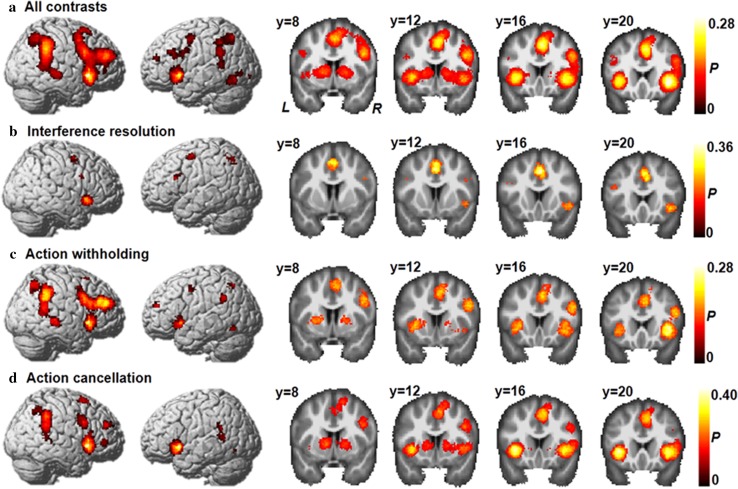
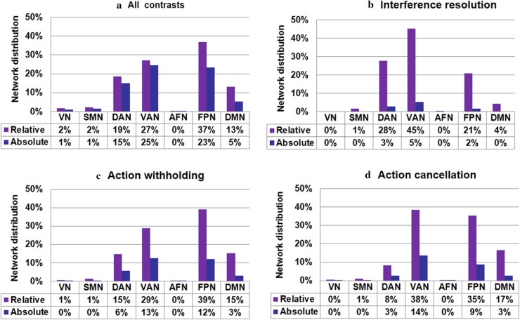
- Across all 225 studies/323 experiments/3453 foci, significant right-hemisphere clusters included frontal cortex, angular gyrus, and supplementary motor area; activated areas were primarily distributed in the fronto-parietal network (36%), ventral attention network (27%), dorsal attention network (18%), and default mode network (13%).
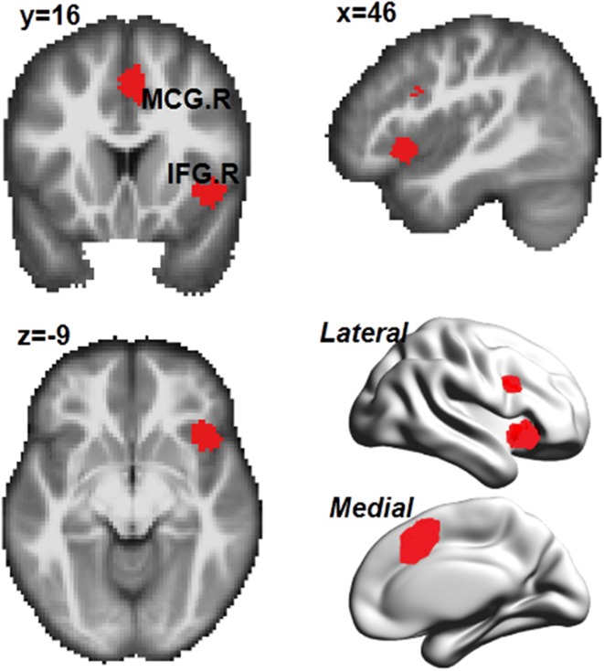
- Common across the three subcategories were the right inferior frontal gyrus extending to insula, right median cingulate and paracingulate gyri, and right superior parietal gyrus; the common network distribution was ventral attention network (61%), fronto-parietal network (26%), default mode network (8%), and dorsal attention network (4%).
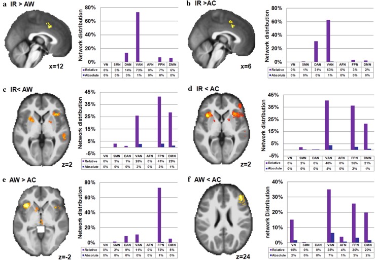
- Subcategory network distributions were: interference resolution — ventral attention network (45%), dorsal attention network (27%), fronto-parietal network (20%); action withholding — fronto-parietal network (39%), ventral attention network (28%), default mode network (15%), dorsal attention network (14%); action cancellation — ventral attention network (38%), fronto-parietal network (35%), default mode network (16%), dorsal attention network (8%).
- Subtraction/chi-square analyses showed interference resolution vs. action withholding differed significantly (χ² = 19.93, df = 6, p = 0.003) and interference resolution vs. action cancellation differed significantly (χ² = 22.37, df = 6, p = 0.001); interference resolution showed stronger ventral attention network activation than action inhibition, while action withholding/cancellation primarily engaged the fronto-striatal circuit.
Methods
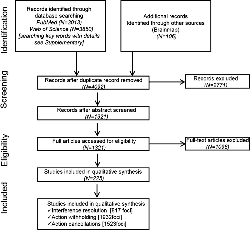
- Literature search used PubMed, Web of Science, and BrainMap; 4092 reports were screened and 225 full articles were included. Experiments were categorized as interference resolution (50 articles, 68 experiments, 817 foci), action withholding (117 articles, 147 contrasts, 1932 foci), and action cancellation (73 articles, 108 experiments, 1523 foci).
- Activation coordinates were analyzed with multilevel kernel density analysis (MKDA) using a 5-mm spherical kernel, sample-size-weighted density maps, and Monte Carlo family-wise error correction (p < 0.05, FWE corrected across the entire brain); non-MNI coordinates were transformed using the Lancaster transformation.
- Significant voxels were overlaid onto seven functional networks (fronto-parietal, dorsal attention, ventral attention, somatomotor, visual, affective, default mode); chi-square tests and subtraction analyses compared subcategories, with validation via leave-one-out cross-validation and random selection of 68 contrasts.
Limitations
- The paper states that synthesizing different interference-resolution tasks (Stroop, Simon, Flanker, etc.) increases heterogeneity of paradigms and designs, although leave-one-out cross-validation showed highly replicable network distribution profiles; future meta-analyses with common stimulus–response incompatibility tasks are needed.
- The paper states that simple and complex Go/NoGo tasks may not be perfectly matched across included studies; the text reports high vs. low NoGo frequency comparisons as not significantly different, but the validation table shows the high-frequency NoGo-only contrast differed significantly (χ² = 43.52, df = 6, p < 0.01).
- The paper states that stop signal tasks may draw more on proactive than reactive control; a supplementary chi-square test found no significant difference between all contrasts and Stop–Go-only contrasts, but heterogeneity of action cancellation tasks remains a concern. Evident from the text: coordinate-based MKDA synthesizes published peak coordinates, so it cannot test individual-level or causal network dynamics.
Model-written summary. Check it against the paper before relying on it.
Integrity signals
Registry
Cites
Statistics
Figures
Verification
Claims the paper makes
54 Not checked yet · 52 Own result · 15 Own interpretation · 12 Own method · 1 Sources not found · 1 No basis given
A result the paper reports from its own data — it cites no other source for it, so there is none to check. The quote shows it is in the paper.
In the paper
“More precisely, cluster activations in the frontal cortex included the insula, inferior frontal gyrus, middle frontal gyrus, and superior frontal gyrus.”
§ Large-scale functional neural network correlates of response inhibition: an fMRI meta-analysis > Results > Meta-analysis of all included response inhibition experiments
plain- Asserted: passes — The authors put it forward as true.
- Checkable: passes — It can be checked against evidence.
- Load-bearing: passes — The paper’s conclusions depend on it.
- Based: passes — The paper gives a reason why it holds.
A result the paper reports from its own data — it cites no other source for it, so there is none to check. The quote shows it is in the paper.
In the paper
“The all-inclusive analysis of the 225 studies showed significant activations of three large clusters in the right hemisphere: the frontal cortex, the angular gyrus, and the supplementary motor area (Fig. 3a and Table S3 in Supplementary Materials).”
§ Large-scale functional neural network correlates of response inhibition: an fMRI meta-analysis > Results > Meta-analysis of all included response inhibition experiments
plaincorrelational- Asserted: passes — The authors put it forward as true.
- Checkable: passes — It can be checked against evidence.
- Load-bearing: passes — The paper’s conclusions depend on it.
- Based: passes — The paper gives a reason why it holds.
A result the paper reports from its own data — it cites no other source for it, so there is none to check. The quote shows it is in the paper.
In the paper
“In addition, several areas in the left hemisphere, including the insula, putamen, and middle frontal gyrus were consistently activated across all response inhibition tasks.”
§ Large-scale functional neural network correlates of response inhibition: an fMRI meta-analysis > Results > Meta-analysis of all included response inhibition experiments
plaincorrelational- Asserted: passes — The authors put it forward as true.
- Checkable: passes — It can be checked against evidence.
- Load-bearing: unclear — It is unclear whether the paper’s conclusions depend on it.
- Based: passes — The paper gives a reason why it holds.
A result the paper reports from its own data — it cites no other source for it, so there is none to check. The quote shows it is in the paper.
In the paper
“The regions activated by interference resolution were located in a subset of the previously mentioned clusters (Fig. 3b), including the left supplementary motor area, the left inferior parietal lobule, the left precentral gyrus, the right insula, the right middle frontal gyrus, and bilateral inferior frontal gyri.”
§ Large-scale functional neural network correlates of response inhibition: an fMRI meta-analysis > Results > Brain activation patterns of each category
plaincorrelational- Asserted: passes — The authors put it forward as true.
- Checkable: passes — It can be checked against evidence.
- Load-bearing: unclear — It is unclear whether the paper’s conclusions depend on it.
- Based: passes — The paper gives a reason why it holds.
A result the paper reports from its own data — it cites no other source for it, so there is none to check. The quote shows it is in the paper.
In the paper
“Furthermore, activation of the angular gyrus cluster extended to the superior temporal gyrus, while activation of the supplementary motor area extended to the middle frontal gyrus.”
§ Large-scale functional neural network correlates of response inhibition: an fMRI meta-analysis > Results > Meta-analysis of all included response inhibition experiments
plaincorrelational- Asserted: passes — The authors put it forward as true.
- Checkable: passes — It can be checked against evidence.
- Load-bearing: unclear — It is unclear whether the paper’s conclusions depend on it.
- Based: passes — The paper gives a reason why it holds.
A result the paper reports from its own data — it cites no other source for it, so there is none to check. The quote shows it is in the paper.
In the paper
“Furthermore, activation of the angular gyrus cluster extended to the superior temporal gyrus, while activation of the supplementary motor area extended to the middle frontal gyrus.”
§ Large-scale functional neural network correlates of response inhibition: an fMRI meta-analysis > Results > Meta-analysis of all included response inhibition experiments
plaincorrelational- Asserted: passes — The authors put it forward as true.
- Checkable: passes — It can be checked against evidence.
- Load-bearing: unclear — It is unclear whether the paper’s conclusions depend on it.
- Based: passes — The paper gives a reason why it holds.
A result the paper reports from its own data — it cites no other source for it, so there is none to check. The quote shows it is in the paper.
In the paper
“By overlapping these clusters with the seven functional networks, we discovered that the activated areas were primarily distributed in the fronto-parietal network (36%), ventral attention network (27%), dorsal attention network (18%), and default mode network (13%) (Table S9 in Supplementary Materials) (Fig. 4a).”
§ Large-scale functional neural network correlates of response inhibition: an fMRI meta-analysis > Results > Meta-analysis of all included response inhibition experiments
plain- Asserted: passes — The authors put it forward as true.
- Checkable: passes — It can be checked against evidence.
- Load-bearing: passes — The paper’s conclusions depend on it.
- Based: passes — The paper gives a reason why it holds.
A result the paper reports from its own data — it cites no other source for it, so there is none to check. The quote shows it is in the paper.
In the paper
“These activations extended to the subcortical regions (the thalamus and pallidum).”
§ Large-scale functional neural network correlates of response inhibition: an fMRI meta-analysis > Results > Meta-analysis of all included response inhibition experiments
plaincorrelational- Asserted: passes — The authors put it forward as true.
- Checkable: passes — It can be checked against evidence.
- Load-bearing: unclear — It is unclear whether the paper’s conclusions depend on it.
- Based: passes — The paper gives a reason why it holds.
A result the paper reports from its own data — it cites no other source for it, so there is none to check. The quote shows it is in the paper.
In the paper
“Furthermore, relative to action cancellation, action withholding primarily recruited the fronto-parietal network.”
§ Large-scale functional neural network correlates of response inhibition: an fMRI meta-analysis > Discussion
plaincorrelational- Asserted: passes — The authors put it forward as true.
- Checkable: passes — It can be checked against evidence.
- Load-bearing: passes — The paper’s conclusions depend on it.
- Based: passes — The paper gives a reason why it holds.
A result the paper reports from its own data — it cites no other source for it, so there is none to check. The quote shows it is in the paper.
In the paper
“Independent of the task type, brain areas including the right inferior frontal gyrus extending to the insula, the right median cingulate, and paracingulate gyri, and the right superior parietal gyrus were activated across all paradigm classes.”
§ Large-scale functional neural network correlates of response inhibition: an fMRI meta-analysis > Discussion
plaincorrelational- Asserted: passes — The authors put it forward as true.
- Checkable: passes — It can be checked against evidence.
- Load-bearing: passes — The paper’s conclusions depend on it.
- Based: passes — The paper gives a reason why it holds.
- It may state two separate claims as one. 33
- The quote only partly states this claim. 1
- Probably not a substantial claim. 24
A result the paper reports from its own data — it cites no other source for it, so there is none to check. The quote shows it is in the paper.
In the paper
“The activations were distributed in the fronto-parietal network (39%), the ventral attention network (28%), the dorsal attention network (14%), and the default mode network (15%) (Fig. 4c).”
§ Large-scale functional neural network correlates of response inhibition: an fMRI meta-analysis > Results > Brain activation patterns of each category
plaincorrelational- Asserted: passes — The authors put it forward as true.
- Checkable: passes — It can be checked against evidence.
- Load-bearing: passes — The paper’s conclusions depend on it.
- Based: passes — The paper gives a reason why it holds.
It may state two separate claims as one.
A result the paper reports from its own data — it cites no other source for it, so there is none to check. The quote shows it is in the paper.
In the paper
“The activation distribution included the fronto-parietal network (35%), the ventral attention network (38%), the default mode network (16%), and the dorsal attention network (8%) (Fig. 4d).”
§ Large-scale functional neural network correlates of response inhibition: an fMRI meta-analysis > Results > Brain activation patterns of each category
plaincorrelational- Asserted: passes — The authors put it forward as true.
- Checkable: passes — It can be checked against evidence.
- Load-bearing: passes — The paper’s conclusions depend on it.
- Based: passes — The paper gives a reason why it holds.
It may state two separate claims as one.
A result the paper reports from its own data — it cites no other source for it, so there is none to check. The quote shows it is in the paper.
In the paper
“Interference resolution was found to draw on the ventral attention network, while fronto-parietal network was implicated in action withholding/cancellation.”
§ Large-scale functional neural network correlates of response inhibition: an fMRI meta-analysis > Discussion > Distinct neural networks
plaincorrelational- Asserted: passes — The authors put it forward as true.
- Checkable: passes — It can be checked against evidence.
- Load-bearing: passes — The paper’s conclusions depend on it.
- Based: passes — The paper gives a reason why it holds.
It may state two separate claims as one.
A result the paper reports from its own data — it cites no other source for it, so there is none to check. The quote shows it is in the paper.
In the paper
“In this study, we found that across all 225 studies with 323 experiments, areas in the dorsal lateral frontal cortex, pre-supplementary motor area, and temporal parietal junction were consistently activated during all response inhibition tasks (Fig. 3a), i.e., activation was mainly distributed in the fronto-parietal network.”
§ Large-scale functional neural network correlates of response inhibition: an fMRI meta-analysis > Discussion > Common neural networks
plaincorrelational- Asserted: passes — The authors put it forward as true.
- Checkable: passes — It can be checked against evidence.
- Load-bearing: passes — The paper’s conclusions depend on it.
- Based: passes — The paper gives a reason why it holds.
It may state two separate claims as one.
A result the paper reports from its own data — it cites no other source for it, so there is none to check. The quote shows it is in the paper.
In the paper
“Furthermore, we observed that interference resolution, which was related to greater activation in the ventral and dorsal attention networks (Fig. 6a, b), involved the left supplementary area, the left precentral gyrus, and the left superior parietal gyrus.”
§ Large-scale functional neural network correlates of response inhibition: an fMRI meta-analysis > Results > Activation differences among different classifications
plaincorrelational- Asserted: passes — The authors put it forward as true.
- Checkable: passes — It can be checked against evidence.
- Load-bearing: passes — The paper’s conclusions depend on it.
- Based: passes — The paper gives a reason why it holds.
It may state two separate claims as one.
The paper states this without a source or data behind it here, so there is nothing to check it against. The quote shows it is in the paper.
In the paper
“Based on the reported characterizations of functional neural networks, these clusters appeared to be located at the ventral attention network (45%), the dorsal attention network (27%), and the fronto-parietal network (20%) (Table 1; Fig. 4b).”
§ Large-scale functional neural network correlates of response inhibition: an fMRI meta-analysis > Results > Brain activation patterns of each category
hedgedcorrelational- Asserted: passes — The authors put it forward as true.
- Checkable: passes — It can be checked against evidence.
- Load-bearing: passes — The paper’s conclusions depend on it.
- Based: passes — The paper gives a reason why it holds.
It may state two separate claims as one.
A result the paper reports from its own data — it cites no other source for it, so there is none to check. The quote shows it is in the paper.
In the paper
“The activated regions included the bilateral insula cortex, which extended to the basal ganglia (e.g., caudate and putamen) and inferior frontal gyrus; the right supplementary motor area, which extended to the median cingulate and paracingulate gyri; the bilateral superior temporal gyri; and the right inferior parietal lobule (Table 3; Fig. 3d).”
§ Large-scale functional neural network correlates of response inhibition: an fMRI meta-analysis > Results > Brain activation patterns of each category
plaincorrelational- Asserted: passes — The authors put it forward as true.
- Checkable: passes — It can be checked against evidence.
- Load-bearing: passes — The paper’s conclusions depend on it.
- Based: passes — The paper gives a reason why it holds.
It may state two separate claims as one. The quote only partly states this claim.
A result the paper reports from its own data — it cites no other source for it, so there is none to check. The quote shows it is in the paper.
In the paper
“Meanwhile, subtraction analyses suggested that some specific neural correlates are involved in different categories of response inhibition tasks”
- Also stated in: § Conclusions · plain
“Subtraction analyses elucidated the distinct neural substrates of interference resolution, action withholding, and action cancellation”
§ Large-scale functional neural network correlates of response inhibition: an fMRI meta-analysis > Discussion > Implications
plaincorrelational- Asserted: passes — The authors put it forward as true.
- Checkable: passes — It can be checked against evidence.
- Load-bearing: passes — The paper’s conclusions depend on it.
- Based: passes — The paper gives a reason why it holds.
It may state two separate claims as one.
- Also stated in: § Conclusions · plain
In the paper
“Indeed, studies have suggested that response inhibition is correlated with the connections between the cingulo-opercular network (salience network) and fronto-parietal network, and that default mode network activity might be actively downregulated by an inhibitory module within the fronto-parietal network (Leech et al. 2011).”
§ Large-scale functional neural network correlates of response inhibition: an fMRI meta-analysis > Introduction > Neural correlates of response inhibition
What the sources say
- Not checked yetcites Leech et al. 2011 source
hedgedcorrelational- Asserted: passes — The authors put it forward as true.
- Checkable: passes — It can be checked against evidence.
- Load-bearing: passes — The paper’s conclusions depend on it.
- Based: passes — The paper gives a reason why it holds.
Probably not a substantial claim.
None of the sources cited for this claim have been checked yet.
In the paper
“Nonetheless, unique neural networks for the three subcategories of processes underpinning response inhibition have also been suggested (Rubia et al. 2001; Sebastian et al. 2013b; Chevrier et al. 2007; van Velzen et al. 2014; Sebastian et al. 2013a).”
§ Large-scale functional neural network correlates of response inhibition: an fMRI meta-analysis > Introduction > Neural correlates of response inhibition
What the sources say
hedgedcorrelational- Asserted: passes — The authors put it forward as true.
- Checkable: passes — It can be checked against evidence.
- Load-bearing: passes — The paper’s conclusions depend on it.
- Based: passes — The paper gives a reason why it holds.
Probably not a substantial claim.
None of the sources cited for this claim have been checked yet.
In the paper
“Dysfunctional response inhibition is a core symptom observed in various mental disorders, including attention deficit hyperactivity disorder (Schachar et al. 2007), schizophrenia (Vercammen et al. 2012), and depression (Palazidou 2012), as well as learning difficulties (Eickhoff et al. 2009) and behavioral problems (Liu et al. 2012).”
§ Large-scale functional neural network correlates of response inhibition: an fMRI meta-analysis > Introduction
What the sources say
plaincorrelational- Asserted: passes — The authors put it forward as true.
- Checkable: passes — It can be checked against evidence.
- Load-bearing: passes — The paper’s conclusions depend on it.
- Based: passes — The paper gives a reason why it holds.
It may state two separate claims as one.
What the paper did — it cites no source for this, so there is none to check. The quote shows it is in the paper.
In the paper
“Random selections of 68 contrasts from action withholding and action cancellation were performed to test the effect of the experiment numbers. We repeated the MKDA analysis using the same settings, and Chi-square analyses were performed to compare the activated voxels in the seven network correlates to those of our main analyses and the random selected studies.”
§ Large-scale functional neural network correlates of response inhibition: an fMRI meta-analysis > Methods and materials > Effect of contrast numbers
plain- Asserted: passes — The authors put it forward as true.
- Checkable: passes — It can be checked against evidence.
- Load-bearing: fails — The paper’s conclusions may not depend on it.
- Based: passes — The paper gives a reason why it holds.
Probably not a substantial claim. The paper’s conclusions may not depend on it. It may state two separate claims as one.
None of the sources cited for this claim have been checked yet.
In the paper
“Paradigms such as the stroop, Flanker, Simon, stimulus response compatibility (SRC), antisaccade tasks, stop signal, and Go/NoGo tasks, which require a response to a target stimulus among irrelevant/distracting stimuli, are commonly regarded as paradigms that involve inhibitory action control (Nigg 2000) and are commonly used for the investigation of response inhibition mechanisms (Nee et al. 2007”
§ Large-scale functional neural network correlates of response inhibition: an fMRI meta-analysis > Introduction > Measurement of response inhibition
What the sources say
plaincorrelational- Asserted: passes — The authors put it forward as true.
- Checkable: passes — It can be checked against evidence.
- Load-bearing: passes — The paper’s conclusions depend on it.
- Based: passes — The paper gives a reason why it holds.
Probably not a substantial claim. It may state two separate claims as one.
What the paper did — it cites no source for this, so there is none to check. The quote shows it is in the paper.
In the paper
“Thus, the reported meta-analytic results in this study represent consistently activated regions across studies: regions in which significant activations were observed in the local neighborhood by more studies than would be expected by chance (p < 0.05, FWE corrected across the entire brain).”
§ Large-scale functional neural network correlates of response inhibition: an fMRI meta-analysis > Methods and materials > Multilevel kernel density analysis (MKDA)
plaincorrelational- Asserted: passes — The authors put it forward as true.
- Checkable: passes — It can be checked against evidence.
- Load-bearing: passes — The paper’s conclusions depend on it.
- Based: passes — The paper gives a reason why it holds.
Probably not a substantial claim.
What the paper did — it cites no source for this, so there is none to check. The quote shows it is in the paper.
In the paper
“The family wise error (FWE) rate was estimated with a Monte Carlo simulation to correct for multiple comparisons, and a natural null hypothesis was that the “activated” regions are randomly distributed throughout the brain.”
§ Large-scale functional neural network correlates of response inhibition: an fMRI meta-analysis > Methods and materials > Multilevel kernel density analysis (MKDA)
plain- Asserted: passes — The authors put it forward as true.
- Checkable: passes — It can be checked against evidence.
- Load-bearing: passes — The paper’s conclusions depend on it.
- Based: passes — The paper gives a reason why it holds.
Probably not a substantial claim. It may state two separate claims as one.
In the paper
“For example, the Simon task (interference resolution) was found to stimulate the pre-motor and parietal regions to a greater extent than action cancellation (stop signals), whereas action cancellation was found to elicit stronger activation in the bilateral posterior inferior frontal gyrus/insula and the right striatum than action withholding (Go/NoGo) (Sebastian et al. 2013b).”
§ Large-scale functional neural network correlates of response inhibition: an fMRI meta-analysis > Introduction > Neural correlates of response inhibition
What the sources say
- Not checked yetcites Sebastian et al. 2013b
plaindirect- Asserted: passes — The authors put it forward as true.
- Checkable: passes — It can be checked against evidence.
- Load-bearing: fails — The paper’s conclusions may not depend on it.
- Based: passes — The paper gives a reason why it holds.
Probably not a substantial claim. The paper’s conclusions may not depend on it.
What the paper did — it cites no source for this, so there is none to check. The quote shows it is in the paper.
In the paper
“The same statistical analyses and thresholding approaches were applied for all meta-analyses in this study.”
§ Large-scale functional neural network correlates of response inhibition: an fMRI meta-analysis > Methods and materials > Multilevel kernel density analysis (MKDA)
plain- Asserted: passes — The authors put it forward as true.
- Checkable: passes — It can be checked against evidence.
- Load-bearing: passes — The paper’s conclusions depend on it.
- Based: passes — The paper gives a reason why it holds.
Probably not a substantial claim.
None of the sources cited for this claim have been checked yet.
In the paper
“We classified stop signal tasks as those that trigger action cancellation (Aron et al. 2004; Schachar et al. 2007).”
§ Large-scale functional neural network correlates of response inhibition: an fMRI meta-analysis > Methods and materials > Experiment categorization
What the sources say
plain- Asserted: passes — The authors put it forward as true.
- Checkable: passes — It can be checked against evidence.
- Load-bearing: passes — The paper’s conclusions depend on it.
- Based: passes — The paper gives a reason why it holds.
Probably not a substantial claim.
In the paper
“For example, the Simon task (interference resolution) was found to stimulate the pre-motor and parietal regions to a greater extent than action cancellation (stop signals), whereas action cancellation was found to elicit stronger activation in the bilateral posterior inferior frontal gyrus/insula and the right striatum than action withholding (Go/NoGo) (Sebastian et al. 2013b).”
§ Large-scale functional neural network correlates of response inhibition: an fMRI meta-analysis > Introduction > Neural correlates of response inhibition
What the sources say
- Not checked yetcites Sebastian et al. 2013b
plaindirect- Asserted: passes — The authors put it forward as true.
- Checkable: passes — It can be checked against evidence.
- Load-bearing: fails — The paper’s conclusions may not depend on it.
- Based: passes — The paper gives a reason why it holds.
Probably not a substantial claim. The paper’s conclusions may not depend on it.
In the paper
“Indeed, studies have suggested that response inhibition is correlated with the connections between the cingulo-opercular network (salience network) and fronto-parietal network, and that default mode network activity might be actively downregulated by an inhibitory module within the fronto-parietal network (Leech et al. 2011).”
§ Large-scale functional neural network correlates of response inhibition: an fMRI meta-analysis > Introduction > Neural correlates of response inhibition
What the sources say
- Not checked yetcites Leech et al. 2011 source
hedgedconditional- Asserted: passes — The authors put it forward as true.
- Checkable: passes — It can be checked against evidence.
- Load-bearing: fails — The paper’s conclusions may not depend on it.
- Based: passes — The paper gives a reason why it holds.
Probably not a substantial claim. The paper’s conclusions may not depend on it.
None of the sources cited for this claim have been checked yet.
In the paper
“Pre-SMA is associated with monitoring and selecting appropriate motor response output (Nachev et al. 2008; Iannaccone et al. 2015) and the superior parietal gyrus has an essential role in re-directing attention by promoting attention allocation to nonspatial properties of stimuli (Mevorach et al. 2009; Wang et al. 2015).”
§ Large-scale functional neural network correlates of response inhibition: an fMRI meta-analysis > Discussion > Distinct neural networks
What the sources say
plaincorrelational- Asserted: passes — The authors put it forward as true.
- Checkable: passes — It can be checked against evidence.
- Load-bearing: passes — The paper’s conclusions depend on it.
- Based: passes — The paper gives a reason why it holds.
It may state two separate claims as one.
A conclusion the paper draws from its own results — it cites no other source for it, so there is none to check. The quote shows it is in the paper.
In the paper
“Based on the previous findings and the current results on the common areas involved in different response inhibition tasks, we argue that when performing these tasks, individuals need to maintain task requirements across trials and goal-corresponding action inhibition through engagement of the right IFG.”
§ Large-scale functional neural network correlates of response inhibition: an fMRI meta-analysis > Discussion > Common neural networks
plaindirect- Asserted: passes — The authors put it forward as true.
- Checkable: passes — It can be checked against evidence.
- Load-bearing: passes — The paper’s conclusions depend on it.
- Based: passes — The paper gives a reason why it holds.
It may state two separate claims as one.
None of the sources cited for this claim have been checked yet.
In the paper
“The activation differences observed between these two processes suggest that action withholding and cancellation may interact at different times within the action generation or action inhibition process, thus jointly influencing motor response inhibition (Sebastian et al. 2013a; Dambacher et al. 2014; Cieslik et al. 2015).”
§ Large-scale functional neural network correlates of response inhibition: an fMRI meta-analysis > Discussion > Distinct neural networks
What the sources say
hedgedconditional- Asserted: passes — The authors put it forward as true.
- Checkable: passes — It can be checked against evidence.
- Load-bearing: passes — The paper’s conclusions depend on it.
- Based: passes — The paper gives a reason why it holds.
It may state two separate claims as one.
A conclusion the paper draws from its own results — it cites no other source for it, so there is none to check. The quote shows it is in the paper.
In the paper
“Action withholding encompasses future action selection and inhibition, whereas action cancellation goes beyond this to demand inhibition of an ongoing response.”
§ Large-scale functional neural network correlates of response inhibition: an fMRI meta-analysis > Discussion > Distinct neural networks
plain- Asserted: passes — The authors put it forward as true.
- Checkable: passes — It can be checked against evidence.
- Load-bearing: passes — The paper’s conclusions depend on it.
- Based: passes — The paper gives a reason why it holds.
Probably not a substantial claim. It may state two separate claims as one.
A conclusion the paper draws from its own results — it cites no other source for it, so there is none to check. The quote shows it is in the paper.
In the paper
“In contrast, the right insula is required to overcome preponderant tendencies, notice the salient stimuli, and coordinate various control mechanisms.”
§ Large-scale functional neural network correlates of response inhibition: an fMRI meta-analysis > Discussion > Common neural networks
plaindirect- Asserted: passes — The authors put it forward as true.
- Checkable: passes — It can be checked against evidence.
- Load-bearing: passes — The paper’s conclusions depend on it.
- Based: passes — The paper gives a reason why it holds.
It may state two separate claims as one.
In the paper
“Nee et al. (2013) reported that there was widespread bilateral fronto-parietal network activation during various types of working memory tasks.”
§ Large-scale functional neural network correlates of response inhibition: an fMRI meta-analysis > Discussion > Common neural networks
What the sources say
- Not checked yetcites Nee et al. (2013) source
plaincorrelational- Asserted: passes — The authors put it forward as true.
- Checkable: passes — It can be checked against evidence.
- Load-bearing: fails — The paper’s conclusions may not depend on it.
- Based: passes — The paper gives a reason why it holds.
Probably not a substantial claim. The paper’s conclusions may not depend on it.
None of the sources cited for this claim have been checked yet.
In the paper
“The right IFG, besides detecting changes in stimulus features (Sharp et al. 2010; Dodds et al. 2010), facilitates infrequent action-related events by activating nondominant but relevant responses while inhibiting automatic but irrelevant actions at the same time.”
§ Large-scale functional neural network correlates of response inhibition: an fMRI meta-analysis > Discussion > Common neural networks
What the sources say
plaindirect- Asserted: passes — The authors put it forward as true.
- Checkable: passes — It can be checked against evidence.
- Load-bearing: passes — The paper’s conclusions depend on it.
- Based: passes — The paper gives a reason why it holds.
It may state two separate claims as one.
In the paper
“Moreover, the MCG has also been implicated in performance monitoring via conflict detection in information processing, reallocation of attentional resources according to task-relevant information, and corresponding action formation (Badzakova-Trajkov et al. 2009).”
§ Large-scale functional neural network correlates of response inhibition: an fMRI meta-analysis > Discussion > Common neural networks
What the sources say
- Not checked yetcites Badzakova-Trajkov et al. 2009 source
plaincorrelational- Asserted: passes — The authors put it forward as true.
- Checkable: passes — It can be checked against evidence.
- Load-bearing: unclear — It is unclear whether the paper’s conclusions depend on it.
- Based: passes — The paper gives a reason why it holds.
It may state two separate claims as one.
What the paper did — it cites no source for this, so there is none to check. The quote shows it is in the paper.
In the paper
“Notably, similar results were also detected when input contrast counts were taken into account (Tables S5, S9), which demonstrated that the neural network involved in response inhibition was stable and robust.”
§ Large-scale functional neural network correlates of response inhibition: an fMRI meta-analysis > Discussion > Common neural networks
boosted- Asserted: passes — The authors put it forward as true.
- Checkable: passes — It can be checked against evidence.
- Load-bearing: passes — The paper’s conclusions depend on it.
- Based: passes — The paper gives a reason why it holds.
It may state two separate claims as one.
In the paper
“Along this line of thought, lesion studies have also indicated that stop signal task performance worsens as the size of an inferior frontal gyrus lesion increases (Aron et al. 2003), which supports an inhibitory role of the inferior frontal gyrus in resolving conflicts during response execution.”
§ Large-scale functional neural network correlates of response inhibition: an fMRI meta-analysis > Discussion > Common neural networks
What the sources say
- Not checked yetcites Aron et al. 2003 source
hedgeddirect- Asserted: passes — The authors put it forward as true.
- Checkable: passes — It can be checked against evidence.
- Load-bearing: passes — The paper’s conclusions depend on it.
- Based: passes — The paper gives a reason why it holds.
It may state two separate claims as one.
In the paper
“By synthesizing semantic and visuospatial analogy tasks, Hobeika et al. (2016) observed that there were domain-oriented regions in the inferior and middle frontal gyri.”
§ Large-scale functional neural network correlates of response inhibition: an fMRI meta-analysis > Discussion > Common neural networks
What the sources say
- Not checked yetcites Hobeika et al. (2016) source
plaincorrelational- Asserted: passes — The authors put it forward as true.
- Checkable: passes — It can be checked against evidence.
- Load-bearing: fails — The paper’s conclusions may not depend on it.
- Based: passes — The paper gives a reason why it holds.
Probably not a substantial claim. The paper’s conclusions may not depend on it.
A conclusion the paper draws from its own results — it cites no other source for it, so there is none to check. The quote shows it is in the paper.
In the paper
“This process may require enhanced consideration of the objective and engagement of resources for conflict management.”
§ Large-scale functional neural network correlates of response inhibition: an fMRI meta-analysis > Discussion > Distinct neural networks
hedgedconditional- Asserted: passes — The authors put it forward as true.
- Checkable: fails — It may be too vague to check against evidence.
- Load-bearing: fails — The paper’s conclusions may not depend on it.
- Based: passes — The paper gives a reason why it holds.
Probably not a substantial claim. It may be too vague to check against evidence. The paper’s conclusions may not depend on it.
In the paper
“Friedman and Miyake 2004 indicated that subjects might experience several different mental processes during response inhibition tasks: maintaining the stimulus, which requires appropriate response in working memory; detecting conflicts for stimuli inconsistent with the goal; overcoming preponderant tendencies; and choosing correct responses.”
§ Large-scale functional neural network correlates of response inhibition: an fMRI meta-analysis > Discussion > Common neural networks
What the sources say
- Not checked yetcites Friedman and Miyake 2004 source
hedged- Asserted: passes — The authors put it forward as true.
- Checkable: passes — It can be checked against evidence.
- Load-bearing: fails — The paper’s conclusions may not depend on it.
- Based: passes — The paper gives a reason why it holds.
Probably not a substantial claim. The paper’s conclusions may not depend on it. It may state two separate claims as one.
In the paper
“This observation is in line with the finding of previous meta-analytic studies on the topic (e.g., Cieslik et al. 2015).”
§ Large-scale functional neural network correlates of response inhibition: an fMRI meta-analysis > Discussion
What the sources say
- Not checked yetcites Cieslik et al. 2015 source
plaincorrelational- Asserted: passes — The authors put it forward as true.
- Checkable: passes — It can be checked against evidence.
- Load-bearing: fails — The paper’s conclusions may not depend on it.
- Based: passes — The paper gives a reason why it holds.
Probably not a substantial claim. The paper’s conclusions may not depend on it.
In the paper
“In this meta-analysis, the experimental paradigms classified as evoking interference resolution were those that required conflict resolution and inhibition of response tendencies for successful responding (Nee et al. 2007).”
§ Large-scale functional neural network correlates of response inhibition: an fMRI meta-analysis > Discussion > Distinct neural networks
What the sources say
- Not checked yetcites Nee et al. 2007 source
plain- Asserted: passes — The authors put it forward as true.
- Checkable: passes — It can be checked against evidence.
- Load-bearing: passes — The paper’s conclusions depend on it.
- Based: passes — The paper gives a reason why it holds.
Probably not a substantial claim.
In the paper
“The right anterior insula has been proposed to represent a hub that controls brain activity across different tasks and stimulus modalities to initiate and adjust cognitive control mechanisms (Cai et al. 2014).”
§ Large-scale functional neural network correlates of response inhibition: an fMRI meta-analysis > Discussion > Common neural networks
What the sources say
- Not checked yetcites Cai et al. 2014 source
hedgeddirect- Asserted: passes — The authors put it forward as true.
- Checkable: passes — It can be checked against evidence.
- Load-bearing: fails — The paper’s conclusions may not depend on it.
- Based: passes — The paper gives a reason why it holds.
Probably not a substantial claim. The paper’s conclusions may not depend on it.
In the paper
“The previous research has demonstrated a linear relationship between the neural activity of the anterior insula and task performance across three response inhibition tasks (Flanker, Go/NoGo etc.) (Wager et al. 2005), which were included in the current meta-analysis.”
§ Large-scale functional neural network correlates of response inhibition: an fMRI meta-analysis > Discussion > Common neural networks
What the sources say
- Not checked yetcites Wager et al. 2005 source
boostedcorrelational- Asserted: passes — The authors put it forward as true.
- Checkable: passes — It can be checked against evidence.
- Load-bearing: fails — The paper’s conclusions may not depend on it.
- Based: passes — The paper gives a reason why it holds.
Probably not a substantial claim. The paper’s conclusions may not depend on it.
None of the sources cited for this claim have been checked yet.
In the paper
“Three stages of general information processing exist: stimulus identification, response selection, and response execution, or the motor stage.”
§ Large-scale functional neural network correlates of response inhibition: an fMRI meta-analysis > Discussion > Distinct neural networks
plain- Asserted: passes — The authors put it forward as true.
- Checkable: passes — It can be checked against evidence.
- Load-bearing: fails — The paper’s conclusions may not depend on it.
- Based: passes — The paper gives a reason why it holds.
Probably not a substantial claim. The paper’s conclusions may not depend on it.
In the paper
“Moreover, Nee et al. (2007) reported that unique neural activity patterns were associated with different response inhibitions tasks, but they did not compare these tasks to each other.”
§ Large-scale functional neural network correlates of response inhibition: an fMRI meta-analysis > Discussion > Implications
What the sources say
- Not checked yetcites Nee et al. (2007) source
plaincorrelational- Asserted: passes — The authors put it forward as true.
- Checkable: passes — It can be checked against evidence.
- Load-bearing: passes — The paper’s conclusions depend on it.
- Based: passes — The paper gives a reason why it holds.
It may state two separate claims as one.
None of the sources cited for this claim have been checked yet.
In the paper
“These studies are limited, however, in that they focus on a single subcategory of response inhibition (e.g., action withholding using the Go/NoGo task only in Simmonds et al. 2008), included a small sample size (Criaud and Boulinguez 2013; Simmonds et al. 2008; Buchsbaum et al. 2005), failed to compare subcategories directly (Nee et al. 2007), or used a less reliable version of GingerALE (Eickhoff”
§ Large-scale functional neural network correlates of response inhibition: an fMRI meta-analysis > Discussion > Implications
What the sources say
plain- Asserted: passes — The authors put it forward as true.
- Checkable: passes — It can be checked against evidence.
- Load-bearing: passes — The paper’s conclusions depend on it.
- Based: passes — The paper gives a reason why it holds.
It may state two separate claims as one.
In the paper
“Herein, the progressive increase of neural activations by interference resolution, action withholding, and action cancellation may imply that response inhibition may contain subcomponents that interact in a sequential fashion; action withholding is an intermediate process within the sequence of interference resolution, action withholding, and action cancellation (Sebastian et al. 2013a, b).”
§ Large-scale functional neural network correlates of response inhibition: an fMRI meta-analysis > Discussion > Distinct neural networks
What the sources say
- Not checked yetcites Sebastian et al. 2013a, b
hedgedconditional- Asserted: passes — The authors put it forward as true.
- Checkable: passes — It can be checked against evidence.
- Load-bearing: passes — The paper’s conclusions depend on it.
- Based: passes — The paper gives a reason why it holds.
It may state two separate claims as one.
A conclusion the paper draws from its own results — it cites no other source for it, so there is none to check. The quote shows it is in the paper.
In the paper
“Instead, response inhibition could be a multidimensional construct consisting of multiple subcategories of cognitive processes, which recruit both common and distinct neural correlates, and hence neural networks.”
- Also stated in: § Conclusions · hedged
“Overall, our results suggest that response inhibition is not a unidimensional construct but consists of subcategories of cognitive processes that engage common as well as distinct neural correlates and networks.”
§ Large-scale functional neural network correlates of response inhibition: an fMRI meta-analysis > Discussion > Implications
hedgedcorrelational- Asserted: passes — The authors put it forward as true.
- Checkable: passes — It can be checked against evidence.
- Load-bearing: passes — The paper’s conclusions depend on it.
- Based: passes — The paper gives a reason why it holds.
It may state two separate claims as one.
- Also stated in: § Conclusions · hedged
In the paper
“This suggests that response inhibition is one core component of cognitive control, and its neural correlates may also co-exist with other components of cognitive control (Duncan 2010).”
§ Large-scale functional neural network correlates of response inhibition: an fMRI meta-analysis > Discussion > Implications
What the sources say
- Not checked yetcites Duncan 2010 source
hedgedcorrelational- Asserted: passes — The authors put it forward as true.
- Checkable: passes — It can be checked against evidence.
- Load-bearing: passes — The paper’s conclusions depend on it.
- Based: passes — The paper gives a reason why it holds.
It may state two separate claims as one.
In the paper
“In a neurophysiological experiment, striatal activity was recorded, while monkeys were performing a Go/NoGo task. The authors found that the striatum could be important for preparing to stop a response (under working memory) and then implementing inhibitory control over a sustained period (Apicella et al. 1992).”
§ Large-scale functional neural network correlates of response inhibition: an fMRI meta-analysis > Discussion > Distinct neural networks
What the sources say
- Not checked yetcites Apicella et al. 1992 source
hedgedconditional- Asserted: passes — The authors put it forward as true.
- Checkable: passes — It can be checked against evidence.
- Load-bearing: unclear — It is unclear whether the paper’s conclusions depend on it.
- Based: passes — The paper gives a reason why it holds.
It may state two separate claims as one.
In the paper
“It has been suggested that the replicability of meta-analyses including less than 30 studies is limited, and the conclusions drawn may, therefore, be questionable (Eickhoff et al. 2016b).”
§ Large-scale functional neural network correlates of response inhibition: an fMRI meta-analysis > Discussion > Implications
What the sources say
- Not checked yetcites Eickhoff et al. 2016b
hedgedconditional- Asserted: passes — The authors put it forward as true.
- Checkable: passes — It can be checked against evidence.
- Load-bearing: passes — The paper’s conclusions depend on it.
- Based: passes — The paper gives a reason why it holds.
Probably not a substantial claim. It may state two separate claims as one.
A conclusion the paper draws from its own results — it cites no other source for it, so there is none to check. The quote shows it is in the paper.
In the paper
“Similar studies have begun to emerge (Johnstone et al. 2009), and precise knowledge of the distinct profiles of response inhibition subcomponents will advance diagnostic accuracy.”
§ Large-scale functional neural network correlates of response inhibition: an fMRI meta-analysis > Discussion > Implications
hedgeddirect- Asserted: passes — The authors put it forward as true.
- Checkable: fails — It may be too vague to check against evidence.
- Load-bearing: fails — The paper’s conclusions may not depend on it.
- Based: passes — The paper gives a reason why it holds.
Probably not a substantial claim. It may be too vague to check against evidence. The paper’s conclusions may not depend on it.
What the paper did — it cites no source for this, so there is none to check. The quote shows it is in the paper.
In the paper
“Our meta-analysis included studies adopting a mixture of inhibitory control tasks, such as the Stroop, Simon, and Flanker tasks, with the consequence of increased heterogeneity of study paradigm and designs.”
§ Large-scale functional neural network correlates of response inhibition: an fMRI meta-analysis > Discussion > Methodological considerations
plaindirect- Asserted: passes — The authors put it forward as true.
- Checkable: passes — It can be checked against evidence.
- Load-bearing: unclear — It is unclear whether the paper’s conclusions depend on it.
- Based: passes — The paper gives a reason why it holds.
It may state two separate claims as one.
A conclusion the paper draws from its own results — it cites no other source for it, so there is none to check. The quote shows it is in the paper.
In the paper
“However, enhanced performance in one or two response inhibition tasks may improve the efficiency of the right inferior frontal gyrus-based inhibition process and produce long-term benefits that affect regulation across multiple response inhibition contexts.”
§ Large-scale functional neural network correlates of response inhibition: an fMRI meta-analysis > Discussion > Implications
hedgedconditional- Asserted: passes — The authors put it forward as true.
- Checkable: passes — It can be checked against evidence.
- Load-bearing: fails — The paper’s conclusions may not depend on it.
- Based: fails — The paper may give no reason why it holds.
Probably not a substantial claim. The paper’s conclusions may not depend on it. The paper may give no reason why it holds. It may state two separate claims as one.
In the paper
“Both action withholding and cancellation are impaired in ADHD individuals.”
§ Large-scale functional neural network correlates of response inhibition: an fMRI meta-analysis > Discussion > Implications
What the sources say
- Not checked yetcites Schachar et al. 2007 source
plaincorrelational- Asserted: passes — The authors put it forward as true.
- Checkable: passes — It can be checked against evidence.
- Load-bearing: unclear — It is unclear whether the paper’s conclusions depend on it.
- Based: passes — The paper gives a reason why it holds.
It may state two separate claims as one.
None of the sources cited for this claim have been checked yet.
In the paper
“For example, a specific deficit in inhibiting proponent motor responses during the stop signal task (Mittner et al. 2014) as well as cognitive inhibition in the Stroop task (Lynn et al. 2014; Ganos et al. 2014) was observed in methamphetamine abusers.”
§ Large-scale functional neural network correlates of response inhibition: an fMRI meta-analysis > Discussion > Implications
What the sources say
plaincorrelational- Asserted: passes — The authors put it forward as true.
- Checkable: passes — It can be checked against evidence.
- Load-bearing: unclear — It is unclear whether the paper’s conclusions depend on it.
- Based: passes — The paper gives a reason why it holds.
It may state two separate claims as one.
The paper relies on this without support.
The majority of previous studies on response inhibition have either explored only a single response inhibition task in small samples with limited statistical power or were challenging to compare because they employed various different stimuli, hindering complete conclusions.
“Although the above findings provide important preliminary evidence of the potential common and distinct neural underpinnings of different response inhibition tasks, the majority of studies have either explored only a single response inhibition task in small samples with limited statistical power or were challenging to compare, because they employed various different stimuli (e.g., visual, auditory”
The paper relies on this without support.
Response inhibition tasks all require the suppression of an inappropriate action and the concurrent initiation and execution of a context-appropriate alternative.
“all require the suppression of an inappropriate action and the concurrent initiation and execution of the context-appropriate alternative”
The paper relies on this without support.
The three subcategories of response inhibition (interference resolution, action withholding, action cancellation) are valid and distinct categories for classifying the included paradigms.
“the three subcategories of processes associated with response inhibition”
Claims other papers make about this one
Each is checked against this paper’s own text: does it say what the citing paper says it does?
In the citing paper
“These networks are differentially engaged by different types of inhibitory control tasks (Hung et al., 2018, Zhang et al., 2017).”
From Inhibitory control in obsessive compulsive disorder: a systematic review and activation likelihood estimation meta-analysis of functional magnetic resonance imaging studies (2022) · cites this paper as Zhang et al., 2017
In the citing paper
“In addition, inhibitory control is not a unitary construct. Rather, inhibitory control is an umbrella term which includes dissociable subprocesses that are underpinned by distinct and only partially overlapping networks (Hung et al., 2018, Swick et al., 2011, Zhang et al., 2017).”
From Inhibitory control in obsessive compulsive disorder: a systematic review and activation likelihood estimation meta-analysis of functional magnetic resonance imaging studies (2022) · cites this paper as Zhang et al., 2017
Self-check
Percentages, sums and reported differences re-computed from the paper’s own numbers.
Each contradiction between two passages listed here was confirmed by an independent check.
Rated major: it would touch a result — a value a result rests on, reported and recomputed p-values on opposite sides of the significance level, two passages that conflict with no sign which is right, or a cited work retracted before this paper was published.
In the paper
“First, we identified those brain regions that showed significant convergence across 225 studies comprising 3453 foci from 323 contrasts.”
§ Large-scale functional neural network correlates of response inhibition: an fMRI meta-analysis > Methods and materials > Multilevel kernel density analysis (MKDA)
Reported 3453 · recomputed 4272
Rated minor: it would not touch a result — a value no result rests on, a p-value that stays on the same side of the significance level, or a conflict that is clerical, resolved elsewhere in the paper, or outside its own findings.
In the paper
§ Large-scale functional neural network correlates of response inhibition: an fMRI meta-analysis > Results > Activation differences among different classifications
“Brain areas in bilateral insula and the right caudate showed greater activation during action withholding than action cancellation (Fig. 6e).”
§ Large-scale functional neural network correlates of response inhibition: an fMRI meta-analysis > Results > Activation differences among different classifications
“| Caudate | L | −10 | 10 | −2 | 0.11 | 36 |”
Citations
Cites
Miyake, A., Friedman, N. P., Emerson, M. J., Witzki, A. H., Howerter, A., & Wager, T. D. (2000) · Cognitive Psychology · doi:10.1006/cogp.1999.0734 · Cited by 15,798
The organization of the human cerebral cortex estimated by intrinsic functional connectivity
Thomas Yeo, B. T., Krienen, F. M., Sepulcre, J., Sabuncu, M. R., Lashkari, D., Hollinshead, M., Roffman, J. L., Smoller, J. W., Zöllei, L., Polimeni, J. R., et al. (2011) · Journal of Neurophysiology · doi:10.1152/jn.00338.2011 · Cited by 9,182
The organization of the human cerebellum estimated by intrinsic functional connectivity
Buckner, R. L., Krienen, F. M., Castellanos, A., Diaz, J. C., & Yeo, B. T. T. (2011) · Journal of Neurophysiology · doi:10.1152/jn.00339.2011 · Cited by 7,304
Correspondence of the brain's functional architecture during activation and rest
Smith, S. M., Fox, P. T., Miller, K. L., Glahn, D. C., Fox, P. M., Mackay, C. E., Filippini, N., Watkins, K. E., Toro, R., Laird, A. R., et al. (2009) · Proceedings of the National Academy of Sciences · doi:10.1073/pnas.0905267106 · Cited by 5,150
Consistent resting-state networks across healthy subjects
Damoiseaux, J. S., Rombouts, S. A. R. B., Barkhof, F., Scheltens, P., Stam, C. J., Smith, S. M., & Beckmann, C. F. (2006) · Proceedings of the National Academy of Sciences · doi:10.1073/pnas.0601417103 · Cited by 4,326
Inhibition and the right inferior frontal cortex
Aron, A. R., Robbins, T. W., & Poldrack, R. A. (2004) · Trends in Cognitive Sciences · doi:10.1016/j.tics.2004.02.010 · Cited by 3,197
The relations among inhibition and interference control functions: A latent-variable analysis.
Friedman, N. P., & Miyake, A. (2004) · Journal of Experimental Psychology: General · doi:10.1037/0096-3445.133.1.101 · Cited by 2,309 · cited 2×
“Friedman and Miyake 2004 indicated that subjects might experience several different mental processes during response inhibition tasks: maintaining the stimulus, which requires appropriate response in working memory; detecting conflicts for stimuli inconsistent with the goal; overcoming preponderant…”
BackgroundMethodThe role of the posterior cingulate cortex in cognition and disease
Leech, R., & Sharp, D. J. (2014) · Brain · doi:10.1093/brain/awt162 · Cited by 2,247
Inhibition and the right inferior frontal cortex: one decade on
Aron, A. R., Robbins, T. W., & Poldrack, R. A. (2014) · Trends in Cognitive Sciences · doi:10.1016/j.tics.2013.12.003 · Cited by 2,166
Nigg, J. T. (2000) · Psychological Bulletin · doi:10.1037/0033-2909.126.2.220 · Cited by 1,941 · cited 2×
“…a response to a target stimulus among irrelevant/distracting stimuli, are commonly regarded as paradigms that involve inhibitory action control (Nigg 2000) and are commonly used for the investigation of response inhibitionmechanisms (Nee et al. 2007; vanVelzen et al. 2014; Swick et al. 2011;…”
Background
Cited by
Sorted by citations
Items 301–397 of 397
Boisvert, M., Pilon, F., Mottron, L., & Potvin, S. (2024) · medRxiv · doi:10.1101/2024.10.11.24315338 · Cited by 2
Chen, X., Xu, Z., Li, T., Wang, L., Li, P., Xu, H., Feng, C., & Liu, C. (2021) · Brain Imaging and Behavior · doi:10.1007/s11682-021-00574-w · Cited by 2
Ikeda, Y., Funayama, T., Okubo, Y., & Suzuki, H. (2022) · Cerebral Cortex · doi:10.1093/cercor/bhac477 · Cited by 1
Zhang, Y., Wu, P., Xie, S., Hou, Y., Wu, H., & Shi, H. (2024) · Scientific Reports · doi:10.1038/s41598-024-58308-z · Cited by 1
Saracaydin, G., van Rooij, D., Deters, R. K., Messchendorp, M., Naaijen, J., Penzol, M. J., Rosa, M., Aggensteiner, P.-M., Baumeister, S., Holz, N. E., et al. (2025) · European Child & Adolescent Psychiatry · doi:10.1007/s00787-025-02638-4 · Cited by 1
Alterations of brain white matter network topological properties in overt hypothyroidism
Tian, J., Zou, Q., Cao, J., Wang, L., Tian, J., Yang, C. D., Ma, W., Huang, G., Tan, J., Zhang, W., et al. (2025) · Endocrine Connections · doi:10.1530/ec-25-0039 · Cited by 1
Clark, C. A. C., Behrendt, M. G., Long, T. M., & Dauer, J. T. (2025) · Trends in Neuroscience and Education · doi:10.1016/j.tine.2025.100253 · Cited by 3
Daviddi, S., & Santangelo, V. (2025) · European Journal of Neuroscience · doi:10.1111/ejn.70244 · Cited by 3
Weber, S., Kemp, S. A., & Hinder, M. R. (2026) · Cognitive Affective & Behavioral Neuroscience · doi:10.3758/s13415-026-01440-2 · Cited by 1
Woodcock, E. A., Kopchick, J. J., King, A., Lundahl, L. H., & Diwadkar, V. A. (2025) · Addiction Neuroscience · doi:10.1016/j.addicn.2025.100235 · Cited by 1
Liu, H., Yi, X., Wang, X., Cai, S., Hua, L., Yao, Z., & Lu, Q. (2025) · BMC Psychiatry · doi:10.1186/s12888-025-07286-1 · Cited by 1
Revisiting motor inhibition in individuals with functional paralysis and spinal cord injury
Vallesi, V., Galléa, C., Sritharan, J., Hurni, E. R., Hoogland, I. E., Gegusch, M., Slotboom, J., Worbe, Y., Roze, E. F., Scheel‐Sailer, A., et al. (2025) · Brain Communications · doi:10.1093/braincomms/fcaf377 · Cited by 1
The brain and aggression: Interventions for reducing interpersonal reactive aggression
Denson, T. F., Blair, J., Wagels, L., Votinov, M., & Habel, U. (2025) · Current Opinion in Psychology · doi:10.1016/j.copsyc.2025.102219 · Cited by 1
Structural covariance network of inhibition and shifting in major depressive disorder
Liu, X., Li, Y., Li, Q., Hu, Y., Fu, C. H. Y., & Gong, Q. (2025) · Cerebral Cortex · doi:10.1093/cercor/bhaf312 · Cited by 2
Sharing errors with human and non-human agents
Musco, M. A., Sacheli, L. M., Leggio, D., Basso, G., & Paulesu, E. (2025) · Cerebral Cortex · doi:10.1093/cercor/bhaf315 · Cited by 1
Brain imaging correlates of food addiction: A systematic review with methodological recommendations
Renaud, I., Thibault, R., & Val‐Laillet, D. (2026) · Progress in Neuro-Psychopharmacology and Biological Psychiatry · doi:10.1016/j.pnpbp.2026.111653 · Cited by 1
Warden, A. C. M., Cruse, D., McAllister, C. J., & MacDonald, H. J. (2026) · Journal of Neuroscience · doi:10.1523/jneurosci.1151-25.2026 · Cited by 1
Feng, Y., Lin, X., Li, Y., Li, H., Zhu, S., Cheng, Y., Wu, J., Saccà, V., Yang, W. F. Z., Ren, B., et al. (2026) · Journal of Behavioral Addictions · doi:10.1556/2006.2025.00563 · Cited by 1
Mattavelli, G. C., Pirone, R., Gianelli, C., & Canessa, N. (2026) · Cortex · doi:10.1016/j.cortex.2026.05.011 · Cited by 1
Koch, K., Gianotti, L. R. R., Hausfeld, J., Studler, M., & Knoch, D. (2021) · Journal of Cognitive Neuroscience · doi:10.1162/jocn_a_01751 · Cited by 1
Czekóová, K., Shaw, D. J., Lamoš, M., Špiláková, B., Salazar, M., Román, R., & Brázdil, M. (2022) · Scandinavian Journal of Psychology · doi:10.1111/sjop.12824 · Cited by 1
Hanley, C., Burns, N., Thomas, H. R., Marstaller, L., & Burianová, H. (2023) · Neurobiology of Aging · doi:10.1016/j.neurobiolaging.2023.07.004 · Cited by 1
Disselhoff, V., András, J., Latal, B., Schnider, B., Wehrle, F. M., Hagmann, C. F., Held, U., Tuura, R., Fauchère, J., & Hüppi, P. S. (2024) · Pediatric Research · doi:10.1038/s41390-024-03241-0 · Cited by 3
Schroeder, P. A., & Svaldi, J. (2025) · Neuromodulation Technology at the Neural Interface · doi:10.1016/j.neurom.2025.06.012 · Cited by 1
Nephew, B. C., Febo, M., & Junior, H. P. D. O. S. (2019) · Behavioral and Brain Sciences · doi:10.1017/s0140525x18001279 · Cited by 1
The moderating effect of BDNF Val66Met polymorphism on inhibitory control in elderly individuals
Audiffren, M. F., André, N., Fagot, D., Chicherio, C., & Albinet, C. T. (2021) · Elsevier eBooks · doi:10.1016/b978-0-12-818000-6.00008-1 · Cited by 1
Kember, J., Hare, C., Tekok-Kılıç, A., Marshall, W., Emrich, S. M., Segalowitz, S. J., & Panda, E. J. (2021) · bioRxiv (Cold Spring Harbor Laboratory) · doi:10.1101/2021.08.04.455077 · Cited by 1
A circuit-based approach to repair hypersexuality in Parkinson’s diseasePreprint
Mata‐Marín, D., Pineda‐Pardo, J. A., Michiels, M., Pagge, C., Ammann, C., Martínez‐Fernández, R., Molina, J. A., Vela, L., Alonso‐Frech, F., & Obeso, I. (2022) · medRxiv · doi:10.1101/2022.01.25.22269798 · Cited by 1
Neural correlates of externalizing disorders
Westerman, H. B., Tomlinson, R. C., & Hyde, L. W. (2022) · Elsevier eBooks · doi:10.1016/b978-0-323-91497-0.00148-x · Cited by 1
Ibrahim, K., Iturmendi-Sabater, I., Vasishth, M., Barron, D. S., Guardavaccaro, M., Funaro, M. C., Holmes, A. J., McCarthy, G., Eickhoff, S. B., & Sukhodolsky, D. G. (2022) · Research Square · doi:10.21203/rs.3.rs-2297758/v1 · Cited by 1
Surani, Z., Turesky, T. K., Sullivan, E., Shama, T., Haque, R., Islam, N., Kakon, S. H., Yu, X., Petri, W. A., Nelson, C. A., et al. (2024) · bioRxiv (Cold Spring Harbor Laboratory) · doi:10.1101/2024.02.05.578942 · Cited by 1
Multi-study fMRI outlooks on subcortical BOLD responses in the stop-signal paradigmPreprint
Isherwood, S. J., Kemp, S. A., Miletić, S., Stevenson, N., Bazin, P., & Forstmann, B. U. (2024) · eLife · doi:10.7554/elife.88652.2 · Cited by 1
Multi-study fMRI outlooks on subcortical BOLD responses in the stop-signal paradigmPreprint
Isherwood, S. J., Kemp, S. A., Miletić, S., Stevenson, N., Bazin, P., & Forstmann, B. U. (2025) · eLife · doi:10.7554/elife.88652.3 · Cited by 1
Young, G. C. (2019) · Springer eBooks · doi:10.1007/978-3-030-02493-2_8
Korzeczek, A. (2021) · eDiss (Georg-August-Universität Göttingen) · doi:10.53846/goediss-8450
The Neuropsychological Basis of Deception
Shao, R., & Lee, T. M. C. (2020) · Elsevier eBooks · doi:10.1016/b978-0-12-809324-5.24122-9
Self-citationCHARACTERISTICS OF NONTELEPHONE HOUSEHOLDS: A SUPPLEMENTAL SURVEY TO TWO TRAVEL-RELATED SURVEYS
Bose, J., Russell, J. N., Giesbrecht, L., & Cohen, M. P. (2003)
Long, J., Song, X., Wang, Y., Wang, C., Huang, R., & Zhang, R. (2022) · Research Square · doi:10.21203/rs.3.rs-1544054/v1
Self-citationUnderstanding the neural basis of deception versus truth-telling
Leow, D. W. Y. (2021) · doi:10.32657/10356/161752
Olié, É., Clain, G., Malestroit, M., Fiedos, D., Cognasse, F., Deverdun, J., Bars, E. L., & Courtet, P. (2023) · Research Square · doi:10.21203/rs.3.rs-2669073/v1
Binge drinking and inhibitory control: a mini-review of fMRI studies
Rossetti, M. G., Longo, C., Perlini, C., & Bellani, M. (2023) · Minerva Psychiatry · doi:10.23736/s2724-6612.22.02392-2
Kuhns, L., Kroon, E., Filbey, F. M., & Cousijn, J. (2023) · Addictive Behaviors Reports · doi:10.1016/j.abrep.2023.100507
Wagels, L., Habel, U., & Nickl‐Jockschat, T. (2023) · Springer eBooks · doi:10.1007/978-3-030-42825-9_31-1
The many faces of control: brain-behavior interactions in development
Gooskens, B. (2024) · Utrecht University Repository (Utrecht University) · doi:10.33540/1183
Parimoo, S., Grady, C. L., & Olsen, R. K. (2024) · Journal of Cognitive Neuroscience · doi:10.1162/jocn_a_02159
The spatiotemporal brain dynamics of cognitive controlPreprint
Mazloum, R., Popovova, J., Rosio, M., Macauda, G., Stämpfli, P., Vuilleumier, P. O., Frühholz, S., Scharnowski, F., Menon, V., & Michels, L. (2024) · Research Square · doi:10.21203/rs.3.rs-4281192/v1
Wang, Y., Ying, Y., Zhu, Z., Wang, L., Yang, Y., Zhang, L., Yu, W., Liang, Y., Ji, Y., & Pan, Y. (2024) · Research Square · doi:10.21203/rs.3.rs-4285302/v1
To Play or Not to Play: An Investigation Into The Effect of Video Game Use on Executive Functions
Hipkiss, S. R. D. (2024) · Cronfa (Swansea University) · doi:10.23889/suthesis.66918
Li, Y., Yang, Q., Liu, Y., Zheng, Y., Li, J., Chen, C., Chen, B., Yao, D., Yu, L., Xu, P., et al. (2024) · IEEE Open Journal of Engineering in Medicine and Biology · doi:10.1109/ojemb.2024.3425855
Biomarkers for Cognitive Control, Response Inhibition, Aggressivity, Impulsivity, and Violence
Hoptman, M. J., Girgis, R. R., & Javitt, D. C. (2024) · Advances in neurobiology · doi:10.1007/978-3-031-69491-2_24
Rosa, P. A. D., Videsott, G., Borsa, V. M., Catricalà, E., Pecco, N., Alemanno, F., Canini, M., Falini, A., Franceschini, R., & Abutalebi, J. (2025) · Brain Sciences · doi:10.3390/brainsci15010054 · Cited by 1
Surgery-induced reduction in inflammation relates to improved neural inhibitory control in obesityPreprint
Kaufmann, L., Custers, E., Vreeken, D., Snabel, J. M., Morrison, M. C., Kleemann, R., Wiesmann, M., Hazebroek, E. J., Aarts, E., & Kiliaan, A. J. (2025) · medRxiv · doi:10.1101/2025.02.21.25322626
Cofresí, R. U., Upton, S., Terry, D., Brown, A. A., Piasecki, T. M., Bartholow, B. D., & Froeliger, B. (2025) · Frontiers in Human Neuroscience · doi:10.3389/fnhum.2025.1557661
Anxiety, brain structure and socioeconomic status in middle-aged and older adultsPreprint
Johns, S., Lea‐Carnall, C., Shryane, N. M., & Maharani, A. (2025) · medRxiv · doi:10.1101/2025.04.14.25325776
Effects of Preparatory Brain States on Motor ProcessingPreprint
Yordanova, J., Nanova, P., Lyamova, L., & Kolev, V. (2025) · SSRN Electronic Journal · doi:10.2139/ssrn.5266777
Youngkin, A. E., Snetkov, G., Grady, C., Puglia, M. H., Frazier, K. F., Pelphrey, K. A., Fairchild, K. D., Goodkin, H. P., & Evans, T. M. (2025) · bioRxiv (Cold Spring Harbor Laboratory) · doi:10.1101/2025.06.24.661281 · Cited by 1
Self-Regulation in Eating Disorders
Shevlin, B. R. K., Chester, M. A., Qiu, J. L., Lou, S., & Berner, L. A. (2025) · Oxford University Press eBooks · doi:10.1093/9780197675212.003.0013
Sánchez-López, J., Silva‐Pereyra, J., Sánchez-Moguel, S. M., Alatorre-Cruz, G. C., González‐López, M., Sigg-Alonso, J., Pérez-Figueroa, M., & Fernández, T. (2025) · Frontiers in Aging Neuroscience · doi:10.3389/fnagi.2025.1602114 · Cited by 1
State-space modeling uncovers brain-behavior dynamics of inhibitory control
Yun, X., Cai, W., Junjun, F., Zhu, X., & Zhao, X. H. (2025) · Cerebral Cortex · doi:10.1093/cercor/bhaf197
Effects of target probability on motor preparation and execution
Yordanova, J., Nanova, P., Lyamova, L., & Kolev, V. (2025) · International Journal of Psychophysiology · doi:10.1016/j.ijpsycho.2025.113261
Mattar, L. S., Shah, S., Chamakura, L., Oswalt, D. N., Zhang, Y., Devara, D., Kang, J. U., Jourahmad, Z., Jafri, R., Liu, G., et al. (2025) · NeuroImage · doi:10.1016/j.neuroimage.2025.121507 · Cited by 2
Pursey, K., Ng, E. S., Skinner, J. A., Whatnall, M. C., Leary, M., Jeganathan, J., Wood, L. G., Williams, E. J., Verdejo‐García, A., Collins, C. E., et al. (2025) · Neuroscience · doi:10.1016/j.neuroscience.2025.12.001
Altered Brain Network Topology during Successful Response Inhibition in Children with Binge EatingPreprint
Martin, E., Schulz, K. P., Hildebrandt, T., Sysko, R., Berner, L. A., & Li, X. (2025) · bioRxiv (Cold Spring Harbor Laboratory) · doi:10.64898/2025.12.09.693025
Seiler, O., Walther, K., Hamer, H. M., & Schwarz, M. (2025) · Epileptic Disorders · doi:10.1002/epd2.70131
Wenigmann, M., Neukel, C., Spieß, K., Bertsch, K., & Herpertz, S. C. (2025) · Psychopathology · doi:10.1159/000549876
Yuk, V., Dunkley, B. T., Anagnostou, E. A., & Taylor, M. J. (2020) · Figshare · doi:10.6084/m9.figshare.13347064
Yuk, V., Dunkley, B. T., Anagnostou, E. A., & Taylor, M. J. (2020) · Figshare · doi:10.6084/m9.figshare.13347058
Yuk, V., Dunkley, B. T., Anagnostou, E. A., & Taylor, M. J. (2020) · Figshare · doi:10.6084/m9.figshare.13347070
Yuk, V., Dunkley, B. T., Anagnostou, E. A., & Taylor, M. J. (2020) · Figshare · doi:10.6084/m9.figshare.13347061
Yuk, V., Dunkley, B. T., Anagnostou, E. A., & Taylor, M. J. (2020) · Figshare · doi:10.6084/m9.figshare.13347067
Sun, J., Niu, Z., Liu, L., Gao, T., Xing, M., Xu, H., Peng, H., Zhao, Q., Wang, J., & Yu, X. (2026) · European Archives of Psychiatry and Clinical Neuroscience · doi:10.1007/s00406-025-02189-4
Targeting aggression with prefrontal high-definition transcranial direct current stimulation
Lasogga, L., Hofhansel, L., Gramegna, C., Habel, U., Mehler, D. M. A., Re, G., Dammers, J., Reif, A., & Weidler, C. (2026) · Scientific Reports · doi:10.1038/s41598-026-39423-5
Similitudes et différences entre le TDAH et le TSA, illustrées par l’étude du défaut d’inhibition
Septier, M. (2019) · theses.fr (ABES) · doi:10.70675/22fe83c2z678fz4b7ezac95z1af4554dfcbf
Bases neuro-fonctionnelles des troubles de l’impulsivité dans la maladie de Parkinson
Meyer, G. M. (2022) · theses.fr (ABES) · doi:10.70675/ebcaf07cz9890z4e15zb160z1746f591e65c
Spatial interference in rats and dorsal striatum involvement in response conflict management
Poitreau, J. (2023) · theses.fr (ABES) · doi:10.70675/575f1970zc847z4b6az8583z73874d93629f
Friehs, M. A., Ferrante, M., Jung, H., Dechant, M., Frings, C., & Hartwigsen, G. (2026) · Cognitive Affective & Behavioral Neuroscience · doi:10.3758/s13415-026-01439-9
Rethinking the link between cognitive control and emotion regulation: A meta-analytic review
Schulze, K., Mueller, I., Holt, D. V., Putz, S., Barnow, S., & Pruessner, L. (2026) · Neuroscience & Biobehavioral Reviews · doi:10.1016/j.neubiorev.2026.106708 · Cited by 1
Guo, M., Zou, Y., Wang, T., Wu, H., Ji, Y., Liu, S., Xu, G., & Wang, P. (2026) · Neuroscience · doi:10.1016/j.neuroscience.2026.05.002
Hwang, G., Blair, N.-O. P., Arpinar, V. E., Ward, B. D., McAuliffe, T., Claesges, S. A., Nencka, A. S., Wang, Y., Reynolds, C. F., Stein, E. A., et al. (2026) · Imaging Neuroscience · doi:10.1162/imag.a.1271
Xu, M.-X., Yang, Y., He, X., Huang, X. J., Yun, X.-Q., Zeng, X.-X., Mao, F.-L., Wang, Y., & Qin, F. (2026) · Frontiers in Psychology · doi:10.3389/fpsyg.2026.1846757
Cruse, D., & Warden, A. C. M. (2026) · OSF Preprints (OSF Preprints)
Distinct neuronal pathways to alcohol use disorder with and without comorbid social anxiety disorderPreprint
Wuellhorst, V., Hildebrandt, M. K., Schwarz, K., & Endraß, T. (2026) · Research Square · doi:10.21203/rs.3.rs-10080409/v1
Why response inhibition research presents unique challenges for unimodal BOLD-fMRI
Wessel, J. R. (2026) · Cortex · doi:10.1016/j.cortex.2026.06.023 · Cited by 1
Quint, P., Schmidt, P.-M., Mackert, S., Aslan, B., Guth, M., von der Emde, L., Lechtenboehmer, R., Faßbender, K., Baumert, P. M., Stoffel‐Wagner, B., et al. (2026) · Human Brain Mapping · doi:10.1002/hbm.70598
Harada, Y., Hino, S., & Yamashita, S. (2026) · Behavioural Brain Research · doi:10.1016/j.bbr.2026.116392
Cognition and Human Performance
Tomporowski, P. D., Perez, T. M., Qazi, A. S., & Sullivan, S. G. B. (2026) · Springer eBooks · doi:10.1007/978-3-032-21528-4_1
The Impact of Cognitive Control Training on Error-Monitoring
Ingels, A., Dousset, C., & Campanella, S. (2026) · Journal of Psychophysiology · doi:10.1027/0269-8803/a000362
Zhao, W., Zhu, D., Zhao, X., Zhu, S., Fang, J., Shen, Y., Zhu, J., & Yu, Y. (2026) · NeuroImage · doi:10.1016/j.neuroimage.2026.122205
The neural basis of explicit and implicit emotion regulation: a coordinate-based meta-analysis
Zhou, Y., Lee, V. T. Y., Chang, D. H. F., & Lam, C. L. M. (2026) · Oxford Open Neuroscience · doi:10.1093/oons/kvag013
Spatial proximity differentially modulates goal-directed action and inhibitionPreprint
Dreher, J., Li, Y., Rostami, F., Tang, Z., O’Connor, D. A., & Derrington, E. A. (2026) · Research Square · doi:10.21203/rs.3.rs-10862265/v1
Böhmer, J., Reinhardt, P., Bottino, M., Baertl, C., Arntz, F., Hildebrandt, M. K., Chen, H., Thill, L., Petzold, J., Ritter, K., et al. (2026) · bioRxiv (Cold Spring Harbor Laboratory) · doi:10.64898/2026.10.05.756706
The Concept of Inhibition in Cognitive Neuroscience: General Principles
Masharipov, R. S., Kokhova, M. V., Didur, М. D., Cherednichenko, D. V., & Kireev, M. V. (2026) · Neuroscience and Behavioral Physiology · doi:10.1007/s11055-026-02086-w
Perkins, S. C., Angstadt, M., Leach, M., & Jonson‐Reid, M. (2026) · Trauma Violence & Abuse · doi:10.1177/15248380261489613
Metadata
- Authors
- Zhang, R., Geng, X., & Lee, T. M. C.
- Affiliations
- University of Hong Kong (HK)
- Corresponding author
- Lee, Tatia M. C.
- Venue
- Brain Structure and Function
- Published
- 2017-05-27
- Volume
- 222
- Issue
- 9
- Pages
- 3973-3990
- ISSN
- 1863-2653 (ISSN-L)
- 1863-2661
- PMC
- PMC5686258
- OpenAlex
- W2620553762
- Semantic Scholar
- 6eb34688ed62fa1af95e749f7530515e077dab88
- Cited by
- 398
- References
- 89
- Citations by year
- 2026: 26 · 2025: 63 · 2024: 61 · 2023: 55 · 2022: 53 · 2021: 59 · 2020: 33 · 2019: 32 · 2018: 14 · 2017: 1
- FWCI
- 12.55
- Type
- journal article
- Language
- en
- Open access
- hybrid
- doi.org
- Locations
- Brain Structure and Function (publishedVersion, cc-by)
- PubMed Central (publishedVersion, cc-by)
- PubMed (publishedVersion)
- The HKU Scholars Hub (University of Hong Kong) (submittedVersion, cc-by-nc-nd)
- Funders
- This project is supported by The University of Hong Kong May Endowed Professorship in Neuropsychology. (NA)
- Topics
- Life Sciences › Neuroscience › Cognitive Neuroscience › Neural and Behavioral Psychology Studies
- Life Sciences › Neuroscience › Cognitive Neuroscience › Neurobiology of Language and Bilingualism
- Life Sciences › Neuroscience › Cognitive Neuroscience › Functional Brain Connectivity Studies
- Keywords
- response inhibition, inhibitory control, interference resolution, action cancellation, right inferior frontal cortex, ventral attention network, frontoparietal network, frontostriatal circuits, right hemisphere activation, insula, paracingulate gyrus, superior parietal gyrus
- MeSH terms
- Brain Mapping, Frontal Lobe, Humans, Image Processing, Computer-Assisted, Inhibition, Psychological, Functional Laterality, Magnetic Resonance Imaging, Neural Pathways, Oxygen
About these checks
Registry, retraction, Cites and statistics results are computed, not judged, with one exception: a printed reference that no index lists is read once by a model before it counts as a citation error. Claims are extracted by a model and kept only when their quote appears verbatim in the text; each cited source is then checked, with a model’s help, for whether it says what the claim relies on. Those verdicts can be wrong — read the claim’s quote and the cited source before relying on one.
A language model drew the claims from the paper on 2026-10-06, under version 3 of the claim definition (model deepseek-v4.1-flash, prompt extract-v3).
A second model labelled each claim on 2026-10-06 (model jev-1.13-20260917, definition revision gold-guideline-v0.2).
Statistics and arithmetic are recomputed, not judged.
Confidence bands are shown only for fields calibrated on our annotated test set. None is calibrated yet, so no band is shown.
What counts as a claim ›How we measure confidence ›How we check this ›
Are you an author, or do you see a problem with this page? Request a review
