

The DOI matches its Crossref record; 11 of the 11 references it cites were found in a registry or an index.
Circulating microRNAs in breast cancer: novel diagnostic and prognostic biomarkers
Abstract
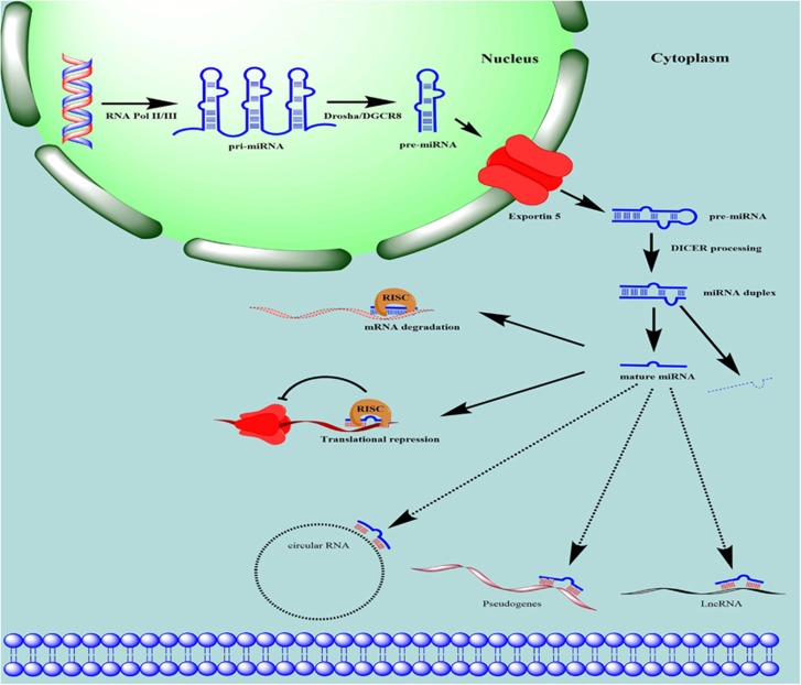
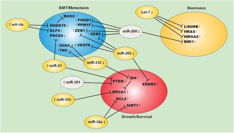
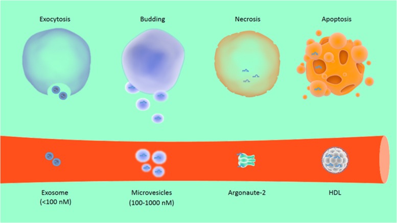
Effective management of breast cancer depends on early diagnosis and proper monitoring of patients’ response to therapy. However, these goals are difficult to achieve because of the lack of sensitive and specific biomarkers for early detection and for disease monitoring. Accumulating evidence in the past several years has highlighted the potential use of peripheral blood circulating nucleic acids such as DNA, mRNA and micro (mi)RNA in breast cancer diagnosis, prognosis and for monitoring response to anticancer therapy. Among these, circulating miRNA is increasingly recognized as a promising biomarker, given the ease with which miRNAs can be isolated and their structural stability under different conditions of sample processing and isolation. In this review, we provide current state-of-the-art of miRNA biogenesis, function and discuss the advantages, limitations, as well as pitfalls of using circulating miRNAs as diagnostic, prognostic or predictive biomarkers in breast cancer management.
Overview
The claims and statistics of this paper haven’t been checked yet.
Run the check — free, no account needed. It reads the open-access text and checks the sources it cites.
Integrity signals
Registry
Cites
Statistics
Figures
Verification
Claims the paper makes
The claims and statistics of this paper haven’t been checked yet.
Run the check — free, no account needed. It reads the open-access text and checks the sources it cites.
Claims other papers make about this one
Each is checked against this paper’s own text: does it say what the citing paper says it does?
In the citing paper
“Especially in the biomarker field, low level of detectability is a very critical issue, since many disease-derived miRNAs circulate in blood at low amounts (7).”
From Depletion of erythropoietic miR-486-5p and miR-451a improves detectability of rare microRNAs in peripheral blood-derived small RNA sequencing libraries (2020) · cites this paper as [7]
Self-check
Percentages, sums and reported differences re-computed from the paper’s own numbers.
The self-check needs the paper’s full text.
Citations
Cites
Valadi, H., Ekström, K., Bossios, A., Sjöstrand, M., Lee, J. J., & Lötvall, J. O. (2007) · Nature Cell Biology · doi:10.1038/ncb1596 · Cited by 12,834
Most mammalian mRNAs are conserved targets of microRNAs
Friedman, R. C., Farh, K. K.-H., Burge, C. B., & Bartel, D. P. (2009) · Genome Research · doi:10.1101/gr.082701.108 · Cited by 8,462
Natural RNA circles function as efficient microRNA sponges
Hansen, T. B., Jensen, T. I., Clausen, B. H., Bramsen, J. B., Finsen, B., Damgaard, C. K., & Kjems, J. (2013) · Nature · doi:10.1038/nature11993 · Cited by 7,985
The impact of microRNAs on protein output
Baek, D., Villén, J., Shin, C., Camargo, F. D., Gygi, S. P., & Bartel, D. P. (2008) · Nature · doi:10.1038/nature07242 · Cited by 3,592
Widespread changes in protein synthesis induced by microRNAs
Selbach, M., Schwanhäusser, B., Thierfelder, N., Fang, Z., Khanin, R., & Rajewsky, N. (2008) · Nature · doi:10.1038/nature07228 · Cited by 3,429
Tumour invasion and metastasis initiated by microRNA-10b in breast cancer
Ma, L., Teruya-Feldstein, J., & Weinberg, R. A. (2007) · Nature · doi:10.1038/nature06174 · Cited by 2,605
Si, M.-L., Zhu, S., Wu, H., Lu, Z., Wu, F., & Mo, Y.-Y. (2007) · Oncogene · doi:10.1038/sj.onc.1210083 · Cited by 1,600
Frankel, L. B., Christoffersen, N. R., Jacobsen, A., Lindow, M., Krogh, A., & Lund, A. H. (2008) · Journal of Biological Chemistry · doi:10.1074/jbc.m707224200 · Cited by 1,141
Functional screening identifies miRNAs inducing cardiac regeneration
Eulalio, A., Mano, M., Ferro, M. D., Zentilin, L., Sinagra, G., Zacchigna, S., & Giacca, M. (2012) · Nature · doi:10.1038/nature11739 · Cited by 1,029
Circulating microRNAs as novel biomarkers for diabetes mellitus
Guay, C., & Regazzi, R. (2013) · Nature Reviews Endocrinology · doi:10.1038/nrendo.2013.86 · Cited by 563
Concordant Regulation of Translation and mRNA Abundance for Hundreds of Targets of a Human microRNA
Hendrickson, D. G., Hogan, D. J., McCullough, H. L., Myers, J. W., Herschlag, D., Ferrell, J. E., & Brown, P. O. (2009) · PLoS Biology · doi:10.1371/journal.pbio.1000238 · Cited by 418
Research works printed but not linked: 127
- Siegel RL, Miller KD, Jemal A . Cancer statistics 2015 CA Cancer J Clin 2015; 65: 5–29. · DOI
- Youlden DR, Cramb SM, Dunn NA, Muller JM, Pyke CM, Baade PD . The descriptive epidemiology of female breast cancer: an international comparison of screening, incidence, survival and mortality. Cancer Epidemiol 2012; 36: 237–248. · DOI
- Jorgensen KJ, Kalager M, Barratt A, Baines C, Zahl PH, Brodersen J et al. Overview of guidelines on breast screening: why recommendations differ and what to do about it. Breast 2016; 31: 261–269. · DOI
- Charles L, Loprinzi FRA, Martee L, Hensley JTR ASCO SEP. 3rd edn. American Society of Clinical Oncology, Alexandria, VA, USA, 2013.
- Ellsworth RE, Blackburn HL, Shriver CD, Soon-Shiong P, Ellsworth DL . Molecular heterogeneity in breast cancer: state of the science and implications for patient care. Semin Cell Dev Biol 2016; 64: 65–72. · DOI
- Paik S, Shak S, Tang G, Kim C, Baker J, Cronin M et al. A multigene assay to predict recurrence of tamoxifen-treated, node-negative breast cancer. N Engl J Med 2004; 351: 2817–2826. · DOI
- Lim LP, Lau NC, Garrett-Engele P, Grimson A, Schelter JM, Castle J et al. Microarray analysis shows that some microRNAs downregulate large numbers of target mRNAs. Nature 2005; 433: 769–773. · DOI · Paper page
- Bushati N, Cohen SM . microRNA functions. Annu Rev Cell Dev Biol 2007; 23: 175–205. · DOI · Paper page
- Carthew RW, Sontheimer EJ . Origins and mechanisms of miRNAs and siRNAs. Cell 2009; 136: 642–655. · DOI
- Davis BN, Hata A . Regulation of microRNA biogenesis: a miRiad of mechanisms. Cell Commun Signal 2009; 7: 18. · DOI
- Filipowicz W, Bhattacharyya SN, Sonenberg N . Mechanisms of post-transcriptional regulation by microRNAs: are the answers in sight? Nat Rev Genet 2008; 9: 102–114. · DOI
- Forman JJ, Legesse-Miller A, Coller HA . A search for conserved sequences in coding regions reveals that the let-7 microRNA targets Dicer within its coding sequence. Proc Natl Acad Sci USA 2008; 105: 14879–14884. · DOI · Paper page
- Hausser J, Landthaler M, Jaskiewicz L, Gaidatzis D, Zavolan M . Relative contribution of sequence and structure features to the mRNA binding of Argonaute/EIF2C-miRNA complexes and the degradation of miRNA targets. Genome Res 2009; 19: 2009–2020. · DOI
- Vasudevan S, Tong Y, Steitz JA . Switching from repression to activation: microRNAs can up-regulate translation. Science 2007; 318: 1931–1934. · DOI
- Winter J, Jung S, Keller S, Gregory RI, Diederichs S . Many roads to maturity: microRNA biogenesis pathways and their regulation. Nat Cell Biol 2009; 11: 228–234. · DOI
- Lee Y, Ahn C, Han J, Choi H, Kim J, Yim J et al. The nuclear RNase III Drosha initiates microRNA processing. Nature 2003; 425: 415–419. · DOI
- Lee Y, Kim M, Han J, Yeom KH, Lee S, Baek SH et al. MicroRNA genes are transcribed by RNA polymerase II. EMBO J 2004; 23: 4051–4060. · DOI
- Zeng Y, Yi R, Cullen BR . Recognition and cleavage of primary microRNA precursors by the nuclear processing enzyme Drosha. EMBO J 2005; 24: 138–148. · DOI
- Lund E, Guttinger S, Calado A, Dahlberg JE, Kutay U . Nuclear export of microRNA precursors. Science 2004; 303: 95–98. · DOI
- Yi R, Qin Y, Macara IG, Cullen BR . Exportin-5 mediates the nuclear export of pre-microRNAs and short hairpin RNAs. Genes Dev 2003; 17: 3011–3016. · DOI
- Ding XC, Weiler J, Grosshans H . Regulating the regulators: mechanisms controlling the maturation of microRNAs. Trends Biotechnol 2009; 27: 27–36. · DOI
- Bosse GD, Simard MJ . A new twist in the microRNA pathway: not Dicer but Argonaute is required for a microRNA production. Cell Res 2010; 20: 735–737. · DOI
- Hutvagner G, Simard MJ . Argonaute proteins: key players in RNA silencing. Nat Rev Mol Cell Biol 2008; 9: 22–32. · DOI
- Giraldez AJ, Mishima Y, Rihel J, Grocock RJ, Van Dongen S, Inoue K et al. Zebrafish MiR-430 promotes deadenylation and clearance of maternal mRNAs. Science 2006; 312: 75–79. · DOI · Paper page
- Pillai RS, Bhattacharyya SN, Artus CG, Zoller T, Cougot N, Basyuk E et al. Inhibition of translational initiation by Let-7 MicroRNA in human cells. Science 2005; 309: 1573–1576. · DOI
- Zeng Y, Cullen BR . Sequence requirements for micro RNA processing and function in human cells. RNA 2003; 9: 112–123. · DOI
- Doench JG, Petersen CP, Sharp PA . siRNAs can function as miRNAs. Genes Dev 2003; 17: 438–442. · DOI
- Li WH, Gojobori T, Nei M . Pseudogenes as a paradigm of neutral evolution. Nature 1981; 292: 237–239. · DOI
- Vanin EF . Processed pseudogenes: characteristics and evolution. Annu Rev Genet 1985; 19: 253–272. · DOI
- Poliseno L, Salmena L, Zhang J, Carver B, Haveman WJ, Pandolfi PP . A coding-independent function of gene and pseudogene mRNAs regulates tumour biology. Nature 2010; 465: 1033–1038. · DOI
- Rinn JL, Kertesz M, Wang JK, Squazzo SL, Xu X, Brugmann SA et al. Functional demarcation of active and silent chromatin domains in human HOX loci by noncoding RNAs. Cell 2007; 129: 1311–1323. · DOI
- Liu XH, Sun M, Nie FQ, Ge YB, Zhang EB, Yin DD et al. Lnc RNA HOTAIR functions as a competing endogenous RNA to regulate HER2 expression by sponging miR-331-3p in gastric cancer. Mol Cancer 2014; 13: 92. · DOI
- Lasda E, Parker R . Circular RNAs: diversity of form and function. RNA 2014; 20: 1829–1842. · DOI
- Kroh EM, Parkin RK, Mitchell PS, Tewari M . Analysis of circulating microRNA biomarkers in plasma and serum using quantitative reverse transcription-PCR (qRT-PCR). Methods 2010; 50: 298–301. · DOI
- Hamam R, Ali AM, Alsaleh KA, Kassem M, Alfayez M, Aldahmash A et al. microRNA expression profiling on individual breast cancer patients identifies novel panel of circulating microRNA for early detection. Sci Rep 2016; 6: 25997. · DOI
- Wu X, Somlo G, Yu Y, Palomares MR, Li AX, Zhou W et al. De novo sequencing of circulating miRNAs identifies novel markers predicting clinical outcome of locally advanced breast cancer. J Transl Med 2012; 10: 42. · DOI
- Oikonomopoulos A, Polytarchou C, Joshi S, Hommes DW, Iliopoulos D . Identification of circulating microRNA signatures in Crohn's disease using the Nanostring nCounter technology. Inflamm Bowel Dis 2016; 22: 2063–2069. · DOI
- Alajez NM, Shi W, Wong D, Lenarduzzi M, Waldron J, Weinreb I et al. Lin28b promotes head and neck cancer progression via modulation of the insulin-like growth factor survival pathway. Oncotarget 2012; 3: 1641–1652. · DOI
- Kirschner MB, Kao SC, Edelman JJ, Armstrong NJ, Vallely MP, van Zandwijk N et al. Haemolysis during sample preparation alters microRNA content of plasma. PLoS ONE 2011; 6: e24145. · DOI · Paper page
- McDonald JS, Milosevic D, Reddi HV, Grebe SK, Algeciras-Schimnich A . Analysis of circulating microRNA: preanalytical and analytical challenges. Clin Chem 2011; 57: 833–840. · DOI
- Quiat D, Olson EN . MicroRNAs in cardiovascular disease: from pathogenesis to prevention and treatment. J Clin Invest 2013; 123: 11–18. · DOI
- Cao L, Lin EJ, Cahill MC, Wang C, Liu X, During MJ . Molecular therapy of obesity and diabetes by a physiological autoregulatory approach. Nat Med 2009; 15: 447–454. · DOI
- Hilton C, Neville MJ, Karpe F . MicroRNAs in adipose tissue: their role in adipogenesis and obesity. Int J Obes (Lond) 2012; 37: 325–332. · DOI
- Dehwah MA, Xu A, Huang Q . MicroRNAs and type 2 diabetes/obesity. J Genet Genomics 2012; 39: 11–18. · DOI
- Croce CM . Causes and consequences of microRNA dysregulation in cancer. Nat Rev Genet 2009; 10: 704–714. · DOI
- Alajez NM, Lenarduzzi M, Ito E, Hui AB, Shi W, Bruce J et al. MiR-218 suppresses nasopharyngeal cancer progression through downregulation of survivin and the SLIT2-ROBO1 pathway. Cancer Res 2011; 71: 2381–2391. · DOI
- Viswanathan SR, Powers JT, Einhorn W, Hoshida Y, Ng TL, Toffanin S et al. Lin28 promotes transformation and is associated with advanced human malignancies. Nat Genet 2009; 41: 843–848. · DOI
- Yu F, Yao H, Zhu P, Zhang X, Pan Q, Gong C et al. let-7 regulates self renewal and tumorigenicity of breast cancer cells. Cell 2007; 131: 1109–1123. · DOI
- Altuvia Y, Landgraf P, Lithwick G, Elefant N, Pfeffer S, Aravin A et al. Clustering and conservation patterns of human microRNAs. Nucleic Acids Res 2005; 33: 2697–2706. · DOI
- Huber MA, Azoitei N, Baumann B, Grunert S, Sommer A, Pehamberger H et al. NF-kappaB is essential for epithelial-mesenchymal transition and metastasis in a model of breast cancer progression. J Clin Invest 2004; 114: 569–581. · DOI
- Yang J, Mani SA, Donaher JL, Ramaswamy S, Itzykson RA, Come C et al. Twist, a master regulator of morphogenesis, plays an essential role in tumor metastasis. Cell 2004; 117: 927–939. · DOI
- Shimono Y, Zabala M, Cho RW, Lobo N, Dalerba P, Qian D et al. Downregulation of miRNA-200c links breast cancer stem cells with normal stem cells. Cell 2009; 138: 592–603. · DOI
- Korpal M, Lee ES, Hu G, Kang Y . The miR-200 family inhibits epithelial-mesenchymal transition and cancer cell migration by direct targeting of E-cadherin transcriptional repressors ZEB1 and ZEB2. J Biol Chem 2008; 283: 14910–14914. · DOI
- Jurmeister S, Baumann M, Balwierz A, Keklikoglou I, Ward A, Uhlmann S et al. MicroRNA-200c represses migration and invasion of breast cancer cells by targeting actin-regulatory proteins FHOD1 and PPM1F. Mol Cell Biol 2012; 32: 633–651. · DOI
- Baffa R, Fassan M, Volinia S, O'Hara B, Liu CG, Palazzo JP et al. MicroRNA expression profiling of human metastatic cancers identifies cancer gene targets. J Pathol 2009; 219: 214–221. · DOI
- Rowland BD, Peeper DS . KLF4, p21 and context-dependent opposing forces in cancer. Nat Rev Cancer 2006; 6: 11–23. · DOI
- Zhu S, Si ML, Wu H, Mo YY . MicroRNA-21 targets the tumor suppressor gene tropomyosin 1 (TPM1). J Biol Chem 2007; 282: 14328–14336. · DOI · Paper page
- Qian B, Katsaros D, Lu L, Preti M, Durando A, Arisio R et al. High miR-21 expression in breast cancer associated with poor disease-free survival in early stage disease and high TGF-beta1. Breast Cancer Res Treat 2009; 117: 131–140. · DOI
- Wickramasinghe NS, Manavalan TT, Dougherty SM, Riggs KA, Li Y, Klinge CM . Estradiol downregulates miR-21 expression and increases miR-21 target gene expression in MCF-7 breast cancer cells. Nucleic Acids Res 2009; 37: 2584–2595. · DOI
- Schramedei K, Morbt N, Pfeifer G, Lauter J, Rosolowski M, Tomm JM et al. MicroRNA-21 targets tumor suppressor genes ANP32A and SMARCA4. Oncogene 2011; 30: 2975–2985. · DOI
- Png KJ, Yoshida M, Zhang XH, Shu W, Lee H, Rimner A et al. MicroRNA-335 inhibits tumor reinitiation and is silenced through genetic and epigenetic mechanisms in human breast cancer. Genes Dev 2011; 25: 226–231. · DOI
- Tavazoie SF, Alarcon C, Oskarsson T, Padua D, Wang Q, Bos PD et al. Endogenous human microRNAs that suppress breast cancer metastasis. Nature 2008; 451: 147–152. · DOI
- Heyn H, Engelmann M, Schreek S, Ahrens P, Lehmann U, Kreipe H et al. MicroRNA miR-335 is crucial for the BRCA1 regulatory cascade in breast cancer development. Int J Cancer 2011; 129: 2797–2806. · DOI
- Shi W, Gerster K, Alajez NM, Tsang J, Waldron L, Pintilie M et al. MicroRNA-301 mediates proliferation and invasion in human breast cancer. Cancer Res 2011; 71: 2926–2937. · DOI
- Faraoni I, Antonetti FR, Cardone J, Bonmassar E . miR-155 gene: a typical multifunctional microRNA. Biochim Biophys Acta 2009; 1792: 497–505. · DOI · Paper page
- O'Donovan PJ, Livingston DM . BRCA1 and BRCA2: breast/ovarian cancer susceptibility gene products and participants in DNA double-strand break repair. Carcinogenesis 2010; 31: 961–967. · DOI
- Szabo CI, King MC . Inherited breast and ovarian cancer. Hum Mol Genet 1995; 4 Spec No: 1811–1817. · DOI
- Jiang S, Zhang HW, Lu MH, He XH, Li Y, Gu H et al. MicroRNA-155 functions as an OncomiR in breast cancer by targeting the suppressor of cytokine signaling 1 gene. Cancer Res 2010; 70: 3119–3127. · DOI · Paper page
- Yamakuchi M, Ferlito M, Lowenstein CJ . miR-34a repression of SIRT1 regulates apoptosis. Proc Natl Acad Sci USA 2008; 105: 13421–13426. · DOI
- Li L, Yuan L, Luo J, Gao J, Guo J, Xie X . MiR-34a inhibits proliferation and migration of breast cancer through down-regulation of Bcl-2 and SIRT1. Clin Exp Med 2013; 13: 109–117. · DOI
- Wu H, Zhu S, Mo YY . Suppression of cell growth and invasion by miR-205 in breast cancer. Cell Res 2009; 19: 439–448. · DOI
- Iorio MV, Casalini P, Piovan C, Di Leva G, Merlo A, Triulzi T et al. microRNA-205 regulates HER3 in human breast cancer. Cancer Res 2009; 69: 2195–2200. · DOI
- Gregory PA, Bert AG, Paterson EL, Barry SC, Tsykin A, Farshid G et al. The miR-200 family and miR-205 regulate epithelial to mesenchymal transition by targeting ZEB1 and SIP1. Nat Cell Biol 2008; 10: 593–601. · DOI
- Vickers KC, Palmisano BT, Shoucri BM, Shamburek RD, Remaley AT . MicroRNAs are transported in plasma and delivered to recipient cells by high-density lipoproteins. Nat Cell Biol 2011; 13: 423–433. · DOI · Paper page
- Pegtel DM, Cosmopoulos K, Thorley-Lawson DA, van Eijndhoven MA, Hopmans ES, Lindenberg JL et al. Functional delivery of viral miRNAs via exosomes. Proc Natl Acad Sci USA 2010; 107: 6328–6333. · DOI
- Turchinovich A, Weiz L, Burwinkel B . Extracellular miRNAs: the mystery of their origin and function. Trends Biochem Sci 2012; 37: 460–465. · DOI
- Al-toub M, Vishnubalaji R, Hamam R, Kassem M, Aldahmash A, Alajez NM . CDH1 and IL1-beta expression dictates FAK and MAPKK-dependent cross-talk between cancer cells and human mesenchymal stem cells. Stem Cell Res Ther 2015; 6: 135. · DOI
- Chen X, Ba Y, Ma L, Cai X, Yin Y, Wang K et al. Characterization of microRNAs in serum: a novel class of biomarkers for diagnosis of cancer and other diseases. Cell Res 2008; 18: 997–1006. · DOI · Paper page
- Gilad S, Meiri E, Yogev Y, Benjamin S, Lebanony D, Yerushalmi N et al. Serum microRNAs are promising novel biomarkers. PLoS ONE 2008; 3: e3148. · DOI
- Mitchell PS, Parkin RK, Kroh EM, Fritz BR, Wyman SK, Pogosova-Agadjanyan EL et al. Circulating microRNAs as stable blood-based markers for cancer detection. Proc Natl Acad Sci USA 2008; 105: 10513–10518. · DOI · Paper page
- Chin LJ, Slack FJ . A truth serum for cancer—microRNAs have major potential as cancer biomarkers. Cell Res 2008; 18: 983–984. · DOI
- Gandhi R, Healy B, Gholipour T, Egorova S, Musallam A, Hussain MS et al. Circulating microRNAs as biomarkers for disease staging in multiple sclerosis. Ann Neurol 2013; 73: 729–740. · DOI
- Khoo SK, Petillo D, Kang UJ, Resau JH, Berryhill B, Linder J et al. Plasma-based circulating MicroRNA biomarkers for Parkinson's disease. J Parkinson's Dis 2012; 2: 321–331. · DOI
- Lawrie CH, Gal S, Dunlop HM, Pushkaran B, Liggins AP, Pulford K et al. Detection of elevated levels of tumour-associated microRNAs in serum of patients with diffuse large B-cell lymphoma. Br J Haematol 2008; 141: 672–675. · DOI
- Resnick KE, Alder H, Hagan JP, Richardson DL, Croce CM, Cohn DE . The detection of differentially expressed microRNAs from the serum of ovarian cancer patients using a novel real-time PCR platform. Gynecol Oncol 2009; 112: 55–59. · DOI
- Ng EK, Chong WW, Jin H, Lam EK, Shin VY, Yu J et al. Differential expression of microRNAs in plasma of patients with colorectal cancer: a potential marker for colorectal cancer screening. Gut 2009; 58: 1375–1381. · DOI · Paper page
- Tanaka M, Oikawa K, Takanashi M, Kudo M, Ohyashiki J, Ohyashiki K et al. Down-regulation of miR-92 in human plasma is a novel marker for acute leukemia patients. PLoS ONE 2009; 4: e5532. · DOI
- Ho AS, Huang X, Cao H, Christman-Skieller C, Bennewith K, Le QT et al. Circulating miR-210 as a novel hypoxia marker in pancreatic cancer. Transl Oncol 2010; 3: 109–113. · DOI
- Wang J, Chen J, Chang P, LeBlanc A, Li D, Abbruzzesse JL et al. MicroRNAs in plasma of pancreatic ductal adenocarcinoma patients as novel blood-based biomarkers of disease. Cancer Prevent Res 2009; 2: 807–813. · DOI
- Wong TS, Ho WK, Chan JY, Ng RW, Wei WI . Mature miR-184 and squamous cell carcinoma of the tongue. Sci World J 2009; 9: 130–132. · DOI
- Yamamoto Y, Kosaka N, Tanaka M, Koizumi F, Kanai Y, Mizutani T et al. MicroRNA-500 as a potential diagnostic marker for hepatocellular carcinoma. Biomarkers 2009; 14: 529–538. · DOI
- Heneghan HM, Miller N, Kelly R, Newell J, Kerin MJ . Systemic miRNA-195 differentiates breast cancer from other malignancies and is a potential biomarker for detecting noninvasive and early stage disease. Oncologist 2010; 15: 673–682. · DOI
- Schwarzenbach H, Milde-Langosch K, Steinbach B, Muller V, Pantel K . Diagnostic potential of PTEN-targeting miR-214 in the blood of breast cancer patients. Breast Cancer Res Treat 2012; 134: 933–941. · DOI
- Cuk K, Zucknick M, Madhavan D, Schott S, Golatta M, Heil J et al. Plasma microRNA panel for minimally invasive detection of breast cancer. PLoS ONE 2013; 8: e76729. · DOI
- Shen J, Hu Q, Schrauder M, Yan L, Wang D, Medico L et al. Circulating miR-148b and miR-133a as biomarkers for breast cancer detection. Oncotarget 2014; 5: 5284–5294. · DOI
- Kodahl AR, Lyng MB, Binder H, Cold S, Gravgaard K, Knoop AS et al. Novel circulating microRNA signature as a potential non-invasive multi-marker test in ER-positive early-stage breast cancer: a case control study. Mol Oncol 2014; 8: 874–883. · DOI
- Zearo S, Kim E, Zhu Y, Zhao JT, Sidhu SB, Robinson BG et al. MicroRNA-484 is more highly expressed in serum of early breast cancer patients compared to healthy volunteers. BMC Cancer 2014; 14: 200. · DOI
- Shimomura A, Shiino S, Kawauchi J, Takizawa S, Sakamoto H, Matsuzaki J et al. Novel combination of serum microRNA for detecting breast cancer in the early stage. Cancer Sci 2016; 107: 326–334. · DOI
- Sochor M, Basova P, Pesta M, Dusilkova N, Bartos J, Burda P et al. Oncogenic microRNAs: miR-155, miR-19a, miR-181b, and miR-24 enable monitoring of early breast cancer in serum. BMC Cancer 2014; 14: 448. · DOI
- Chan M, Liaw CS, Ji SM, Tan HH, Wong CY, Thike AA et al. Identification of circulating microRNA signatures for breast cancer detection. Clin Cancer Res 2013; 19: 4477–4487. · DOI
- Matamala N, Vargas MT, Gonzalez-Campora R, Minambres R, Arias JI, Menendez P et al. Tumor microRNA expression profiling identifies circulating microRNAs for early breast cancer detection. Clin Chem 2015; 61: 1098–1106. · DOI
- Li XX, Gao SY, Wang PY, Zhou X, Li YJ, Yu Y et al. Reduced expression levels of let-7c in human breast cancer patients. Oncol Lett 2015; 9: 1207–1212. · DOI
- Wang PY, Gong HT, Li BF, Lv CL, Wang HT, Zhou HH et al. Higher expression of circulating miR-182 as a novel biomarker for breast cancer. Oncol Lett 2013; 6: 1681–1686. · DOI
- Waters PS, Dwyer RM, Brougham C, Glynn CL, Wall D, Hyland P et al. Impact of tumour epithelial subtype on circulating microRNAs in breast cancer patients. PLoS ONE 2014; 9: e90605. · DOI
- Blenkiron C, Goldstein LD, Thorne NP, Spiteri I, Chin SF, Dunning MJ et al. MicroRNA expression profiling of human breast cancer identifies new markers of tumor subtype. Genome Biol 2007; 8: R214. · DOI
- Iorio MV, Ferracin M, Liu CG, Veronese A, Spizzo R, Sabbioni S et al. MicroRNA gene expression deregulation in human breast cancer. Cancer Res 2005; 65: 7065–7070. · DOI · Paper page
- Zhu W, Qin W, Atasoy U, Sauter ER . Circulating microRNAs in breast cancer and healthy subjects. BMC Res Notes 2009; 2: 89. · DOI
- Wang F, Zheng Z, Guo J, Ding X . Correlation and quantitation of microRNA aberrant expression in tissues and sera from patients with breast tumor. Gynecol Oncol 2010; 119: 586–593. · DOI
- Joosse SA, Muller V, Steinbach B, Pantel K, Schwarzenbach H . Circulating cell-free cancer-testis MAGE-A RNA, BORIS RNA, let-7b and miR-202 in the blood of patients with breast cancer and benign breast diseases. Br J Cancer 2014; 111: 909–917. · DOI
- Mangolini A, Ferracin M, Zanzi MV, Saccenti E, Ebnaof SO, Poma VV et al. Diagnostic and prognostic microRNAs in the serum of breast cancer patients measured by droplet digital PCR. Biomark Res 2015; 3: 12. · DOI
- Kleivi Sahlberg K, Bottai G, Naume B, Burwinkel B, Calin GA, Borresen-Dale AL et al. A serum microRNA signature predicts tumor relapse and survival in triple-negative breast cancer patients. Clin Cancer Res 2015; 21: 1207–1214. · DOI
- Chen W, Cai F, Zhang B, Barekati Z, Zhong XY . The level of circulating miRNA-10b and miRNA-373 in detecting lymph node metastasis of breast cancer: potential biomarkers. Tumour Biol 2013; 34: 455–462. · DOI
- Roth C, Rack B, Muller V, Janni W, Pantel K, Schwarzenbach H . Circulating microRNAs as blood-based markers for patients with primary and metastatic breast cancer. Breast Cancer Res 2010; 12: R90. · DOI
- Shaker O, Maher M, Nassar Y, Morcos G, Gad Z . Role of microRNAs -29b-2, -155, -197 and -205 as diagnostic biomarkers in serum of breast cancer females. Gene 2015; 560: 77–82. · DOI
- Si H, Sun X, Chen Y, Cao Y, Chen S, Wang H et al. Circulating microRNA-92a and microRNA-21 as novel minimally invasive biomarkers for primary breast cancer. J Cancer Res Clin Oncol 2013; 139: 223–229. · DOI
- Huo D, Clayton WM, Yoshimatsu TF, Chen J, Olopade OI . Identification of a circulating MicroRNA signature to distinguish recurrence in breast cancer patients. Oncotarget 2016; ; 7: 55231–55248. · DOI
- Eichelser C, Flesch-Janys D, Chang-Claude J, Pantel K, Schwarzenbach H . Deregulated serum concentrations of circulating cell-free microRNAs miR-17, miR-34a, miR-155, and miR-373 in human breast cancer development and progression. Clin Chem 2013; 59: 1489–1496. · DOI
- Wang H, Tan G, Dong L, Cheng L, Li K, Wang Z et al. Circulating MiR-125b as a marker predicting chemoresistance in breast cancer. PLoS ONE 2012; 7: e34210. · DOI
- Sun Y, Wang M, Lin G, Sun S, Li X, Qi J et al. Serum microRNA-155 as a potential biomarker to track disease in breast cancer. PLoS ONE 2012; 7: e47003. · DOI
- Witwer KW . Circulating microRNA biomarker studies: pitfalls and potential solutions. Clin Chem 2015; 61: 56–63. · DOI · Paper page
- Wang K, Yuan Y, Cho JH, McClarty S, Baxter D, Galas DJ . Comparing the MicroRNA spectrum between serum and plasma. PLoS ONE 2012; 7: e41561. · DOI
- Diener Y, Walenda T, Jost E, Brummendorf TH, Bosio A, Wagner W et al. MicroRNA expression profiles of serum from patients before and after chemotherapy. Genomics Data 2015; 6: 125–127. · DOI
- Hansen TF, Carlsen AL, Heegaard NH, Sorensen FB, Jakobsen A . Changes in circulating microRNA-126 during treatment with chemotherapy and bevacizumab predicts treatment response in patients with metastatic colorectal cancer. Br J Cancer 2015; 112: 624–629. · DOI
- Ponomaryova AA, Morozkin ES, Rykova EY, Zaporozhchenko IA, Skvortsova TE, Dobrodeev capital AC et al. Dynamic changes in circulating miRNA levels in response to antitumor therapy of lung cancer. Exp Lung Res 2016; 42: 95–102. · DOI
- Li Y, Kowdley KV . Method for microRNA isolation from clinical serum samples. Anal Biochem 2012; 431: 69–75. · DOI
- Mathivanan S, Lim JW, Tauro BJ, Ji H, Moritz RL, Simpson RJ . Proteomics analysis of A33 immunoaffinity-purified exosomes released from the human colon tumor cell line LIM1215 reveals a tissue-specific protein signature. Mol Cell Proteomics 2010; 9: 197–208. · DOI
- Taylor DD, Gercel-Taylor C . MicroRNA signatures of tumor-derived exosomes as diagnostic biomarkers of ovarian cancer. Gynecol Oncol 2008; 110: 13–21. · DOI
Cited by
Sorted by citations
Juzenas, S., Lindqvist, C. M., Ito, G., Dolshanskaya, Y., Halfvarson, J., Franke, A., & Hemmrich-Stanisak, G. (2020) · NAR Genomics and Bioinformatics · doi:10.1093/nargab/lqaa008 · Cited by 17
Metadata
- Authors
- Hamam, R., Hamam, D., Alsaleh, K. A., Kassem, M., Zaher, W., Alfayez, M., Aldahmash, A., & Alajez, N. M.
- Venue
- Cell Death & Disease
- Published
- 2017-09-07
- Volume
- 8
- Issue
- 9
- Pages
- e3045-e3045
- ISSN
- 2041-4889
- PMC
- PMC5636984
- Cited by
- 322
- References
- 11
- Language
- en
About these checks
Registry, retraction, Cites and statistics results are computed, not judged, with one exception: a printed reference that no index lists is read once by a model before it counts as a citation error. Claims are extracted by a model and kept only when their quote appears verbatim in the text; each cited source is then checked, with a model’s help, for whether it says what the claim relies on. Those verdicts can be wrong — read the claim’s quote and the cited source before relying on one.
Statistics and arithmetic are recomputed, not judged.
Confidence bands are shown only for fields calibrated on our annotated test set. None is calibrated yet, so no band is shown.
What counts as a claim ›How we measure confidence ›How we check this ›
Are you an author, or do you see a problem with this page? Request a review
