

The DOI matches its Crossref record; 36 of the 36 references it cites were found in a registry or an index; 17 of the 34 sources its claims cite were checked for support.
Lipid Nanoparticle Delivery of siRNA to Silence Neuronal Gene Expression in the Brain
Open accessAbstract
Manipulation of gene expression in the brain is fundamental for understanding the function of proteins involved in neuronal processes. In this article, we show a method for using small interfering RNA (siRNA) in lipid nanoparticles (LNPs) to efficiently silence neuronal gene expression in cell culture and in the brain in vivo through intracranial injection. We show that neurons accumulate these LNPs in an apolipoprotein E-dependent fashion, resulting in very efficient uptake in cell culture (100%) with little apparent toxicity. In vivo, intracortical or intracerebroventricular (ICV) siRNA-LNP injections resulted in knockdown of target genes either in discrete regions around the injection site or in more widespread areas following ICV injections with no apparent toxicity or immune reactions from the LNPs. Effective targeted knockdown was demonstrated by showing that intracortical delivery of siRNA against GRIN1 (encoding GluN1 subunit of the NMDA receptor (NMDAR)) selectively reduced synaptic NMDAR currents in vivo as compared with synaptic AMPA receptor currents. Therefore, LNP delivery of siRNA rapidly manipulates expression of proteins involved in neuronal processes in vivo, possibly enabling the development of gene therapies for neurological disorders.Molecular Therapy-Nucleic Acids (2013) 2, e136; doi:10.1038/mtna.2013.65; published online 3 December 2013.
Overview
AI snapshot
Written from the paper’s full text.
TL;DR
Lipid nanoparticles (LNPs) deliver siRNA efficiently to neurons in culture and in the brain after intracranial or intracerebroventricular injection, silencing target genes with little apparent toxicity. Uptake is apolipoprotein E–dependent, and GRIN1 knockdown functionally reduces synaptic NMDA receptor currents in vivo.
Key findings
- In cultured hippocampal neurons, 100% of neurons took up DiI-labeled LNPs after 24 hours; PTEN siRNA (246 nmol/l, 48 hours) reduced PTEN/β-actin by 80% versus control (P < 0.001), and 0.7 nmol/l still reduced PTEN/β-actin by 59% (P < 0.001), with no toxicity by LDH release or nuclear morphology.
- Intracortical LNP-PTEN siRNA reduced PTEN/β-actin by 72% versus noninjected and 69% versus luc siRNA–injected controls (P < 0.001); knockdown was significant within 1 mm of the injection tract but not 1–3 mm away, and was sustained for at least 15 days (91% reduction, P < 0.001).
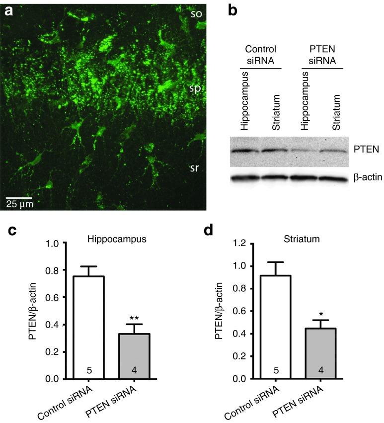
- Bilateral intracerebroventricular injection of 2 µl PTEN siRNA-LNPs produced widespread uptake and reduced PTEN/β-actin by 55.8% in hippocampus and 51.2% in striatum versus control siRNA–injected rats.
- GRIN1 siRNA reduced GluN1/β-actin by 51% versus noninjected and 54% versus luc siRNA-LNP controls (P < 0.01 and P < 0.05), decreased GluN1 cluster density in vitro by 41% (P < 0.001), and selectively reduced the synaptic NMDAR/AMPAR current ratio in vivo; TNF-α was not significantly increased by LNP-siRNA in slices or injected tissue (P > 0.05).
Methods
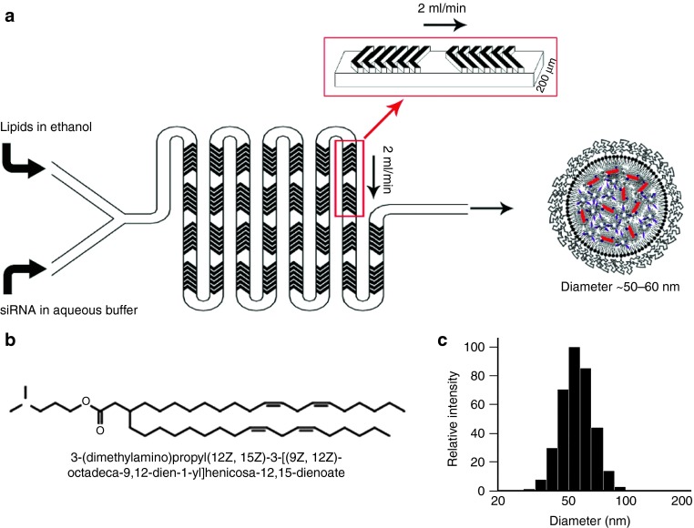
- LNPs were prepared by microfluidic mixing with DMAP-BLP/DSPC/cholesterol/PEG-DMG at 50/10/37.5/1.5 mol% plus 1 mol% DiOC18 or DiIC18; PTEN siRNA-LNPs had diameter 55 ± 11 nm, and in vivo LNPs were 50–60 nm.
- Primary rat hippocampal neuron cultures (DIV 12–14) with an astrocyte feeder layer were used for uptake, toxicity, and knockdown assays; Sprague–Dawley rats (P22–P26) received intracortical injections (500 nl at 5 mg/ml siRNA) or bilateral ICV injections (2 µl), with brain slices prepared about 5 days later.
- Main analyses included Western blot, immunohistochemistry/immunocytochemistry, LDH assay, DiI uptake assay, TNF-α ELISA, and whole-cell patch-clamp measurement of NMDAR/AMPAR excitatory postsynaptic currents; statistics used t-test or ANOVA with Newman–Keuls multiple comparison test.
Limitations
- The paper states that no further optimization was done to increase LNP-siRNA efficiency in this study, and that the therapeutic utility of direct intracranial LNP-siRNA administration for neurological disorders remains to be established.
- The paper notes that LNP systems cannot cross the blood–brain barrier, that local intracortical knockdown was limited to about 1 mm from the injection tract, and that the longest time point tested for PTEN suppression was 15 days.
- Evident from the text: findings are limited to primary rat neuronal cultures and rats, with no primate or human data; the in vivo functional experiments also contain a likely typo referring to “GluR1 siRNA” where GluN1/GRIN1 siRNA was intended.
Model-written summary. Check it against the paper before relying on it.
Integrity signals
Registry
Cites
Statistics
Figures
Verification
Claims the paper makes
36 Own result · 16 Own interpretation · 11 Own method · 7 Partly supported · 7 No full text of the sources · 5 Supported · 3 Not supported · 1 Supported so far · 1 Sources not found · 1 Not checked yet · accuracy not yet measured
The cited sources support only part of this claim — read each source’s verdict below.
In the paper
“which usually result in transfection rates of 1–10%.13”
§ Lipid Nanoparticle Delivery of siRNA to Silence Neuronal Gene Expression in the Brain > Results > LNP-mediated neuronal gene silencing in vitro
What the sources say
- Partly supportedcites [13] sourceExperimental — accuracy not yet measured
The calcium-phosphate table entry gives typical efficiencies of 5–10% in postmitotic neurons, and the Lipofection section gives 1–5% for plasmid-based transfection. Those figures do not establish the claimed usual rate for siRNA delivery: the Lipofection section instead describes efficient siRNA transfer, reaching up to 83% in primary rat hippocampal neurons. (LLM, conf=0.86)
plaindirect- Asserted: passes — The authors put it forward as true.
- Checkable: passes — It can be checked against evidence.
- Load-bearing: passes — The paper’s conclusions depend on it.
- Based: passes — The paper gives a reason why it holds.
- Partly supported
Every source the paper cites for this claim was checked and supports it.
In the paper
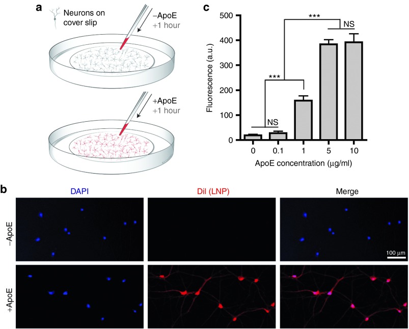
“We found that LNP uptake reached saturation at ˜5 µg ApoE/ml (Figure 3c), in line with measurements from human cerebrospinal fluid (CSF) in vivo of 9.09 µg/ml.16”
§ Lipid Nanoparticle Delivery of siRNA to Silence Neuronal Gene Expression in the Brain > Results > LNP-mediated neuronal gene silencing in vitro
What the sources say
- Supportedcites [16] source
The Results section, “ApoE levels and stability in human CSF,” reports a mean apoE concentration of 9.09 μg/ml in CSF samples from 168 people. The citation supports that comparison value; the LNP uptake saturation finding is presented as the citing authors’ own result (Figure 3c), not as a finding from this source. (LLM, conf=0.94)
plaincorrelational- Asserted: passes — The authors put it forward as true.
- Checkable: passes — It can be checked against evidence.
- Load-bearing: passes — The paper’s conclusions depend on it.
- Based: passes — The paper gives a reason why it holds.
- Supported
The cited sources support only part of this claim — read each source’s verdict below.
In the paper
“In the brain and neuronal cultures, ApoE is synthesized in astrocytes.14,15”
§ Lipid Nanoparticle Delivery of siRNA to Silence Neuronal Gene Expression in the Brain > Discussion
What the sources say
- Not checked — no full text of the sourcecites [14] source
- Partly supportedcites [15] sourceExperimental — accuracy not yet measured
The Biochemical properties section states that astrocytes are the predominant source of ApoE in the brain and describes ApoE particles secreted by cultured astrocytes. It does not specifically establish where ApoE is synthesized in neuronal cultures, and it notes that microglia also synthesize some ApoE. Page numbers are unavailable in our copy. (LLM, conf=0.82)
plain- Asserted: passes — The authors put it forward as true.
- Checkable: passes — It can be checked against evidence.
- Load-bearing: passes — The paper’s conclusions depend on it.
- Based: passes — The paper gives a reason why it holds.
The cited sources support only part of this claim — read each source’s verdict below.
In the paper
“Viral delivery is the most effective available way to transfect neurons.13,24”
§ Lipid Nanoparticle Delivery of siRNA to Silence Neuronal Gene Expression in the Brain > Discussion
What the sources say
- Partly supportedcites [13] sourceExperimental — accuracy not yet measured
The “Virus-based transfection methods” section describes viral delivery as extremely efficient and often the only viable option for efficient delivery to specific cell types in vivo. It does not call viral delivery the most effective method for neurons generally; the “Electrical transfection methods” section instead calls nucleofection the method of choice when high transfection efficiency is essential. (LLM, conf=0.85)
- Not checked — no full text of the sourcecites [24] source
plain- Asserted: passes — The authors put it forward as true.
- Checkable: passes — It can be checked against evidence.
- Load-bearing: passes — The paper’s conclusions depend on it.
- Based: passes — The paper gives a reason why it holds.
- Partly supported
Every source the paper cites for this claim was checked and supports it.
In the paper
“Expression of GluN1 is necessary for the formation of a functional NMDAR channel in the plasma membrane.19”
§ Lipid Nanoparticle Delivery of siRNA to Silence Neuronal Gene Expression in the Brain > Results > LNP-mediated knockdown of GluN1 in cell culture
What the sources say
- Supportedcites [19] source
The section on subunit assembly states that functional NMDA receptors require two GluN1 subunits. The section on synaptic and extrasynaptic NMDA receptors also calls GluN1 an obligate subunit for channel function, supporting its necessity for a functional receptor at the cell surface. (LLM, conf=0.93)
plaindirect- Asserted: passes — The authors put it forward as true.
- Checkable: passes — It can be checked against evidence.
- Load-bearing: passes — The paper’s conclusions depend on it.
- Based: passes — The paper gives a reason why it holds.
- Supported
The cited sources support only part of this claim — read each source’s verdict below.
In the paper
“The concentrations of ApoE found in the brain are similar to the concentrations we found to be effective in cell culture.16”
§ Lipid Nanoparticle Delivery of siRNA to Silence Neuronal Gene Expression in the Brain > Discussion
What the sources say
- Partly supportedcites [16] sourceExperimental — accuracy not yet measured
The Results and Discussion report human cerebrospinal-fluid ApoE concentrations averaging 9.09 μg/ml and ranging from 2 to 16 μg/ml. The paper does not report concentrations measured in brain tissue; the comparison with effective cell-culture concentrations is the citing authors’ own finding, not one this source can establish. (LLM, conf=0.83)
plain- Asserted: passes — The authors put it forward as true.
- Checkable: passes — It can be checked against evidence.
- Load-bearing: passes — The paper’s conclusions depend on it.
- Based: passes — The paper gives a reason why it holds.
- Partly supported
The cited sources support only part of this claim — read each source’s verdict below.
In the paper
“With regard to dose levels, ICV infusion of free ASOs against superoxide dismutase by osmotic pump at 100 µg/day per rat over 28 days (total of 2.8 mg of ASO) resulted in ˜50% superoxide dismutase mRNA silencing.28”
§ Lipid Nanoparticle Delivery of siRNA to Silence Neuronal Gene Expression in the Brain > Discussion
What the sources say
- Partly supportedcites [28] sourceExperimental — accuracy not yet measured
The Results and Figure 3 caption describe SOD1-targeted ASOs infused into rats’ lateral ventricles by osmotic pump for 28 days at the stated daily dose. But the Results report SOD1 mRNA at 25–40% of normal (a 60–75% reduction); the approximately 50% reduction was in SOD1 protein, not mRNA. Page numbers are unavailable in our copy. (LLM, conf=0.88)
plaindirect- Asserted: passes — The authors put it forward as true.
- Checkable: passes — It can be checked against evidence.
- Load-bearing: passes — The paper’s conclusions depend on it.
- Based: passes — The paper gives a reason why it holds.
- Partly supported
A result the paper reports from its own data — it cites no other source for it, so there is none to check. The quote shows it is in the paper.
In the paper
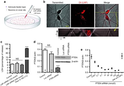
“Incubation of primary rat neuronal cultures with LNPs containing PTEN siRNA (246 nmol/l) for 48 hours resulted in robust knockdown of PTEN protein indicated by Western blot (PTEN/β-actin reduced by 80% compared with control; P < 0.001, Figure 2d).”
§ Lipid Nanoparticle Delivery of siRNA to Silence Neuronal Gene Expression in the Brain > Results > LNP-mediated neuronal gene silencing in vitro
hedgeddirect- Asserted: passes — The authors put it forward as true.
- Checkable: passes — It can be checked against evidence.
- Load-bearing: passes — The paper’s conclusions depend on it.
- Based: passes — The paper gives a reason why it holds.
A result the paper reports from its own data — it cites no other source for it, so there is none to check. The quote shows it is in the paper.
In the paper
“In addition, neurons treated with nonencapsulated PTEN siRNA (246 nmol/l) showed no significant change in PTEN/β-actin compared with control (Figure 2e).”
§ Lipid Nanoparticle Delivery of siRNA to Silence Neuronal Gene Expression in the Brain > Results > LNP-mediated neuronal gene silencing in vitro
plain- Asserted: passes — The authors put it forward as true.
- Checkable: passes — It can be checked against evidence.
- Load-bearing: passes — The paper’s conclusions depend on it.
- Based: passes — The paper gives a reason why it holds.
A result the paper reports from its own data — it cites no other source for it, so there is none to check. The quote shows it is in the paper.
In the paper
“Of note, in control experiments, incubation of cultures with LNPs containing siRNA against luciferase (luc), which is not found in the mammalian genome, had no effect on levels of PTEN protein (Figure 2d).”
§ Lipid Nanoparticle Delivery of siRNA to Silence Neuronal Gene Expression in the Brain > Results > LNP-mediated neuronal gene silencing in vitro
plain- Asserted: passes — The authors put it forward as true.
- Checkable: passes — It can be checked against evidence.
- Load-bearing: passes — The paper’s conclusions depend on it.
- Based: passes — The paper gives a reason why it holds.
- Rests on an assumption: Luciferase siRNA is not found in the mammalian genome.
- It may state two separate claims as one. 21
- Probably not a substantial claim. 20
At least one cited source doesn’t support this claim or points to a different paper, and none confirmed it — see the verdicts below.
In the paper
“These results indicate that LNPs are much more efficient and less toxic than current methods used to deliver siRNA to cultured neurons such as electroporation, calcium phosphate, or lipofection, which usually result in transfection rates of 1–10%.13”
§ Lipid Nanoparticle Delivery of siRNA to Silence Neuronal Gene Expression in the Brain > Results > LNP-mediated neuronal gene silencing in vitro
What the sources say
- Disputedcites [13] sourceExperimental — accuracy not yet measured
The Lipofection section reports siRNA delivery into postmitotic neurons at up to 83% efficiency, contrary to the cited sentence’s characterization of siRNA-delivery methods as usually achieving 1–10%. Its low lipofection rates of typically 1–5% are explicitly for plasmid-based vectors, not siRNA. The paper does not compare LNP efficiency or toxicity with these methods. (LLM, conf=0.88)
Checked by a reasoning model — may be wrong.
hedgeddirect- Asserted: passes — The authors put it forward as true.
- Checkable: passes — It can be checked against evidence.
- Load-bearing: passes — The paper’s conclusions depend on it.
- Based: passes — The paper gives a reason why it holds.
It may state two separate claims as one.
- Disputed
At least one cited source doesn’t support this claim or points to a different paper, and none confirmed it — see the verdicts below.
In the paper
“By contrast, current nonviral vectors exhibit transfection efficiencies of 10% or less and are often accompanied by cell damage.13”
§ Lipid Nanoparticle Delivery of siRNA to Silence Neuronal Gene Expression in the Brain > Discussion
What the sources say
- Disputedcites [13] sourceExperimental — accuracy not yet measured
The Electrical transfection methods section directly contradicts the claim’s 10% ceiling: it reports nucleofection rates averaging 60–80% after optimization, with up to 95% in embryonic rat neurons and better cell survival than conventional electroporation. The paper does report cell damage with some nonviral techniques, including biolistics, but does not characterize nonviral methods as uniformly low-efficiency and damaging. Page numbers were not available in our copy. (LLM, conf=0.90)
Checked by a reasoning model — may be wrong.
plaincorrelational- Asserted: passes — The authors put it forward as true.
- Checkable: passes — It can be checked against evidence.
- Load-bearing: passes — The paper’s conclusions depend on it.
- Based: passes — The paper gives a reason why it holds.
It may state two separate claims as one.
- Disputed
At least one cited source doesn’t support this claim or points to a different paper, and none confirmed it — see the verdicts below.
In the paper
“Furthermore, a number of clinical trials are now in progress for treatments of serious neurological diseases that rely on continuous infusion of ASOs into the CSF.27,28”
§ Lipid Nanoparticle Delivery of siRNA to Silence Neuronal Gene Expression in the Brain > Discussion
What the sources say
- Not checked — no full text of the sourcecites [27] source
- Disputedcites [28] sourceExperimental — accuracy not yet measured
The Discussion (page unavailable) describes a Phase I trial in human familial ALS patients as planned, not in progress, and says safety studies were underway in animals. The Results describe continuous ASO infusion in rats and a nonhuman primate, but the paper does not report multiple ongoing clinical trials. (LLM, conf=0.88)
Checked by a reasoning model — may be wrong.
plain- Asserted: passes — The authors put it forward as true.
- Checkable: passes — It can be checked against evidence.
- Load-bearing: fails — The paper’s conclusions may not depend on it.
- Based: passes — The paper gives a reason why it holds.
Probably not a substantial claim. The paper’s conclusions may not depend on it.
The sources with a verdict support this claim; the rest weren’t checked yet, have no full text we could read, or their verdicts aren’t rated yet.
In the paper
“Previous work has shown that LNP systems with this lipid composition can silence target genes in hepatocytes following IV injection at dose levels as low as 0.01 mg siRNA/kg body weight8,9 in an apolipoprotein E (ApoE)–dependent fashion.10”
§ Lipid Nanoparticle Delivery of siRNA to Silence Neuronal Gene Expression in the Brain > Results > Encapsulation of siRNA in LNPs
What the sources say
- Supportedcites [8] source
The Results (page not supplied) report 50% factor VII silencing at 0.01 mg siRNA/kg in mice, and Materials and Methods (page not supplied) specify intravenous administration. The ApoE-dependent mechanism is attached to citation 10 in the citing sentence, not to this source. (LLM, conf=0.91)
- Supportedcites [9] source
The Figure 7 results report hepatic gene silencing in mice with an ED50 of 0.005 mg siRNA/kg—below the claimed 0.01 mg/kg—and the paper describes the experiments as using intravenous administration. Its mechanism discussion states that ApoE mediates hepatocyte uptake of these LNPs. Page numbers are not available in our copy. (LLM, conf=0.90)
- Not checked — no full text of the sourcecites [10] source
plaindirect- Asserted: passes — The authors put it forward as true.
- Checkable: passes — It can be checked against evidence.
- Load-bearing: fails — The paper’s conclusions may not depend on it.
- Based: passes — The paper gives a reason why it holds.
Probably not a substantial claim. The paper’s conclusions may not depend on it.
- Rests on an assumption: ApoE is the dominant lipoprotein in the brain.
- Supported
Every source the paper cites for this claim was checked and supports it.
In the paper
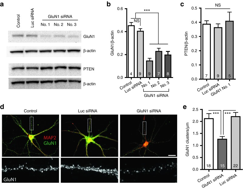
“The NMDAR ion channel is heteromeric, composed of two obligatory GluN1 subunits and two GluN2 or GluN3 subunits.”
§ Lipid Nanoparticle Delivery of siRNA to Silence Neuronal Gene Expression in the Brain > Results > LNP-mediated knockdown of GluN1 in cell culture
What the sources say
- Supportedcites [19] source
In “C. Synaptic and Extrasynaptic N-Methyl-D-aspartate Receptors” (page not supplied), the review describes NMDA receptors as heterotetramers with two GluN1 subunits and two GluN2 or GluN3 subunits. The same passage calls GluN1 an obligate subunit for channel function. (LLM, conf=0.94)
plain- Asserted: passes — The authors put it forward as true.
- Checkable: passes — It can be checked against evidence.
- Load-bearing: passes — The paper’s conclusions depend on it.
- Based: passes — The paper gives a reason why it holds.
Probably not a substantial claim.
- Supported
Every source the paper cites for this claim was checked and supports it.
In the paper
“Injection of ASOs into the CSF represents another promising method to silence neuronal gene expression in vivo.25”
§ Lipid Nanoparticle Delivery of siRNA to Silence Neuronal Gene Expression in the Brain > Discussion
What the sources say
- Supportedcites [25] source
The Results section reports that ASOs infused into the cerebrospinal fluid of rhesus monkeys reached neurons and produced sustained reductions in huntingtin mRNA across brain and spinal cord regions. The Discussion describes the approach as promising for human therapy. The paper used continuous infusion rather than a single injection, but this does not change the claim's point about CSF delivery of ASOs in vivo. (LLM, conf=0.93)
plainconditional- Asserted: passes — The authors put it forward as true.
- Checkable: passes — It can be checked against evidence.
- Load-bearing: fails — The paper’s conclusions may not depend on it.
- Based: passes — The paper gives a reason why it holds.
Probably not a substantial claim. The paper’s conclusions may not depend on it.
- Supported
The cited sources support only part of this claim — read each source’s verdict below.
In the paper
“With respect to the mechanism of action, the LNP delivery systems used here contain the ionizable cationic lipid DMAP-BLP, a lipid that exhibits optimized bilayer destabilizing and pKa properties leading to highly potent gene silencing in hepatocytes following IV administration (Supplementary Data) that is similar to “gold standard” lipids such as DLinMC3-DMA.8”
§ Lipid Nanoparticle Delivery of siRNA to Silence Neuronal Gene Expression in the Brain > Discussion
What the sources say
- Partly supportedcites [8] sourceExperimental — accuracy not yet measured
Split into 2 atoms — 0 confirmed, 1 partially supported, 0 disputed, 0 misattributed, 1 unverifiable. Expand the claim to see per-atom verdicts.
plaindirect- Asserted: passes — The authors put it forward as true.
- Checkable: passes — It can be checked against evidence.
- Load-bearing: unclear — It is unclear whether the paper’s conclusions depend on it.
- Based: passes — The paper gives a reason why it holds.
It may state two separate claims as one.
- Partly supported
Every source the paper cites for this claim was checked and supports it.
In the paper
“In addition, there are now well-established methods to reduce or eliminate siRNA immunogenicity by adjusting siRNA chemistry.29”
§ Lipid Nanoparticle Delivery of siRNA to Silence Neuronal Gene Expression in the Brain > Discussion
What the sources say
- Supportedcites [29] source
The section “Abrogating TLR7/8 activation by chemical modification” describes chemical substitutions that reduce siRNA immunogenicity and says some effectively abrogate TLR interaction. The “Quick guide to chemical siRNA design” adds that 2′-OMe modification may fully abrogate TLR7/8 responses. (LLM, conf=0.92)
boosteddirect- Asserted: passes — The authors put it forward as true.
- Checkable: passes — It can be checked against evidence.
- Load-bearing: fails — The paper’s conclusions may not depend on it.
- Based: passes — The paper gives a reason why it holds.
Probably not a substantial claim. The paper’s conclusions may not depend on it.
- Supported
The cited sources support only part of this claim — read each source’s verdict below.
In the paper
“However, two major problems associated with ASOs are the high doses required to achieve gene silencing effects and immunostimulatory effects.26,27”
§ Lipid Nanoparticle Delivery of siRNA to Silence Neuronal Gene Expression in the Brain > Discussion
What the sources say
- Partly supportedcites [26] sourceExperimental — accuracy not yet measured
The Cellular Delivery and Associated Safety section says phosphorothioate antisense oligonucleotides (AONs) can have immunostimulatory effects, while noting that appropriate modifications can suppress them. Neither that section nor the AON Mechanisms section says high doses are required to achieve gene silencing; the paper instead discusses dosage control and possible dose-dependent toxicity. Our copy provides no page numbers for these passages. (LLM, conf=0.87)
- Not checked — no full text of the sourcecites [27] source
plaincorrelational- Asserted: passes — The authors put it forward as true.
- Checkable: passes — It can be checked against evidence.
- Load-bearing: passes — The paper’s conclusions depend on it.
- Based: passes — The paper gives a reason why it holds.
It may state two separate claims as one.
- Partly supported
A result the paper reports from its own data — it cites no other source for it, so there is none to check. The quote shows it is in the paper.
In the paper
“In vivo, intracortical or intracerebroventricular (ICV) siRNA-LNP injections resulted in knockdown of target genes either in discrete regions around the injection site or in more widespread areas following ICV injections with no apparent toxicity or immune reactions from the LNPs.”
§ Lipid Nanoparticle Delivery of siRNA to Silence Neuronal Gene Expression in the Brain > Abstract
plain- Asserted: passes — The authors put it forward as true.
- Checkable: passes — It can be checked against evidence.
- Load-bearing: passes — The paper’s conclusions depend on it.
- Based: passes — The paper gives a reason why it holds.
It may state two separate claims as one.
A result the paper reports from its own data — it cites no other source for it, so there is none to check. The quote shows it is in the paper.
In the paper
“LNPs were nontoxic at the concentrations used, with no observable differences in cell death relative to controls measured either by comparing nuclear morphology or by lactate dehydrogenase (LDH) release (Figure 2c).”
§ Lipid Nanoparticle Delivery of siRNA to Silence Neuronal Gene Expression in the Brain > Results > LNP-mediated neuronal gene silencing in vitro
plain- Asserted: passes — The authors put it forward as true.
- Checkable: passes — It can be checked against evidence.
- Load-bearing: passes — The paper’s conclusions depend on it.
- Based: passes — The paper gives a reason why it holds.
It may state two separate claims as one.
A result the paper reports from its own data — it cites no other source for it, so there is none to check. The quote shows it is in the paper.
In the paper
“In this article, we show a method for using small interfering RNA (siRNA) in lipid nanoparticles (LNPs) to efficiently silence neuronal gene expression in cell culture and in the brain in vivo through intracranial injection.”
- Also stated in: § Introduction · boosted
“In this article, we report the conditions under which LNP delivery of siRNA is a remarkably efficient method for silencing neuronal gene expression in both primary neuronal culture and following intracranial injection in vivo.”
§ Lipid Nanoparticle Delivery of siRNA to Silence Neuronal Gene Expression in the Brain > Abstract
boosteddirect- Asserted: passes — The authors put it forward as true.
- Checkable: passes — It can be checked against evidence.
- Load-bearing: passes — The paper’s conclusions depend on it.
- Based: passes — The paper gives a reason why it holds.
It may state two separate claims as one.
- Also stated in: § Introduction · boosted
A result the paper reports from its own data — it cites no other source for it, so there is none to check. The quote shows it is in the paper.
In the paper
“With no added ApoE, there was no evidence of neuronal LNP uptake, whereas graded uptake was observed when ApoE concentrations were increased above 0.1μg/ml to 1 μg/ml and 5 µg/ml ApoE.”
§ Lipid Nanoparticle Delivery of siRNA to Silence Neuronal Gene Expression in the Brain > Discussion
plaincorrelational- Asserted: passes — The authors put it forward as true.
- Checkable: passes — It can be checked against evidence.
- Load-bearing: passes — The paper’s conclusions depend on it.
- Based: passes — The paper gives a reason why it holds.
It may state two separate claims as one.
A result the paper reports from its own data — it cites no other source for it, so there is none to check. The quote shows it is in the paper.
In the paper
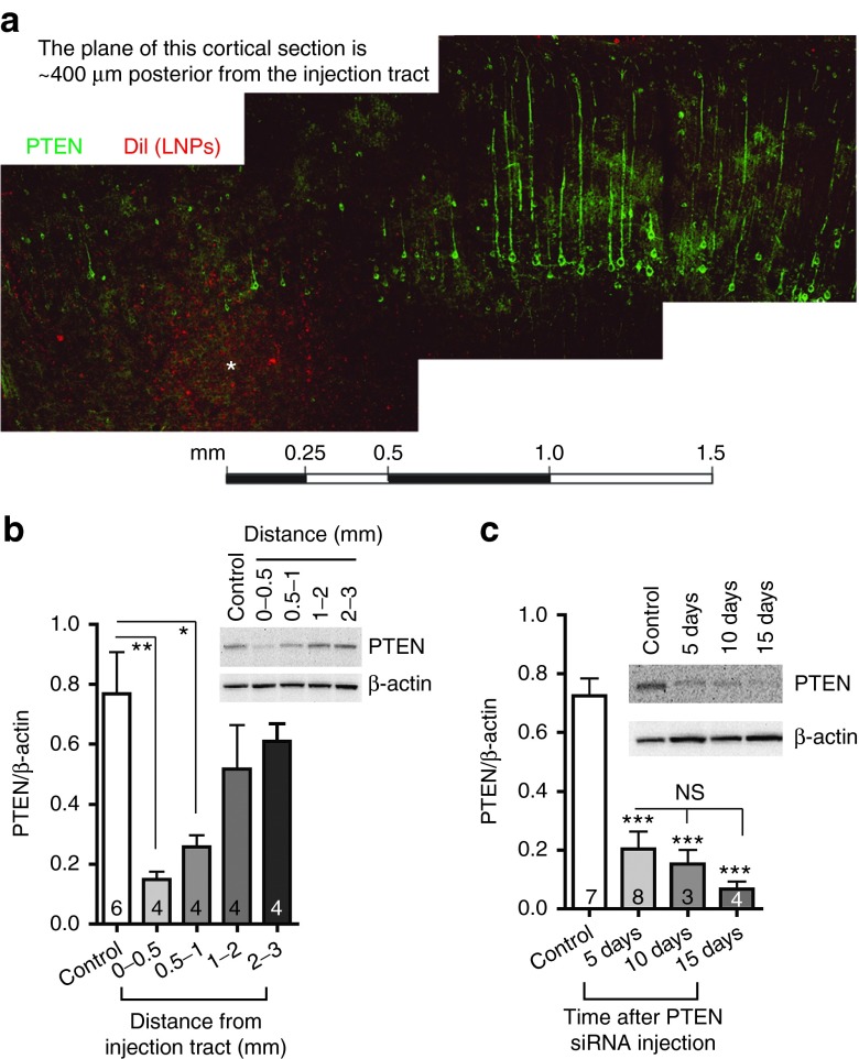
“Western blot analysis, which revealed that PTEN protein levels were significantly reduced at distances within 1 mm of the injection tract but not 1–3 mm away (Figure 5b).”
§ Lipid Nanoparticle Delivery of siRNA to Silence Neuronal Gene Expression in the Brain > Results > Time course and distance analysis of PTEN knockdown in vivo
plain- Asserted: passes — The authors put it forward as true.
- Checkable: passes — It can be checked against evidence.
- Load-bearing: passes — The paper’s conclusions depend on it.
- Based: passes — The paper gives a reason why it holds.
It may state two separate claims as one.
A result the paper reports from its own data — it cites no other source for it, so there is none to check. The quote shows it is in the paper.
In the paper
“As a positive control, cultures treated with 1% Triton X-100 showed a large increase in LDH release (90.21 ± 0.62%).”
§ Lipid Nanoparticle Delivery of siRNA to Silence Neuronal Gene Expression in the Brain > Results > LNP-mediated neuronal gene silencing in vitro
plaindirect- Asserted: passes — The authors put it forward as true.
- Checkable: passes — It can be checked against evidence.
- Load-bearing: fails — The paper’s conclusions may not depend on it.
- Based: passes — The paper gives a reason why it holds.
Probably not a substantial claim. The paper’s conclusions may not depend on it.
A result the paper reports from its own data — it cites no other source for it, so there is none to check. The quote shows it is in the paper.
In the paper
“In this article, we demonstrate that a single LNP-siRNA injection of 2.5 µg of siRNA per rat by intracortical administration results in pronounced (up to 91%) gene silencing that is sustained for at least 15 days after injection.”
§ Lipid Nanoparticle Delivery of siRNA to Silence Neuronal Gene Expression in the Brain > Discussion
boosteddirect- Asserted: passes — The authors put it forward as true.
- Checkable: passes — It can be checked against evidence.
- Load-bearing: passes — The paper’s conclusions depend on it.
- Based: passes — The paper gives a reason why it holds.
It may state two separate claims as one.
A result the paper reports from its own data — it cites no other source for it, so there is none to check. The quote shows it is in the paper.
In the paper
“In experiments in vivo, localized injection of siRNA-LNPs within the cortex or ICV injections to cause more diffuse and widespread LNP uptake led to consistent decreases in both mRNA and protein expression again with no indication of cell loss or damage from the uptake of LNPs.”
§ Lipid Nanoparticle Delivery of siRNA to Silence Neuronal Gene Expression in the Brain > Discussion
plaindirect- Asserted: passes — The authors put it forward as true.
- Checkable: passes — It can be checked against evidence.
- Load-bearing: passes — The paper’s conclusions depend on it.
- Based: passes — The paper gives a reason why it holds.
It may state two separate claims as one.
A result the paper reports from its own data — it cites no other source for it, so there is none to check. The quote shows it is in the paper.
In the paper
“however, sequence number 1 resulted in the most robust knockdown, and it was therefore used for the subsequent experiments in vivo.”
§ Lipid Nanoparticle Delivery of siRNA to Silence Neuronal Gene Expression in the Brain > Results > LNP-mediated knockdown of GluN1 in cell culture
plaindirect- Asserted: passes — The authors put it forward as true.
- Checkable: passes — It can be checked against evidence.
- Load-bearing: passes — The paper’s conclusions depend on it.
- Based: passes — The paper gives a reason why it holds.
It may state two separate claims as one.
A result the paper reports from its own data — it cites no other source for it, so there is none to check. The quote shows it is in the paper.
In the paper
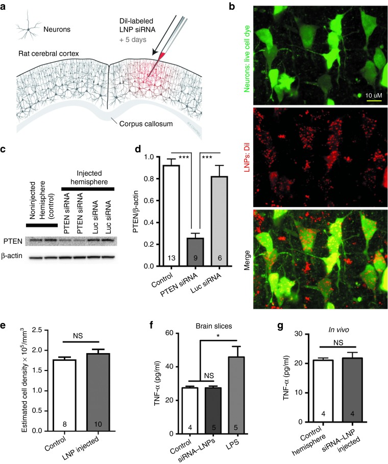
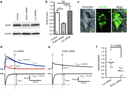
“The results suggest that there was no immunostimulatory effect when luc siRNA-LNPs (246 nmol/l) were applied to brain slices. TNF-α levels in the luc siRNA-LNP–treated group were not significantly different from the control group (Figure 4f, P > 0.05).”
§ Lipid Nanoparticle Delivery of siRNA to Silence Neuronal Gene Expression in the Brain > Results > LNP-mediated neuronal gene silencing in vivo
hedged- Asserted: passes — The authors put it forward as true.
- Checkable: passes — It can be checked against evidence.
- Load-bearing: passes — The paper’s conclusions depend on it.
- Based: passes — The paper gives a reason why it holds.
It may state two separate claims as one.
What the paper did — it cites no source for this, so there is none to check. The quote shows it is in the paper.
In the paper
“To get the NMDA component, we measured the amplitude of the outward current at Vh + 40 mV 50 ms after the stimulation artifact, a time point when the AMPAR current had returned to baseline (this was verified by subtracting the trace in the presence of the NMDAR antagonist, DL-2-amino-5-phosphonopentanoic acid (APV) to show the outward INMDAR (blue) and IAMPAR (red) components separately).”
§ Lipid Nanoparticle Delivery of siRNA to Silence Neuronal Gene Expression in the Brain > Results > Functional knockdown of NMDAR currents in vivo
plain- Asserted: passes — The authors put it forward as true.
- Checkable: passes — It can be checked against evidence.
- Load-bearing: passes — The paper’s conclusions depend on it.
- Based: passes — The paper gives a reason why it holds.
Probably not a substantial claim.
In the paper
“In ApoE knockout mice, LNP-siRNA gene silencing activity is inhibited in hepatocytes and can be reestablished by incubating the LNPs with ApoE before IV administration.”
§ Lipid Nanoparticle Delivery of siRNA to Silence Neuronal Gene Expression in the Brain > Discussion
What the sources say
- Not checked — no full text of the sourcecites [10] source
plaindirect- Asserted: passes — The authors put it forward as true.
- Checkable: passes — It can be checked against evidence.
- Load-bearing: passes — The paper’s conclusions depend on it.
- Based: passes — The paper gives a reason why it holds.
It may state two separate claims as one.
A claim is checked only against the cited paper’s full text, never its abstract, and none is available for its sources.
In the paper
“NMDARs are typically arranged in clusters at synaptic and extrasynaptic sites.20,21”
§ Lipid Nanoparticle Delivery of siRNA to Silence Neuronal Gene Expression in the Brain > Results > LNP-mediated knockdown of GluN1 in cell culture
What the sources say
plain- Asserted: passes — The authors put it forward as true.
- Checkable: passes — It can be checked against evidence.
- Load-bearing: fails — The paper’s conclusions may not depend on it.
- Based: passes — The paper gives a reason why it holds.
Probably not a substantial claim. The paper’s conclusions may not depend on it.
What the paper did — it cites no source for this, so there is none to check. The quote shows it is in the paper.
In the paper
“The AMPA component was measured as the inward current when the cell was held at −70 mV (Figure 8d).”
§ Lipid Nanoparticle Delivery of siRNA to Silence Neuronal Gene Expression in the Brain > Results > Functional knockdown of NMDAR currents in vivo
plain- Asserted: passes — The authors put it forward as true.
- Checkable: passes — It can be checked against evidence.
- Load-bearing: passes — The paper’s conclusions depend on it.
- Based: passes — The paper gives a reason why it holds.
Probably not a substantial claim.
A claim is checked only against the cited paper’s full text, never its abstract, and none is available for its sources.
In the paper
“Therefore, a common protocol to test the NMDAR/AMPAR ratio in response to the synaptic release of glutamate is to measure the ratio of the evoked excitatory postsynaptic current when the NMDAR is blocked by holding the cell at −70 mV to obtain a pure AMPAR component versus when the cell is held at +40 mV to get a mixed AMPAR + NMDAR component (Figure 8d).22,23”
§ Lipid Nanoparticle Delivery of siRNA to Silence Neuronal Gene Expression in the Brain > Results > Functional knockdown of NMDAR currents in vivo
What the sources say
plain- Asserted: passes — The authors put it forward as true.
- Checkable: passes — It can be checked against evidence.
- Load-bearing: passes — The paper’s conclusions depend on it.
- Based: passes — The paper gives a reason why it holds.
Probably not a substantial claim.
- Rests on an assumption: Glutamate is the major excitatory neurotransmitter in the brain and acts on both AMPAR and NMDAR at synapses.
A conclusion the paper draws from its own results — it cites no other source for it, so there is none to check. The quote shows it is in the paper.
In the paper
“These results demonstrate that when administered directly into the brain, LNPs are capable of diffusing through the extracellular milieu to deliver siRNA to neurons and induce gene silencing at sites distant from the site of injection.”
§ Lipid Nanoparticle Delivery of siRNA to Silence Neuronal Gene Expression in the Brain > Results > LNP-mediated neuronal gene silencing in vivo
boosteddirect- Asserted: passes — The authors put it forward as true.
- Checkable: passes — It can be checked against evidence.
- Load-bearing: passes — The paper’s conclusions depend on it.
- Based: passes — The paper gives a reason why it holds.
It may state two separate claims as one.
What the paper did — it cites no source for this, so there is none to check. The quote shows it is in the paper.
In the paper
“Neurons that were chosen for experiments were within 500 µm of the injection site and were verified to have taken up LNPs based on punctate intracellular DiI fluorescence (Figure 8c), and the ratio of NMDAR/AMPAR was measured.”
§ Lipid Nanoparticle Delivery of siRNA to Silence Neuronal Gene Expression in the Brain > Results > Functional knockdown of NMDAR currents in vivo
plain- Asserted: passes — The authors put it forward as true.
- Checkable: passes — It can be checked against evidence.
- Load-bearing: passes — The paper’s conclusions depend on it.
- Based: passes — The paper gives a reason why it holds.
It may state two separate claims as one.
A conclusion the paper draws from its own results — it cites no other source for it, so there is none to check. The quote shows it is in the paper.
In the paper
“The use of LNPs can circumvent these issues, and provides a method to rapidly test various siRNA constructs against the expression of different target proteins, and then examine the protein's function.”
§ Lipid Nanoparticle Delivery of siRNA to Silence Neuronal Gene Expression in the Brain > Discussion
plaindirect- Asserted: passes — The authors put it forward as true.
- Checkable: passes — It can be checked against evidence.
- Load-bearing: passes — The paper’s conclusions depend on it.
- Based: passes — The paper gives a reason why it holds.
It may state two separate claims as one.
- Rests on an assumption: The disadvantage of viral delivery is the requirement for constructing virus vectors that take added time, potentially cause immune responses, and raise safety concerns.
What the paper did — it cites no source for this, so there is none to check. The quote shows it is in the paper.
In the paper
“This is a region with robust LNP uptake and gene knockdown but not damaged from the injection needle itself, suggesting that LNPs themselves are nontoxic in vivo.”
§ Lipid Nanoparticle Delivery of siRNA to Silence Neuronal Gene Expression in the Brain > Results > LNP-mediated neuronal gene silencing in vivo
hedged- Asserted: passes — The authors put it forward as true.
- Checkable: passes — It can be checked against evidence.
- Load-bearing: passes — The paper’s conclusions depend on it.
- Based: passes — The paper gives a reason why it holds.
It may state two separate claims as one.
A conclusion the paper draws from its own results — it cites no other source for it, so there is none to check. The quote shows it is in the paper.
In the paper
“The results presented in this article show that LNP-siRNA systems can exhibit efficient gene silencing properties in neurons both in vitro and in vivo that lead to functional consequences without inducing significant toxicity.”
§ Lipid Nanoparticle Delivery of siRNA to Silence Neuronal Gene Expression in the Brain > Discussion
plaindirect- Asserted: passes — The authors put it forward as true.
- Checkable: passes — It can be checked against evidence.
- Load-bearing: passes — The paper’s conclusions depend on it.
- Based: passes — The paper gives a reason why it holds.
It may state two separate claims as one.
In the paper
“Clinical studies suggest that the immunostimulatory effects of LNP-siRNA injections are infrequent and readily managed, see http://www.alnylam.com/capella/wp-content/uploads/2012/07/Alnylam-ALN-TTR02-PhaseI-Results-120716.pdf.”
§ Lipid Nanoparticle Delivery of siRNA to Silence Neuronal Gene Expression in the Brain > Discussion
What the sources say
- Not matchedcites http://www.alnylam.com/capella/wp-content/uploads/2012/07/Alnylam-ALN-TTR02-PhaseI-Results-120716.pdf
Couldn’t be matched to a paper in Crossref, so it can’t be checked.
hedged- Asserted: passes — The authors put it forward as true.
- Checkable: passes — It can be checked against evidence.
- Load-bearing: unclear — It is unclear whether the paper’s conclusions depend on it.
- Based: passes — The paper gives a reason why it holds.
It may state two separate claims as one.
- Not matched
What the paper did — it cites no source for this, so there is none to check. The quote shows it is in the paper.
In the paper
“Cell density was measured in 10 µm z-stacks of 200 × 200 µm, and all cells including partial cells were counted, therefore resulting in an overestimate of cell density/mm3.”
§ Lipid Nanoparticle Delivery of siRNA to Silence Neuronal Gene Expression in the Brain > Materials and methods
plaindirect- Asserted: passes — The authors put it forward as true.
- Checkable: passes — It can be checked against evidence.
- Load-bearing: fails — The paper’s conclusions may not depend on it.
- Based: passes — The paper gives a reason why it holds.
Probably not a substantial claim. The paper’s conclusions may not depend on it.
What the paper did — it cites no source for this, so there is none to check. The quote shows it is in the paper.
In the paper
“The extracellular solutions were supplemented with 50 µmol/l picrotoxin (Sigma) and 8 mmol/l of each MgCl2 and CaCl2 to block GABAA synaptic potentials, block epileptiform activity, and minimize polysynaptic responses.”
§ Lipid Nanoparticle Delivery of siRNA to Silence Neuronal Gene Expression in the Brain > Materials and methods
plaindirect- Asserted: passes — The authors put it forward as true.
- Checkable: passes — It can be checked against evidence.
- Load-bearing: fails — The paper’s conclusions may not depend on it.
- Based: passes — The paper gives a reason why it holds.
Probably not a substantial claim. The paper’s conclusions may not depend on it. It may state two separate claims as one.
What the paper did — it cites no source for this, so there is none to check. The quote shows it is in the paper.
In the paper
“Cells were allowed to dialyze for at least 15 minutes before starting recordings.”
§ Lipid Nanoparticle Delivery of siRNA to Silence Neuronal Gene Expression in the Brain > Materials and methods
plain- Asserted: passes — The authors put it forward as true.
- Checkable: passes — It can be checked against evidence.
- Load-bearing: fails — The paper’s conclusions may not depend on it.
- Based: passes — The paper gives a reason why it holds.
Probably not a substantial claim. The paper’s conclusions may not depend on it.
A conclusion the paper draws from its own results — it cites no other source for it, so there is none to check. The quote shows it is in the paper.
In the paper
“The LNP platform is highly flexible, potentially allowing delivery of other materials to neurons such as drugs, fluorescent dyes and plasmids.”
§ Lipid Nanoparticle Delivery of siRNA to Silence Neuronal Gene Expression in the Brain > Discussion
hedgedconditional- Asserted: passes — The authors put it forward as true.
- Checkable: passes — It can be checked against evidence.
- Load-bearing: fails — The paper’s conclusions may not depend on it.
- Based: fails — The paper may give no reason why it holds.
Probably not a substantial claim. The paper’s conclusions may not depend on it. The paper may give no reason why it holds.
In the paper
“A lipid-based formulation of cytarabine has gained clinical acceptance for treatment of brain cancer following direct administration into CSF.34”
§ Lipid Nanoparticle Delivery of siRNA to Silence Neuronal Gene Expression in the Brain > Discussion
What the sources say
- Not checked — no full text of the sourcecites [34] source
plaindirect- Asserted: passes — The authors put it forward as true.
- Checkable: passes — It can be checked against evidence.
- Load-bearing: fails — The paper’s conclusions may not depend on it.
- Based: passes — The paper gives a reason why it holds.
Probably not a substantial claim. The paper’s conclusions may not depend on it.
A conclusion the paper draws from its own results — it cites no other source for it, so there is none to check. The quote shows it is in the paper.
In the paper
“The therapeutic utility of direct intracranial administration of LNP-siRNA for treatment of neurological disorders remains to be established; however, there are now a number of clinically accepted therapies relying on direct pumping of therapeutics into the CSF.32,33”
§ Lipid Nanoparticle Delivery of siRNA to Silence Neuronal Gene Expression in the Brain > Discussion
plain- Asserted: passes — The authors put it forward as true.
- Checkable: passes — It can be checked against evidence.
- Load-bearing: fails — The paper’s conclusions may not depend on it.
- Based: passes — The paper gives a reason why it holds.
Probably not a substantial claim. The paper’s conclusions may not depend on it.
What the paper did — it cites no source for this, so there is none to check. The quote shows it is in the paper.
In the paper
“Vhold was then returned to −70 mV at the end of the experiment to verify that there was no change in the baseline.”
§ Lipid Nanoparticle Delivery of siRNA to Silence Neuronal Gene Expression in the Brain > Materials and methods
plain- Asserted: passes — The authors put it forward as true.
- Checkable: passes — It can be checked against evidence.
- Load-bearing: fails — The paper’s conclusions may not depend on it.
- Based: passes — The paper gives a reason why it holds.
Probably not a substantial claim. The paper’s conclusions may not depend on it.
What the paper did — it cites no source for this, so there is none to check. The quote shows it is in the paper.
In the paper
“Access resistance was continuously monitored during the experiments.”
§ Lipid Nanoparticle Delivery of siRNA to Silence Neuronal Gene Expression in the Brain > Materials and methods
plain- Asserted: passes — The authors put it forward as true.
- Checkable: passes — It can be checked against evidence.
- Load-bearing: fails — The paper’s conclusions may not depend on it.
- Based: passes — The paper gives a reason why it holds.
Probably not a substantial claim. The paper’s conclusions may not depend on it.
What the paper did — it cites no source for this, so there is none to check. The quote shows it is in the paper.
In the paper
“Media containing Triton X-100 (1%) was used as a positive control for cell death.”
§ Lipid Nanoparticle Delivery of siRNA to Silence Neuronal Gene Expression in the Brain > Materials and methods
plain- Asserted: passes — The authors put it forward as true.
- Checkable: passes — It can be checked against evidence.
- Load-bearing: fails — The paper’s conclusions may not depend on it.
- Based: passes — The paper gives a reason why it holds.
Probably not a substantial claim. The paper’s conclusions may not depend on it.
What the paper did — it cites no source for this, so there is none to check. The quote shows it is in the paper.
In the paper
“As a negative control experiment, primary antibody was omitted during the immunostaining.”
§ Lipid Nanoparticle Delivery of siRNA to Silence Neuronal Gene Expression in the Brain > Materials and methods
plain- Asserted: passes — The authors put it forward as true.
- Checkable: passes — It can be checked against evidence.
- Load-bearing: fails — The paper’s conclusions may not depend on it.
- Based: passes — The paper gives a reason why it holds.
Probably not a substantial claim. The paper’s conclusions may not depend on it.
The paper relies on this without support.
ApoE is the dominant lipoprotein in the brain.
“Because ApoE is the dominant lipoprotein in the brain, we tested whether ApoE facilitates uptake of LNPs by neurons in cell culture.”
The paper relies on this without support.
LNP systems cannot cross the blood–brain barrier.
“Due to the inability of LNP systems to cross the blood–brain barrier, the potency of these systems for silencing genes in brain tissue has not been investigated.”
The paper relies on this without support.
Luciferase siRNA is not found in the mammalian genome.
“Of note, in control experiments, incubation of cultures with LNPs containing siRNA against luciferase (luc), which is not found in the mammalian genome, had no effect on levels of PTEN protein (Figure 2d).”
The paper relies on this without support.
The disadvantage of viral delivery is the requirement for constructing virus vectors that take added time, potentially cause immune responses, and raise safety concerns.
“The disadvantage of viral delivery is the requirement for constructing virus vectors that take added time, potentially cause immune responses, and raise safety concerns.”
The paper relies on this without support.
Glutamate is the major excitatory neurotransmitter in the brain and acts on both AMPAR and NMDAR at synapses.
“Glutamate is the major excitatory neurotransmitter in the brain and acts on both AMPAR and NMDAR at synapses.”
The paper relies on this without support.
Rapid dissociation of PEG-DMG from lipid nanoparticles upon dilution allows association of proteins such as ApoE.
“The LNP formulation employed here used PEG-DMG, which is a type of PEG-lipid with short acyl chains that allow it to dissociate rapidly30 from LNPs upon dilution, allowing association of proteins such as ApoE.”
The paper relies on this without support.
The phosphorothioate (P=S) linkage in the siRNA provides protection from exonuclease degradation.
“The P=S linkage provided protection from exonuclease degradation, and the 2′-OMe modifications reduced potential for immunostimulatory activity and provided stability toward endonucleolytic degradation.”
The paper relies on this without support.
The 2′-OMe modifications in the siRNA reduce potential for immunostimulatory activity and provide stability toward endonucleolytic degradation.
“The P=S linkage provided protection from exonuclease degradation, and the 2′-OMe modifications reduced potential for immunostimulatory activity and provided stability toward endonucleolytic degradation.”
Claims other papers make about this one
Each is checked against this paper’s own text: does it say what the citing paper says it does?
The cited source says what the claim attributes to it.
In the citing paper
“Rungta et al. (2013) demonstrated that LNPs encapsulating siRNA have been injected into the brain after a direct intracerebral or intracerebroventricular (ICV) injection, and were able to efficiently knock down neuronal gene expression (16).”
From Lipid Nanoparticles Deliver mRNA to the Brain after an Intracerebral Injection (2023) · cites this paper as [16]
Self-check
Percentages, sums and reported differences re-computed from the paper’s own numbers.
Each contradiction between two passages listed here was confirmed by an independent check.
Rated minor: it would not touch a result — a value no result rests on, a p-value that stays on the same side of the significance level, or a conflict that is clerical, resolved elsewhere in the paper, or outside its own findings.
In the paper
§ Lipid Nanoparticle Delivery of siRNA to Silence Neuronal Gene Expression in the Brain > Results > LNP-mediated neuronal gene silencing in vitro
“we tested the dose dependence of ApoE versus LNP uptake by measuring the total DiI fluorescence from ruptured cells 1 hour after treatment.”
§ Lipid Nanoparticle Delivery of siRNA to Silence Neuronal Gene Expression in the Brain > Materials and methods
“Cortical neuronal cultures (3 × 10⁴ cells in 12-well plate) were treated with luc siRNA-LNP (at 1.6 µg/ml) conjugated with DiI for 2 hr in the absence and presence of recombinant ApoE4 (Peprotech, Rocky Hill, NJ) at different concentrations (0.1, 1, 5, 10 µg/ml).”
Rated minor: it would not touch a result — a value no result rests on, a p-value that stays on the same side of the significance level, or a conflict that is clerical, resolved elsewhere in the paper, or outside its own findings.
In the paper
§ Lipid Nanoparticle Delivery of siRNA to Silence Neuronal Gene Expression in the Brain > Results > Functional knockdown of NMDAR currents in vivo
“Rats were injected with LNPs containing GluR1 siRNA or luc siRNA, and the NMDAR/AMPAR ratio was measured in cortical brain slices 4–6 days later”
§ Lipid Nanoparticle Delivery of siRNA to Silence Neuronal Gene Expression in the Brain > Results > Functional knockdown of NMDAR currents in vivo
“we found that neurons treated with LNPs that contained GluN1 siRNA had significantly decreased NMDAR/AMPAR ratios compared with neurons with LNPs from luc siRNA–injected rats”
Citations
Cites
Potent and specific genetic interference by double-stranded RNA in Caenorhabditis elegans
Fire, A., Xu, S., Montgomery, M. K., Kostas, S. A., Driver, S. E., & Mello, C. C. (1998) · Nature · doi:10.1038/35888 · Cited by 15,429
Glutamate Receptor Ion Channels: Structure, Regulation, and Function
Traynelis, S. F., Wollmuth, L. P., McBain, C. J., Menniti, F. S., Vance, K. M., Ogden, K. K., Hansen, K. B., Yuan, H., Myers, S. J., & Dingledine, R. (2010) · Pharmacological Reviews · doi:10.1124/pr.109.002451 · Cited by 3,568
Rational design of cationic lipids for siRNA delivery
Semple, S. C., Akinc, A., Chen, J., Sandhu, A. P., Mui, B. L., Cho, C. K., Sah, D. W. Y., Stebbing, D., Crosley, E. J., Yaworski, E., et al. (2010) · Nature Biotechnology · doi:10.1038/nbt.1602 · Cited by 1,822
Self-citationRNAi-mediated gene silencing in non-human primates
Zimmermann, T. S., Lee, A. C. H., Akinc, A., Bramlage, B., Bumcrot, D., Fedoruk, M. N., Harborth, J., Heyes, J. A., Jeffs, L. B., John, M., et al. (2006) · Nature · doi:10.1038/nature04688 · Cited by 1,357
Maximizing the potency of siRNA lipid nanoparticles for hepatic gene silencing in vivo
Jayaraman, M., Ansell, S. M., Mui, B. L., Tam, Y. K., Chen, J., Du, X., Butler, D., Eltepu, L., Matsuda, S., Narayanannair, J. K., et al. (2012) · Angewandte Chemie International Edition · doi:10.1002/anie.201203263 · Cited by 1,267
Self-citationTargeted delivery of RNAi therapeutics with endogenous and exogenous ligand-based mechanisms
Akinc, A., Querbes, W., De, S., Qin, J., Frank-Kamenetsky, M., Jayaprakash, K. N., Jayaraman, M., Rajeev, K. G., Cantley, W. L., Dorkin, J. R., et al. (2010) · Molecular Therapy · doi:10.1038/mt.2010.85 · Cited by 1,261
Apolipoprotein E and its receptors in Alzheimer's disease: pathways, pathogenesis and therapy
Bu, G. (2009) · Nature Reviews Neuroscience · doi:10.1038/nrn2620 · Cited by 1,168
A non-disruptive technique for loading calcium buffers and indicators into cells
Tsien, R. Y. (1981) · Nature · doi:10.1038/290527a0 · Cited by 1,132
Pten Regulates Neuronal Arborization and Social Interaction in Mice
Kwon, C.-H., Luikart, B. W., Powell, C. M., Zhou, J., Matheny, S. A., Zhang, W., Li, Y., Baker, S. J., & Parada, L. F. (2006) · Neuron · doi:10.1016/j.neuron.2006.03.023 · Cited by 970
Lipid-like materials for low-dose, in vivo gene silencing
Love, K. T., Mahon, K. P., Levins, C. G., Whitehead, K. A., Querbes, W., Dorkin, J. R., Qin, J., Cantley, W., Qin, L. L., Racie, T., et al. (2010) · Proceedings of the National Academy of Sciences · doi:10.1073/pnas.0910603106 · Cited by 969
Cited by
Sorted by citations
Advances in oligonucleotide drug delivery
Roberts, T. C., Langer, R., & Wood, M. J. A. (2020) · Nature Reviews Drug Discovery · doi:10.1038/s41573-020-0075-7 · Cited by 2,121
Lipid Nanoparticle Systems for Enabling Gene Therapies
Cullis, P. R., & Hope, M. J. (2017) · Molecular Therapy · doi:10.1016/j.ymthe.2017.03.013 · Cited by 1,208
Self-citationLipids and Lipid Derivatives for RNA Delivery
Zhang, Y., Sun, C., Wang, C., Jankovic, K. E., & Dong, Y. (2021) · Chemical Reviews · doi:10.1021/acs.chemrev.1c00244 · Cited by 649
Lipid nanoparticles for nucleic acid delivery: Current perspectives
Samaridou, E., Heyes, J. A., & Lutwyche, P. (2020) · Advanced Drug Delivery Reviews · doi:10.1016/j.addr.2020.06.002 · Cited by 609
Lipid nanoparticles enabling gene therapies: from concepts to clinical utility
Kulkarni, J. A., Cullis, P. R., & van der Meel, R. (2018) · Nucleic Acid Therapeutics · doi:10.1089/nat.2018.0721 · Cited by 592
“The first demonstration of LNP-siRNA activity in neurons was targeting the GluN1 subunit of the N -methyl-D-Aspartate receptor following intracortical and intracerebroventricular administration [71].”
Self-citationThe 60-year evolution of lipid nanoparticles for nucleic acid delivery
Cullis, P. R., & Felgner, P. L. (2024) · Nature Reviews Drug Discovery · doi:10.1038/s41573-024-00977-6 · Cited by 583
“Direct injection of LNP RNA systems into the eye 165 and brain 166–168 are increasingly enabling delivery to a variety of tissues in these organs.”
Self-citationRecent advances in siRNA delivery mediated by lipid-based nanoparticles
Yonezawa, S., Koide, H., & Asai, T. (2020) · Advanced Drug Delivery Reviews · doi:10.1016/j.addr.2020.07.022 · Cited by 394
“In 2013, it was reported that ionizable cationic lipid-LNP containing siRNA spread widely in the brain and showed significant knockdown of target mRNA (GRIN1, coding GluN1 subunit of NMDA [N-methyl-Daspartate] receptor) in the brain after intracortical or intracerebroventricular injection without an immune response [2…”
Influence of particle size on the in vivo potency of lipid nanoparticle formulations of siRNA
Chen, S., Tam, Y. Y. C., Lin, P. J. C., Sung, M. M. H., Tam, Y. K., & Cullis, P. R. (2016) · Journal of Controlled Release · doi:10.1016/j.jconrel.2016.05.059 · Cited by 366
Self-citationMicrofluidic Mixing: A General Method for Encapsulating Macromolecules in Lipid Nanoparticle Systems
Leung, A. K. K., Tam, Y. Y. C., Chen, S., Hafez, I. M., & Cullis, P. R. (2015) · The Journal of Physical Chemistry B · doi:10.1021/acs.jpcb.5b02891 · Cited by 359
Self-citationMaeki, M., Kimura, N., Sato, Y., Harashima, H., & Tokeshi, M. (2018) · Advanced Drug Delivery Reviews · doi:10.1016/j.addr.2018.03.008 · Cited by 329
Lipid nanoparticles for delivery of messenger RNA to the back of the eye
Patel, S., Ryals, R. C., Weller, K. K., Pennesi, M. E., & Sahay, G. (2019) · Journal of Controlled Release · doi:10.1016/j.jconrel.2019.04.015 · Cited by 260
“Ionizable LNPs (such as MC3, KC2, and DODMA) have been previously demonstrated to enter hepatocytes and neurons via the low-density lipoprotein (LDL) receptor in an ApoEdependent manner [16,55].”
BackgroundThe Cellular Mechanisms of Neuronal Swelling Underlying Cytotoxic Edema
Rungta, R. L., Choi, H. B., Tyson, J. R., Malik, A., Dissing‐Olesen, L., Lin, P. J. C., Cain, S. M., Cullis, P. R., Snutch, T. P., & MacVicar, B. A. (2015) · Cell · doi:10.1016/j.cell.2015.03.029 · Cited by 255
“We recently reported the development of an efficient LNP-mediated delivery system to introduce siRNAs against specific molecular targets into CNS neurons both in vivo and in vitro (Rungta et al., 2013).”
InfluentialSelf-citationSmall interfering RNA for cancer treatment: overcoming hurdles in delivery
Charbe, N. B., Amnerkar, N. D., Ramesh, B., Tambuwala, M. M., Bakshi, H. A., Aljabali, A. A. A., Khadse, S. C., Satheeshkumar, R., Satija, S., Metha, M., et al. (2020) · Acta Pharmaceutica Sinica B · doi:10.1016/j.apsb.2020.10.005 · Cited by 250
“Rungta et al.(311) have developed the siRNAelipid nanoparticle systems to deliver siRNA to the neurons.”
BackgroundDesign of lipid nanoparticles for in vitro and in vivo delivery of plasmid DNA
Kulkarni, J. A., Myhre, J. L., Chen, S., Tam, Y. Y. C., Danescu, A., Richman, J. M., & Cullis, P. R. (2016) · Nanomedicine Nanotechnology Biology and Medicine · doi:10.1016/j.nano.2016.12.014 · Cited by 243
“To study the role of ApoE, wild-type (C57BL/6) and ApoE -/- (B6.”
BackgroundInfluentialSelf-citationIn vivo somatic cell base editing and prime editing
Newby, G. A., & Liu, D. R. (2021) · Molecular Therapy · doi:10.1016/j.ymthe.2021.09.002 · Cited by 195
Lipid nanoparticle-mediated drug delivery to the brain
Khare, P., Edgecomb, S. X., Hamadani, C. M., Tanner, E. E. L., & S Manickam, D. (2023) · Advanced Drug Delivery Reviews · doi:10.1016/j.addr.2023.114861 · Cited by 187
Cohen, Z. R., Ramishetti, S., Peshes-Yaloz, N., Goldsmith, M., Wohl, A., Zibly, Z., & Peer, D. (2015) · ACS Nano · doi:10.1021/nn506248s · Cited by 182
Chen, S., Tam, Y. Y. C., Lin, P. J. C., Leung, A. K. K., Tam, Y. K., & Cullis, P. R. (2014) · Journal of Controlled Release · doi:10.1016/j.jconrel.2014.09.025 · Cited by 160
“As the lipid composition employed here is different than used previously, we examined the size distribution achieved for LNP– siRNA systems composed of DMAP-BLP, DSPC, cholesterol and PEG- DMG in the molar ratios 50:10:(39.75 − x):(0.25 + x), where x was varied from 0 to 4.75, encapsulating siRNA at a N/P (+/-) charge…”
InfluentialSelf-citationConformation-sensitive targeting of lipid nanoparticles for RNA therapeutics
Dammes, N., Goldsmith, M., Ramishetti, S., Dearling, J. L. J., Veiga, N., Packard, A. B., & Peer, D. (2021) · Nature Nanotechnology · doi:10.1038/s41565-021-00928-x · Cited by 159
Helmschrodt, C., Höbel, S., Schöniger, S., Bauer, A., Bonicelli, J., Gringmuth, M., Fietz, S. A., Aigner, A. M., Richter, A., & Richter, F. (2017) · Molecular Therapy — Nucleic Acids · doi:10.1016/j.omtn.2017.08.013 · Cited by 154
“For example, 5 mg siRNA complexed with branched PEIs achieved similar knockdown of N-methyl-Daspartate (NMDA) receptor NR2B subunit in spinal cord after intrathecal injection,(51) while higher dosages of 10 mg siRNA in lipid nanoparticles were infused ICVbilaterally in rats in order to knock down an endogenous target…”
BackgroundMethodRecent advances in mesenchymal stem cell membrane-coated nanoparticles for enhanced drug delivery
Wang, M., Xin, Y., Cao, H., Li, W., Hua, Y., Webster, T. J., Zhang, C., Tang, W., & Liu, Z. (2020) · Biomaterials Science · doi:10.1039/d0bm01164a · Cited by 144
Neuron-Targeted Nanoparticle for siRNA Delivery to Traumatic Brain Injuries
Kwon, E. J., Skalak, M., Bu, R. L., & Bhatia, S. N. (2016) · ACS Nano · doi:10.1021/acsnano.6b03858 · Cited by 136
Lipid Nanoparticles as Delivery Vehicles for Inhaled Therapeutics
Leong, E. W. X., & Ge, R. (2022) · Biomedicines · doi:10.3390/biomedicines10092179 · Cited by 134
“reported silencing of the GluN1 subunit of the neuronal N-methyl-D-aspartate receptor after delivering GluN1 siRNA in LNPs to mice via intracranial injection [48].”
MethodTargeting specific cells in the brain with nanomedicines for CNS therapies
Zhang, F., Lin, Y.-A., Kannan, S., & Kannan, R. M. (2015) · Journal of Controlled Release · doi:10.1016/j.jconrel.2015.12.013 · Cited by 113
“In some studies, neuronspecific targeting was achieved by taking advantage of the vehicle’s intrinsic ability to interact with neurons (Table 3) [122, 123].”
BackgroundTargeted Imaging of Brain Tumors with a Framework Nucleic Acid Probe
Tian, T., Li, J., Xie, C., et al. (2018) · ACS Applied Materials & Interfaces · doi:10.1021/acsami.7b17927 · Cited by 110
The Future of Tissue-Targeted Lipid Nanoparticle-Mediated Nucleic Acid Delivery
Kularatne, R. N., Crist, R. M., & Stern, S. T. (2022) · Pharmaceuticals · doi:10.3390/ph15070897 · Cited by 109
“058 PDI (ZP not specified) intracranial PTEN, luciferase and GRIN1 siRNA * 26–30-day-old (sex not specified) Sprague Dawley rats CNS neurons [45]”
BackgroundMicrofluidics Synthesis of Gene Silencing Cubosomes
Kim, H., Sung, J., Chang, Y., Alfeche, A., & Leal, C. (2018) · ACS Nano · doi:10.1021/acsnano.8b03770 · Cited by 93
Lipid nanoparticle-mediated siRNA delivery for safe targeting of human CML in vivo
Jyotsana, N., Sharma, A. K., Chaturvedi, A., Budida, R., Scherr, M., Kuchenbauer, F. C., Lindner, R. M., Noyan, F., Sühs, K., Stangel, M., et al. (2019) · Annals of Hematology · doi:10.1007/s00277-019-03713-y · Cited by 97
Self-citationGene Therapy for the Nervous System: Challenges and New Strategies
Maguire, C. A., Ramirez, S. H., Merkel, S. F., Sena‐Esteves, M., & Breakefield, X. O. (2014) · Neurotherapeutics · doi:10.1007/s13311-014-0299-5 · Cited by 99
“[77] used siRNA encapsulated in liposomes to knock-down specific mRNA targets in the brain after direct intracranial injection.”
MethodWaggoner, L. E., Miyasaki, K. F., & Kwon, E. J. (2023) · Biomaterials Science · doi:10.1039/d2bm01846b · Cited by 108
Tanaka, H., Nakatani, T., Furihata, T., Tange, K., Nakai, Y., Yoshioka, H., Harashima, H., & Akita, H. (2018) · Molecular Pharmaceutics · doi:10.1021/acs.molpharmaceut.7b01084 · Cited by 82
Critical Review of Lipid-Based Nanoparticles as Carriers of Neuroprotective Drugs and Extracts
Fernandes, F., Dias-Teixeira, M., Delerue-Matos, C. M., & Grosso, C. (2021) · Nanomaterials · doi:10.3390/nano11030563 · Cited by 81
“[43] used small interfering RNA (siRNA) in lipid nanoparticles (LNPs) to efficiently silence the gene GRIN1 in hippocampal neuronal cultures and in in vivo through intracortical or ICV injections.”
MethodClinical advances of RNA therapeutics for treatment of neurological and neuromuscular diseases
Holm, A., Hansen, S. N., Klitgaard, H., & Kauppinen, S. (2022) · RNA Biology · doi:10.1080/15476286.2022.2066334 · Cited by 92
Ralvenius, W. T., Andresen, J. L., Huston, M. M., Penney, J., Bonner, J. M., Fenton, O. S., Langer, R., & Tsai, L. (2024) · Advanced Materials · doi:10.1002/adma.202309225 · Cited by 78
Sahel, D. K., Vora, L. K., Saraswat, A. L., Sharma, S., Monpara, J. D., D’souza, A. A., Mishra, D., Tryphena, K. P., Kawakita, S., Khan, S. U., et al. (2023) · Advanced Science · doi:10.1002/advs.202207512 · Cited by 79
Spontaneous, solvent-free entrapment of siRNA within lipid nanoparticles
Kulkarni, J. A., Thomson, S. B., Zaifman, J., Leung, J., Wagner, P. K., Hill, A. M., Tam, Y. Y. C., Cullis, P. R., Petkau, T. L., & Leavitt, B. R. (2020) · Nanoscale · doi:10.1039/d0nr06816k · Cited by 73
Self-citationYamamoto, Y., Lin, P. J. C., Beraldi, E., Zhang, F., Kawai, Y., Leong, J., Katsumi, H., Fazli, L., Fraser, R., Cullis, P. R., et al. (2015) · Clinical Cancer Research · doi:10.1158/1078-0432.ccr-15-0866 · Cited by 75
“Although DMAP-BLP has been shown to be potent and safe in the brain (30), full toxicity profile of this lipid has yet to be characterized.”
BackgroundMethodSelf-citationMicrofluidics: a transformational tool for nanomedicine development and production
Garg, S., Heuck, G., Ip, S. Y., & Ramsay, E. (2016) · Journal of drug targeting · doi:10.1080/1061186x.2016.1198354 · Cited by 76
“ApoE dependence of the uptake of LNPs by neurons is shown by an increase in DiI fluorescence [30].”
BackgroundMethodInfluentialMendonça, M. C. P., Kont, A., Aburto, M. R., Cryan, J. F., & O’Driscoll, C. M. (2021) · Molecular Pharmaceutics · doi:10.1021/acs.molpharmaceut.0c01238 · Cited by 75
LNP-mediated delivery of CRISPR RNP for wide-spread in vivo genome editing in mouse cornea
Mohanna, S. Z. M., Djaksigulova, D., Hill, A. M., Wagner, P. K., Simpson, E. M., & Leavitt, B. R. (2022) · Journal of Controlled Release · doi:10.1016/j.jconrel.2022.08.042 · Cited by 73
Negishi, Y., Yamane, M., Kurihara, N., Endo‐Takahashi, Y., Sashida, S., Takagi, N., Suzuki, R., & Maruyama, K. (2015) · Pharmaceutics · doi:10.3390/pharmaceutics7030344 · Cited by 68
Chen, S., Zaifman, J., Kulkarni, J. A., Zhigaltsev, I. V., Tam, Y. K., Ciufolini, M. A., Tam, Y. Y. C., & Cullis, P. R. (2018) · Journal of Controlled Release · doi:10.1016/j.jconrel.2018.07.026 · Cited by 68
Self-citationKala, S., Mak, A. S. C., Liu, X., Posocco, P., Pricl, S., Peng, L., & Wong, A. S. T. (2014) · Journal of Medicinal Chemistry · doi:10.1021/jm401907z · Cited by 67
Lipid Nanoparticles Deliver mRNA to the Brain after an Intracerebral Injection
Tuma, J., Chen, Y.-J., Collins, M. G., Paul, A., Li, J., Han, H., Sharma, R., Murthy, N., & Lee, H. Y. (2023) · Biochemistry · doi:10.1021/acs.biochem.3c00371 · Cited by 65
siRNA as a tool to improve the treatment of brain diseases: Mechanism, targets and delivery
Gomes, M. J., Martins, S., & Sarmento, B. F. C. C. (2015) · Ageing Research Reviews · doi:10.1016/j.arr.2015.03.001 · Cited by 59
Lee, J. B., Zhang, K., Tam, Y. Y. C., Quick, J., Tam, Y. K., Lin, P. J. C., Chen, S., Liu, Y., Nair, J. K., Zlatev, I., et al. (2016) · Molecular Therapy — Nucleic Acids · doi:10.1038/mtna.2016.43 · Cited by 61
Self-citationCain, S. M., Tyson, J. R., Choi, H., Ko, R. W. Y., Lin, P. J. C., LeDue, J. M., Powell, K. L., Bernier, L., Rungta, R. L., Yang, Y., et al. (2018) · Epilepsia · doi:10.1111/epi.14018 · Cited by 57
“2 transcripts together with a recently developed methodology involving packaged lipid nanoparticles (LNPs) previously demonstrated as an efficacious vector for in vivo knockdown.(24,25) Initially, several distinct DsiRNAs were screened for their ability to attenuate exogenous CaV3.”
BackgroundSelf-citationReichenstein, I., Eitan, C., Díaz-García, S., Haim, G., Magen, I., Siany, A., Hoye, M. L., Rivkin, N., Olender, T., Tóth, B., et al. (2019) · Science Translational Medicine · doi:10.1126/scitranslmed.aav5264 · Cited by 57
Destination Brain: the Past, Present, and Future of Therapeutic Gene Delivery
Joshi, C. R., Labhasetwar, V., & Ghorpade, A. (2017) · Journal of Neuroimmune Pharmacology · doi:10.1007/s11481-016-9724-3 · Cited by 55
“Arginine-modified polyplexes (Morris and Labhasetwar 2015), transactivator of transcription-conjugated NPs (Rao et al. 2008) and AAV vector serotype 9 (Foust et al. 2009) crossed the BBB when injected intravenously in rodent models, indicating that tailor-made NPs with surface modifications and genetically modified vi…”
InfluentialAkita, H., Tamaru, M., Kajimoto, K., Sato, Y., Hatakeyama, H., Harashima, H., & Nakatani, T. (2014) · International Journal of Nanomedicine · doi:10.2147/ijn.s65402 · Cited by 55
“17 Uno et al also reported that α -tocopherol-conjugated siRNA accumulated efficiently in neuronal cells after incorporation into high-density lipoprotein after intracerebroventricular injection.”
Kiprowska, M., Степанова, А., Todaro, D. R., Galkin, A. S., Haas, A. L., Wilson, S. M., & Figueiredo‐Pereira, M. E. (2017) · Biochimica et Biophysica Acta (BBA) - Molecular Basis of Disease · doi:10.1016/j.bbadis.2017.03.017 · Cited by 55
Synthetic SiRNA Delivery: Progress and Prospects
Roberts, T. C., Ezzat, K., Andaloussi, S. E., & Weinberg, M. S. (2015) · Methods in molecular biology · doi:10.1007/978-1-4939-3112-5_23 · Cited by 55
Lipid nanoparticles for RNA delivery: Self-assembling vs driven-assembling strategies
Nele, V., Campani, V., Moosavian, S. A., & Rosa, G. D. (2024) · Advanced Drug Delivery Reviews · doi:10.1016/j.addr.2024.115291 · Cited by 58
mRNA lipid nanoparticle formulation, characterization and evaluation
Ma, Y., VanKeulen-Miller, R., & Fenton, O. S. (2025) · Nature Protocols · doi:10.1038/s41596-024-01134-4 · Cited by 54
“There are also other techniques to quantify the in vitro protein expression, such as western blotting 67,68 , mass spectrometry 69 and ELISA 70,71 .”
Comprehensive analysis of lipid nanoparticle formulation and preparation for RNA delivery
Haque, M. A., Shrestha, A., Mikelis, C. M., & Mattheolabakis, G. (2024) · International Journal of Pharmaceutics X · doi:10.1016/j.ijpx.2024.100283 · Cited by 52
“Overall, the cholesterol content in LNP formulations has been predominately at ~37 – 40 % of total molecular lipid content in multiple LNP formulations with different ionizable lipids (Ball et al., 2018a; Rungta et al., 2013; Yamamoto et al., 2015).”
BackgroundLipid-based nanocarriers for delivery of small interfering RNA for therapeutic use
Parashar, D., Rajendran, V., Shukla, R., & Sistla, R. (2019) · European Journal of Pharmaceutical Sciences · doi:10.1016/j.ejps.2019.105159 · Cited by 49
D’souza, A. A., Nozohouri, S., Bleier, B. S., & Amiji, M. M. (2022) · Pharmaceutical Research · doi:10.1007/s11095-022-03433-5 · Cited by 49
“Lipid nanoparticles also exhibit neuronal uptake facilitated by astrocyte-secreted apolipoprotein E (ApoE), recognized by low-density lipoprotein receptors on neurons [182].”
BackgroundReceptor-Assisted Nanotherapeutics for Overcoming the Blood–Brain Barrier
Mhaske, A. S., Shukla, S., Ahirwar, K., Singh, K. K., & Shukla, R. (2024) · Molecular Neurobiology · doi:10.1007/s12035-024-04015-9 · Cited by 54
“Particles undergo endocytosis and thereby attain neuronal uptake [47].”
BackgroundTherapeutic Applications of Spherical Nucleic Acids
Barnaby, S. N., Sita, T. L., Petrosko, S. H., Stegh, A. H., & Mirkin, C. A. (2015) · Cancer treatment and research · doi:10.1007/978-3-319-16555-4_2 · Cited by 46
“These in vivo barriers to oligonucleotide delivery have slowed the translation of nucleic acid-based therapeutics to the clinic and mandated the use of oligonucleotide-carrier systems, such as polymers/polyplexes [24, 25], dendrimers [26], and lipids [27, 28].”
BackgroundRecent Developments in Microfluidic Technologies for Central Nervous System Targeted Studies
Teixeira, M. I., Amaral, M. H., Costa, P. C., Lopes, C. M., & Lamprou, D. A. (2020) · Pharmaceutics · doi:10.3390/pharmaceutics12060542 · Cited by 44
“The results show therefore that the siRNA lipid formulation was capable of manipulating the expression of molecular targets involved in neuronal processes, potentially facilitating the development of gene therapies for NDs [119].”
BackgroundLipid-based nanoparticles and RNA as innovative neuro-therapeutics
Tsakiri, M., Zivko, C., Demetzos, C., & Mahairaki, V. (2022) · Frontiers in Pharmacology · doi:10.3389/fphar.2022.900610 · Cited by 39
“Furthermore, no toxicity to neurons was observed (Rungta et al., 2013).”
BackgroundOligonucleotide-based therapy for neurodegenerative diseases
Magen, I., & Hornstein, E. (2014) · Brain Research · doi:10.1016/j.brainres.2014.04.005 · Cited by 40
lncRNA in the liver: Prospects for fundamental research and therapy by RNA interference
Smekalova, E. M., Kotelevtsev, Y. V., Leboeuf, D., Shcherbinina, E. Y., Fefilova, A. S., Zatsepin, T. S., & Koteliansky, V. (2016) · Biochimie · doi:10.1016/j.biochi.2016.06.007 · Cited by 41
Enhanced target cell specificity and uptake of lipid nanoparticles using RNA aptamers and peptides
Ray, R. M., Hansen, A. H., Taskova, M., Jandl, B., Hansen, J. T., Soemardy, C., Morris, K. V., & Astakhova, K. (2021) · Beilstein Journal of Organic Chemistry · doi:10.3762/bjoc.17.75 · Cited by 40
Development of Therapeutics for C9ORF72 ALS/FTD-Related Disorders
Mis, M. S. C., Brajkovic, S., Tafuri, F., Bresolin, N., Comi, G. P., & Corti, S. P. (2016) · Molecular Neurobiology · doi:10.1007/s12035-016-9993-0 · Cited by 40
“Recently, lipid nanoparticles were demonstrated to have high delivery efficiency with little toxicity in the central nervous system (CNS) [44].”
BackgroundsiRNA delivery to lymphatic endothelial cells via ApoE-mediated uptake by lipid nanoparticles
Sakurai, Y., Yoshikawa, K., Arai, K., Kazaoka, A., Aoki, S., Ito, K., Nakai, Y., Tange, K., Furihata, T., Tanaka, H., et al. (2022) · Journal of Controlled Release · doi:10.1016/j.jconrel.2022.11.036 · Cited by 36
Byrnes, A. E., Dominguez, S. L., Yen, C.-W., Laufer, B. I., Foreman, O., Reichelt, M., Lin, H., Sagolla, M., Hötzel, K., Ngu, H., et al. (2023) · Molecular Therapy - Nucleic Acids · doi:10.1016/j.omtn.2023.05.005 · Cited by 38
Allele-Selective Inhibition of Mutant Atrophin-1 Expression by Duplex and Single-Stranded RNAs
Hu, J., Liu, J., Jayaprakash, K. N., Lackey, J. G., Kuchimanchi, S., Rajeev, K. G., Manoharan, M., Swayze, E. E., Lima, W. F., Prakash, T. P., et al. (2014) · Biochemistry · doi:10.1021/bi500610r · Cited by 35
Self-citationSharma, N. M., Cunningham, C., Zheng, H., Liu, X., & Patel, K. P. (2016) · Circulation Heart Failure · doi:10.1161/circheartfailure.116.003423 · Cited by 37
Selective Xi reactivation and alternative methods to restore MECP2 function in Rett syndrome
Grimm, N.-B., & Lee, J. T. (2022) · Trends in Genetics · doi:10.1016/j.tig.2022.01.007 · Cited by 34
Yardeni, T., Fine, R., Joshi, Y., Gradus, T., Kozer, N., Reichenstein, I., Yanowski, E., Nevo, S., Weiss-Tishler, H., Eisenberg‐Bord, M., et al. (2018) · Scientific Reports · doi:10.1038/s41598-017-17878-x · Cited by 34
Sabbir, M. G. (2018) · Frontiers in Molecular Biosciences · doi:10.3389/fmolb.2018.00099 · Cited by 34
“Knockdown of CaMKK2 Knockdown of CaMKK2 in cultured DRG neurons was achieved by transfecting cells with lipid nanoparticles (LNP) encapsulated cocktail of 3 siRNAs specific to exon 5, 8, and 12 of CaMKK2 gene (Table 1) (Rungta et al., 2013).”
BackgroundMethodInfluentialNag, K., Sarker, M. E. H., Kumar, S., Khan, H., Chakraborty, S., Islam, M. J., Baray, J. C., Khan, M. M. R., Mahmud, A., Barman, U., et al. (2022) · Scientific Reports · doi:10.1038/s41598-022-12100-z · Cited by 33
Fluorometric Sensor for Mercury Ion Detection in a Fluidic MEMS Device
Karthikeyan, K., & Sujatha, L. (2018) · IEEE Sensors Journal · doi:10.1109/jsen.2018.2840331 · Cited by 33
“during fluid flow and also complete mixing achieved at high flow rates [34]–[38].”
BackgroundNon-Viral Nucleic Acid Delivery Strategies to the Central Nervous System
Tan, J. Y., Sellers, D. L., Pham, B., Pun, S. H., & Horner, P. J. (2016) · Frontiers in Molecular Neuroscience · doi:10.3389/fnmol.2016.00108 · Cited by 34
“An apropos administration route must be chosen to maximize therapeutics at the treatment site (i.e., direct injections); however, caution must be heeded in avoiding unnecessary damage to healthy tissues by direct administration into the CNS (i.e., intravenous and peripheral administration).”
BackgroundsiRNA-nanoparticle conjugate in gene silencing: A future cure to deadly diseases?
Acharya, R., Saha, S., Ray, S., Hazra, S., Mitra, M. K., & Chakraborty, J. (2017) · Materials Science and Engineering C · doi:10.1016/j.msec.2017.03.009 · Cited by 33
Nanocarriers for Delivery of Oligonucleotides to the CNS
Male, D. K., & Gromnicova, R. (2022) · International Journal of Molecular Sciences · doi:10.3390/ijms23020760 · Cited by 32
“siRNA has been incorporated into the nanoparticles to silence the NMDA receptor in neurons following intracerebral injection [65].”
BackgroundMethodAllele-specific silencing of mutant p53 attenuates dominant-negative and gain-of-function activities
Iyer, S. V., Parrales, A., Begani, P., Narkar, A., Adhikari, A. S., Martinez, L. A., & Iwakuma, T. (2015) · Oncotarget · doi:10.18632/oncotarget.6634 · Cited by 31
“Methodologies that specifically deliver siRNAs into tumors in vivo, such as surface-modified nanoparticle-mediated siRNA delivery, should be further developed to improve the efficacy and specificity of targeted cancer therapies by siRNAs [48-51].”
BackgroundSato, Y., Sakurai, Y., Kajimoto, K., Nakamura, T., Yamada, Y., Akita, H., & Harashima, H. (2016) · Macromolecular Bioscience · doi:10.1002/mabi.201600179 · Cited by 32
Hirunagi, T., Sahashi, K., Tachikawa, K., Leu, A. I., Nguyen, M., Mukthavaram, R., Karmali, P. P., Chivukula, P., Tohnai, G., Iida, M., et al. (2021) · Molecular Therapy — Nucleic Acids · doi:10.1016/j.omtn.2021.02.007 · Cited by 31
Overcoming Barriers: Clinical Translation of siRNA Nanomedicines
Tieu, T., Wei, Y., Cifuentes‐Rius, A., & Voelcker, N. H. (2021) · Advanced Therapeutics · doi:10.1002/adtp.202100108 · Cited by 31
Nanomedicine based strategies for oligonucleotide traversion across the blood–brain barrier
Amulya, E., Sikder, A., Vambhurkar, G. B., Shah, S., Khatri, D. K., Raghuvanshi, R. S., Singh, S. B., & Srivastava, S. (2023) · Journal of Controlled Release · doi:10.1016/j.jconrel.2023.01.031 · Cited by 30
Akita, H., Nakatani, T., Kuroki, K., Maenaka, K., Tange, K., Nakai, Y., & Harashima, H. (2015) · International Journal of Pharmaceutics · doi:10.1016/j.ijpharm.2015.05.043 · Cited by 30
Page, S., Khan, T. A., Kühl, P. G., Schwach, G., Storch, K., & Chokshi, H. P. (2022) · The Annual Review of Pharmacology and Toxicology · doi:10.1146/annurev-pharmtox-052120-093517 · Cited by 28
Use, exposure and omics characterisation of potential hazard in nanomaterials
Ling, N., Zhang, N., Yong, W. T. L., Misbah, S., Hashim, F., Soon, C. F., Lim, G. P., Peng, W., & Sonne, C. (2023) · Materials Today Advances · doi:10.1016/j.mtadv.2023.100341 · Cited by 31
An Overview of Nanomedicines for Neuron Targeting
García-Chica, J., Paraiso, W. K. D., Tanabe, S., Serra, D., Herrero, L., Casals, N., García, J., Ariza, X., Quader, S., & Rodríguez‐Rodríguez, R. (2020) · Nanomedicine · doi:10.2217/nnm-2020-0088 · Cited by 28
Multifunctional Nanocarriers for Alzheimer’s Disease: Befriending the Barriers
Ansari, M. A., Tripathi, T., Venkidasamy, B., Monziani, A., Rajakumar, G., Alomary, M. N., Alyahya, S. A., Onimus, O., D’souza, N., Barkat, M. A., et al. (2023) · Molecular Neurobiology · doi:10.1007/s12035-023-03730-z · Cited by 29
Lipid-based nanoparticles for drug delivery in Parkinson’s disease
Cai, H., Liu, D., Xue, W.-W., Ma, L., Xie, H.-T., & Ning, K. (2024) · Translational Neuroscience · doi:10.1515/tnsci-2022-0359 · Cited by 26
“The authors employed liposomal delivery of siRNA targeting the GluN1 subunit of NMDA receptors and demonstrated its e ff ective and selective reduction of synaptic NMDA receptor currents compared to synaptic α -amino-3-hydroxy-5-methyl-4-isoxazole-pro- pionic acid receptor currents [115].”
Geng, L., Kato, N., Kodama, Y., Mukai, H., & Kawakami, S. (2023) · International Journal of Pharmaceutics · doi:10.1016/j.ijpharm.2023.122896 · Cited by 28
Phospholipid‐Free Small Unilamellar Vesicles for Drug Targeting to Cells in the Liver
Zhang, W., Böttger, R., Qin, Z., Kulkarni, J. A., Vogler, J., Cullis, P. R., & Li, S. (2019) · Small · doi:10.1002/smll.201901782 · Cited by 27
Self-citationSaleh, A. J., Sabbir, M. G., Aghanoori, M., Smith, D. R., Chowdhury, S. R., Tessler, L., Brown, J., Gedarevich, E., Kassahun, M. Z., Frizzi, K. E., et al. (2020) · Molecular Neurobiology · doi:10.1007/s12035-020-01900-x · Cited by 29
RNA therapies for CNS diseases
Francesco, V. D., Chua, A. J. K., Huang, D., D’souza, A. A., Yang, A., Bleier, B. S., & Amiji, M. M. (2024) · Advanced Drug Delivery Reviews · doi:10.1016/j.addr.2024.115283 · Cited by 27
Nuts and Bolts: Microfluidics for the Production of Biomaterials
Fontana, F., Martins, J. P., Torrieri, G., & Santos, H. A. (2019) · Advanced Materials Technologies · doi:10.1002/admt.201800611 · Cited by 25
Harnessing mRNA-lipid nanoparticles as innovative therapies for autoimmune diseases
Razavi, R., Kegel, M., Muscat-Rivera, J., Weissman, D., & Melamed, J. R. (2025) · Molecular Therapy — Methods & Clinical Development · doi:10.1016/j.omtm.2025.101566 · Cited by 28
“173”
Oliveira, C. L., Silveira, I., Veiga, F., & Ribeiro, A. J. (2014) · Expert Opinion on Drug Delivery · doi:10.1517/17425247.2014.945421 · Cited by 23
“More recently, this mechanism has also been described for efficient uptake of siRNA by neurons in the brain [42].”
BackgroundTrileucine residues in a ligand-CPP-based siRNA delivery platform improve endosomal escape of siRNA
Ullah, I., Chung, K., Beloor, J., Kim, J.-K., Cho, M., Kim, N., Lee, K. Y., Kumar, P., & Lee, S. (2016) · Journal of drug targeting · doi:10.1080/1061186x.2016.1258566 · Cited by 23
Gene therapies for high-grade gliomas: from the bench to the bedside.
Lucifero, A. G., Luzzi, S., Brambilla, I., Guarracino, C., Mosconi, M., Foiadelli, T., & Savasta, S. (2020) · Acta bio-medica dell'Ateneo parmense/Acta biomedica · doi:10.23750/abm.v91i7-s.9953 · Cited by 23
“Table 3 summarizes the clinical trials on novel gene therapies for HGGs. (Table 3).”
BackgroundInfluentialRNA interference efficiently targets human leukemia driven by a fusion oncogene in vivo
Jyotsana, N., Sharma, A. K., Chaturvedi, A., Scherr, M., Kuchenbauer, F. C., Sajti, L., Barchanski, A., Lindner, R. M., Noyan, F., Sühs, K., et al. (2017) · Leukemia · doi:10.1038/leu.2017.269 · Cited by 23
Self-citationArshad, M., Atochina‐Vasserman, E. N., Maurya, D. S., Lu, J., Ona, N. A., Vasserman, J. A., McWilliams, B. C., Ni, H., Berkihiser, S., Park, W., et al. (2025) · Journal of the American Chemical Society · doi:10.1021/jacs.5c07232 · Cited by 20
Current knowledge on the tissue distribution of mRNA nanocarriers for therapeutic protein expression
Zadory, M., Lopez, E., Babity, S., Gravel, S., & Brambilla, D. (2022) · Biomaterials Science · doi:10.1039/d2bm00859a · Cited by 18
Metadata
- Authors
- Rungta, R. L., Choi, H. B., Lin, P. J., Ko, R. W., Ashby, D., Nair, J., Manoharan, M., Cullis, P. R., & MacVicar, B. A.
- Affiliations
- University of British Columbia (CA)
- Alnylam Pharmaceuticals (United States) (US)
- Corresponding author
- MacVicar, Brian A
- ORCID
- Venue
- Molecular Therapy—Nucleic Acids
- Publisher
- Elsevier BV
- Published
- 2013-12-03
- Volume
- 2
- Issue
- 11
- Pages
- e136
- ISSN
- 2162-2531 (ISSN-L)
- PMC
- PMC3889191
- OpenAlex
- W2166483989
- Semantic Scholar
- ef56b67d26457c62fc9e18d60b3153ef94defeb9
- Cited by
- 165
- References
- 36
- Citations by year
- 2026: 6 · 2025: 15 · 2024: 18 · 2023: 16 · 2022: 16 · 2021: 10 · 2020: 12 · 2019: 8 · 2018: 12 · 2017: 8 · 2016: 16 · 2015: 16 · 2014: 12
- FWCI
- 5.92
- Type
- journal article
- Language
- en
- Open access
- gold
- doi.org
- Locations
- Molecular Therapy — Nucleic Acids (publishedVersion)
- PubMed (publishedVersion)
- Europe PMC (PubMed Central) (submittedVersion, cc-by-nc-nd)
- PubMed Central (submittedVersion, cc-by-nc-nd)
- Topics
- Life Sciences › Biochemistry, Genetics and Molecular Biology › Molecular Biology › RNA Interference and Gene Delivery
- Physical Sciences › Materials Science › Biomaterials › Nanoparticle-Based Drug Delivery
- Life Sciences › Neuroscience › Neurology › Barrier Structure and Function Studies
- Keywords
- lipid nanoparticles, siRNA delivery, apolipoprotein E, intracerebroventricular administration, intracortical injection, GRIN1, NMDA receptor, AMPA receptors, synaptic currents, neurotoxicity
About these checks
Registry, retraction, Cites and statistics results are computed, not judged, with one exception: a printed reference that no index lists is read once by a model before it counts as a citation error. Claims are extracted by a model and kept only when their quote appears verbatim in the text; each cited source is then checked, with a model’s help, for whether it says what the claim relies on. Those verdicts can be wrong — read the claim’s quote and the cited source before relying on one.
A language model drew the claims from the paper on 2026-10-20, under version 3 of the claim definition (model deepseek-v4.1-flash, prompt extract-v3).
A second model labelled each claim on 2026-10-20 (model jev-1.13-20260917, definition revision gold-guideline-v0.2).
Cited sources are checked with a language model’s help (gpt-6-sol).
Statistics and arithmetic are recomputed, not judged.
Confidence bands are shown only for fields calibrated on our annotated test set. None is calibrated yet, so no band is shown.
What counts as a claim ›How we measure confidence ›How we check this ›
Are you an author, or do you see a problem with this page? Request a review
