

Decoding tumour phenotype by noninvasive imaging using a quantitative radiomics approach
Open accessThe DOI matches its Crossref record; 29 of the 29 references it cites were found in a registry or an index.
Abstract
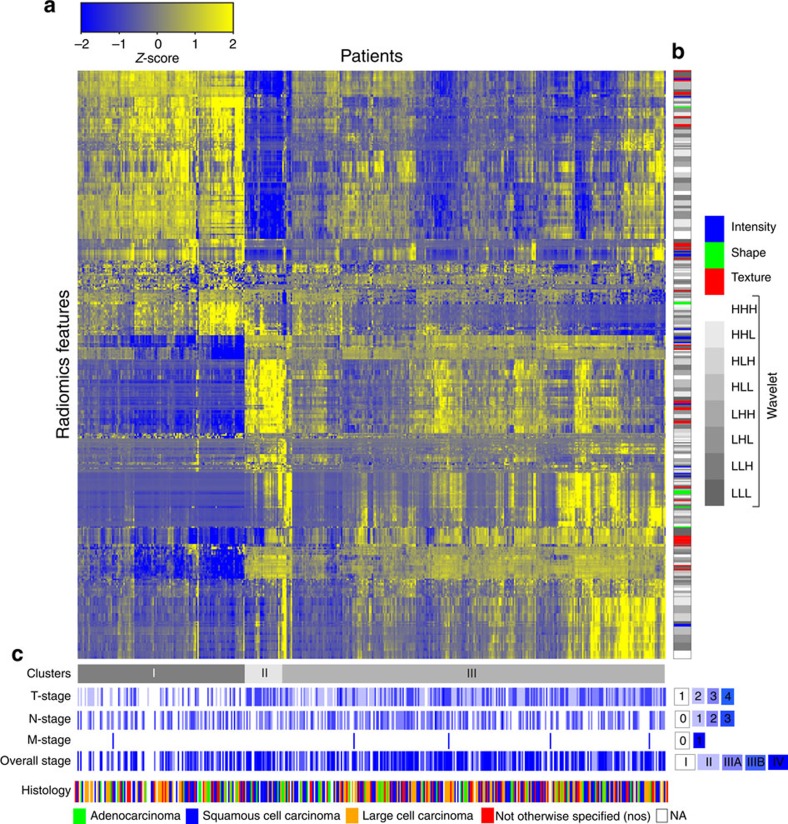
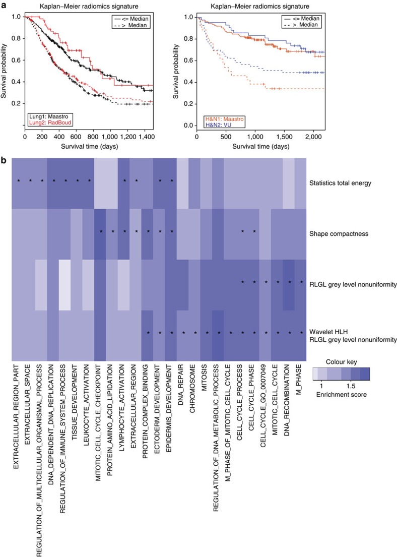
Human cancers exhibit strong phenotypic differences that can be visualized noninvasively by medical imaging. Radiomics refers to the comprehensive quantification of tumour phenotypes by applying a large number of quantitative image features. Here we present a radiomic analysis of 440 features quantifying tumour image intensity, shape and texture, which are extracted from computed tomography data of 1,019 patients with lung or head-and-neck cancer. We find that a large number of radiomic features have prognostic power in independent data sets of lung and head-and-neck cancer patients, many of which were not identified as significant before. Radiogenomics analysis reveals that a prognostic radiomic signature, capturing intratumour heterogeneity, is associated with underlying gene-expression patterns. These data suggest that radiomics identifies a general prognostic phenotype existing in both lung and head-and-neck cancer. This may have a clinical impact as imaging is routinely used in clinical practice, providing an unprecedented opportunity to improve decision-support in cancer treatment at low cost.
Overview
The claims and statistics of this paper haven’t been checked yet.
Free: it reads the open-access text and checks the first 5 claims that cite a source we can check. Create a free account to check every claim.
Integrity signals
Registry
Cites
Statistics
Figures


Verification
Claims the paper makes
The claims and statistics of this paper haven’t been checked yet.
Free: it reads the open-access text and checks the first 5 claims that cite a source we can check. Create a free account to check every claim.
Claims other papers make about this one
Each is checked against this paper’s own text: does it say what the citing paper says it does?
The source backs part of the claim, not all of it — or backs it for a different population, condition or comparison.
In the citing paper
“Notably, the signature was comprised of the top features from four classes (size, shape, texture, and wavelets) that were prioritized from a database of 442 features by using test-retest reproducibility and intersubject range.”
From Radiomics: Images Are More than Pictures, They Are Data (2016) · cites this paper as [47]
Experimental — accuracy not yet measuredThe source backs part of the claim, not all of it — or backs it for a different population, condition or comparison.
In the citing paper
“Many studies have shown that imaging features are significantly associated with patterns of gene expression and genetic mutations, demonstrating that radiogenomic analysis can identify different biological mechanisms by means of mathematical and computational devices, enabling the decoding of disease phenotypes by noninvasive methods(48,49).”
From Artificial intelligence, machine learning, computer-aided diagnosis, and radiomics: advances in imaging towards to precision medicine (2019) · cites this paper as [49]
Experimental — accuracy not yet measuredThe cited source says what the claim attributes to it.
In the citing paper
“A study by Aerts et al (47) showed that a radiomic signature could be used to predict outcome in completely independent cohorts of patients with lung cancer from two separate institutions.”
From Radiomics: Images Are More than Pictures, They Are Data (2016) · cites this paper as [47]
The cited source says what the claim attributes to it.
In the citing paper
“Further, this same signature could be applied to cohorts of patients with head and neck cancer with equivalent prognostic power.”
From Radiomics: Images Are More than Pictures, They Are Data (2016) · cites this paper as [47]
In the citing paper
“We used scans from 26 patients from the RIDER dataset19, routinely used for test–retest robustness analysis in tumour imaging19–21.”
From Foundation Model for Cancer Imaging Biomarkers (2024) · cites this paper as [20]
3 more claims about this paper
Create a free account to see every claim other papers make about this one.
Self-check
Percentages, sums and reported differences re-computed from the paper’s own numbers.
The self-check needs the paper’s full text.
Citations
Cites
Subramanian, A., Tamayo, P., Mootha, V. K., Mukherjee, S., Ebert, B. L., Gillette, M. A., Paulovich, A., Pomeroy, S. L., Golub, T. R., Lander, E. S., et al. (2005) · Proceedings of the National Academy of Sciences · doi:10.1073/pnas.0506580102 · Cited by 53,625
Harrell, F. E. (2001) · Springer Series in Statistics · doi:10.1007/978-1-4757-3462-1 · Cited by 9,195
Intratumor Heterogeneity and Branched Evolution Revealed by Multiregion Sequencing
Gerlinger, M., Rowan, A. J., Horswell, S., Larkin, J., Endesfelder, D., Gronroos, E., Martinez, P., Matthews, N., Stewart, A., Tarpey, P., et al. (2012) · New England Journal of Medicine · doi:10.1056/nejmoa1113205 · Cited by 7,774 · cited 2×
“It is known that multiple subclonal populations coexist within tumours, reflecting extensive intratumoral ‘somatic evolution’ 15,16 .”
BackgroundMolecular signatures database (MSigDB) 3.0
Liberzon, A., Subramanian, A., Pinchback, R., Thorvaldsdóttir, H., Tamayo, P., & Mesirov, J. P. (2011) · Bioinformatics · doi:10.1093/bioinformatics/btr260 · Cited by 7,111
Harrell (2015) · Springer Series in Statistics · doi:10.1007/978-3-319-19425-7 · Cited by 6,878
Radiomics: extracting more information from medical images using advanced feature analysis
Lambin, P., Rios-Velazquez, E., Leijenaar, R., Carvalho, S., van Stiphout, R. G. P. M., Granton, P., Zegers, C. M. L., Gillies, R., Boellard, R., Dekker, A., et al. (2012) · European Journal of Cancer · doi:10.1016/j.ejca.2011.11.036 · Cited by 6,084
Self-citationDistant metastasis occurs late during the genetic evolution of pancreatic cancer
Yachida, S., Jones, S., Bozic, I., Antal, T., Leary, R., Fu, B., Kamiyama, M., Hruban, R. H., Eshleman, J. R., Nowak, M. A., et al. (2010) · Nature · doi:10.1038/nature09515 · Cited by 2,502 · cited 1×
“It is known that multiple subclonal populations coexist within tumours, reflecting extensive intratumoral ‘somatic evolution’ 15,16 .”
BackgroundRadiomics: the process and the challenges
Kumar, V., Gu, Y., Basu, S., Berglund, A., Eschrich, S. A., Schabath, M. B., Forster, K., Aerts, H. J. W. L., Dekker, A., Fenstermacher, D., et al. (2012) · Magnetic Resonance Imaging · doi:10.1016/j.mri.2012.06.010 · Cited by 2,258
Self-citationTixier, F., Le Rest, C. C., Hatt, M., Albarghach, N., Pradier, O., Metges, J.-P., Corcos, L., & Visvikis, D. (2011) · Journal of Nuclear Medicine · doi:10.2967/jnumed.110.082404 · Cited by 696
Decoding global gene expression programs in liver cancer by noninvasive imaging
Segal, E., Sirlin, C. B., Ooi, C., Adler, A. S., Gollub, J., Chen, X., Chan, B. K., Matcuk, G. R., Barry, C. T., Chang, H. Y., et al. (2007) · Nature Biotechnology · doi:10.1038/nbt1306 · Cited by 629
Cited by
Items 701–800 of 1,023
Yu, W. J., Tang, C. Z., Hobbs, B. P., Li, X., Koay, E. J., Wistuba, I. I., Sepesi, B., Behrens, C., Canales, J. R., Parra, E. R., et al. (2017) · International Journal of Radiation Oncology*Biology*Physics · doi:10.1016/j.ijrobp.2017.10.046 · Cited by 73
Cozzi, L., Comito, T., Fogliata, A., Franzese, C., Franceschini, D., Bonifacio, C., Tozzi, A., Di Brina, L., Clerici, E., Tomatis, S., et al. (2019) · PLOS ONE · doi:10.1371/journal.pone.0210758 · Cited by 73
Wu, J., Zhang, Q., Zhao, Y., Liu, Y., Chen, A., Li, X., Wu, T., Li, J., Guo, Y., & Liu, A. (2019) · Frontiers in Oncology · doi:10.3389/fonc.2019.01250 · Cited by 75
Mukherjee, P., Cintra, M. B., Huang, C., Zhou, M., Zhu, S., Colevas, A. D., Fischbein, N. J., & Gevaert, O. (2020) · Radiology Imaging Cancer · doi:10.1148/rycan.2020190039 · Cited by 76
“C.H. and S.Z. were supported by the China Scholarship Council (201606320087) and the China Medical Board (CMB) Collaborating Program (15-216).”
MethodShiri, I., Hajianfar, G., Sohrabi, A., Abdollahi, H., Shayesteh, S. P., Geramifar, P., Zaidi, H., Oveisi, M., & Rahmim, A. (2020) · Medical Physics · doi:10.1002/mp.14368 · Cited by 77
Towards precision medicine: from quantitative imaging to radiomics
Acharya, U. R., Hagiwara, Y., Sudarshan, V. K., Chan, W. Y., & Ng, K. H. (2018) · Journal of Zhejiang University SCIENCE B · doi:10.1631/jzus.b1700260 · Cited by 81
“Nevertheless, studies have demonstrated that there is a correlation between genomics, proteomics, and radiomics (Segal et al., 2007; Zinn et al., 2011; Aerts et al., 2014; Sala et al., 2017).”
BackgroundNovel radiomic signature as a prognostic biomarker for locally advanced rectal cancer
Meng, Y., Zhang, Y., Dong, D., Li, C., Liang, X., Zhang, C., Wan, L., Zhao, X., Xu, K., Zhou, C., et al. (2018) · Journal of Magnetic Resonance Imaging · doi:10.1002/jmri.25968 · Cited by 78
Owens, C. A., Peterson, C. B., Tang, C. Z., Koay, E. J., Yu, W. J., Mackin, D., Li, J., Salehpour, M., Fuentes, D., Court, L. E., et al. (2018) · PLoS ONE · doi:10.1371/journal.pone.0205003 · Cited by 83
Bologna, M., Corino, V., & Mainardi, L. T. (2019) · Medical Physics · doi:10.1002/mp.13834 · Cited by 77
Beik, J., Shiran, M. B., Abed, Z., Shiri, I., Ghadimi‐Daresajini, A., Farkhondeh, F., Ghaznavi, H., & Shakeri‐Zadeh, A. (2018) · Medical Physics · doi:10.1002/mp.13100 · Cited by 72
MRI Radiomic Features Are Independently Associated With Overall Survival in Soft Tissue Sarcoma
Spraker, M. B., Wootton, L. S., Hippe, D. S., Ball, K. C., Peeken, J. C., Macomber, M. W., Chapman, T. R., Hoff, M. N., Kim, E. Y., Pollack, S. M., et al. (2019) · Advances in Radiation Oncology · doi:10.1016/j.adro.2019.02.003 · Cited by 77
“Tumor volume (cm(3)) 153 (1-3694) 147 (4-918) .”
CT radiomics and PET radiomics: ready for clinical implementation?
Bogowicz, M., Vuong, D., Huellner, M. W., Pavic, M., Andratschke, N. H., Gabryś, H. S., Gückenberger, M., & Tanadini‐Lang, S. (2019) · The Quarterly Journal of Nuclear Medicine and Molecular Imaging · doi:10.23736/s1824-4785.19.03192-3 · Cited by 81
Wang, H., Zhang, J., Bao, S., Liu, J., Hou, F., Huang, Y., Chen, H., Duan, S., Hao, D., & Liu, J. (2020) · Journal of Magnetic Resonance Imaging · doi:10.1002/jmri.27111 · Cited by 78
Fang, M., Kan, Y., Dong, D., Yu, T., Zhao, N., Jiang, W., Zhong, L., Hu, C., Luo, Y., & Tian, J. (2020) · Frontiers in Oncology · doi:10.3389/fonc.2020.00563 · Cited by 79
Wagner, M. W., Hainc, N., Khalvati, F., Namdar, K., Nobre, L. F., Sheng, M., Laughlin, S., Shroff, M. M., Bouffet, É., Tabori, U. Y., et al. (2021) · American Journal of Neuroradiology · doi:10.3174/ajnr.a6998 · Cited by 72
“Details of preprocessing and radiomic feature extraction in 3D Slicer and other software have been described elsewhere.(12,16,24)”
MethodRadiomics and Digital Image Texture Analysis in Oncology (Review)
Litvin, A., Burkin, D., Kropinov, A. A., & Paramzin, F. (2021) · Современные технологии в медицине · doi:10.17691/stm2021.13.2.11 · Cited by 81
“Higher-order features describe the statistical characteristics of images obtained from the original ones by applying various mathematical methods: Fourier transform, wavelet analysis, as well as various filters [34].”
MethodHe, D., Wang, X., Fu, C., Wei, X., Bao, J., Ji, X., Bai, H., Xia, W., Gao, X., Huang, Y., et al. (2021) · Cancer Imaging · doi:10.1186/s40644-021-00414-6 · Cited by 72
Liu, S.-C., Lai, J., Huang, J.-Y., Cho, C.-F., Lee, P. H., Lu, M.-H., Yeh, C., Yu, J., & Lin, W. (2021) · Cancer Imaging · doi:10.1186/s40644-021-00425-3 · Cited by 78
“It has gained increasing attention in recent years because of its capability in decoding tissue pathology and unveiling the “hidden” data which may be invisible to radiologists’ or clinicians’ eyes [32, 33].”
BackgroundTomassini, S., Falcionelli, N., Sernani, P., Burattini, L., & Dragoni, A. F. (2022) · Computers in Biology and Medicine · doi:10.1016/j.compbiomed.2022.105691 · Cited by 86
Colen, R., Foster, I., Gatenby, R., Giger, M. E., Gillies, R., Gutman, D., Heller, M., Jain, R., Madabhushi, A., Madhavan, S., et al. (2014) · Translational Oncology · doi:10.1016/j.tranon.2014.07.007 · Cited by 72
“Mathematical approaches for dimension reduction and the validation of these approaches using clinical data are urgently needed in order to integrate these disparate datasets [2 – 4].”
MethodSelf-citationDeep Learning in Nuclear Medicine and Molecular Imaging: Current Perspectives and Future Directions
Choi, H. (2017) · Nuclear Medicine and Molecular Imaging · doi:10.1007/s13139-017-0504-7 · Cited by 80
Al‐Ajmi, E., Forghani, B., Reinhold, C., Bayat, M., & Forghani, R. (2018) · European Radiology · doi:10.1007/s00330-017-5214-0 · Cited by 77
Sun, Y., Hu, P., Wang, J., Shen, L., Xia, F., Gan, Q. Y., Hu, W., Zhang, Z., Xin, C., Peng, W., et al. (2018) · Journal of Magnetic Resonance Imaging · doi:10.1002/jmri.25969 · Cited by 72
“In addition to anatomical information, many classes of image features exist in medical images, including texture features, which are a set of metrics calculated during image processing and are designed to quantify the perceived texture of an image, wavelet features, and fractal features.(14) Radiomics is an emerging f…”
BackgroundRadiogenomics of breast cancer using dynamic contrast enhanced MRI and gene expression profiling
Yeh, A. C., Li, H., Zhu, Y., Zhang, J., Khramtsova, G. F., Drukker, K., Edwards, A. V., McGregor, S. M., Yoshimatsu, T. F., Zheng, Y., et al. (2019) · Cancer Imaging · doi:10.1186/s40644-019-0233-5 · Cited by 78
Lv, W., Yuan, Q., Wang, Q., Ma, J., Jiang, J., Yang, W., Feng, Q., Chen, W., Rahmim, A., & Lu, L. (2018) · European Radiology · doi:10.1007/s00330-018-5343-0 · Cited by 71
“constructed asymmetrical GLCM to derive the texture features [13].”
MethodArshad, M., Thornton, A., Lu, H., Tam, H., Wallitt, K. L., Rodgers, N., Scarsbrook, A. F., McDermott, G. M., Cook, G., Landau, D., et al. (2018) · European Journal of Nuclear Medicine and Molecular Imaging · doi:10.1007/s00259-018-4139-4 · Cited by 76
“These included intensity features, shape features, and texture features [gray level cooccurrence matrix (GLCM), gray level run length matrix (GLRLM) and neighbourhood gray difference matrix (NGTDM)] with or without wavelet transformation, as previously reported [5, 6].”
MethodZhang, Y., Zhang, B., Liang, F., Liang, S., Zhang, Y., Yan, P., Ma, C., Liu, A., Guo, F., & Jiang, C. (2018) · European Radiology · doi:10.1007/s00330-018-5747-x · Cited by 71
Tino, R., Leary, M., Yeo, A. U., Kyriakou, E., Kron, T., & Brandt, M. (2020) · International Journal of Extreme Manufacturing · doi:10.1088/2631-7990/ab70af · Cited by 75
Zhou, H., Wang, K., & Tian, J. (2020) · IEEE Transactions on Biomedical Engineering · doi:10.1109/tbme.2020.2971065 · Cited by 72
“…based on machine learning can extract and analyze thousands of quantitatively calculated image features (also called radiomics features) from medical images, which has the potential to uncover disease characteristics that is impossible for human to recognize by naked eyes in clinical practice [13].”
BackgroundZhang, L., Kang, L., Li, G., Zhang, X., Ren, J., Shi, Z., Li, J., & Yu, S. (2020) · La radiologia medica · doi:10.1007/s11547-020-01138-6 · Cited by 72
“Quantitative radiomics feature extraction was conducted, as previously described [23].”
MethodEffects of MRI image normalization techniques in prostate cancer radiomics
Isaksson, L. J., Raimondi, S., Botta, F., Pepa, M., Gugliandolo, S. G., Angelis, S. P. D., Marvaso, G., Petralia, G., COBELLI, O. D., Gandini, S., et al. (2020) · Physica Medica · doi:10.1016/j.ejmp.2020.02.007 · Cited by 72
Machine learning helps identifying volume-confounding effects in radiomics
Traverso, A., Kazmierski, M., Zhovannik, I., Welch, M. L., Wee, L., Jaffray, D. A., Dekker, A. L. A. J., & Hope, A. J. (2020) · Physica Medica · doi:10.1016/j.ejmp.2020.02.010 · Cited by 74
“Radiomics, the automated extraction of quantitative descriptors from medical images, has demonstrated promising prognostic and predictive results for overall survival [1], distant metastases [4] and cancer biology [6].”
BackgroundSelf-citationHwang, E. J., & Park, C. M. (2020) · Korean Journal of Radiology · doi:10.3348/kjr.2019.0821 · Cited by 71
Jiang, W., Song, Y., Sun, Z., Qiu, J., & Shi, L. (2021) · International Journal of Radiation Oncology*Biology*Physics · doi:10.1016/j.ijrobp.2021.01.049 · Cited by 71
Sun, C., van Soest, J., & Dumontier, M. (2023) · Journal of Biomedical Informatics · doi:10.1016/j.jbi.2023.104404 · Cited by 97
Methylation analyses in liquid biopsy
Lissa, D., & Robles, A. I. (2016) · Translational Lung Cancer Research · doi:10.21037/tlcr.2016.10.03 · Cited by 77
Zhang, B., Ouyang, F., Gu, D., Dong, Y., Zhang, L., Mo, X., Huang, W., & Zhang, S. (2017) · Oncotarget · doi:10.18632/oncotarget.19799 · Cited by 72
“By converting medical images into high-dimensional, mineable, and quantitative features via high-throughput extraction of data-characterization algorithms, radiomics provides an unprecedented opportunity to improve decision- support in oncology at low cost and noninvasively [6, 7].”
BackgroundRadiomics and liquid biopsy in oncology: the holons of systems medicine
Neri, E., Re, M. D., Paiar, F., Erba, P. A., Cocuzza, P., Regge, D., & Danesi, R. (2018) · Insights into Imaging · doi:10.1007/s13244-018-0657-7 · Cited by 76
“The basis of radiomics is that such extracted features are the phenotype, the image quantitative expression of pathophysiological processes that can also be expressed by other Bomics^ including genomics, transcriptomics, metabolomics, and proteomics [3, 4].”
BackgroundRadiation Therapy Quality Assurance Tasks and Tools: The Many Roles of Machine Learning
Kalet, A. M., Luk, S. M. H., & Phillips, M. H. (2019) · Medical Physics · doi:10.1002/mp.13445 · Cited by 78
Cobo, M., Fernández‐Miranda, P. M., Bastarrika, G., & Iglesias, L. L. (2023) · Scientific Data · doi:10.1038/s41597-023-02641-x · Cited by 88
Zhang, H., Han, H., Liang, Z., Hu, Y., Liu, Y., Moore, W. H., Ma, J., & Lu, H. (2015) · IEEE Transactions on Medical Imaging · doi:10.1109/tmi.2015.2498148 · Cited by 70
Selecting radiomic features from FDG-PET images for cancer treatment outcome prediction
Lian, C., Ruan, S., Denœux, T., Jardin, F., & Véra, P. (2016) · Medical Image Analysis · doi:10.1016/j.media.2016.05.007 · Cited by 70
“tative image features, provide an unprecedented opportunity to improve personalized treatment assessment (Aerts et al., 2014).”
BackgroundQiu, Q., Duan, J., Duan, Z., Meng, X., Ma, C., Zhu, J., Lu, J., Liu, T., & Yin, Y. (2019) · Quantitative Imaging in Medicine and Surgery · doi:10.21037/qims.2019.03.02 · Cited by 70
“The definitions and interpretation of these features have been described previously (7,9).”
BackgroundZhang, L., Huang, M., Yan, L., Liang, J.-H., Gao, T.-S., Deng, B., Yao, J., Li, L., Chen, F., Huang, X., et al. (2019) · EBioMedicine · doi:10.1016/j.ebiom.2019.03.050 · Cited by 75
“The central hypothesis of radiomics is that medical imaging features can capture crucial information regarding the intrinsic biological and physiological characteristics of tumors, making it possible to characterize intratumor heterogeneity [8,19,20].”
BackgroundNakanishi, R., Akiyoshi, T., Toda, S., Murakami, Y., Taguchi, S., Oba, K., Hanaoka, Y., Nagasaki, T., Yamaguchi, T., Konishi, T., et al. (2020) · Annals of Surgical Oncology · doi:10.1245/s10434-020-08974-w · Cited by 76
Targeted transfer learning to improve performance in small medical physics datasets
Romero, M., Interian, Y., Solberg, T. D., & Valdés, G. (2020) · Medical Physics · doi:10.1002/mp.14507 · Cited by 87
“…from the images: grey level co-occurrence matrix (GLCM) features, with 1; 2; 4; 6 pixel offsets and 0; 45 and 90-degree angles (72 features/image), grey image mean, standard deviation and second, third, fourth and fifth moment (6 features/image), and the image entropy (1 feature/image) 27,31,32 .”
A Comprehensive Review on Radiomics and Deep Learning for Nasopharyngeal Carcinoma Imaging
Li, S., Deng, Y., Zhu, Z., Hua, H., & Tao, Z. (2021) · Diagnostics · doi:10.3390/diagnostics11091523 · Cited by 91
Amini, M., Nazari, M., Shiri, I., Hajianfar, G., Deevband, M. R., Abdollahi, H., Arabi, H., Rahmim, A., & Zaidi, H. (2021) · Physics in Medicine and Biology · doi:10.1088/1361-6560/ac287d · Cited by 78
Si, N., Shi, K., Li, N., Dong, X., Zhu, C., Guo, Y., Hu, J., Cui, J., Yang, F., & Zhang, T. (2022) · European Radiology · doi:10.1007/s00330-022-08812-5 · Cited by 75
“Oncology radiomics studies have revealed that certain texture-related patterns are related to the underlying tumor biology and clinical prognosis independently [19].”
BackgroundLam, L. H. T., Do, D. T., Diep, D. T. N., Nguyet, D. L. N., Truong, Q. D., Tri, T. T., Thanh, H. N., & Le, N. Q. K. (2022) · NMR in Biomedicine · doi:10.1002/nbm.4792 · Cited by 79
“This study used radiomics 20 – 22 to analyze the brain tumors from MR images.”
MethodEertink, J. J., Heymans, M. W., Zwezerijnen, G. J. C., Zijlstra, J. M., de Vet, H. C. W., & Boellaard, R. (2022) · EJNMMI Research · doi:10.1186/s13550-022-00931-w · Cited by 78
The lung cancers: staging and response, CT, 18F-FDG PET/CT, MRI, DWI: review and new perspectives
Owens, C., Hindocha, S., Lee, R. W., Millard, T., & Sharma, B. (2023) · British Journal of Radiology · doi:10.1259/bjr.20220339 · Cited by 89
Multi-scale radiomic analysis of sub-cortical regions in MRI related to autism, gender and age
Chaddad, A., Desrosiers, C., & Toews, M. (2017) · Scientific Reports · doi:10.1038/srep45639 · Cited by 82
Disease Definition for Schizophrenia by Functional Connectivity Using Radiomics Strategy
Cui, L.-B., Liu, L., Wang, H.-N., Wang, L.-X., Guo, F., Xi, Y.-B., Liu, T.-T., Li, C., Tian, P., Liu, K., et al. (2018) · Schizophrenia Bulletin · doi:10.1093/schbul/sby007 · Cited by 70
Guo, J., Liu, Z., Shen, C., Zheng, L., Yan, F., Tian, J., & Xian, J. (2018) · European Radiology · doi:10.1007/s00330-018-5381-7 · Cited by 70
Du, R., Lee, V. H., Yuan, H., Lam, K.-O., Pang, H. H., Chen, Y., Lam, E. Y., Khong, P.-L., Lee, A. W., Kwong, D. L., et al. (2019) · Radiology: Artificial Intelligence · doi:10.1148/ryai.2019180075 · Cited by 69
“Radiomic features based on the study by Aerts et al (27) were selected and evaluated.”
MethodResultPredicting survival time of lung cancer patients using radiomic analysis
Chaddad, A., Desrosiers, C., Toews, M., & Abdulkarim, B. (2017) · Oncotarget · doi:10.18632/oncotarget.22251 · Cited by 75
Novel Quantitative Imaging for Predicting Response to Therapy: Techniques and Clinical Applications
Bera, K., Velcheti, V., & Madabhushi, A. (2018) · American Society of Clinical Oncology Educational Book · doi:10.1200/edbk_199747 · Cited by 75
Zhou, H., Jiang, J., Lu, J., Wang, M., Zhang, H., Zuo, C., & Initiative, A. D. N. (2019) · Frontiers in Neuroscience · doi:10.3389/fnins.2018.01045 · Cited by 75
“Radiomics has been applied to various cancers, such as glioma (Li et al., 2018), lung cancer (Aerts et al., 2014; Tang et al., 2018), hepatocellular carcinoma (Cozzi et al., 2017), rectal cancer (Meng et al., 2018), head and neck cancer (Aerts et al., 2014; M. D. Anderson Cancer Center Head and…”
BackgroundMethodInfluentialChen, D., She, Y., Wang, T., Xie, H., Li, J., Jiang, G., Chen, Y., Zhang, L., Xie, D., & Chen, C. (2020) · European Journal of Cardio-Thoracic Surgery · doi:10.1093/ejcts/ezaa011 · Cited by 81
Li, M., Chen, T., Zhao, W., Wei, C., Li, X., Duan, S., Ji, L., Lu, Z., & Shen, J. (2020) · Quantitative Imaging in Medicine and Surgery · doi:10.21037/qims.2019.12.06 · Cited by 77
“Radiomics is defined as the high-throughput extraction of large numbers of medical imaging features that are then converted to mineable highdimensional data, quantitative analysis of these data can provide an unprecedented opportunity to improve clinical decision-making (13-15).”
BackgroundValentinuzzi, D., Vrankar, M., Boc, N., Ahac, V., Zupancic, Z., Unk, M., Škalic, K., Žagar, I., Studen, A., Simončič, U., et al. (2020) · Radiology and Oncology · doi:10.2478/raon-2020-0042 · Cited by 72
Wesdorp, N. J., Hellingman, T., Jansma, E. P., van Waesberghe, J. T. M., Boellaard, R., Punt, C. J. A., Huiskens, J., & Geert Kazemier, M. (2020) · European Journal of Nuclear Medicine and Molecular Imaging · doi:10.1007/s00259-020-05142-w · Cited by 70
Machine and Deep Learning Prediction Of Prostate Cancer Aggressiveness Using Multiparametric MRI
Bertelli, E., Mercatelli, L., Marzi, C., Pachetti, E., Baccini, M., Barucci, A., Colantonio, S., Gherardini, L., Lattavo, L., Pascali, M. A., et al. (2022) · Frontiers in Oncology · doi:10.3389/fonc.2021.802964 · Cited by 77
“In this view, radiomics deals with the extraction of highdimensional quantitative features from clinical images using advanced mathematical algorithms (20, 21).”
BackgroundEvolutionary route of nasopharyngeal carcinoma metastasis and its clinical significance
Mei, L., Zhang, X.-L., You, R., Liu, Y., Cai, H., Liu, L., Liu, X., Zou, X., Xie, Y., Zou, R., et al. (2023) · Nature Communications · doi:10.1038/s41467-023-35995-2 · Cited by 79
Salmanpour, M. R., Hosseinzadeh, M., Rezaeijo, S. M., & Rahmim, A. (2023) · Computer Methods and Programs in Biomedicine · doi:10.1016/j.cmpb.2023.107714 · Cited by 74
Wu, J., Liu, W., Qiu, X., Li, J., Song, K., Shen, S., Huo, L., Chen, L., Xu, M., Wang, H., et al. (2023) · Phenomics · doi:10.1007/s43657-023-00136-8 · Cited by 80
“Medical imaging plays a vital role in the diagnosis of diseases since its discovery, especially in oncologic diagnosis and treatment guidance (Aerts et al. 2014).”
BackgroundLeung, K., Rowe, S. P., Sadaghiani, M. S., Leal, J. P., Mena, E., Choyke, P. L., Du, Y., & Pomper, M. G. (2024) · Journal of Nuclear Medicine · doi:10.2967/jnumed.123.267048 · Cited by 85
“Dataset 4 had clinical information on overall staging (by the cancer staging manual of the American Joint Committee on Cancer [AJCC], seventh edition), surgery, chemotherapy, radiotherapy duration, and overall survival for 74 head and neck cancer patients with no tumor annotations ( 9 ).”
MethodVithayathil, M. K., Koku, D., Campani, C., Nault, J., Sutter, O., Ganne‐Carrié, N., Aboagye, E. O., & Sharma, R. (2025) · Journal of Hepatology · doi:10.1016/j.jhep.2025.04.017 · Cited by 82
“20,21 Previously described radiomic features relating to volume, intensity, heterogeneity and wavelet transformations were extracted, 21 including Image Biomarker Standardisation Initiative (IBSI) Chapter 2-derived features 22 (Table S1).”
3D texture analysis for classification of second harmonic generation images of human ovarian cancer
Wen, B. L., Campbell, K. R., Tilbury, K. B., Nadiarnykh, O., BREWER, M. A., Patankar, M. S., Singh, V., Eliceiri, K. W., & Campagnola, P. J. (2016) · Scientific Reports · doi:10.1038/srep35734 · Cited by 72
Xu, X., Liu, Y., Zhang, X., Tian, Q., Wu, Y., Zhang, G., Meng, J., Yang, Z., & Lu, H. (2017) · Abdominal Radiology · doi:10.1007/s00261-017-1079-6 · Cited by 69
“The radiomic features extracted and employed in this process mainly include image signal intensity, morphological, and textural features [5, 10, 11].”
BackgroundMethodFoley, K., Hills, R. K., Berthon, B., Marshall, C., Parkinson, C., Lewis, W. G., Crosby, T. D. L., Spezi, E., & Roberts, S. A. (2017) · European Radiology · doi:10.1007/s00330-017-4973-y · Cited by 74
“Radiomic data were combined with genomic data to produce a prognostic signature resulting in improved prognostic performance compared to traditional Tumour Node Metastasis (TNM) staging alone [1].”
InfluentialMachine learning: from radiomics to discovery and routine
Langs, G., Röhrich, S., Hofmanninger, J., Prayer, F., Pan, J., Herold, C., & Prosch, H. (2018) · Der Radiologe · doi:10.1007/s00117-018-0407-3 · Cited by 69
“At this time, the main field of application of radiomics is oncology and the capture of fine-grained characteristics of lesions, leading to insights such as a correlation of radiomics features and the gene expression type of the imaged lung cancer [1].”
BackgroundGuo, C., Zhuge, X., Wang, Z., Wang, Q., Sun, K., Feng, Z., & Chen, X. (2018) · Abdominal Radiology · doi:10.1007/s00261-018-1763-1 · Cited by 68
Bianconi, F., Palumbo, I., Fravolini, M. L., Chiari, R., Minestrini, M., Brunese, L., & Palumbo, B. (2019) · Molecular Imaging and Biology · doi:10.1007/s11307-019-01336-3 · Cited by 73
“620], entropy [26], and uniformity [8].”
Quantitative MRI Biomarkers of Stereotactic Radiotherapy Outcome in Brain Metastasis
Karami, E., Soliman, H., Ruschin, M., Sahgal, A., Myrehaug, S. D., Tseng, C., Czarnota, G. J., Maralani, P. J., Chugh, B. P., Lau, A. Z., et al. (2019) · Scientific Reports · doi:10.1038/s41598-019-56185-5 · Cited by 76
“5Department of Radiation Oncology, University of Toronto, Toronto, ON, Canada.”
BackgroundInfluentialPark, K. J., Lee, J., Yoon, S., Heo, C., Park, B. W., & Kim, J. K. (2020) · European Radiology · doi:10.1007/s00330-020-06847-0 · Cited by 72
Du, D., Gu, J., Chen, X., Lv, W., Feng, Q., Rahmim, A., Wu, H., & Lu, L. (2020) · Molecular Imaging and Biology · doi:10.1007/s11307-020-01550-4 · Cited by 76
“A fixed bin width of 25 HU was used since it has been suggested for CT images [27, 45]; a bin width of 0.”
MethodAn Efficient DA-Net Architecture for Lung Nodule Segmentation
Maqsood, M., Yasmin, S., Mehmood, I., Bukhari, M., & Kim, M. (2021) · Mathematics · doi:10.3390/math9131457 · Cited by 73
“Computed tomography (CT) plays a vital part in the diagnosis as well as treatment of lung nodule cancer [6].”
BackgroundHuang, Y., He, L., Dong, D., Yang, C., Liang, C., Chen, X., Ma, Z., Huang, X., Yao, S., Liang, C., et al. (2018) · Chinese Journal of Cancer Research · doi:10.21147/j.issn.1000-9604.2018.01.05 · Cited by 69
He, Q., Li, X., Kim, D. W. N., Jia, X., Gu, X., Zhen, X., & Zhou, L. (2019) · Information Fusion · doi:10.1016/j.inffus.2019.09.001 · Cited by 74
Bleker, J., Kwee, T. C., Dierckx, R. A. J. O., de Jong, I. J., Huisman, H. J., & Yakar, D. (2019) · European Radiology · doi:10.1007/s00330-019-06488-y · Cited by 78
“Previous work by Aerts et al and Zwanenburg et al provide full feature names and their mathematical descriptions [16, 29].”
BackgroundMethodBukowski, M., Farkas, R., Beyan, O., Moll, L., Hahn, H., Kiessling, F., & Schmitz-Rode, T. (2020) · European Radiology · doi:10.1007/s00330-020-06874-x · Cited by 67
“Radiomics is a part of CD and describes the extraction of quantifiable features from radiologic images and its analysis by machine or deep learning [68, 69].”
BackgroundKaur, A., Singh, Y., Neeru, N., Kaur, L., & Singh, A. (2022) · Archives of Computational Methods in Engineering · doi:10.1007/s11831-021-09649-9 · Cited by 79
Zhuo, E., Zhang, W., Li, H., Zhang, G.-Y., Jing, B., Zhou, J. H., Cui, C., Chen, M., Sun, Y. H., Liu, L., et al. (2019) · European Radiology · doi:10.1007/s00330-019-06075-1 · Cited by 67
Mak, R. H., Endres, M. G., Paik, J. H., Sergeev, R. A., Aerts, H., Williams, C. L., Lakhani, K. R., & Guinan, E. C. (2019) · JAMA Oncology · doi:10.1001/jamaoncol.2019.0159 · Cited by 67
Self-citationLi, K., Yao, Q., Xiao, J., Li, M., Yang, J., Hou, W., Du, M., Chen, K., Qu, Y., Li, L., et al. (2020) · Cancer Imaging · doi:10.1186/s40644-020-0288-3 · Cited by 67
Lee, S. H., Han, P., Hales, R. K., Voong, K. R., Noro, K., Sugiyama, S., Haller, J., McNutt, T., & Lee, J. (2020) · Physics in Medicine and Biology · doi:10.1088/1361-6560/ab8531 · Cited by 78
“Radiomics plays an important role in characterizing gray-level distributions in radiographic images and deriving quantitative imaging biomarkers to improve predictive modeling (Aerts et al 2014).”
BackgroundWang, L., Gao, Z., Li, C., Sun, L., Li, J., Yu, J., & Meng, X. Z. (2021) · International Journal of Radiation Oncology*Biology*Physics · doi:10.1016/j.ijrobp.2021.04.047 · Cited by 63
Gong, J., Zhang, W., Huang, W., Liao, Y. H., Yin, Y., Shi, M., Qin, W., & Zhao, L. (2022) · Radiotherapy and Oncology · doi:10.1016/j.radonc.2022.06.010 · Cited by 67
MicroRNA in radiotherapy: miRage or miRador?
Korpela, E., Vesprini, D., & Liu, S. K. (2015) · British Journal of Cancer · doi:10.1038/bjc.2015.6 · Cited by 72
“Future implementation of miR biomarkers might combine additional genomic and non-invasive imaging approaches (i.e., radiogenomics) to optimise personalised clinical management decisions (Aerts et al, 2014).”
BackgroundYin, Q., Hung, S.-C., Wang, L., Lin, W., Fielding, J. R., Rathmell, W. K., Khandani, A. H., Woods, M. E., Milowsky, M. I., Brooks, S. A., et al. (2017) · Scientific Reports · doi:10.1038/srep43356 · Cited by 67
Data Science in Radiology: A Path Forward
Aerts, H. J. W. L. (2018) · Clinical Cancer Research · doi:10.1158/1078-0432.ccr-17-2804 · Cited by 67
Self-citationMcGarry, S. D., Hurrell, S. L., Iczkowski, K. A., Hall, W. A., Kaczmarowski, A. L., Banerjee, A., Keuter, T., Jacobsohn, K. M., Bukowy, J. D., Nevalainen, M. T., et al. (2018) · International Journal of Radiation Oncology*Biology*Physics · doi:10.1016/j.ijrobp.2018.04.044 · Cited by 67
“Radiomics describes the field of study in which images are treated as mineable databases to solve classification problems (9, 10).”
BackgroundMethodDense Convolutional Binary-Tree Networks for Lung Nodule Classification
Liu, Y., Hao, P., Zhang, P., Xu, X., Wu, J. F., & Chen, W. (2018) · IEEE Access · doi:10.1109/access.2018.2865544 · Cited by 71
Radiomics approach for preoperative identification of stages I−II and III−IV of esophageal cancer
Wu, L., Wang, C., Tan, X.-Z., Cheng, Z., Zhao, K., Yan, L., Liang, Y., Liu, Z., & Liang, C. (2018) · Chinese Journal of Cancer Research · doi:10.21147/j.issn.1000-9604.2018.04.02 · Cited by 67
Jiang, Y., Wang, W., Chen, C., Zhang, X., Zha, X., Lv, W., Xie, J., Huang, W., Sun, Z., Hu, Y., et al. (2019) · Frontiers in Oncology · doi:10.3389/fonc.2019.00340 · Cited by 69
“Recent years, radiomics increasingly attracts attention, and it is the process of the conversion of imaging data into high dimensional mineable data via automatically extracting a large number of quantitative image features, followed by further data analysis for clinical decision support (15, 16).”
BackgroundMa, S., Xie, H., Wang, H., Han, C., Yang, J., Lin, Z., Li, Y., He, Q., Wang, R., Cui, Y., et al. (2019) · Journal of Magnetic Resonance Imaging · doi:10.1002/jmri.26777 · Cited by 71
“As an extension of texture analysis, radiomics enable medical images to be converted into highdimensional, mineable, and quantitative features by using highthroughput extraction algorithms of these characterizations.(12,13) Previous studies on breast cancer,(14,15) colorectal cancer,(16) and lung cancer(17) have shown…”
MethodResultCao, H., Bernard, S., Heutte, L., & Sabourin, R. (2018) · Lecture notes in computer science · doi:10.1007/978-3-319-93000-8_88 · Cited by 68
Wu, X., Li, Y., Chen, X., Huang, Y., He, L., Zhao, K., Huang, X., Zhang, W., Huang, Y., Li, Y., et al. (2020) · Academic Radiology · doi:10.1016/j.acra.2019.12.007 · Cited by 75
Metadata
- Authors
- Aerts, H. J. W. L., Velazquez, E. R., Leijenaar, R. T. H., Parmar, C., Grossmann, P., Carvalho, S., Bussink, J., Monshouwer, R., Haibe-Kains, B., Rietveld, D., Hoebers, F., Rietbergen, M. M., Leemans, C. R., Dekker, A., Quackenbush, J., Gillies, R. J., & Lambin, P.
- Affiliations
- Brigham and Women's Hospital (US)
- Maastricht University (NL)
- Radboud University Medical Center (NL)
- Amsterdam UMC Location Vrije Universiteit Amsterdam (NL)
- Dana-Farber Cancer Institute (US)
- Moffitt Cancer Center (US)
- Corresponding author
- Aerts, Hugo J. W. L.
- ORCID
- Venue
- Nature Communications
- Published
- 2014-06-03
- Volume
- 5
- Issue
- 1
- Pages
- 4006-4006
- ISSN
- 2041-1723 (ISSN-L)
- PMC
- PMC4059926
- OpenAlex
- W2103004421
- Semantic Scholar
- 3b002068646831a6f59fabb6daeb0209ed54049d
- Cited by
- 5,132
- References
- 29
- Citations by year
- 2026: 415 · 2025: 460 · 2024: 458 · 2023: 510 · 2022: 567 · 2021: 638 · 2020: 587 · 2019: 552 · 2018: 388 · 2017: 283 · 2016: 175 · 2015: 82 · 2014: 11
- FWCI
- 142.84
- Type
- journal article
- Language
- en
- Update notices
- correction (2014-08-07)
- Open access
- hybrid
- doi.org
- Licence
- Locations
- Nature Communications (publishedVersion, cc-by-nc-nd)
- PubMed Central (publishedVersion, cc-by-nc-nd)
- PubMed (publishedVersion)
- Pure Amsterdam UMC (publishedVersion)
- Amsterdam UMC (VU Amsterdam) - Institutional Repository (publishedVersion)
- Radboud Repository (Radboud University) (submittedVersion)
- Digital Access to Scholarship at Harvard (DASH) (Harvard University) (submittedVersion, cc-by)
- RePEc: Research Papers in Economics (submittedVersion)
- Research Publications (Maastricht University) (submittedVersion, cc-by-nc-nd)
- Radboud Repository (Radboud University) (submittedVersion, other-oa)
- Research Publications (Maastricht University) (submittedVersion, other-oa)
- Data Archiving and Networked Services (DANS) (submittedVersion)
- Funders
- European Federation of Pharmaceutical Industries and Associations
- KWF Kankerbestrijding (UM 2009-4454, 2011-5020, KWF UM 2009-4454, KWF UM 2011-5020, 2009-4454)
- Center for Translational Molecular Medicine (grant 030-103, 030-103)
- Innovative Medicines Initiative (115151)
- National Institutes of Health (U01 CA)
- National Cancer Institute (NIH-USA U01CA 143062-01, U01CA143062)
- Interreg
- Topics
- Health Sciences › Medicine › Radiology, Nuclear Medicine and Imaging › Radiomics and Machine Learning in Medical Imaging
- Physical Sciences › Computer Science › Computer Vision and Pattern Recognition › Image Retrieval and Classification Techniques
- Health Sciences › Medicine › Radiology, Nuclear Medicine and Imaging › Medical Imaging Techniques and Applications
- Keywords
- radiomics, tumor phenotype, lung cancer, head and neck cancer, radiogenomics, intratumoral heterogeneity, computed tomography, image texture features, gene expression, non-invasive imaging, treatment decision support
- MeSH terms
- Adenocarcinoma, Carcinoma, Non-Small-Cell Lung, Carcinoma, Squamous Cell, Female, Head and Neck Neoplasms, Humans, Lung Neoplasms, Male, Phenotype, Prognosis, Tomography, X-Ray Computed, Tumor Burden, Positron-Emission Tomography, Multimodal Imaging
About these checks
Registry, retraction, Cites and statistics results are computed, not judged, with one exception: a printed reference that no index lists is read once by a model before it counts as a citation error. Claims are extracted by a model and kept only when their quote appears verbatim in the text; each cited source is then checked, with a model’s help, for whether it says what the claim relies on. Those verdicts can be wrong — read the claim’s quote and the cited source before relying on one.
Statistics and arithmetic are recomputed, not judged.
Confidence bands are shown only for fields calibrated on our annotated test set. None is calibrated yet, so no band is shown.
What counts as a claim ›How we measure confidence ›How we check this ›
Are you an author, or do you see a problem with this page? Request a review
Data sources
Bibliographic data: Semantic Scholar, OpenAlex, Crossref.
Contains information from the Semantic Scholar Academic Graph Datasets, made available under the ODC Attribution License (ODC-BY 1.0).
Semantic Scholar’s data is described in: Kinney, R., et al. (2023). The Semantic Scholar Open Data Platform. arXiv:2301.10140.
