

Decoding tumour phenotype by noninvasive imaging using a quantitative radiomics approach
Open accessThe DOI matches its Crossref record; 29 of the 29 references it cites were found in a registry or an index.
Abstract
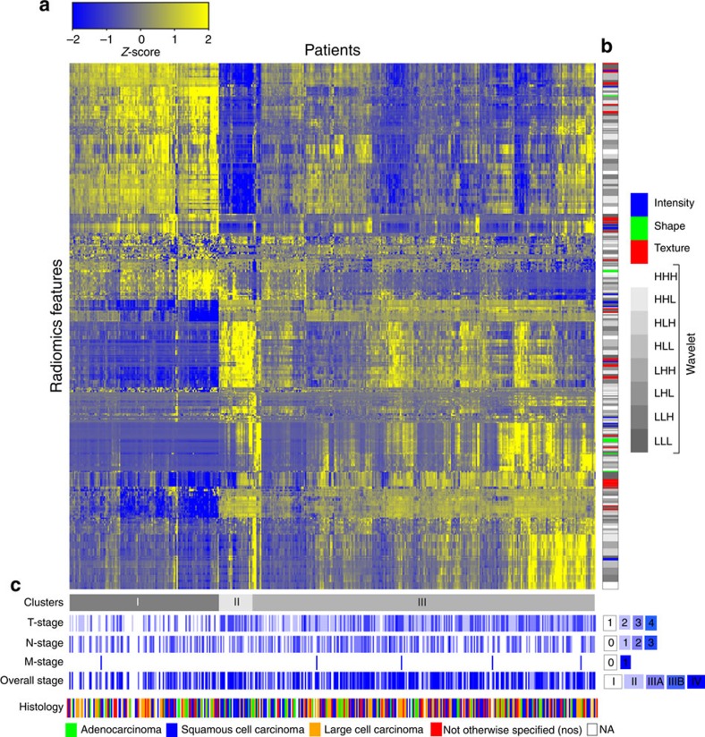
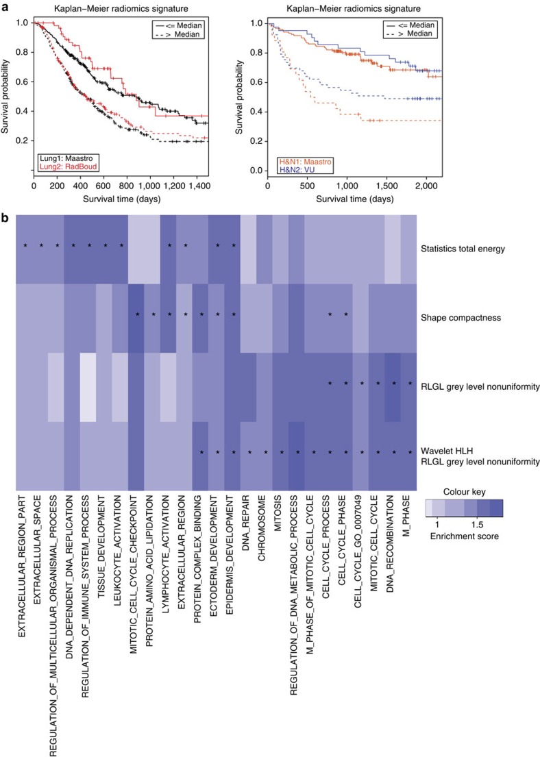
Human cancers exhibit strong phenotypic differences that can be visualized noninvasively by medical imaging. Radiomics refers to the comprehensive quantification of tumour phenotypes by applying a large number of quantitative image features. Here we present a radiomic analysis of 440 features quantifying tumour image intensity, shape and texture, which are extracted from computed tomography data of 1,019 patients with lung or head-and-neck cancer. We find that a large number of radiomic features have prognostic power in independent data sets of lung and head-and-neck cancer patients, many of which were not identified as significant before. Radiogenomics analysis reveals that a prognostic radiomic signature, capturing intratumour heterogeneity, is associated with underlying gene-expression patterns. These data suggest that radiomics identifies a general prognostic phenotype existing in both lung and head-and-neck cancer. This may have a clinical impact as imaging is routinely used in clinical practice, providing an unprecedented opportunity to improve decision-support in cancer treatment at low cost.
Overview
The claims and statistics of this paper haven’t been checked yet.
Free: it reads the open-access text and checks the first 5 claims that cite a source we can check. Create a free account to check every claim.
Integrity signals
Registry
Cites
Statistics
Figures


Verification
Claims the paper makes
The claims and statistics of this paper haven’t been checked yet.
Free: it reads the open-access text and checks the first 5 claims that cite a source we can check. Create a free account to check every claim.
Claims other papers make about this one
Each is checked against this paper’s own text: does it say what the citing paper says it does?
The source backs part of the claim, not all of it — or backs it for a different population, condition or comparison.
In the citing paper
“Notably, the signature was comprised of the top features from four classes (size, shape, texture, and wavelets) that were prioritized from a database of 442 features by using test-retest reproducibility and intersubject range.”
From Radiomics: Images Are More than Pictures, They Are Data (2016) · cites this paper as [47]
Experimental — accuracy not yet measuredThe source backs part of the claim, not all of it — or backs it for a different population, condition or comparison.
In the citing paper
“Many studies have shown that imaging features are significantly associated with patterns of gene expression and genetic mutations, demonstrating that radiogenomic analysis can identify different biological mechanisms by means of mathematical and computational devices, enabling the decoding of disease phenotypes by noninvasive methods(48,49).”
From Artificial intelligence, machine learning, computer-aided diagnosis, and radiomics: advances in imaging towards to precision medicine (2019) · cites this paper as [49]
Experimental — accuracy not yet measuredThe cited source says what the claim attributes to it.
In the citing paper
“A study by Aerts et al (47) showed that a radiomic signature could be used to predict outcome in completely independent cohorts of patients with lung cancer from two separate institutions.”
From Radiomics: Images Are More than Pictures, They Are Data (2016) · cites this paper as [47]
The cited source says what the claim attributes to it.
In the citing paper
“Further, this same signature could be applied to cohorts of patients with head and neck cancer with equivalent prognostic power.”
From Radiomics: Images Are More than Pictures, They Are Data (2016) · cites this paper as [47]
In the citing paper
“We used scans from 26 patients from the RIDER dataset19, routinely used for test–retest robustness analysis in tumour imaging19–21.”
From Foundation Model for Cancer Imaging Biomarkers (2024) · cites this paper as [20]
3 more claims about this paper
Create a free account to see every claim other papers make about this one.
Self-check
Percentages, sums and reported differences re-computed from the paper’s own numbers.
The self-check needs the paper’s full text.
Citations
Cites
Subramanian, A., Tamayo, P., Mootha, V. K., Mukherjee, S., Ebert, B. L., Gillette, M. A., Paulovich, A., Pomeroy, S. L., Golub, T. R., Lander, E. S., et al. (2005) · Proceedings of the National Academy of Sciences · doi:10.1073/pnas.0506580102 · Cited by 53,625
Harrell, F. E. (2001) · Springer Series in Statistics · doi:10.1007/978-1-4757-3462-1 · Cited by 9,195
Intratumor Heterogeneity and Branched Evolution Revealed by Multiregion Sequencing
Gerlinger, M., Rowan, A. J., Horswell, S., Larkin, J., Endesfelder, D., Gronroos, E., Martinez, P., Matthews, N., Stewart, A., Tarpey, P., et al. (2012) · New England Journal of Medicine · doi:10.1056/nejmoa1113205 · Cited by 7,774 · cited 2×
“It is known that multiple subclonal populations coexist within tumours, reflecting extensive intratumoral ‘somatic evolution’ 15,16 .”
BackgroundMolecular signatures database (MSigDB) 3.0
Liberzon, A., Subramanian, A., Pinchback, R., Thorvaldsdóttir, H., Tamayo, P., & Mesirov, J. P. (2011) · Bioinformatics · doi:10.1093/bioinformatics/btr260 · Cited by 7,111
Harrell (2015) · Springer Series in Statistics · doi:10.1007/978-3-319-19425-7 · Cited by 6,878
Radiomics: extracting more information from medical images using advanced feature analysis
Lambin, P., Rios-Velazquez, E., Leijenaar, R., Carvalho, S., van Stiphout, R. G. P. M., Granton, P., Zegers, C. M. L., Gillies, R., Boellard, R., Dekker, A., et al. (2012) · European Journal of Cancer · doi:10.1016/j.ejca.2011.11.036 · Cited by 6,084
Self-citationDistant metastasis occurs late during the genetic evolution of pancreatic cancer
Yachida, S., Jones, S., Bozic, I., Antal, T., Leary, R., Fu, B., Kamiyama, M., Hruban, R. H., Eshleman, J. R., Nowak, M. A., et al. (2010) · Nature · doi:10.1038/nature09515 · Cited by 2,502 · cited 1×
“It is known that multiple subclonal populations coexist within tumours, reflecting extensive intratumoral ‘somatic evolution’ 15,16 .”
BackgroundRadiomics: the process and the challenges
Kumar, V., Gu, Y., Basu, S., Berglund, A., Eschrich, S. A., Schabath, M. B., Forster, K., Aerts, H. J. W. L., Dekker, A., Fenstermacher, D., et al. (2012) · Magnetic Resonance Imaging · doi:10.1016/j.mri.2012.06.010 · Cited by 2,258
Self-citationTixier, F., Le Rest, C. C., Hatt, M., Albarghach, N., Pradier, O., Metges, J.-P., Corcos, L., & Visvikis, D. (2011) · Journal of Nuclear Medicine · doi:10.2967/jnumed.110.082404 · Cited by 696
Decoding global gene expression programs in liver cancer by noninvasive imaging
Segal, E., Sirlin, C. B., Ooi, C., Adler, A. S., Gollub, J., Chen, X., Chan, B. K., Matcuk, G. R., Barry, C. T., Chang, H. Y., et al. (2007) · Nature Biotechnology · doi:10.1038/nbt1306 · Cited by 629
Cited by
Items 901–1,000 of 1,023
Yip, S. S. F., Coroller, T. P., Sanford, N. N., Mamon, H. J., Aerts, H. J. W. L., & Berbeco, R. I. (2016) · Frontiers in Oncology · doi:10.3389/fonc.2016.00072 · Cited by 61
Self-citationLi, S., Wang, K., Hou, Z., Yang, J., Ren, W., Gao, S., Meng, F., Wu, P., Liu, B., Liu, J., et al. (2018) · Frontiers in Oncology · doi:10.3389/fonc.2018.00648 · Cited by 60
Mao, J., Jin, F., Duan, X., Yang, Z., Cao, M., Zhang, F., Lu, L., Zhang, X., Wu, X., Ding, Y., et al. (2019) · European Radiology · doi:10.1007/s00330-018-5961-6 · Cited by 60
Wei, W., Liu, Z., Rong, Y., Zhou, B., Bai, Y., Wei, W., Wang, S., Wang, M., Guo, Y., & Tian, J. (2019) · Frontiers in Oncology · doi:10.3389/fonc.2019.00255 · Cited by 65
“However, radiomic analysis might allow for an additional method by which tumor heterogeneity can be characterized (9).”
BackgroundMethodRadiomics Analysis for Clinical Decision Support in Nuclear Medicine
Ibrahim, A., Vallières, M., Woodruff, H. C., Primakov, S. P., Beheshti, M., Keek, S. A., Refaee, T., Sanduleanu, S., Walsh, S., Morin, O., et al. (2019) · Seminars in Nuclear Medicine · doi:10.1053/j.semnuclmed.2019.06.005 · Cited by 68
Self-citationComputed Tomography-based Radiomics for Risk Stratification in Prostate Cancer
Osman, S. O. S., Leijenaar, R. T. H., Cole, A., Lyons, C. A., Hounsell, A. R., Prise, K. M., O’Sullivan, J. M., Lambin, P., McGarry, C. K., & Jain, S. (2019) · International Journal of Radiation Oncology*Biology*Physics · doi:10.1016/j.ijrobp.2019.06.2504 · Cited by 64
“A detailed explanation of the extracted features and their mathematical definitions are published elsewhere.(6,22) In this analysis, geometric features (except for prostate volume) were excluded because the entire prostate gland was contoured and analyzed rather than tumor specific regions.”
Self-citationSaltz, J. H., Almeida, J. S., Gao, Y., Sharma, A., Bremer, E., DiPrima, T., Saltz, M. M., Kalpathy–Cramer, J., & Kurç, T. (2017) · PubMed Central · Cited by 62
Xu, S., Yao, Q., Liu, G., Jin, D., Chen, H., Xu, J., Li, Z., & Wu, G. (2019) · European Radiology · doi:10.1007/s00330-019-06484-2 · Cited by 60
Integrating radiomics into holomics for personalised oncology: from algorithms to bedside
Gatta, R., Depeursinge, A., Ratib, O. M., Michielin, O. A., & Leimgruber, A. (2020) · European Radiology Experimental · doi:10.1186/s41747-019-0143-0 · Cited by 63
“The pioneer work of Gevaert et al. in 2012 opened the field of radiogenomic by studying the correlation of gene clusters with radiomics features in 26 non-small cell lung cancer [27].”
BackgroundDesideri, I., Loi, M., Francolini, G., Becherini, C., Livi, L., & Bonomo, P. (2020) · Frontiers in Oncology · doi:10.3389/fonc.2020.01708 · Cited by 59
“Several applications of radiomics in the field of precision radiation oncology have been identified, providing insights in terms of stage discrimination (14, 15), molecular stratification (16–18), prognostic impact (19, 20), and prediction of response to treatment (21–23).”
BackgroundRadiomics in gliomas: clinical implications of computational modeling and fractal-based analysis
Jang, K., Russo, C., & Ieva, A. D. (2020) · Neuroradiology · doi:10.1007/s00234-020-02403-1 · Cited by 62
Mao, N., Yin, P., Li, Q., Wang, Q., Liu, M., Ma, H., Dong, J., Che, K., Wang, Z., Duan, S., et al. (2019) · European Radiology · doi:10.1007/s00330-020-07016-z · Cited by 59
Radiomics in hepatic metastasis by colorectal cancer
Granata, V., Fusco, R., Barretta, M. L., Picone, C., Avallone, A., Belli, A., Patrone, R., Ferrante, M., Cozzi, D., Grassi, R., et al. (2021) · Infectious Agents and Cancer · doi:10.1186/s13027-021-00379-y · Cited by 60
Zhao, K., Zheng, Q., Che, T., Dyrba, M., Li, Q., Ding, Y., Zheng, Y., Liu, Y., & Li, S. (2021) · Network Neuroscience · doi:10.1162/netn_a_00200 · Cited by 69
“Texture features can quantify the variations in intensity or patterns, including those features that are imperceptible to the human visual system (Aerts et al., 2014).”
InfluentialMachine intelligence in non-invasive endocrine cancer diagnostics
Thomasian, N. M., Kamel, I. R., & Bai, H. X. (2021) · Nature Reviews Endocrinology · doi:10.1038/s41574-021-00543-9 · Cited by 66
Su, G.-H., Xiao, Y., Jiang, L., Zheng, R., Wang, H., Chen, Y., Gu, Y., You, C., & Shao, Z.-M. (2022) · Journal of Translational Medicine · doi:10.1186/s12967-022-03688-x · Cited by 69
Radiomics Applications in Head and Neck Tumor Imaging: A Narrative Review
Tortora, M., Gemini, L., Scaravilli, A., Ugga, L., Ponsiglione, A., Stanzione, A., D’Arco, F., D’Anna, G., & Cuocolo, R. (2023) · Cancers · doi:10.3390/cancers15041174 · Cited by 68
“Decoding tumour phenotype by noninvasive imaging using a quantitative radiomics approach [28] 1019 Lung or headand-neck cancer CT Characterization 440 features Some radiomic features had prognostic power associated with underlying gene expression patterns.”
BackgroundMethodHuang, H., Chen, H., Zheng, D., Chen, C., Wang, Y., Xu, L., Wang, Y., He, X., Yang, Y., & Li, W. (2024) · Cancer Imaging · doi:10.1186/s40644-024-00692-w · Cited by 65
Wootton, L. S., Nyflot, M. J., Chaovalitwongse, W. A., & Ford, E. (2018) · International Journal of Radiation Oncology*Biology*Physics · doi:10.1016/j.ijrobp.2018.05.033 · Cited by 61
Random forest dissimilarity based multi-view learning for Radiomics application
Cao, H., Bernard, S., Sabourin, R., & Heutte, L. (2018) · Pattern Recognition · doi:10.1016/j.patcog.2018.11.011 · Cited by 69
An investigation of machine learning methods in delta-radiomics feature analysis
Chang, Y., Lafata, K. J., Sun, W., Wang, C., Chang, Z., Kirkpatrick, J. P., & Yin, F.-F. (2019) · PLoS ONE · doi:10.1371/journal.pone.0226348 · Cited by 63
“, pre-treatment)[6, 16, 17], and (2) delta-radiomics, where features are extracted from a time series of images (e.”
BackgroundMRI-derived radiomics: methodology and clinical applications in the field of pelvic oncology
Schick, U., Lucia, F., Dissaux, G., Visvikis, D., Badic, B., Masson, I., Pradier, O., Bourbonne, V., & Hatt, M. (2019) · British Journal of Radiology · doi:10.1259/bjr.20190105 · Cited by 65
Kimura, Y., Kadoya, N., Tomori, S., Oku, Y., & Jingu, K. (2020) · Physica Medica · doi:10.1016/j.ejmp.2020.03.022 · Cited by 59
Gu, D., Xie, Y., Wei, J., Li, W., Ye, Z., Zhu, Z., Tian, J., & Li, X. (2020) · Journal of Magnetic Resonance Imaging · doi:10.1002/jmri.27199 · Cited by 65
“These features may have the potential to capture intratumor heterogeneity, which can reflect the tumor tissue microenvironment and the cancer phenotype.(15) Recently, magnetic resonance imaging (MRI) has been successfully applied in predicting the pathological grading of tumor, assessing the malignancy and treatment r…”
BackgroundLiu, Q., Li, J., Liu, F., Yang, W., Ding, J., Chen, W., Wei, Y., Li, B., & Zheng, L. (2020) · Cancer Imaging · doi:10.1186/s40644-020-00360-9 · Cited by 65
Development and Validation of an Automated Radiomic CT Signature for Detecting COVID-19
Guiot, J., Vaidyanathan, A., Deprez, L., Zerka, F., Danthine, D., Frix, A.-N., Thys, M., Henket, M. A., Canivet, G., Mathieu, S., et al. (2020) · Diagnostics · doi:10.3390/diagnostics11010041 · Cited by 60
Self-citationQin, H., Que, Q., Lin, P., Li, X., Wang, X., He, Y., Chen, J., & Yang, H. (2021) · La radiologia medica · doi:10.1007/s11547-021-01393-1 · Cited by 62
The application of radiomics in predicting gene mutations in cancer
Qi, Y., Zhao, T., & Han, M. (2022) · European Radiology · doi:10.1007/s00330-021-08520-6 · Cited by 59
“Radiomics is an emerging field designed to automatically extract quantitative features from medical images and thus provide a large amount of information about tumour phenotypes [10, 11].”
BackgroundShang, Z.-F., Chauhan, V., Devi, K., & Patil, S. (2024) · Journal of Multidisciplinary Healthcare · doi:10.2147/jmdh.s482757 · Cited by 36
“15,16 Recently, a study demonstrated that AI-assisted tools have reduced the burden of radiologists to three-fifths in screening for breast cancer while avoiding false negatives by over 25%.”
BackgroundApostolova, I. I., Ego, K., Steffen, I. G., Buchert, R., Wertzel, H., Achenbach, H. J., Riedel, S., Schreiber, J., Schultz, M., Furth, C., et al. (2016) · European Journal of Nuclear Medicine and Molecular Imaging · doi:10.1007/s00259-016-3452-z · Cited by 58
Hao, H., Zhou, Z., Li, S., Maquilan, G. M., Folkert, M. R., Iyengar, P., Westover, K. D., Albuquerque, K., Liu, F., Choy, H., et al. (2018) · Physics in Medicine and Biology · doi:10.1088/1361-6560/aabb5e · Cited by 61
Cha, Y. J., Jang, W. I., Kim, M., Yoo, H. J., Paik, E. K., Jeong, H. K., & Youn, S.-M. (2018) · Anticancer Research · doi:10.21873/anticanres.12875 · Cited by 61
Cai, W., He, B., Hu, M., Zhang, W., Xiao, D., Yu, H., Song, Q., Xiang, N., Yang, J., He, S., et al. (2018) · Surgical Oncology · doi:10.1016/j.suronc.2018.11.013 · Cited by 61
Lafata, K. J., Hong, J. C., Geng, R., Ackerson, B. G., Liu, J., Zhou, Z., Torok, J. A., Kelsey, C. R., & Yin, F.-F. (2018) · Physics in Medicine and Biology · doi:10.1088/1361-6560/aaf5a5 · Cited by 58
Zhu, Y., Mohamed, A., Lai, S. Y., Yang, S., Kanwar, A., Wei, L., Kamal, M. J., Sengupta, S., Elhalawani, H. M., Skinner, H. D., et al. (2019) · JCO Clinical Cancer Informatics · doi:10.1200/cci.18.00073 · Cited by 68
“The radiomic features were grouped into five categories: gray-level cooccurrence matrix, gray-level run length matrix, neighbor intensity difference, intensity direct, and size/shape.(17) The Data Supplement introduces how the radiomic features were generated.”
Gleason Probability Maps: A Radiomics Tool for Mapping Prostate Cancer Likelihood in MRI Space
McGarry, S. D., Bukowy, J. D., Iczkowski, K. A., Unteriner, J., Duvnjak, P., Lowman, A. K., Jacobsohn, K. M., Hohenwalter, M. D., Griffin, M. O., Barrington, A., et al. (2019) · Tomography · doi:10.18383/j.tom.2018.00033 · Cited by 58
“In addition, by creating a database of aligned pathology or genetics with clinical imaging, it becomes feasible to find radiologic patterns of tumor phenotype which may provide critical predictive information (19-21).”
BackgroundLee, S. M., Kim, H. S., Lee, S., & Lee, J. W. (2019) · World Journal of Gastroenterology · doi:10.3748/wjg.v25.i11.1289 · Cited by 64
Jahani, N., Cohen, E. A., Hsieh, M.-K., Weinstein, S. P., Pantalone, L., Hylton, N. M., Newitt, D. C., Davatzikos, C. A., & Kontos, D. (2019) · Scientific Reports · doi:10.1038/s41598-019-48465-x · Cited by 61
Application of Radiomics in Predicting the Malignancy of Pulmonary Nodules in Different Sizes
Xu, Y., Lü, L., Linning, E., Lian, W., Yang, H., Schwartz, L. H., Yang, Z., & Zhao, B. (2019) · American Journal of Roentgenology · doi:10.2214/ajr.19.21490 · Cited by 60
Cardiac Computed Tomography: Assessment of Coronary Inflammation and Other Plaque Features
Oikonomou, E. K., West, H. W., & Antoniades, C. A. (2019) · Arteriosclerosis Thrombosis and Vascular Biology · doi:10.1161/atvbaha.119.312899 · Cited by 65
Cao, W., Wu, R., Cao, G., & He, Z. (2020) · IEEE Access · doi:10.1109/access.2020.3018666 · Cited by 59
Sun, R.-J., Fang, M., Tang, L., Li, X.-T., Lu, Q.-Y., Dong, D., Tian, J., & Sun, Y. (2019) · European Journal of Radiology · doi:10.1016/j.ejrad.2020.109277 · Cited by 59
Rathore, S., Bakas, S., Pati, S., Akbari, H., Kalarot, R., Sridharan, P., Rozycki, M., Bergman, M., Tunc, B., Verma, R., et al. (2018) · Lecture Notes in Computer Science · doi:10.1007/978-3-319-75238-9_12 · Cited by 57
“…result in non-invasive imaging signatures with diagnostic, prognostic and predictive value for many types of cancer, such as lung [38, 39], neck [38] and brain [2, 6– 10 , Specifically about the intensity-based features offered in brain-CaPTk, the histogram distributions capture anatomical and…”
BackgroundMyocardial Radiomics in Cardiac MRI
Hassani, C., Saremi, F., Varghese, B. A., & Duddalwar, V. A. (2019) · American Journal of Roentgenology · doi:10.2214/ajr.19.21986 · Cited by 60
Radiomic prediction of mutation status based on MR imaging of lung cancer brain metastases
Chen, B. T., Jin, T., Ye, N., Mambetsariev, I., Daniel, E., Wang, T., Wong, C. W., Rockne, R. C., Colen, R. R., Holodny, A. I., et al. (2020) · Magnetic Resonance Imaging · doi:10.1016/j.mri.2020.03.002 · Cited by 58
Integrating the Tumor Microenvironment into Cancer Therapy
Sanegre, S., Lucantoni, F., Burgos‐Panadero, R., de la Cruz‐Merino, L., Noguera, R., & Naranjo, T. Á. (2020) · Cancers · doi:10.3390/cancers12061677 · Cited by 61
ISANET: Non-small cell lung cancer classification and detection based on CNN and attention mechanism
Xu, Z., Ren, H., Zhou, W., & Liu, Z. (2022) · Biomedical Signal Processing and Control · doi:10.1016/j.bspc.2022.103773 · Cited by 67
Bian, X., Du, S., Yue, Z., Si, G., Zhao, R., Huang, G., Guo, L., Peng, C., & Zhang, L. (2023) · Journal of Magnetic Resonance Imaging · doi:10.1002/jmri.28628 · Cited by 57
“The high-dimensional extracted features from MRI, termed as radiomics features, could obtain a comprehensive characterization and detect potential pathological features of tumors.(15) The single T1CE radiomics model based on intratumoral features achieved a moderate diagnostic performance to distinguish HER2-positive…”
BackgroundCousin, F., Louis, T., Dheur, S., Nana, F. A., Ghaye, B., Occhipinti, M., Vos, W. G., Bottari, F., Paulus, A., Sibille, A., et al. (2023) · Cancers · doi:10.3390/cancers15071968 · Cited by 61
Prediction of malignant and benign of lung tumor using a quantitative radiomic method
Wang, J., Liu, X., Dong, D., Song, J., Xu, M., Zang, Y., & Tian, J. (2016) · Annual International Conference of the IEEE Engineering in Medicine and Biology Society (EMBC) · doi:10.1109/embc.2016.7590938 · Cited by 60
“portions of tumor tissue [2], [3].”
Pham, T. D., Watanabe, Y., Higuchi, M., & Suzuki, H. Y. (2017) · Scientific Reports · doi:10.1038/srep43209 · Cited by 57
“Radiomics 19–22 refers to the extraction of large amounts of quantitative imaging features from medical images obtained with CT, PET or magnetic resonance imaging (MRI).”
BackgroundZhai, T., van Dijk, L. V., Huang, B.-T., Lin, Z., Ribeiro, C. O., Brouwer, C. L., Oosting, S. F., Halmos, G. B., Witjes, M. J. H., Langendijk, J. A., et al. (2017) · Radiotherapy and Oncology · doi:10.1016/j.radonc.2017.07.013 · Cited by 62
“This observation is supported by other studies [11,34,35].”
BackgroundResultHartgerink, D., van der Heijden, B., Ruysscher, D. D., Postma, A. A., Ackermans, L. P. C. M., Hoeben, A., Anten, M. H. M. E., Lambin, P., Terhaag, K., Jochems, A., et al. (2018) · Frontiers in Oncology · doi:10.3389/fonc.2018.00154 · Cited by 56
Self-citationGao, Y., Liu, Y., Wang, Y., Shi, Z., & Yu, J. (2019) · IEEE Transactions on Medical Imaging · doi:10.1109/tmi.2019.2894692 · Cited by 57
“With the rapid development of techniques such as big data, deep learning and Radiomics [1]–[3], many studies have been conducted to reveal the underlying diagnosis and treatment information from MR images, such as noninvasive disease diagnosis [4], [5], differential diagnosis [6], [7], treatment prescription selection…”
Noninvasive molecular diagnosis of craniopharyngioma with MRI-based radiomics approach
Chen, X., Tong, Y., Shi, Z., Chen, H., Yang, Z. H., Wang, Y., Chen, L., & Yu, J. (2019) · BMC Neurology · doi:10.1186/s12883-018-1216-z · Cited by 62
Shi, L., Rong, Y., Daly, M. E., Dyer, B. A., Benedict, S., Qiu, J., & Yamamoto, T. (2019) · Physics in Medicine and Biology · doi:10.1088/1361-6560/ab3247 · Cited by 59
“A total of 658 radiomics features (categorized into shape, intensity, texture and Laplacian of Gaussian (LoG) filter) were extracted using an open source software tool, IBEX (Aerts et al 2014, Zhang et al 2015).”
MethodLiu, B., Cheng, J., Guo, D., He, X., Luo, Y. D., Zeng, Y., & Li, C. M. (2019) · Clinical Radiology · doi:10.1016/j.crad.2019.07.011 · Cited by 57
Technical Note: Ontology‐guided radiomics analysis workflow (O‐RAW)
Shi, Z., Traverso, A., van Soest, J., Dekker, A. L. A. J., & Wee, L. (2019) · Medical Physics · doi:10.1002/mp.13844 · Cited by 59
“5,6 Radiomics requires a high-throughput computerized tumor feature extraction process that can operate on vast quantities of digital imaging data.”
Self-citationElevating pancreatic cystic lesion stratification: Current and future pancreatic cancer biomarker(s)
Carmicheal, J., Patel, A., Dalal, V., Atri, P., Dhaliwal, A., Wittel, U. A., Malafa, M. P., Talmon, G. A., Swanson, B. J., Singh, S., et al. (2019) · Biochimica et Biophysica Acta (BBA) - Reviews on Cancer · doi:10.1016/j.bbcan.2019.188318 · Cited by 61
“The core premise of radiomics is that the differences in size, shape, texture, and greyness of a tumor contoured from a radiological image can reflect the variations in histological phenotype and genotype of the tumor [215].”
BackgroundLue, K., Wu, Y., Liu, S., Hsieh, T., Chuang, K., Lin, H., & Chen, Y. (2019) · Academic Radiology · doi:10.1016/j.acra.2019.10.015 · Cited by 60
Starke, S., Leger, S., Zwanenburg, A., Leger, K., Lohaus, F., Linge, A., Schreiber, A., Kalinauskaitė, G., Tinhofer, I., Guberina, N., et al. (2020) · Scientific Reports · doi:10.1038/s41598-020-70542-9 · Cited by 58
Huemer, F., Leisch, M., Geisberger, R., Melchardt, T., Rinnerthaler, G., Zaborsky, N., & Greil, R. (2020) · International Journal of Molecular Sciences · doi:10.3390/ijms21082856 · Cited by 62
Integrating tumor and nodal radiomics to predict lymph node metastasis in gastric cancer
Yang, J., Wu, Q., Xu, L., Wang, Z., Su, K., Liu, R., Yen, E. A., Liu, S., Qin, J., Rong, Y., et al. (2020) · Radiotherapy and Oncology · doi:10.1016/j.radonc.2020.06.004 · Cited by 57
Qian, J., Herman, M., Brinkmann, D. H., Laack, N. N., Kemp, B. J., Hunt, C. H., Lowe, V. J., & Pafundi, D. (2020) · International Journal of Radiation Oncology*Biology*Physics · doi:10.1016/j.ijrobp.2020.06.073 · Cited by 59
“Radiomics can be used as a possible tool to extract pathological or genomic information medical images [6, 7].”
MethodYang, H., Wu, C., Lee, C., Huang, H.-E., Lee, W.-K., Chung, W., Wu, H., Guo, W., Wu, Y., & Lu, C. (2020) · Radiotherapy and Oncology · doi:10.1016/j.radonc.2020.10.041 · Cited by 57
Yang, X., Dong, X., Wang, J., Li, W., Gu, Z., Gao, D., Zhong, N., & Guan, Y. (2019) · The Oncologist · doi:10.1634/theoncologist.2018-0706 · Cited by 59
“Radiomics, which uses advanced image analysis algorithms in the artificial intelligence domain, makes it possible to reproducibly quantify the imaging phenotypes automatically by extracting a large number of image features and can reflect the voxel-by-voxel genetic information for complete, heterogeneous tumors [13, 1…”
BackgroundHu, X., Sun, X., Hu, F., Liu, F., Ruan, W., Wu, T., An, R. F., & Lan, X. (2021) · European Journal of Nuclear Medicine and Molecular Imaging · doi:10.1007/s00259-021-05325-z · Cited by 58
“A higher diagnostic efficacy also proves that multidisciplinary approaches, including radiomics analysis, make sense for the diagnosis and treatment of diseases [49].”
BackgroundRadiomics and Magnetic Resonance Imaging of Rectal Cancer: From Engineering to Clinical Practice
Coppola, F., Giannini, V., Gabelloni, M., Panić, J., Defeudis, A., Monaco, S. L., Cattabriga, A., Cocozza, M. A., Pastore, L. V., Polici, M., et al. (2021) · Diagnostics · doi:10.3390/diagnostics11050756 · Cited by 59
“One approach is based on lev raging radiomics variability as a featu e sel ction tool by using test–retest analysis and eliminating radiomics features with a high variability [23,32].”
MethodBruixola, G., Remacha, E., Jiménez-Pastor, A., Dualde, D., Viala, A., Montón-Bueno, J., Ibarrola‐Villava, M., Alberich‐Bayarri, Á., & Cervantes, A. M. R. (2021) · Cancer Treatment Reviews · doi:10.1016/j.ctrv.2021.102263 · Cited by 61
Astaraki, M., Yang, G., Zakko, Y., Toma-Daşu, I., Smedby, Ö., & Wang, C. (2021) · Frontiers in Oncology · doi:10.3389/fonc.2021.737368 · Cited by 56
“Comparing the fi rst column of Table 4 with Tables 2 – 4 in Supplementary Material indicates that end-to-end training the deep classi fi ers with the unbalanced dataset could predict the class labels of the lung nodules more accurately than radiomics analyses over the imbalanced feature set without applying the FS metho…”
BackgroundInfluentialGhaffari Laleh, N., Ligero, M., Perez-Lopez, R., & Kather, J. N. (2023) · Clinical Cancer Research · doi:10.1158/1078-0432.ccr-22-0390 · Cited by 56
Cui, Y., Song, J., Pollom, E. L., Alagappan, M., Shirato, H., Chang, D. T., Koong, A. C., & Li, R. (2016) · International Journal of Radiation Oncology*Biology*Physics · doi:10.1016/j.ijrobp.2016.04.034 · Cited by 57
Texture analysis in radiology: Does the emperor have no clothes?
Summers, R. (2017) · Abdominal Radiology · doi:10.1007/s00261-016-0950-1 · Cited by 57
Stability of radiomic features in CT perfusion maps
Bogowicz, M., Riesterer, O., Bundschuh, R. A., Veit‐Haibach, P., Hüllner, M. W., Studer, G., Stieb, S., Glatz, S., Pruschy, M. N., Gückenberger, M., et al. (2016) · Physics in Medicine and Biology · doi:10.1088/1361-6560/61/24/8736 · Cited by 62
Blanc‐Durand, P., Gucht, A. V. D., Jreige, M., Lalonde, M. N., Silva-Monteiro, M., Prior, J. O., Denys, A. L., Depeursinge, A., & Schaefer, N. G. (2017) · Oncotarget · doi:10.18632/oncotarget.23423 · Cited by 60
Wang, H., Nie, P., Wang, Y., Xu, W., Duan, S., Chen, H., Hao, D., & Liu, J. (2019) · Journal of Magnetic Resonance Imaging · doi:10.1002/jmri.26818 · Cited by 57
“It can be used on a variety of medical images to assist in making a diagnosis and selecting treatment and is free of some of the deficiencies of traditional diagnostic methods.(8,9) Zhang et al(23) found that five radiomics features on MRI predicted the histopathological grade (low or high) of soft tissue FIGURE 5: Pe…”
BackgroundPark, J. E., Kim, H. S., Park, S. Y., Nam, S. J., Chun, S., Jo, Y., & Kim, J. H. (2019) · Radiology · doi:10.1148/radiol.2019190913 · Cited by 62
Predicting Therapeutic Antibody Delivery into Human Head and Neck Cancers
Lu, G., Fakurnejad, S., Martin, B. A., van den Berg, N. S., van Keulen, S., Nishio, N., Zhu, A., Chirita, S. U., Zhou, Q., Gao, R. W., et al. (2020) · Clinical Cancer Research · doi:10.1158/1078-0432.ccr-19-3717 · Cited by 57
Kagiyama, N., Shrestha, S., Cho, J. S., Khalil, M., Singh, Y., Challa, A., Casaclang‐Verzosa, G., & Sengupta, P. P. (2020) · EBioMedicine · doi:10.1016/j.ebiom.2020.102726 · Cited by 59
Arieno, A., Chan, A., & Destounis, S. V. (2019) · American Journal of Roentgenology · doi:10.2214/ajr.18.20391 · Cited by 55
Prognostic Value of Pretreatment Radiomic Features of 18F-FDG PET in Patients With Hodgkin Lymphoma
Lue, K., Wu, Y., Liu, S., Hsieh, T., Chuang, K., Lin, H., & Chen, Y. (2019) · Clinical Nuclear Medicine · doi:10.1097/rlu.0000000000002732 · Cited by 56
“Copyright © 2019 Wolters Kluwer H Evidence reveals that intratumor heterogeneity is associated with underlying gene-expression patterns.(23) Tumor FDG uptake reflects enhanced glycolytic metabolism in cancer cells through the Warburg effect.”
BackgroundZhang, L., Zhou, H., Gu, D., Tian, J., Zhang, B., Dong, D., Mo, X., Liu, J., Luo, X., Pei, S., et al. (2019) · Journal of Cancer · doi:10.7150/jca.33345 · Cited by 57
“Radiomics, a rapidly emerging field, transforms medical images into mineable high-dimensional quantitative features via a large number of automatically extracted data-characterization algorithms [23, 24].”
Zhang, X., Yang, Z., Cui, W., Zheng, C., Li, H., Li, Y., Lu, L., Mao, J., Zeng, W., Yang, X., et al. (2021) · European Radiology · doi:10.1007/s00330-020-07674-z · Cited by 58
“Radiomics can assess intra-tumoral heterogeneity that cannot be objectively evaluated by visual interpretation [32].”
BackgroundA deep survival interpretable radiomics model of hepatocellular carcinoma patients
Wei, L., Owen, D., Rosen, B., Guo, X., Cuneo, K. C., Lawrence, T. S., Haken, R. K. T., & Naqa, I. E. (2021) · Physica Medica · doi:10.1016/j.ejmp.2021.02.013 · Cited by 65
“Various studies have suggested that heterogeneity of tumors is associated with genomic heterogeneity and tumoral microenvironment [52, 53], thus plays an important role in the cancer prognosis, which is found in our work as well.”
BackgroundLong, L., Sun, J., Jiang, L., Hu, Y., Li, L., Tan, Y., Cao, M., Lan, X., & Zhang, J. (2021) · Diagnostic and Interventional Imaging · doi:10.1016/j.diii.2021.02.008 · Cited by 59
Perivascular fat imaging by computed tomography (CT): a virtual guide
Kotanidis, C. P., & Antoniades, C. A. (2021) · British Journal of Pharmacology · doi:10.1111/bph.15634 · Cited by 58
Ubaldi, L., Valenti, V., Borgese, R. F., Collura, G., Fantacci, M. E., Ferrera, G., Iacoviello, G., Abbate, B. F., Laruina, F., Tripoli, A., et al. (2021) · Physica Medica · doi:10.1016/j.ejmp.2021.08.015 · Cited by 66
Antonopoulos, A. S., Boutsikou, M., Simantiris, S., Angelopoulos, A., Lazaros, G. A., Panagiotopoulos, I. A., Oikonomou, E. K., Kanoupaki, M., Tousoulis, D., Mohiaddin, R. H., et al. (2021) · Scientific Reports · doi:10.1038/s41598-021-02971-z · Cited by 65
The unmet potential of artificial intelligence in veterinary medicine
Basran, P. S., & Appleby, R. B. (2022) · American Journal of Veterinary Research · doi:10.2460/ajvr.22.03.0038 · Cited by 68
Within-Modality Synthesis and Novel Radiomic Evaluation of Brain MRI Scans
Rezaeijo, S. M., Chegeni, N., Naeini, F. B., Makris, D., & Bakas, S. (2023) · Cancers · doi:10.3390/cancers15143565 · Cited by 58
Moscoso, A., Ruibal, Á., Domínguez‐Prado, I., Fernández‐Ferreiro, A., Herranz, M., Albaina, L., Argibay, S., Silva‐Rodríguez, J., Pardo‐Montero, J., & Aguiar, P. (2017) · European Journal of Nuclear Medicine and Molecular Imaging · doi:10.1007/s00259-017-3830-1 · Cited by 61
Quantitative texture analysis in the prediction of IDH status in low-grade gliomas
Jakola, A. S., Zhang, Y., Skjulsvik, A. J., Solheim, O. S., Bø, H. K., Berntsen, E. M., Reinertsen, I., Gulati, S., Förander, P., & Brismar, T. B. (2017) · Clinical Neurology and Neurosurgery · doi:10.1016/j.clineuro.2017.12.007 · Cited by 57
A Radiomics Approach to Computer-Aided Diagnosis with Cardiac Cine-MRI
Cetin, I., Sanroma, G., Petersen, S. E., Napel, S. A., Cámara, Ó., Ballester, M.-A. G., & Lekadir, K. (2018) · Lecture notes in computer science · doi:10.1007/978-3-319-75541-0_9 · Cited by 55
“Radiomics is the task of calculating a large number of imaging descriptors from delineated images, which has been widely used in cancer imaging [11].”
BackgroundMethodTu, S., Wang, C., Pan, K., Wu, Y., & Wu, C. (2018) · Physics in Medicine and Biology · doi:10.1088/1361-6560/aaafab · Cited by 56
“…medical imaging for advanced image analysis in which quantitative features of tumor phenotypes are extracted from an imaging modality such as computed tomography, magnetic resonance imaging, or positron emission tomography (Kumar et al 2012, Lambin et al 2012, Aerts et al 2014, Zhang et al 2015).”
BackgroundMethodRavanelli, M., Grammatica, A., Tononcelli, E., Morello, R., Leali, M., Battocchio, S., Agazzi, G. M., Buglione, M., Maroldi, R., Nicolai, P., et al. (2018) · American Journal of Neuroradiology · doi:10.3174/ajnr.a5792 · Cited by 55
“The technique has been recently introduced in medical imaging research and has provided promising results for cancer prognostication(14-19) and a noninvasive signature of relevant genotypic and phenotypic tumor patterns.(20-22) Bogowicz et al(23) recently demonstrated that a radiomic CT signature can predict HPV statu…”
MethodConnectome-Based Patterns of First-Episode Medication-Naïve Patients With Schizophrenia
Cui, L., Wei, Y., Xi, Y.-B., Griffa, A., de Lange, S. C., Kahn, R. S., Yin, H. J., & van den Heuvel, M. P. (2019) · Schizophrenia Bulletin · doi:10.1093/schbul/sbz014 · Cited by 55
CT radiomics associations with genotype and stromal content in pancreatic ductal adenocarcinoma
Attiyeh, M. A., Chakraborty, J., McIntyre, C. A., Kappagantula, R., Chou, Y., Askan, G., Seier, K., Gonen, M., Basturk, O., Balachandran, V. P., et al. (2019) · Abdominal Radiology · doi:10.1007/s00261-019-02112-1 · Cited by 55
Fan, Y., Hua, M., Mou, A., Wu, M., Liu, X., Bao, X., Wang, R., & Feng, M. (2019) · Frontiers in Endocrinology · doi:10.3389/fendo.2019.00403 · Cited by 56
Kim, J. Y., Yoon, M. J., Park, J. E., Choi, E. J., Lee, J., & Kim, H. S. (2019) · Neuroradiology · doi:10.1007/s00234-019-02255-4 · Cited by 57
Jeong, J., Wang, L., Ji, B., Lei, Y., Ali, A. N., Liu, T., Curran, W. J., Mao, H. Z., & Yang, X. (2019) · Quantitative Imaging in Medicine and Surgery · doi:10.21037/qims.2019.07.01 · Cited by 56
Metadata
- Authors
- Aerts, H. J. W. L., Velazquez, E. R., Leijenaar, R. T. H., Parmar, C., Grossmann, P., Carvalho, S., Bussink, J., Monshouwer, R., Haibe-Kains, B., Rietveld, D., Hoebers, F., Rietbergen, M. M., Leemans, C. R., Dekker, A., Quackenbush, J., Gillies, R. J., & Lambin, P.
- Affiliations
- Brigham and Women's Hospital (US)
- Maastricht University (NL)
- Radboud University Medical Center (NL)
- Amsterdam UMC Location Vrije Universiteit Amsterdam (NL)
- Dana-Farber Cancer Institute (US)
- Moffitt Cancer Center (US)
- Corresponding author
- Aerts, Hugo J. W. L.
- ORCID
- Venue
- Nature Communications
- Published
- 2014-06-03
- Volume
- 5
- Issue
- 1
- Pages
- 4006-4006
- ISSN
- 2041-1723 (ISSN-L)
- PMC
- PMC4059926
- OpenAlex
- W2103004421
- Semantic Scholar
- 3b002068646831a6f59fabb6daeb0209ed54049d
- Cited by
- 5,132
- References
- 29
- Citations by year
- 2026: 415 · 2025: 460 · 2024: 458 · 2023: 510 · 2022: 567 · 2021: 638 · 2020: 587 · 2019: 552 · 2018: 388 · 2017: 283 · 2016: 175 · 2015: 82 · 2014: 11
- FWCI
- 142.84
- Type
- journal article
- Language
- en
- Update notices
- correction (2014-08-07)
- Open access
- hybrid
- doi.org
- Licence
- Locations
- Nature Communications (publishedVersion, cc-by-nc-nd)
- PubMed Central (publishedVersion, cc-by-nc-nd)
- PubMed (publishedVersion)
- Pure Amsterdam UMC (publishedVersion)
- Amsterdam UMC (VU Amsterdam) - Institutional Repository (publishedVersion)
- Radboud Repository (Radboud University) (submittedVersion)
- Digital Access to Scholarship at Harvard (DASH) (Harvard University) (submittedVersion, cc-by)
- RePEc: Research Papers in Economics (submittedVersion)
- Research Publications (Maastricht University) (submittedVersion, cc-by-nc-nd)
- Radboud Repository (Radboud University) (submittedVersion, other-oa)
- Research Publications (Maastricht University) (submittedVersion, other-oa)
- Data Archiving and Networked Services (DANS) (submittedVersion)
- Funders
- European Federation of Pharmaceutical Industries and Associations
- KWF Kankerbestrijding (UM 2009-4454, 2011-5020, KWF UM 2009-4454, KWF UM 2011-5020, 2009-4454)
- Center for Translational Molecular Medicine (grant 030-103, 030-103)
- Innovative Medicines Initiative (115151)
- National Institutes of Health (U01 CA)
- National Cancer Institute (NIH-USA U01CA 143062-01, U01CA143062)
- Interreg
- Topics
- Health Sciences › Medicine › Radiology, Nuclear Medicine and Imaging › Radiomics and Machine Learning in Medical Imaging
- Physical Sciences › Computer Science › Computer Vision and Pattern Recognition › Image Retrieval and Classification Techniques
- Health Sciences › Medicine › Radiology, Nuclear Medicine and Imaging › Medical Imaging Techniques and Applications
- Keywords
- radiomics, tumor phenotype, lung cancer, head and neck cancer, radiogenomics, intratumoral heterogeneity, computed tomography, image texture features, gene expression, non-invasive imaging, treatment decision support
- MeSH terms
- Adenocarcinoma, Carcinoma, Non-Small-Cell Lung, Carcinoma, Squamous Cell, Female, Head and Neck Neoplasms, Humans, Lung Neoplasms, Male, Phenotype, Prognosis, Tomography, X-Ray Computed, Tumor Burden, Positron-Emission Tomography, Multimodal Imaging
About these checks
Registry, retraction, Cites and statistics results are computed, not judged, with one exception: a printed reference that no index lists is read once by a model before it counts as a citation error. Claims are extracted by a model and kept only when their quote appears verbatim in the text; each cited source is then checked, with a model’s help, for whether it says what the claim relies on. Those verdicts can be wrong — read the claim’s quote and the cited source before relying on one.
Statistics and arithmetic are recomputed, not judged.
Confidence bands are shown only for fields calibrated on our annotated test set. None is calibrated yet, so no band is shown.
What counts as a claim ›How we measure confidence ›How we check this ›
Are you an author, or do you see a problem with this page? Request a review
Data sources
Bibliographic data: Semantic Scholar, OpenAlex, Crossref.
Contains information from the Semantic Scholar Academic Graph Datasets, made available under the ODC Attribution License (ODC-BY 1.0).
Semantic Scholar’s data is described in: Kinney, R., et al. (2023). The Semantic Scholar Open Data Platform. arXiv:2301.10140.
