

Decoding tumour phenotype by noninvasive imaging using a quantitative radiomics approach
Open accessThe DOI matches its Crossref record; 29 of the 29 references it cites were found in a registry or an index.
Abstract
Human cancers exhibit strong phenotypic differences that can be visualized noninvasively by medical imaging. Radiomics refers to the comprehensive quantification of tumour phenotypes by applying a large number of quantitative image features. Here we present a radiomic analysis of 440 features quantifying tumour image intensity, shape and texture, which are extracted from computed tomography data of 1,019 patients with lung or head-and-neck cancer. We find that a large number of radiomic features have prognostic power in independent data sets of lung and head-and-neck cancer patients, many of which were not identified as significant before. Radiogenomics analysis reveals that a prognostic radiomic signature, capturing intratumour heterogeneity, is associated with underlying gene-expression patterns. These data suggest that radiomics identifies a general prognostic phenotype existing in both lung and head-and-neck cancer. This may have a clinical impact as imaging is routinely used in clinical practice, providing an unprecedented opportunity to improve decision-support in cancer treatment at low cost.
Overview
The claims and statistics of this paper haven’t been checked yet.
Free: it reads the open-access text and checks the first 5 claims that cite a source we can check. Create a free account to check every claim.
Integrity signals
Registry
Cites
Statistics
Figures


Verification
Claims the paper makes
The claims and statistics of this paper haven’t been checked yet.
Free: it reads the open-access text and checks the first 5 claims that cite a source we can check. Create a free account to check every claim.
Claims other papers make about this one
Each is checked against this paper’s own text: does it say what the citing paper says it does?
The source backs part of the claim, not all of it — or backs it for a different population, condition or comparison.
In the citing paper
“Notably, the signature was comprised of the top features from four classes (size, shape, texture, and wavelets) that were prioritized from a database of 442 features by using test-retest reproducibility and intersubject range.”
From Radiomics: Images Are More than Pictures, They Are Data (2016) · cites this paper as [47]
Experimental — accuracy not yet measuredThe source backs part of the claim, not all of it — or backs it for a different population, condition or comparison.
In the citing paper
“Many studies have shown that imaging features are significantly associated with patterns of gene expression and genetic mutations, demonstrating that radiogenomic analysis can identify different biological mechanisms by means of mathematical and computational devices, enabling the decoding of disease phenotypes by noninvasive methods(48,49).”
From Artificial intelligence, machine learning, computer-aided diagnosis, and radiomics: advances in imaging towards to precision medicine (2019) · cites this paper as [49]
Experimental — accuracy not yet measuredThe cited source says what the claim attributes to it.
In the citing paper
“A study by Aerts et al (47) showed that a radiomic signature could be used to predict outcome in completely independent cohorts of patients with lung cancer from two separate institutions.”
From Radiomics: Images Are More than Pictures, They Are Data (2016) · cites this paper as [47]
The cited source says what the claim attributes to it.
In the citing paper
“Further, this same signature could be applied to cohorts of patients with head and neck cancer with equivalent prognostic power.”
From Radiomics: Images Are More than Pictures, They Are Data (2016) · cites this paper as [47]
In the citing paper
“We used scans from 26 patients from the RIDER dataset19, routinely used for test–retest robustness analysis in tumour imaging19–21.”
From Foundation Model for Cancer Imaging Biomarkers (2024) · cites this paper as [20]
3 more claims about this paper
Create a free account to see every claim other papers make about this one.
Self-check
Percentages, sums and reported differences re-computed from the paper’s own numbers.
The self-check needs the paper’s full text.
Citations
Cites
Subramanian, A., Tamayo, P., Mootha, V. K., Mukherjee, S., Ebert, B. L., Gillette, M. A., Paulovich, A., Pomeroy, S. L., Golub, T. R., Lander, E. S., et al. (2005) · Proceedings of the National Academy of Sciences · doi:10.1073/pnas.0506580102 · Cited by 53,625
Harrell, F. E. (2001) · Springer Series in Statistics · doi:10.1007/978-1-4757-3462-1 · Cited by 9,195
Intratumor Heterogeneity and Branched Evolution Revealed by Multiregion Sequencing
Gerlinger, M., Rowan, A. J., Horswell, S., Larkin, J., Endesfelder, D., Gronroos, E., Martinez, P., Matthews, N., Stewart, A., Tarpey, P., et al. (2012) · New England Journal of Medicine · doi:10.1056/nejmoa1113205 · Cited by 7,774 · cited 2×
“It is known that multiple subclonal populations coexist within tumours, reflecting extensive intratumoral ‘somatic evolution’ 15,16 .”
BackgroundMolecular signatures database (MSigDB) 3.0
Liberzon, A., Subramanian, A., Pinchback, R., Thorvaldsdóttir, H., Tamayo, P., & Mesirov, J. P. (2011) · Bioinformatics · doi:10.1093/bioinformatics/btr260 · Cited by 7,111
Harrell (2015) · Springer Series in Statistics · doi:10.1007/978-3-319-19425-7 · Cited by 6,878
Radiomics: extracting more information from medical images using advanced feature analysis
Lambin, P., Rios-Velazquez, E., Leijenaar, R., Carvalho, S., van Stiphout, R. G. P. M., Granton, P., Zegers, C. M. L., Gillies, R., Boellard, R., Dekker, A., et al. (2012) · European Journal of Cancer · doi:10.1016/j.ejca.2011.11.036 · Cited by 6,084
Self-citationDistant metastasis occurs late during the genetic evolution of pancreatic cancer
Yachida, S., Jones, S., Bozic, I., Antal, T., Leary, R., Fu, B., Kamiyama, M., Hruban, R. H., Eshleman, J. R., Nowak, M. A., et al. (2010) · Nature · doi:10.1038/nature09515 · Cited by 2,502 · cited 1×
“It is known that multiple subclonal populations coexist within tumours, reflecting extensive intratumoral ‘somatic evolution’ 15,16 .”
BackgroundRadiomics: the process and the challenges
Kumar, V., Gu, Y., Basu, S., Berglund, A., Eschrich, S. A., Schabath, M. B., Forster, K., Aerts, H. J. W. L., Dekker, A., Fenstermacher, D., et al. (2012) · Magnetic Resonance Imaging · doi:10.1016/j.mri.2012.06.010 · Cited by 2,258
Self-citationTixier, F., Le Rest, C. C., Hatt, M., Albarghach, N., Pradier, O., Metges, J.-P., Corcos, L., & Visvikis, D. (2011) · Journal of Nuclear Medicine · doi:10.2967/jnumed.110.082404 · Cited by 696
Decoding global gene expression programs in liver cancer by noninvasive imaging
Segal, E., Sirlin, C. B., Ooi, C., Adler, A. S., Gollub, J., Chen, X., Chan, B. K., Matcuk, G. R., Barry, C. T., Chang, H. Y., et al. (2007) · Nature Biotechnology · doi:10.1038/nbt1306 · Cited by 629
Cited by
nnU-Net: a self-configuring method for deep learning-based biomedical image segmentation
Isensee, F., Jaeger, P. F., Kohl, S. A. A., Petersen, J., & Maier-Hein, K. H. (2021) · Nature Methods · doi:10.1038/s41592-020-01008-z · Cited by 10,477
Radiomics: Images Are More than Pictures, They Are Data
Gillies, R. J., Kinahan, P. E., & Hricak, H. (2016) · Radiology · doi:10.1148/radiol.2015151169 · Cited by 8,421
Self-citationComputational Radiomics System to Decode the Radiographic Phenotype
(2017) · doi:10.1158/0008-5472.can-17-0339 · Cited by 6,593
Self-citationRadiomics: the bridge between medical imaging and personalized medicine
Lambin, P., Leijenaar, R. T. H., Deist, T. M., Peerlings, J., de Jong, E. E. C., van Timmeren, J., Sanduleanu, S., Larue, R. T. H. M., Even, A. J. G., Jochems, A., et al. (2017) · Nature Reviews Clinical Oncology · doi:10.1038/nrclinonc.2017.141 · Cited by 5,688
Self-citationConvolutional neural networks: an overview and application in radiology
Yamashita, R., Nishio, M., Do, R. K. G., & Togashi, K. (2018) · Insights into Imaging · doi:10.1007/s13244-018-0639-9 · Cited by 4,682
“Most recent radiomics studies use hand-crafted feature extraction techniques, such as texture analysis, followed by conventional machine learning classifiers, such as random forests and support vector machines [15, 16].”
MethodZwanenburg, A., Vallières, M., Abdalah, M. A., Aerts, H. J. W. L., Andrearczyk, V., Apte, A., Ashrafinia, S., Bakas, S., Beukinga, R. J., Boellaard, R., et al. (2020) · Radiology · doi:10.1148/radiol.2020191145 · Cited by 4,177
“(2) Subsequently, the ROI is re-segmented to only contain values in the range [1, 6].”
Self-citationArtificial intelligence in radiology
Hosny, A., Parmar, C., Quackenbush, J., Schwartz, L. H., & Aerts, H. J. W. L. (2018) · Nature Reviews Cancer · doi:10.1038/s41568-018-0016-5 · Cited by 3,803
“Within oncology, multiple efforts have successfully explored radiomics tools for assisting clinical decision making related to the diagnosis and risk stratification of different cancers 15,16 .”
Self-citationBakas, S., Akbari, H., Sotiras, A., Bilello, M., Rozycki, M., Kirby, J. S., Freymann, J. B., Farahani, K., & Davatzikos, C. (2017) · Scientific Data · doi:10.1038/sdata.2017.117 · Cited by 3,049
Clonal heterogeneity and tumor evolution: past, present, and the future
McGranahan, N., & Swanton, C. (2017) · Cell · doi:10.1016/j.cell.2017.01.018 · Cited by 2,840
“Aerts and colleagues applied radiomics, which refers to the quantification of tumor phenotype using multiple imaging features, to head and neck and lung cancers (Aerts et al., 2014).”
Artificial intelligence in cancer imaging: Clinical challenges and applications
Bi, W. L., Hosny, A., Schabath, M. B., Giger, M. L., Birkbak, N. J., Mehrtash, A., Allison, T., Arnaout, O., Abbosh, C., Dunn, I. F., et al. (2019) · CA: A Cancer Journal for Clinicians · doi:10.3322/caac.21552 · Cited by 1,846
Self-citationHuang, Y.-Q., Liang, C.-H., He, L., Tian, J., Liang, C.-S., Chen, X., Ma, Z.-L., & Liu, Z.-Y. (2016) · Journal of Clinical Oncology · doi:10.1200/jco.2015.65.9128 · Cited by 1,765
“The term radiomics has attracted increased attention in recent years, and it is the process of the conversion of medical images into high-dimensional, mineable data via high-throughput extraction of quantitative features, followed by subsequent data analysis for decision support.(12,13) Advances in pattern recognition…”
BackgroundSecure, privacy-preserving and federated machine learning in medical imaging
Kaissis, G. A., Makowski, M. R., Rückert, D., & Braren, R. F. (2020) · Nature Machine Intelligence · doi:10.1038/s42256-020-0186-1 · Cited by 1,676
Radiomics in medical imaging—“how-to” guide and critical reflection
van Timmeren, J. E., Cester, D., Tanadini-Lang, S., Alkadhi, H., & Baessler, B. (2020) · Insights into Imaging · doi:10.1186/s13244-020-00887-2 · Cited by 1,316
Deep Learning in Medical Imaging: General Overview
Lee, J.-G., Jun, S., Cho, Y.-W., Lee, H., Kim, G. B., Seo, J. B., & Kim, N. (2017) · Korean Journal of Radiology · doi:10.3348/kjr.2017.18.4.570 · Cited by 1,276
“Many radiomic studies have correlated imaging biomarkers with the genomic expression or clinical outcome (99, 100).”
BackgroundApplications and limitations of radiomics
Yip, S. S. F., & Aerts, H. J. W. L. (2016) · Physics in Medicine and Biology · doi:10.1088/0031-9155/61/13/r150 · Cited by 1,210
“In human patients with head-and-neck cancer, tumours that responded poorly to chemoradiotherapy demonstrated a significantly greater increase in average ADC and higher values in kurtosis and skewness on mid-treatment diffusion weighted MR (DW-MR) than tumours with a better therapeutic response (King et al 2013). K-tra…”
BackgroundMethodResultInfluentialSelf-citationSun, R., Limkin, E. J., Vakalopoulou, M., Dercle, L., Champiat, S., Han, S. R., Verlingue, L., Brandao, D., Lancia, A., Ammari, S., et al. (2018) · The Lancet Oncology · doi:10.1016/s1470-2045(18)30413-3 · Cited by 1,236
Kather, J. N., Krisam, J., Charoentong, P., Luedde, T., Herpel, E., Weis, C.-A., Gaiser, T., Marx, A., Valous, N. A., Ferber, D., et al. (2019) · PLOS Medicine · doi:10.1371/journal.pmed.1002730 · Cited by 1,082
Biological and therapeutic impact of intratumor heterogeneity in cancer evolution
McGranahan, N., & Swanton, C. (2015) · Cancer Cell · doi:10.1016/j.ccell.2014.12.001 · Cited by 1,077
“…of Tumor Evolution and Cooperation between Tumor Subclones Longitudinal (Johnson et al., 2014; Mullighan et al., 2008; Shah et al., 2009), spatial (Aerts et al., 2014; Bashashati et al., 2013; Campbell et al., 2010; Gerlinger et al., 2012, 2014; Haffner et al., 2013; Navin et al., 2010, 2011;…”
BackgroundMachine Learning methods for Quantitative Radiomic Biomarkers
Parmar, C., Grossmann, P., Bussink, J., Lambin, P., & Aerts, H. J. W. L. (2015) · Scientific Reports · doi:10.1038/srep13087 · Cited by 1,019
“Different studies have shown the discriminating capabilities of radiomic features for the stratification of tumor histology 6 , tumor grades or stages 11 , and clinical outcomes 8,12,13 .”
InfluentialSelf-citationLiu, Z., Wang, S., Dong, D., Wei, J., Fang, C., Zhou, X., Sun, K., Li, L., Li, B., Wang, M., et al. (2019) · Theranostics · doi:10.7150/thno.30309 · Cited by 967
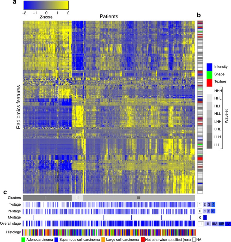
“Aerts et al. found the clusters of radiomic features were associated with the prognosis of lung cancer [4].”
BackgroundMethodInfluentialImaging biomarker roadmap for cancer studies
O'Connor, J. P. B., Aboagye, E. O., Adams, J. E., Aerts, H. J. W. L., Barrington, S. F., Beer, A. J., Boellaard, R., Bohndiek, S. E., Brady, M., Brown, G., et al. (2017) · Nature Reviews Clinical Oncology · doi:10.1038/nrclinonc.2016.162 · Cited by 1,091
Self-citationRepeatability and Reproducibility of Radiomic Features: A Systematic Review
Traverso, A., Wee, L., Dekker, A., & Gillies, R. (2018) · International Journal of Radiation Oncology*Biology*Physics · doi:10.1016/j.ijrobp.2018.05.053 · Cited by 903
“Three studies evaluated the reproducibility of radiomic features with respect to multiple manual segmentations in the same patient (interobserver sensitivity) by use of PET (4, 39, 43).”
InfluentialSelf-citationVallières, M., Freeman, C. R., Skamene, S. R., & El Naqa, I. (2015) · Physics in Medicine and Biology · doi:10.1088/0031-9155/60/14/5471 · Cited by 897
“More than 50 sub-types are recognized by the World Health Organization (WHO). STSs are relatively uncommon, representing approximately 0.7% of new adult malignancies in the United States (Siegel et al 2014). The majority of new cases are either intermediate or high-grade tumours and may arise in virtually all sites, w…”
MethodInfluentialPesapane, F., Codari, M., & Sardanelli, F. (2018) · European Radiology Experimental · doi:10.1186/s41747-018-0061-6 · Cited by 833
Radiation oncology in the era of precision medicine
Baumann, M., Krause, M., Overgaard, J., Debus, J., Bentzen, S. M., Daartz, J., Richter, C., Zips, D., & Bortfeld, T. (2016) · Nature Reviews Cancer · doi:10.1038/nrc.2016.18 · Cited by 812
Innovative approaches for cancer treatment: current perspectives and new challenges
Pucci, C., Martinelli, C., & Ciofani, G. (2019) · ecancermedicalscience · doi:10.3332/ecancer.2019.961 · Cited by 814
“Radiomics is intended as the high throughput quantification of tumour properties obtained from the analysis of medical images [14, 15, 231].”
BackgroundLimkin, E. J., Sun, R., Dercle, L., Zacharaki, E. I., Robert, C., Reuzé, S., Schernberg, A., Paragios, N., Deutsch, E., & Ferté, C. (2017) · Annals of Oncology · doi:10.1093/annonc/mdx034 · Cited by 757
“Radiomics has immense potential to improve knowledge in tumor biology and guide the management of patients at bedside [4].”
BackgroundMethodInfluentialHuang, Y., Liu, Z., He, L., Chen, X., Pan, D., Ma, Z., Liang, C., Tian, J., & Liang, C. (2016) · Radiology · doi:10.1148/radiol.2016152234 · Cited by 743
AI in Medical Imaging Informatics: Current Challenges and Future Directions
Panayides, A. S., Amini, A., Filipovic, N. D., Sharma, A., Tsaftaris, S. A., Young, A., Foran, D., Do, N., Golemati, S., Kurc, T., et al. (2020) · IEEE Journal of Biomedical and Health Informatics · doi:10.1109/jbhi.2020.2991043 · Cited by 713
“Through the construction of imaging signatures (i.e., fusing shape, texture, morphology, intensity, etc., features) and their subsequent association to clinical outcomes, devising robust predictive models (or quantitative imaging biomarkers) is achieved [225].”
AI applications to medical images: From machine learning to deep learning
Castiglioni, I., Rundo, L., Codari, M., Di Leo, G., Salvatore, C., Interlenghi, M., Gallivanone, F., Cozzi, A., D'Amico, N. C., & Sardanelli, F. (2021) · Physica Medica · doi:10.1016/j.ejmp.2021.02.006 · Cited by 710
Xu, X., Zhang, H.-L., Liu, Q.-P., Sun, S.-W., Zhang, J., Zhu, F.-P., Yang, G., Yan, X., Zhang, Y.-D., & Liu, X.-S. (2019) · Journal of Hepatology · doi:10.1016/j.jhep.2019.02.023 · Cited by 706
CT-based radiomic signature predicts distant metastasis in lung adenocarcinoma
Coroller, T. P., Grossmann, P., Hou, Y., Rios Velazquez, E., Leijenaar, R. T. H., Hermann, G., Lambin, P., Haibe-Kains, B., Mak, R. H., & Aerts, H. J. W. L. (2015) · Radiotherapy and Oncology · doi:10.1016/j.radonc.2015.02.015 · Cited by 695
“However, in comparison with a recent study[20] investigating clinical outcomes from another center, patient characteristics or outcomes were comparable.”
Self-citationImaging Intratumor Heterogeneity: Role in Therapy Response, Resistance, and Clinical Outcome
O'Connor, J. P. B., Rose, C. J., Waterton, J. C., Carano, R. A. D., Parker, G. J. M., & Jackson, A. (2015) · Clinical Cancer Research · doi:10.1158/1078-0432.ccr-14-0990 · Cited by 677
“This "radiomic" approach (60) places heterogeneity parameters at its center, but the sheer number of parameters under investigation incurs statistical dangers due to large numbers of multiple comparisons.”
BackgroundThe Potential of Radiomic-Based Phenotyping in Precision Medicine: A Review
Aerts, H. J. W. L. (2016) · JAMA Oncology · doi:10.1001/jamaoncol.2016.2631 · Cited by 669
Self-citationMeasuring Computed Tomography Scanner Variability of Radiomics Features
Mackin, D., Fave, X., Zhang, L., Fried, D., Yang, J., Taylor, B., Rodriguez-Rivera, E., Dodge, C., Jones, A. K., & Court, L. (2015) · Investigative Radiology · doi:10.1097/rli.0000000000000180 · Cited by 651
Deep Learning Predicts Lung Cancer Treatment Response from Serial Medical Imaging
Xu, Y., Hosny, A., Zeleznik, R., Parmar, C., Coroller, T., Franco, I., Mak, R. H., & Aerts, H. J. W. L. (2019) · Clinical Cancer Research · doi:10.1158/1078-0432.ccr-18-2495 · Cited by 628
“To compute handcrafted radiomic features, an accurate tumor delineation is required (9), which is susceptible to inter-reader segmentation variability and also is time-consuming.”
MethodSelf-citationA deep learning-based radiomics model for prediction of survival in glioblastoma multiforme
Lao, J., Chen, Y., Li, Z.-C., Li, Q., Zhang, J., Liu, J., & Zhai, G. (2017) · Scientific Reports · doi:10.1038/s41598-017-10649-8 · Cited by 630
Multi-scale Convolutional Neural Networks for Lung Nodule Classification
Shen, W., Zhou, M., Yang, F., Yang, C., & Tian, J. (2015) · Lecture Notes in Computer Science · doi:10.1007/978-3-319-19992-4_46 · Cited by 592
“Image-based lung nodule analysis is normally performed with nodule segmentation [5], feature extraction [2], and labelling nodule categories [8, 17, 19].”
Giger (2018) · doi:10.1016/j.jacr.2017.12.028 · Cited by 634
Machine and deep learning methods for radiomics
Avanzo, M., Wei, L., Stancanello, J., Vallières, M., Rao, A., Morin, O., Mattonen, S. A., & El Naqa, I. (2020) · Medical Physics · doi:10.1002/mp.13678 · Cited by 605
Robust Radiomics Feature Quantification Using Semiautomatic Volumetric Segmentation
Parmar, C., Rios Velazquez, E., Leijenaar, R., Jermoumi, M., Carvalho, S., Mak, R. H., Mitra, S., Shankar, B. U., Kikinis, R., Haibe-Kains, B., et al. (2014) · PLoS ONE · doi:10.1371/journal.pone.0102107 · Cited by 611
Self-citationHofmanninger, J., Prayer, F., Pan, J., Röhrich, S., Prosch, H., & Langs, G. (2020) · European Radiology Experimental · doi:10.1186/s41747-020-00173-2 · Cited by 595
“ity areas that are at risk of being excluded by threshold- or registrationbased methods, when they are close to the lung border. We utilized the publicly available, previously published Lung1 dataset [38] to quantify the model’s ability to cover tumor areas within the lung. The collection contains scans of 318 non-sma…”
MethodDeep learning for lung cancer prognostication: A retrospective multi-cohort radiomics study
Hosny, A., Parmar, C., Coroller, T. P., Grossmann, P., Zeleznik, R., Kumar, A., Bussink, J., Gillies, R. J., Mak, R. H., & Aerts, H. J. W. L. (2018) · PLOS Medicine · doi:10.1371/journal.pmed.1002711 · Cited by 579
“Consequently, in this study, we benchmarked the deep learning networks against random forest models built on engineered features, with the performance of the random forest models being within previously observed ranges [10].”
BackgroundMethodSelf-citationIntrinsic dependencies of CT radiomic features on voxel size and number of gray levels
Shafiq‐ul‐Hassan, M., Zhang, G. G., Latifi, K., Ullah, G., Hunt, D. C., Balagurunathan, Y., Abdalah, M. A., Schabath, M. B., Goldgof, D. G., Mackin, D., et al. (2017) · Medical Physics · doi:10.1002/mp.12123 · Cited by 585
“log n P; T ð Þ 1⁄2 (3) where V (P,T) is the volume of an individual voxel, P is the pixel spacing (0.”
InfluentialSelf-citationThe Biological Meaning of Radiomic Features
Tomaszewski, M. R., & Gillies, R. J. (2021) · Radiology · doi:10.1148/radiol.2021202553 · Cited by 549
Self-citationPredicting response to cancer immunotherapy using noninvasive radiomic biomarkers
Trebeschi, S., Drago, S. G., Birkbak, N. J., Kurilova, I., Cǎlin, A. M., Delli Pizzi, A., Lalezari, F., Lambregts, D. M. J., Rohaan, M. W., Parmar, C., et al. (2019) · Annals of Oncology · doi:10.1093/annonc/mdz108 · Cited by 566
“profiles and tumor microenvironments in NSCLC [23, 32, 41, 42].”
Self-citationLiu, Z., Zhang, X.-Y., Shi, Y.-J., Wang, L., Zhu, H.-T., Tang, Z., Wang, S., Li, X.-T., Tian, J., & Sun, Y.-S. (2017) · Clinical Cancer Research · doi:10.1158/1078-0432.ccr-17-1038 · Cited by 547
“Radiomics analysis of large imaging datasets has been successfully employed in the field of oncology for noninvasively profiling tumor heterogeneity (19), and there is a growing interest within the field in devising maps that display the associations between tumor heterogeneity and imaging features (20).”
BackgroundMethodRadiomics strategies for risk assessment of tumour failure in head-and-neck cancer
Vallières, M., Kay-Rivest, E., Perrin, L. J., Liem, X., Furstoss, C., Aerts, H. J. W. L., Khaouam, N., Nguyen-Tan, P. F., Wang, C.-S., Sultanem, K., et al. (2017) · Scientific Reports · doi:10.1038/s41598-017-10371-5 · Cited by 542
“→ The best predictive/ prognostic and balanced models for each outcome (final models) are identified in italic and are fully described in Supplementary Table S4. aBinary prediction of outcomes using logistic regression/random forest output responses. bConcordance-index between cox regression/random forest output respo…”
MethodInfluentialSelf-citationOikonomou, E. K., Williams, M. C., Kotanidis, C. P., Desai, M. Y., Marwan, M., Antonopoulos, A. S., Thomas, K. E., Thomas, S., Akoumianakis, I., Fan, L. M., et al. (2019) · European Heart Journal · doi:10.1093/eurheartj/ehz592 · Cited by 534
“Imaging studies in oncology have shown that specific shape- and texture-related patterns generate tumour phenotypes that are independently linked to the underlying tumour biology and to clinical prognosis.(35) Such patterns, often derived from complex mathematic formulae, are invisible to the naked eye of experienced…”
BackgroundReproducibility of radiomics for deciphering tumor phenotype with imaging
Zhao, B., Tan, Y., Tsai, W.-Y., Qi, J., Xie, C., Lu, L., & Schwartz, L. H. (2016) · Scientific Reports · doi:10.1038/srep23428 · Cited by 509
“95 35 (39) 46 (52) 34 (38) 44 (49) 23 (26) 34 (38) 3 (3) 3 (3) 6 (7)”
Deep learning for image-based cancer detection and diagnosis − A survey
Hu, Z., Tang, J., Wang, Z., Zhang, K., Zhang, L., & Sun, Q. (2018) · Pattern Recognition · doi:10.1016/j.patcog.2018.05.014 · Cited by 509
Salahuddin, Z., Woodruff, H. C., Chatterjee, A., & Lambin, P. (2022) · Computers in Biology and Medicine · doi:10.1016/j.compbiomed.2021.105111 · Cited by 508
Self-citationZhang, B., Tian, J., Dong, D., Gu, D., Dong, Y., Zhang, L., Lian, Z., Liu, J., Luo, X., Pei, S., et al. (2017) · Clinical Cancer Research · doi:10.1158/1078-0432.ccr-16-2910 · Cited by 494
“Recently, the most widely used imaging modality in radiomics research has been CT, which can quantify tissue density (14).”
BackgroundMethodLiu, Z., Li, Z., Qu, J., Zhang, R., Zhou, X., Li, L., Sun, K., Tang, Z., Jiang, H., Li, H., et al. (2019) · Clinical Cancer Research · doi:10.1158/1078-0432.ccr-18-3190 · Cited by 491
“Based on the concept that biomedical images contain information that may reflect underlying pathophysiology and their relationships could be revealed via quantitative image analyses (17), radiomics turns medical images into minable data to improve diagnostic (20, 21), prognostic (22), and predictive (23) accuracy, bri…”
BackgroundMethodBeyond imaging: The promise of radiomics
Avanzo, M., Stancanello, J., & El Naqa, I. (2017) · Physica Medica · doi:10.1016/j.ejmp.2017.05.071 · Cited by 483
“CT The most widely used imaging modality in radiomics studies is CT, which assesses tissue density, shape and texture of tumor and lymph-nodes [16,21,29].”
BackgroundMethodInfluentialWang, S., Zhou, M., Liu, Z., Liu, Z., Gu, D., Zang, Y., Dong, D., Gevaert, O., & Tian, J. (2017) · Medical Image Analysis · doi:10.1016/j.media.2017.06.014 · Cited by 478
“Treatment therapy monitoring and lung nodule analysis (Aerts et al., 2014) using computed tomography (CT) images are important strategies for early lung cancer diagnosis and survival time improvement.”
MethodCharacterization of PET/CT images using texture analysis: the past, the present… any future?
Hatt, M., Tixier, F., Pierce, L., Kinahan, P. E., Le Rest, C. C., & Visvikis, D. (2017) · European Journal of Nuclear Medicine and Molecular Imaging · doi:10.1007/s00259-016-3427-0 · Cited by 475
“Regarding the low-dose CT component of PET/CT, usually without contrast enhancement, heterogeneity refers to the variability in tissue density, which may result from spatially varying vascularization, necrosis or cellularity, as well as the proportions of fat, air and water [7].”
BackgroundMethodDeepLung: Deep 3D Dual Path Nets for Automated Pulmonary Nodule Detection and Classification
Zhu, W., Liu, C., Fan, W., & Xie, X. (2018) · 2018 IEEE Winter Conference on Applications of Computer Vision (WACV) · doi:10.1109/wacv.2018.00079 · Cited by 449
Radiomics and radiogenomics in lung cancer: A review for the clinician
Thawani, R., McLane, M., Beig, N., Ghose, S., Prasanna, P., Velcheti, V., & Madabhushi, A. (2018) · Lung Cancer · doi:10.1016/j.lungcan.2017.10.015 · Cited by 450
Kickingereder, P., Burth, S., Wick, A., Götz, M., Eidel, O., Schlemmer, H.-P., Maier-Hein, K. H., Wick, W., Bendszus, M., Radbruch, A., et al. (2016) · Radiology · doi:10.1148/radiol.2016160845 · Cited by 446
Val-Laillet, D., Aarts, E., Weber, B., Ferrari, M., Quaresima, V., Stoeckel, L. E., Alonso-Alonso, M., Audette, M., Malbert, C. H., & Stice, E. (2015) · NeuroImage: Clinical · doi:10.1016/j.nicl.2015.03.016 · Cited by 442
“Radiomics has been initially developed to decode tumor phenotypes (Aerts et al., 2014 ), including brain tumors (Coquery et al., 2014), but could be applied to other medical fi elds than oncology, such as eating disorders and obesity.”
Wang, S., Shi, J., Ye, Z., Dong, D., Yu, D., Zhou, M., Liu, Y., Gevaert, O., Wang, K., Zhu, Y., et al. (2019) · European Respiratory Journal · doi:10.1183/13993003.00986-2018 · Cited by 435
“Recent studies revealed that features extracted from lung cancer CT images were related to gene expression patterns [13 – 16] and showed predictive power on EGFR profiles [17 – 19].”
BackgroundRadiomic feature clusters and Prognostic Signatures specific for Lung and Head & Neck cancer
Parmar, C., Leijenaar, R. T. H., Grossmann, P., Rios Velazquez, E., Bussink, J., Rietveld, D., Rietbergen, M. M., Haibe-Kains, B., Lambin, P., & Aerts, H. J. W. L. (2015) · Scientific Reports · doi:10.1038/srep11044 · Cited by 434
Self-citationArtificial intelligence in radiation oncology
Huynh, E., Hosny, A., Guthier, C., Bitterman, D. S., Petit, S. F., Haas-Kogan, D. A., Kann, B., Aerts, H. J. W. L., & Mak, R. H. (2020) · Nature Reviews Clinical Oncology · doi:10.1038/s41571-020-0417-8 · Cited by 419
Self-citationCancer Diagnosis Using Deep Learning: A Bibliographic Review
Munir, K., Elahi, H., Ayub, A., Frezza, F., & Rizzi, A. (2019) · Cancers · doi:10.3390/cancers11091235 · Cited by 422
Leijenaar, R. T. H., Nalbantov, G., Carvalho, S., van Elmpt, W. J. C., Troost, E. G. C., Boellaard, R., Aerts, H. J. W. L., Gillies, R. J., & Lambin, P. (2015) · Scientific Reports · doi:10.1038/srep11075 · Cited by 426
“assigned to bin j ( ( , ) P i j GLCM ), (2) part of a consecutive run of l voxels assigned to bin i ( ( , ) P i l GLRLM ) or (3) part of a connected neighborhood of v voxels ( ( , ) P i v GLSZM ).”
BackgroundSelf-citationDong, D., Fang, M.-J., Tang, L., Shan, X.-H., Gao, J.-B., Giganti, F., Wang, R.-P., Chen, X., Wang, X.-X., Palumbo, D., et al. (2020) · Annals of Oncology · doi:10.1016/j.annonc.2020.04.003 · Cited by 416
Zhou, M., Scott, J., Chaudhury, B., Hall, L., Goldgof, D., Yeom, K. W., Iv, M., Ou, Y., Kalpathy-Cramer, J., Napel, S., et al. (2018) · American Journal of Neuroradiology · doi:10.3174/ajnr.a5391 · Cited by 415
“14,31 Examples of texture features include gray-level co-occurrence matrices 32 and gray-level size zone matrices, 33 which examine the spatial relationships of pixels through a series of statistical measures.”
BackgroundSelf-citationValidation of A Method to Compensate Multicenter Effects Affecting CT Radiomics
Orlhac, F., Frouin, F., Nioche, C., Ayache, N., & Buvat, I. (2019) · Radiology · doi:10.1148/radiol.2019182023 · Cited by 411
“One of the most widely cited studies (13) in radiomics, which included 1019 patients, used different CT imaging protocols involving different CT scanners, pixel size, and section thickness with or without intravenous contrast enhancement, and without accounting for this variability in the data analysis.”
BackgroundCapsule Networks for Brain Tumor Classification Based on MRI Images and Coarse Tumor Boundaries
Afshar, P., Plataniotis, K. N., & Mohammadi, A. (2019) · ICASSP 2019 - 2019 IEEE International Conference on Acoustics, Speech and Signal Processing (ICASSP) · doi:10.1109/icassp.2019.8683759 · Cited by 380
Radiomic Machine-Learning Classifiers for Prognostic Biomarkers of Head and Neck Cancer
Parmar, C., Grossmann, P., Rietveld, D., Rietbergen, M. M., Lambin, P., & Aerts, H. J. W. L. (2015) · Frontiers in Oncology · doi:10.3389/fonc.2015.00272 · Cited by 407
“Some studies have investigated the reproducibility/variability of radiomic features across different clinical settings (2, 12–14).”
BackgroundMethodInfluentialSelf-citationHuang, S. H., Xu, W., Waldron, J., Siu, L., Shen, X., Tong, L., Ringash, J., Bayley, A., Kim, J., Hope, A., et al. (2015) · Journal of Clinical Oncology · doi:10.1200/jco.2014.58.6412 · Cited by 405
Vanguri, R. S., Luo, J., Aukerman, A. T., Egger, J. V., Fong, C. J., Horvat, N., Pagano, A., Araujo-Filho, J. D. A. B., Geneslaw, L., Rizvi, H., et al. (2022) · Nature Cancer · doi:10.1038/s43018-022-00416-8 · Cited by 395
“PD-L1 expression 0 114 (46) – 0 (0) 1–49% 51 (21) – 13 (25) ≥50% 82 (33) – 39 (75) Tissue site – Lung 109 (44) – 25 (48) Pleura/pleural fluid 19 (8) – 1 (2) Lymph node 45 (18) – 10 (19) Liver 11 (4) – 2 (4) Bone 16 (7) – 3 (6) Adrenal 11 (4) – 1 (2) Other 36 (15) 10 (19) TMB ≥10 mutations per Mb 155 (63) 9 (29) 22 (58…”
Somatic Mutations Drive Distinct Imaging Phenotypes in Lung Cancer
Rios Velazquez, E., Parmar, C., Liu, Y., Coroller, T. P., Cruz, G., Stringfield, O., Ye, Z., Makrigiorgos, M., Fennessy, F., Mak, R. H., et al. (2017) · Cancer Research · doi:10.1158/0008-5472.can-17-0122 · Cited by 391
“Radiomic biomarkers have been shown to be associated with several clinical endpoints, including survival (11, 14–16), nodule malignancy (17, 18), pathologic response (19, 20), recurrence, and distant metastasis (21–23), as well as tumor gene expression patterns (11, 14, 21).”
BackgroundSelf-citationPark, J. E., Park, S. Y., Kim, H. J., & Kim, H. S. (2019) · Korean Journal of Radiology · doi:10.3348/kjr.2018.0070 · Cited by 388
“The study methodology differs for 1) intra-individual test-retest repeatability (29-38), 2) multi-machine reproducibility (33, 39-41), and 3) image-acquisition (33, 41, 42) and reconstruction parameters (42-44).”
MethodFehr, D., Veeraraghavan, H., Wibmer, A., Gondo, T., Matsumoto, K., Vargas, H. A., Sala, E., Hricak, H., & Deasy, J. O. (2015) · Proceedings of the National Academy of Sciences · doi:10.1073/pnas.1505935112 · Cited by 382
Radiomics in Oncology: A Practical Guide
Shur, J. D., Doran, S. J., Kumar, S., ap Dafydd, D., Downey, K., O’Connor, J. P. B., Papanikolaou, N., Messiou, C., Koh, D.-M., & Orton, M. R. (2021) · RadioGraphics · doi:10.1148/rg.2021210037 · Cited by 378
“Radiomics refers to the extraction of mineable high-dimensional data from radiologic images (1–3) and has been applied within oncology to improve diagnosis and prognostication (4,5) with the aim of delivering precision medicine.”
BackgroundRadiomics: from qualitative to quantitative imaging
Rogers, W., Thulasi Seetha, S., Refaee, T. A. G., Lieverse, R. I. Y., Granzier, R. W. Y., Ibrahim, A., Keek, S. A., Sanduleanu, S., Primakov, S. P., Beuque, M. P. L., et al. (2020) · The British Journal of Radiology · doi:10.1259/bjr.20190948 · Cited by 372
Self-citationExploratory Study to Identify Radiomics Classifiers for Lung Cancer Histology
Wu, W., Parmar, C., Grossmann, P., Quackenbush, J., Lambin, P., Bussink, J., Mak, R., & Aerts, H. J. W. L. (2016) · Frontiers in Oncology · doi:10.3389/fonc.2016.00071 · Cited by 378
“Radiomic Features Tumor phenotypic characteristics were quantified by extracting 440 3D radiomic features from the segmented tumor regions of pretreatment CT images (32).”
BackgroundMethodInfluentialSelf-citationDefining the biological basis of radiomic phenotypes in lung cancer
Grossmann, P., Stringfield, O., El-Hachem, N., Bui, M. M., Rios Velazquez, E., Parmar, C., Leijenaar, R. T., Haibe-Kains, B., Lambin, P., Gillies, R. J., et al. (2017) · eLife · doi:10.7554/elife.23421 · Cited by 365
“…for precision medicine, an increasing number of studies have investigated associations between imaging and tumor biology in different cancer types (Aerts et al., 2014; Gevaert et al., 2012; Diehn et al., 2008; Grossmann et al., 2016; Gutman et al., 2015; Segal et al., 2007; Li et al., 2016; Yoon…”
BackgroundMethodResultInfluentialSelf-citationRadiomics: a new application from established techniques
Parekh, V., & Jacobs, M. A. (2016) · Expert Review of Precision Medicine and Drug Development · doi:10.1080/23808993.2016.1164013 · Cited by 369
“complete set of 11 features derived from GLRL matrix [11].”
BackgroundMethodInfluentialSala, E., Mema, E., Himoto, Y., Veeraraghavan, H., Brenton, J. D., Snyder, A., Weigelt, B., & Vargas, H. A. (2017) · Clinical Radiology · doi:10.1016/j.crad.2016.09.013 · Cited by 369
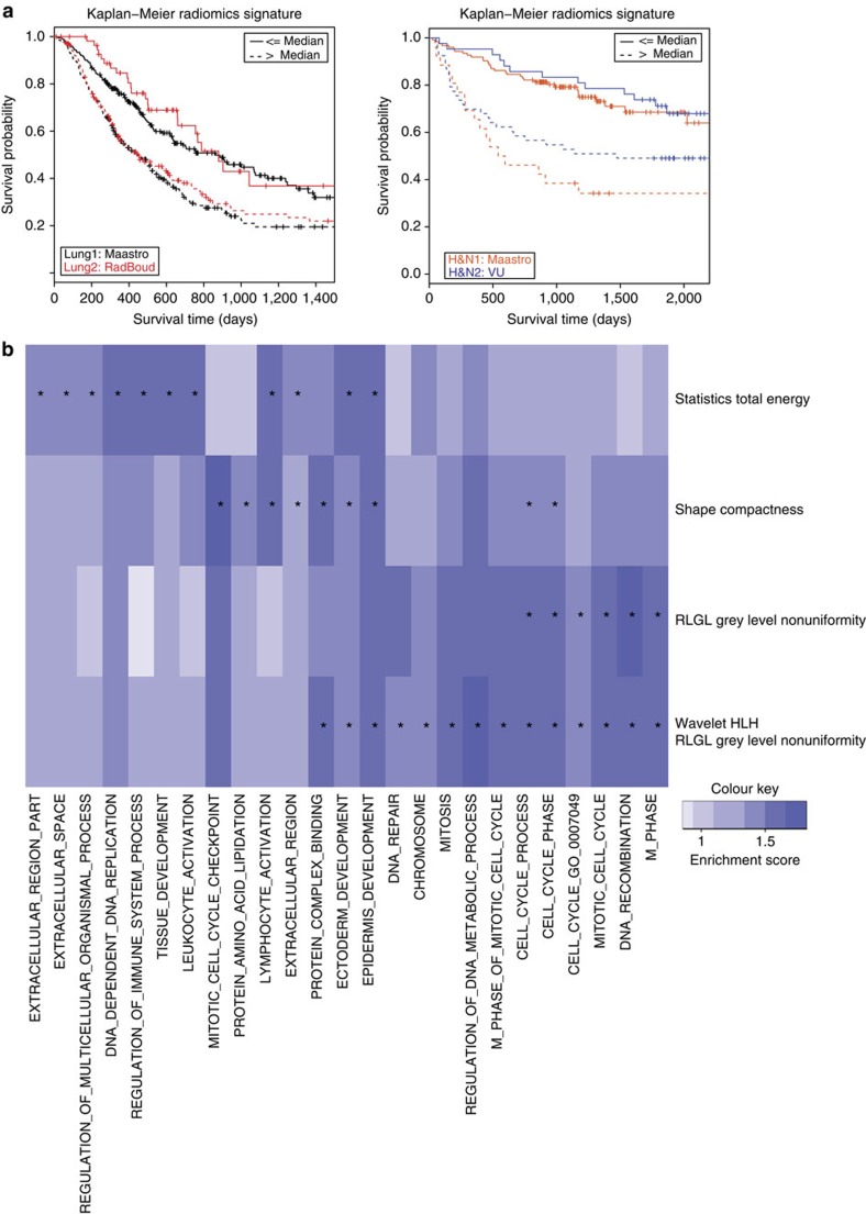
“Additionally, this same signature could be applied to patients with head and neck cancer with equivalent prognostic power (23).”
BackgroundDeep Learning in Medical Imaging
Kim, M., Yun, J., Cho, Y., Shin, K., Jang, R., Bae, H.-J., & Kim, N. (2019) · Neurospine · doi:10.14245/ns.1938396.198 · Cited by 351
Larue, R. T. H. M., Defraene, G., De Ruysscher, D., Lambin, P., & van Elmpt, W. (2017) · The British Journal of Radiology · doi:10.1259/bjr.20160665 · Cited by 363
“Size (volume and maximal diameter) and shape measures (sphericity, compactness and surface-to-volume ratio) have been described in radiomics research.(37,61) Finally, features relating to tumour location (e.”
BackgroundMethodInfluentialSelf-citationMRI features predict survival and molecular markers in diffuse lower-grade gliomas
Zhou, H., Vallières, M., Bai, H. X., Su, C., Tang, H., Oldridge, D., Zhang, Z., Xiao, B., Liao, W., Tao, Y., et al. (2017) · Neuro-Oncology · doi:10.1093/neuonc/now256 · Cited by 354
Delta-radiomics features for the prediction of patient outcomes in non–small cell lung cancer
Fave, X., Zhang, L., Yang, J., Mackin, D., Balter, P., Gomez, D., Followill, D., Jones, A. K., Stingo, F., Liao, Z., et al. (2017) · Scientific Reports · doi:10.1038/s41598-017-00665-z · Cited by 353
False Discovery Rates in PET and CT Studies with Texture Features: A Systematic Review
Chalkidou, A., O’Doherty, M. J., & Marsden, P. K. (2015) · PLOS ONE · doi:10.1371/journal.pone.0124165 · Cited by 344
“There are recent examples in the field of image-derived biomarkers where cancer stratification models were developed by combining clinical, imaging and gene expression data using large multicentre datasets, with multiple external validation sets and from various cancer sites to reduce the risk of type-I errors [66].”
BackgroundA review in radiomics: Making personalized medicine a reality via routine imaging
Guiot, J., Vaidyanathan, A., Deprez, L., Zerka, F., Danthine, D., Frix, A., Lambin, P., Bottari, F., Tsoutzidis, N., Miraglio, B., et al. (2022) · Medicinal Research Reviews · doi:10.1002/med.21846 · Cited by 337
Self-citationFrom Handcrafted to Deep-Learning-Based Cancer Radiomics: Challenges and Opportunities
Afshar, P., Mohammadi, A., Plataniotis, K. N., Oikonomou, A., & Benali, H. (2019) · IEEE Signal Processing Magazine · doi:10.1109/msp.2019.2900993 · Cited by 294
“The KMS curve [2], [3] represents a trajectory for measuring the probability of survival S ( t ) in given points of time t , i.e.,”
BackgroundMethodInfluentialLi, Z., Wang, Y., Yu, J., Guo, Y., & Cao, W. (2017) · Scientific Reports · doi:10.1038/s41598-017-05848-2 · Cited by 332
Radiomic phenotype features predict pathological response in non-small cell lung cancer
Coroller, T. P., Agrawal, V., Narayan, V., Hou, Y., Grossmann, P., Lee, S. W., Mak, R. H., & Aerts, H. J. W. L. (2016) · Radiotherapy and Oncology · doi:10.1016/j.radonc.2016.04.004 · Cited by 327
“Furthermore, we have previously demonstrated the reproducibility [7] of the radiomics features using the RIDER [34] test / retest dataset and we resampled of imaging using a common pixel spacing (3×3×3mm3) to limit variability across patients.”
MethodSelf-citationPeng, H., Dong, D., Fang, M.-J., Li, L., Tang, L.-L., Chen, L., Li, W.-F., Mao, Y.-P., Fan, W., Liu, L.-Z., et al. (2019) · Clinical Cancer Research · doi:10.1158/1078-0432.ccr-18-3065 · Cited by 327
“Radiomics has recently emerged as a promising field in oncology, and is basedon the premise thatmedical imaging canprovide important information on tumor physiology (15, 16).”
BackgroundThe Biological Meaning of Radiomic Features
Tomaszewski, M. R., & Gillies, R. J. (2021) · Radiology · doi:10.1148/radiol.2021219005 · Cited by 327
Self-citationAdvanced phenotyping and phenotype data analysis for the study of plant growth and development
Rahaman, M. M., Chen, D., Gillani, Z., Klukas, C., & Chen, M. (2015) · Frontiers in Plant Science · doi:10.3389/fpls.2015.00619 · Cited by 323
“Therefore, it is possible to find the significant association between trait profiles or pairs of the same groups or between groups of genotypes and phenotypes using correlation coefficient and test statistic (e.g., χ2 test, onesided Mann–Whitney U-test; Aerts et al., 2014; Chen et al., 2014b).”
BackgroundMethodInfluentialVulnerabilities of radiomic signature development: The need for safeguards
Welch, M. L., McIntosh, C., Haibe-Kains, B., Milosevic, M. F., Wee, L., Dekker, A., Huang, S. H., Purdie, T. G., O'Sullivan, B., Aerts, H. J. W. L., et al. (2019) · Radiotherapy and Oncology · doi:10.1016/j.radonc.2018.10.027 · Cited by 320
Self-citationibex: An open infrastructure software platform to facilitate collaborative work in radiomics
Zhang, L., Fried, D. V., Fave, X. J., Hunter, L. A., Yang, J., & Court, L. E. (2015) · Medical Physics · doi:10.1118/1.4908210 · Cited by 316
Yang, L., Gu, D., Wei, J., Yang, C., Rao, S., Wang, W., Chen, C., Ding, Y., Tian, J., & Zeng, M. (2019) · Liver Cancer · doi:10.1159/000494099 · Cited by 316
Artificial intelligence and machine learning in cancer imaging
Koh, D.-M., Papanikolaou, N., Bick, U., Illing, R., Kahn, C. E., Kalpathi-Cramer, J., Matos, C., Martí-Bonmatí, L., Miles, A., Mun, S. K., et al. (2022) · Communications Medicine · doi:10.1038/s43856-022-00199-0 · Cited by 311
“…image texture, shape, margin sharpness) 7 – 9 and/or using automated learning for task-relevant features; and (5) building a classi fi er that uses the computed features to predict a clinical state, e.g., probability of a speci fi c gene mutation, response to therapy or overall survival 10,11 .”
A radiogenomic dataset of non-small cell lung cancer
Bakr, S., Gevaert, O., Echegaray, S., Ayers, K., Zhou, M., Shafiq, M., Zheng, H., Benson, J. A., Zhang, W., Leung, A. N. C., et al. (2018) · Scientific Data · doi:10.1038/sdata.2018.202 · Cited by 311
A Review of Deep Learning on Medical Image Analysis
Wang, J., Zhu, H., Wang, S.-H., & Zhang, Y.-D. (2021) · Mobile Networks and Applications · doi:10.1007/s11036-020-01672-7 · Cited by 303
Metadata
- Authors
- Aerts, H. J. W. L., Velazquez, E. R., Leijenaar, R. T. H., Parmar, C., Grossmann, P., Carvalho, S., Bussink, J., Monshouwer, R., Haibe-Kains, B., Rietveld, D., Hoebers, F., Rietbergen, M. M., Leemans, C. R., Dekker, A., Quackenbush, J., Gillies, R. J., & Lambin, P.
- Affiliations
- Brigham and Women's Hospital (US)
- Maastricht University (NL)
- Radboud University Medical Center (NL)
- Amsterdam UMC Location Vrije Universiteit Amsterdam (NL)
- Dana-Farber Cancer Institute (US)
- Moffitt Cancer Center (US)
- Corresponding author
- Aerts, Hugo J. W. L.
- ORCID
- Venue
- Nature Communications
- Published
- 2014-06-03
- Volume
- 5
- Issue
- 1
- Pages
- 4006-4006
- ISSN
- 2041-1723 (ISSN-L)
- PMC
- PMC4059926
- OpenAlex
- W2103004421
- Semantic Scholar
- 3b002068646831a6f59fabb6daeb0209ed54049d
- Cited by
- 5,132
- References
- 29
- Citations by year
- 2026: 415 · 2025: 460 · 2024: 458 · 2023: 510 · 2022: 567 · 2021: 638 · 2020: 587 · 2019: 552 · 2018: 388 · 2017: 283 · 2016: 175 · 2015: 82 · 2014: 11
- FWCI
- 142.84
- Type
- journal article
- Language
- en
- Update notices
- correction (2014-08-07)
- Open access
- hybrid
- doi.org
- Licence
- Locations
- Nature Communications (publishedVersion, cc-by-nc-nd)
- PubMed Central (publishedVersion, cc-by-nc-nd)
- PubMed (publishedVersion)
- Pure Amsterdam UMC (publishedVersion)
- Amsterdam UMC (VU Amsterdam) - Institutional Repository (publishedVersion)
- Radboud Repository (Radboud University) (submittedVersion)
- Digital Access to Scholarship at Harvard (DASH) (Harvard University) (submittedVersion, cc-by)
- RePEc: Research Papers in Economics (submittedVersion)
- Research Publications (Maastricht University) (submittedVersion, cc-by-nc-nd)
- Radboud Repository (Radboud University) (submittedVersion, other-oa)
- Research Publications (Maastricht University) (submittedVersion, other-oa)
- Data Archiving and Networked Services (DANS) (submittedVersion)
- Funders
- European Federation of Pharmaceutical Industries and Associations
- KWF Kankerbestrijding (UM 2009-4454, 2011-5020, KWF UM 2009-4454, KWF UM 2011-5020, 2009-4454)
- Center for Translational Molecular Medicine (grant 030-103, 030-103)
- Innovative Medicines Initiative (115151)
- National Institutes of Health (U01 CA)
- National Cancer Institute (NIH-USA U01CA 143062-01, U01CA143062)
- Interreg
- Topics
- Health Sciences › Medicine › Radiology, Nuclear Medicine and Imaging › Radiomics and Machine Learning in Medical Imaging
- Physical Sciences › Computer Science › Computer Vision and Pattern Recognition › Image Retrieval and Classification Techniques
- Health Sciences › Medicine › Radiology, Nuclear Medicine and Imaging › Medical Imaging Techniques and Applications
- Keywords
- radiomics, tumor phenotype, lung cancer, head and neck cancer, radiogenomics, intratumoral heterogeneity, computed tomography, image texture features, gene expression, non-invasive imaging, treatment decision support
- MeSH terms
- Adenocarcinoma, Carcinoma, Non-Small-Cell Lung, Carcinoma, Squamous Cell, Female, Head and Neck Neoplasms, Humans, Lung Neoplasms, Male, Phenotype, Prognosis, Tomography, X-Ray Computed, Tumor Burden, Positron-Emission Tomography, Multimodal Imaging
About these checks
Registry, retraction, Cites and statistics results are computed, not judged, with one exception: a printed reference that no index lists is read once by a model before it counts as a citation error. Claims are extracted by a model and kept only when their quote appears verbatim in the text; each cited source is then checked, with a model’s help, for whether it says what the claim relies on. Those verdicts can be wrong — read the claim’s quote and the cited source before relying on one.
Statistics and arithmetic are recomputed, not judged.
Confidence bands are shown only for fields calibrated on our annotated test set. None is calibrated yet, so no band is shown.
What counts as a claim ›How we measure confidence ›How we check this ›
Are you an author, or do you see a problem with this page? Request a review
Data sources
Bibliographic data: Semantic Scholar, OpenAlex, Crossref.
Contains information from the Semantic Scholar Academic Graph Datasets, made available under the ODC Attribution License (ODC-BY 1.0).
Semantic Scholar’s data is described in: Kinney, R., et al. (2023). The Semantic Scholar Open Data Platform. arXiv:2301.10140.
