

Decoding tumour phenotype by noninvasive imaging using a quantitative radiomics approach
Open accessThe DOI matches its Crossref record; 29 of the 29 references it cites were found in a registry or an index.
Abstract
Human cancers exhibit strong phenotypic differences that can be visualized noninvasively by medical imaging. Radiomics refers to the comprehensive quantification of tumour phenotypes by applying a large number of quantitative image features. Here we present a radiomic analysis of 440 features quantifying tumour image intensity, shape and texture, which are extracted from computed tomography data of 1,019 patients with lung or head-and-neck cancer. We find that a large number of radiomic features have prognostic power in independent data sets of lung and head-and-neck cancer patients, many of which were not identified as significant before. Radiogenomics analysis reveals that a prognostic radiomic signature, capturing intratumour heterogeneity, is associated with underlying gene-expression patterns. These data suggest that radiomics identifies a general prognostic phenotype existing in both lung and head-and-neck cancer. This may have a clinical impact as imaging is routinely used in clinical practice, providing an unprecedented opportunity to improve decision-support in cancer treatment at low cost.
Overview
The claims and statistics of this paper haven’t been checked yet.
Free: it reads the open-access text and checks the first 5 claims that cite a source we can check. Create a free account to check every claim.
Integrity signals
Registry
Cites
Statistics
Figures


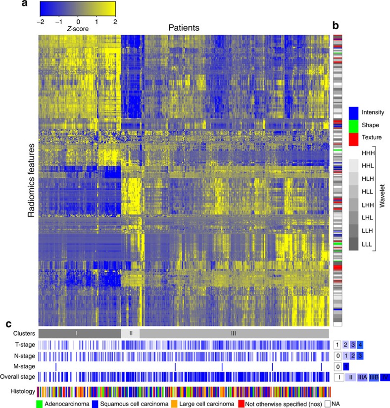
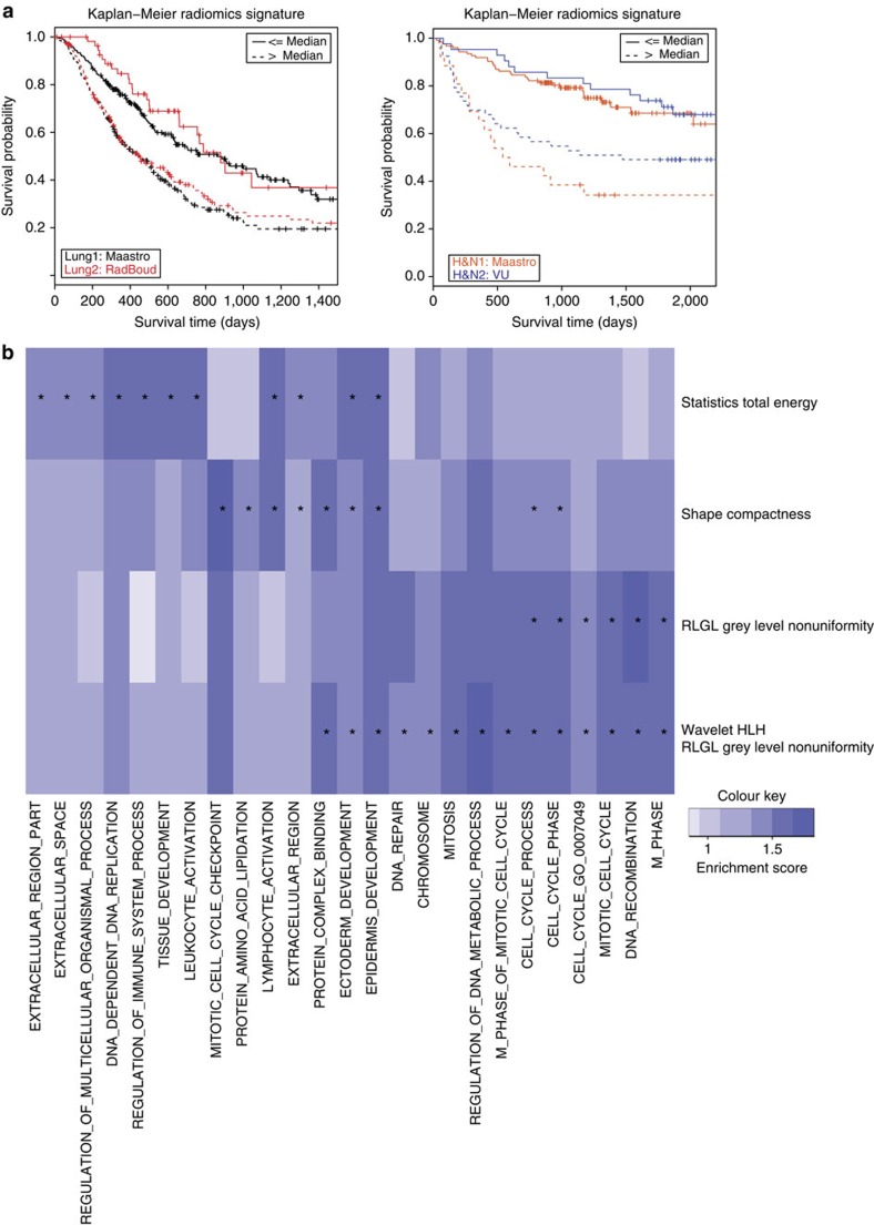
Verification
Claims the paper makes
The claims and statistics of this paper haven’t been checked yet.
Free: it reads the open-access text and checks the first 5 claims that cite a source we can check. Create a free account to check every claim.
Claims other papers make about this one
Each is checked against this paper’s own text: does it say what the citing paper says it does?
The source backs part of the claim, not all of it — or backs it for a different population, condition or comparison.
In the citing paper
“Notably, the signature was comprised of the top features from four classes (size, shape, texture, and wavelets) that were prioritized from a database of 442 features by using test-retest reproducibility and intersubject range.”
From Radiomics: Images Are More than Pictures, They Are Data (2016) · cites this paper as [47]
Experimental — accuracy not yet measuredThe source backs part of the claim, not all of it — or backs it for a different population, condition or comparison.
In the citing paper
“Many studies have shown that imaging features are significantly associated with patterns of gene expression and genetic mutations, demonstrating that radiogenomic analysis can identify different biological mechanisms by means of mathematical and computational devices, enabling the decoding of disease phenotypes by noninvasive methods(48,49).”
From Artificial intelligence, machine learning, computer-aided diagnosis, and radiomics: advances in imaging towards to precision medicine (2019) · cites this paper as [49]
Experimental — accuracy not yet measuredThe cited source says what the claim attributes to it.
In the citing paper
“A study by Aerts et al (47) showed that a radiomic signature could be used to predict outcome in completely independent cohorts of patients with lung cancer from two separate institutions.”
From Radiomics: Images Are More than Pictures, They Are Data (2016) · cites this paper as [47]
The cited source says what the claim attributes to it.
In the citing paper
“Further, this same signature could be applied to cohorts of patients with head and neck cancer with equivalent prognostic power.”
From Radiomics: Images Are More than Pictures, They Are Data (2016) · cites this paper as [47]
In the citing paper
“We used scans from 26 patients from the RIDER dataset19, routinely used for test–retest robustness analysis in tumour imaging19–21.”
From Foundation Model for Cancer Imaging Biomarkers (2024) · cites this paper as [20]
3 more claims about this paper
Create a free account to see every claim other papers make about this one.
Self-check
Percentages, sums and reported differences re-computed from the paper’s own numbers.
The self-check needs the paper’s full text.
Citations
Cites
Subramanian, A., Tamayo, P., Mootha, V. K., Mukherjee, S., Ebert, B. L., Gillette, M. A., Paulovich, A., Pomeroy, S. L., Golub, T. R., Lander, E. S., et al. (2005) · Proceedings of the National Academy of Sciences · doi:10.1073/pnas.0506580102 · Cited by 53,625
Harrell, F. E. (2001) · Springer Series in Statistics · doi:10.1007/978-1-4757-3462-1 · Cited by 9,195
Intratumor Heterogeneity and Branched Evolution Revealed by Multiregion Sequencing
Gerlinger, M., Rowan, A. J., Horswell, S., Larkin, J., Endesfelder, D., Gronroos, E., Martinez, P., Matthews, N., Stewart, A., Tarpey, P., et al. (2012) · New England Journal of Medicine · doi:10.1056/nejmoa1113205 · Cited by 7,774 · cited 2×
“It is known that multiple subclonal populations coexist within tumours, reflecting extensive intratumoral ‘somatic evolution’ 15,16 .”
BackgroundMolecular signatures database (MSigDB) 3.0
Liberzon, A., Subramanian, A., Pinchback, R., Thorvaldsdóttir, H., Tamayo, P., & Mesirov, J. P. (2011) · Bioinformatics · doi:10.1093/bioinformatics/btr260 · Cited by 7,111
Harrell (2015) · Springer Series in Statistics · doi:10.1007/978-3-319-19425-7 · Cited by 6,878
Radiomics: extracting more information from medical images using advanced feature analysis
Lambin, P., Rios-Velazquez, E., Leijenaar, R., Carvalho, S., van Stiphout, R. G. P. M., Granton, P., Zegers, C. M. L., Gillies, R., Boellard, R., Dekker, A., et al. (2012) · European Journal of Cancer · doi:10.1016/j.ejca.2011.11.036 · Cited by 6,084
Self-citationDistant metastasis occurs late during the genetic evolution of pancreatic cancer
Yachida, S., Jones, S., Bozic, I., Antal, T., Leary, R., Fu, B., Kamiyama, M., Hruban, R. H., Eshleman, J. R., Nowak, M. A., et al. (2010) · Nature · doi:10.1038/nature09515 · Cited by 2,502 · cited 1×
“It is known that multiple subclonal populations coexist within tumours, reflecting extensive intratumoral ‘somatic evolution’ 15,16 .”
BackgroundRadiomics: the process and the challenges
Kumar, V., Gu, Y., Basu, S., Berglund, A., Eschrich, S. A., Schabath, M. B., Forster, K., Aerts, H. J. W. L., Dekker, A., Fenstermacher, D., et al. (2012) · Magnetic Resonance Imaging · doi:10.1016/j.mri.2012.06.010 · Cited by 2,258
Self-citationTixier, F., Le Rest, C. C., Hatt, M., Albarghach, N., Pradier, O., Metges, J.-P., Corcos, L., & Visvikis, D. (2011) · Journal of Nuclear Medicine · doi:10.2967/jnumed.110.082404 · Cited by 696
Decoding global gene expression programs in liver cancer by noninvasive imaging
Segal, E., Sirlin, C. B., Ooi, C., Adler, A. S., Gollub, J., Chen, X., Chan, B. K., Matcuk, G. R., Barry, C. T., Chang, H. Y., et al. (2007) · Nature Biotechnology · doi:10.1038/nbt1306 · Cited by 629
Cited by
Items 801–900 of 1,023
Mahajan, A., Goh, V., Basu, S. K., Vaish, R., Weeks, A. J., Thakur, M., & Cook, G. (2015) · Clinical Radiology · doi:10.1016/j.crad.2015.06.082 · Cited by 71
Multi‐subtype classification model for non‐small cell lung cancer based on radiomics: SLS model
Liu, J., Cui, J., Liu, F., Yuan, Y., Guo, F., & Zhang, G. (2019) · Medical Physics · doi:10.1002/mp.13551 · Cited by 73
Reliability of CT‐based texture features: Phantom study
Varghese, B. A., Hwang, D., Cen, S., Levy, J. R., Liu, D. H., Lau, C., Rivas, M., Desai, B., Goodenough, D. J., & Duddalwar, V. A. (2019) · Journal of Applied Clinical Medical Physics · doi:10.1002/acm2.12666 · Cited by 66
“With the technological advancements in medical imaging, radiomics, defined as the high‐throughput extraction of quantitative features from routine medical images to create a mineable database of imaging metrics, has emerged as a promising tool for decision support.(1,2) Radiomic metrics assessing tumor shape, nonunifo…”
BackgroundLi, L., Wang, K., Ma, X., Liu, Z., Wang, S., Du, J., Tian, K., Zhou, X., Wei, W., Sun, K., et al. (2019) · European Journal of Radiology · doi:10.1016/j.ejrad.2019.07.006 · Cited by 66
“Radio-oncomics”: The potential of radiomics in radiation oncology
Peeken, J. C., Nüsslin, F., & Combs, S. E. (2017) · Strahlentherapie und Onkologie · doi:10.1007/s00066-017-1175-0 · Cited by 68
“The authors analyzed CT-based feature sets in multiple other studies, showing correlations with distant failure and pathologic complete response (pCR) in NSCLC patients receiving neoadjuvant radiochemotherapy [8, 27, 31].”
MethodResultSun, K., Hu, H.-T., Chen, S., Ye, J., Li, G., Chen, L., Peng, J., Feng, S., Yuan, Y., Hou, X., et al. (2020) · BMC Cancer · doi:10.1186/s12885-020-06970-7 · Cited by 71
Liu, Q., Xu, X., Zhu, F.-P., Zhang, Y., & Liu, X.-S. (2020) · EClinicalMedicine · doi:10.1016/j.eclinm.2020.100379 · Cited by 69
Navarro, F. A., Dapper, H., Asadpour, R., Knebel, C., Spraker, M. B., Schwarze, V., Schaub, S. K., Mayr, N. A., Specht, K., Woodruff, H. C., et al. (2021) · Cancers · doi:10.3390/cancers13122866 · Cited by 69
Self-citationZhu, H., Jiang, L., Zhang, H., Luo, L., Chen, Y., & Chen, Y. (2021) · NeuroImage Clinical · doi:10.1016/j.nicl.2021.102744 · Cited by 75
de Farias, E. C., di Noia, C., Han, C., Sala, E., Castelli, M., & Rundo, L. (2021) · Scientific Reports · doi:10.1038/s41598-021-00898-z · Cited by 78
“…features were calculated without any image filters applied from the following categories: Gray-Level Co-occurrence Matrix features (GLCM) 34–36 (24), Gray-Level Dependence Matrix (GLDM) 37 (14), Gray-Level Run Length Matrix (GLRLM) 38 (16), Gray-Level Size Zone Matrix (GLSZM) 39 (16) and…”
BackgroundMethodGao, W., Wang, W., Song, D., Yang, C., Zhu, K., Zeng, M., Rao, S., & Wang, M. (2022) · La radiologia medica · doi:10.1007/s11547-021-01445-6 · Cited by 68
“Moreover, radiomics features are explicitly designed and superficial [18], which can limit image heterogeneity expression.”
BackgroundThe Integration of Radiomics and Artificial Intelligence in Modern Medicine
Maniaci, A., Lavalle, S., Gagliano, C., Lentini, M., Masiello, E., Parisi, F. M., Iannella, G., Cilia, N. D., Salerno, V. M., Cusumano, G., et al. (2024) · Life · doi:10.3390/life14101248 · Cited by 84
“In order to improve clinical decision-making and customized medicine, the discipline of radiomics—the extraction and analysis of quantitative information from medical images—has drawn a lot of interest recently [1–3].”
BackgroundInfluentialIntegration of multiple “OMIC” biomarkers: A precision medicine strategy for lung cancer
Robles, A. I., & Harris, C. C. (2016) · Lung Cancer · doi:10.1016/j.lungcan.2016.06.003 · Cited by 70
CT-based Radiomics Signature to Discriminate High-grade From Low-grade Colorectal Adenocarcinoma
Huang, X., Cheng, Z., Huang, Y., Liang, C., He, L., Ma, Z., Chen, X., Wu, X., Li, Y., Liang, C., et al. (2018) · Academic Radiology · doi:10.1016/j.acra.2018.01.020 · Cited by 69
“However, as tumors were heterogeneous in space (16), the discrepancy may occur between biopsy and surgical specimen for evaluating histologic grade, because biopsy samples may be superficial, inadequate, or poorly oriented (2,17,18).”
BackgroundLi, Y., Jiang, J., Lu, J., Jiang, J., Zhang, H., & Zuo, C. (2019) · Therapeutic Advances in Neurological Disorders · doi:10.1177/1756286419838682 · Cited by 74
“8,9 11–14 Although current radiomics research studies have been mainly focused on oncology, considerations have been recently extended to numerous medical applications.”
BackgroundBanna, G. L., Olivier, T., Rundo, F., Malapelle, U., Fraggetta, F., Libra, M., & Addeo, A. (2019) · Frontiers in Medicine · doi:10.3389/fmed.2019.00172 · Cited by 69
“They could find many radiomic features having a prognosis value and built a prognostic radiomic signature, which was found to be correlated with underlying gene-expression patterns (32).”
BackgroundHou, L., Gupta, R., Arnam, J. S. V., Zhang, Y., Sivalenka, K., Samaras, D., Kurç, T., & Saltz, J. H. (2020) · Scientific Data · doi:10.1038/s41597-020-0528-1 · Cited by 69
“Segmentation of nuclei is the first step in extracting interpretable features that provide valuable diagnostic and prognostic cancer indicators [11, 12, 28, 15, 1], and thus is a crucial step for precision medicine [13, 8].”
BackgroundGrading of Gliomas by Using Radiomic Features on Multiple Magnetic Resonance Imaging (MRI) Sequences
Qin, J.-B., Liu, Z., Zhang, H., Shen, C., Wang, X., Tan, Y. Q., Wang, S., Wu, X.-F., & Tian, J. (2017) · Medical Science Monitor · doi:10.12659/msm.901270 · Cited by 67
Pan, C., Liu, J., Tang, J., Chen, X., Fang, C., Wu, Y., Geng, Y., Xu, C., Zhang, X., Wu, Z., et al. (2018) · Radiotherapy and Oncology · doi:10.1016/j.radonc.2018.07.011 · Cited by 72
Traverso, A., Kazmierski, M., Shi, Z., Kalendralis, P., Welch, M. L., Nissen, H. D., Jaffray, D. A., Dekker, A. L. A. J., & Wee, L. (2019) · Physica Medica · doi:10.1016/j.ejmp.2019.04.009 · Cited by 73
“Radiomics refers to computerized extraction of a large number of quantitative image metrics from medical images, that may reveal a deeper level of detail than is accessible to an unaided human eye, with the intent of defining tumour sub-types [12].”
BackgroundSelf-citationFeature-Guided Deep Radiomics for Glioblastoma Patient Survival Prediction
Shboul, Z. A., Alam, M., Vidyaratne, L., Pei, L., Elbakary, M. I., & Iftekharuddin, K. M. (2019) · Frontiers in Neuroscience · doi:10.3389/fnins.2019.00966 · Cited by 78
“The heterogeneity in Glioblastoma may be quantified using texture and histogram-graph features; while the shape of the tumor may be effectively captured using volumetric and Euler characteristic features (Pope et al., 2005; Aerts et al., 2014; Rathore et al., 2016).”
Methodvan Griethuysen, J. J. M., Lambregts, D. M. J., Trebeschi, S., Lahaye, M. J., Bakers, F. C. H., Vliegen, R. F. A., Beets, G. L., Aerts, H. J. W. L., & Beets‐Tan, R. G. H. (2019) · Abdominal Radiology · doi:10.1007/s00261-019-02321-8 · Cited by 64
Self-citationPesapane, F., Suter, M. B., Rotili, A., Penco, S., Nigro, O., Cremonesi, M., Bellomi, M., Jereczek‐Fossa, B. A., Pinotti, G., & Cassano, E. (2020) · Medical Oncology · doi:10.1007/s12032-020-01353-1 · Cited by 72
Spohn, S. K. B., Bettermann, A. S., Bamberg, F., Benndorf, M., Mix, M. D., Nicolay, N. H., Fechter, T., Hölscher, T., Grosu, R., Chiti, A., et al. (2021) · Theranostics · doi:10.7150/thno.61207 · Cited by 77
“The time period considered in this literature review was from 1st of January 2014 [64] to 1st of January 2021.”
BackgroundAmini, M., Hajianfar, G., Avval, A. H., Nazari, M., Deevband, M. R., Oveisi, M., Shiri, I., & Zaidi, H. (2021) · Clinical Oncology · doi:10.1016/j.clon.2021.11.014 · Cited by 68
Alabi, R. O., Elmusrati, M. S., Leivo, I. V. V., Almangush, A., & Mäkitie, A. A. (2024) · International Journal of Medical Informatics · doi:10.1016/j.ijmedinf.2024.105464 · Cited by 75
Bagher‐Ebadian, H., Siddiqui, F., Liu, C., Movsas, B., & Chetty, I. J. (2017) · Medical Physics · doi:10.1002/mp.12188 · Cited by 73
Henderson, S. A., Purdie, C. A., Michie, C. O., Evans, A. J., Lerski, R. A., Johnston, M., Vinnicombe, S., & Thompson, A. M. (2017) · European Radiology · doi:10.1007/s00330-017-4850-8 · Cited by 68
“Recently, heterogeneity, as assessed using TA, has found a role in the diagnosis, characterization [25] and treatment response assessment of various cancer types [26, 27], with initial reports suggesting an association with patient survival [28].”
BackgroundOpen access image repositories: high-quality data to enable machine learning research
Prior, F., Almeida, J. S., Kathiravelu, P., Kurç, T., Smith, K., FitzGerald, T. J., & Saltz, J. H. (2019) · Clinical Radiology · doi:10.1016/j.crad.2019.04.002 · Cited by 77
Kothari, G., Korte, J. C., Lehrer, E. J., Zaorsky, N. G., Lazarakis, S., Kron, T., Hardcastle, N., & Siva, S. (2020) · Radiotherapy and Oncology · doi:10.1016/j.radonc.2020.10.023 · Cited by 69
Ferreira, M. S., Lovinfosse, P., Hermesse, J., Cuypere, M. D., Rousseau, C., Lucia, F., Schick, U., Reinhold, C., Robin, P., Hatt, M., et al. (2021) · European Journal of Nuclear Medicine and Molecular Imaging · doi:10.1007/s00259-021-05303-5 · Cited by 73
Self-citationDistributed radiomics as a signature validation study using the Personal Health Train infrastructure
Shi, Z., Zhovannik, I., Traverso, A., Dankers, F. J. W. M., Deist, T. M., Kalendralis, P., Monshouwer, R., Bussink, J., Fijten, R., Aerts, H. J. W. L., et al. (2019) · Scientific Data · doi:10.1038/s41597-019-0241-0 · Cited by 69
Self-citationMachine Learning and Imaging Informatics in Oncology
Tseng, H.-H., Wei, L., Cui, S., Luo, Y., Ten Haken, R. K., & El Naqa, I. (2020) · Oncology · doi:10.1159/000493575 · Cited by 64
“Transfer learning, data augmentation, generative adversarial nets [39], semisupervised learning among others have been proposed to address the data limitation problem by providing additional data for training, while deep learning interpretation is still in its infancy.”
BackgroundInfluential3D-MCN: A 3D Multi-scale Capsule Network for Lung Nodule Malignancy Prediction
Afshar, P., Oikonomou, A., Naderkhani, F., Tyrrell, P. N., Plataniotis, K. N., Farahani, K., & Mohammadi, A. (2020) · Scientific Reports · doi:10.1038/s41598-020-64824-5 · Cited by 74
“The aforementioned shortcomings of human-centered cancer diagnosis have encouraged the development of a relatively new field in medical imaging, referred to as “radiomics” 6–8 , which has shown promising results in cancer diagnosis 9 .”
BackgroundChen, H., Zhang, X., Wang, X., Quan, X., Deng, Y., Lu, M., Wei, Q., Ye, Q., Zhou, Q., Xiang, Z., et al. (2021) · European Radiology · doi:10.1007/s00330-021-07748-6 · Cited by 68
Contemporary issues in the implementation of lung cancer screening
Lam, S. C., & Tammemägi, M. C. (2021) · European Respiratory Review · doi:10.1183/16000617.0288-2020 · Cited by 66
Le, V.-H., Kha, Q.-H., Hung, T. N. K., & Le, N. Q. K. (2021) · Cancers · doi:10.3390/cancers13143616 · Cited by 76
“[13] found four feature signatures representing four main groups of features: original, tumor intensity, first order, and wavelet, and used these four features in turn to evaluate the effectiveness of predicting survival in NSCLC patients in the training set.”
MethodInfluentialShi, J., Dong, Y., Jiang, W., Qin, F., Wang, X., Cui, L., Liu, Y., Jin, Y., Luo, Y., & Jiang, X. (2021) · Magnetic Resonance Imaging · doi:10.1016/j.mri.2021.12.008 · Cited by 70
Gitto, S., Bologna, M., Corino, V., Emili, I., Albano, D., Messina, C., Armiraglio, E., Parafioriti, A., Luzzati, A., Mainardi, L. T., et al. (2022) · La radiologia medica · doi:10.1007/s11547-022-01468-7 · Cited by 64
“FOS and textural features were extracted from both the original volumes and the 8 volumes obtained by first-level wavelet decomposition [16].”
MethodSai, S., Chamola, V., Choo, K. R., Sikdar, B., & Rodrigues, J. J. P. C. (2022) · IEEE Internet of Things Journal · doi:10.1109/jiot.2022.3232793 · Cited by 87
“Aerts et al. [138] demonstrated their proposed system (named C-DistriM, chained distributed ML) by predicting two-year lung cancer survival using the NSCLC-Radiomics data.”
MethodZhang, Y., Zhou, C., Guo, S., Wang, C., Yang, J., Yang, Z., Wang, R., Zhang, X., & Zhou, F. (2024) · Journal of Cancer Research and Clinical Oncology · doi:10.1007/s00432-023-05574-5 · Cited by 74
“They play a crucial role in solving a range of clinical issues, including precise disease diagnosis, treatment evaluation, and prognosis prediction (Lambin et al. 2017; Aerts et al. 2014; Gillies et al. 2016).”
BackgroundClonal cooperativity in heterogenous cancers
Zhou, H., Neelakantan, D., & Ford, H. L. (2016) · Seminars in Cell and Developmental Biology · doi:10.1016/j.semcdb.2016.08.028 · Cited by 74
Giganti, F., Marra, P., Ambrosi, A., Salerno, A., Antunes, S., Chiari, D., Orsenigo, E., Esposito, A., Mazza, E. A., Albarello, L., et al. (2017) · European Journal of Radiology · doi:10.1016/j.ejrad.2017.02.043 · Cited by 69
Belli, M. L., Mori, M., Broggi, S., Cattaneo, G. M., Bettinardi, V., Dell’Oca, I., Fallanca, F., Passoni, P., Vanoli, E. G., Calandrino, R., et al. (2018) · Physica Medica · doi:10.1016/j.ejmp.2018.05.013 · Cited by 67
Helden, E. J. V., Vacher, Y. J. L., van Wieringen, W. N., van Velden, F. H. P., Verheul, H. M. W., Hoekstra, O. S., Boellaard, R., & van Oordt, C. W. M. D. H. (2018) · European Journal of Nuclear Medicine and Molecular Imaging · doi:10.1007/s00259-018-4100-6 · Cited by 64
Big Data in Head and Neck Cancer
Resteghini, C., Trama, A., Borgonovi, E., Hosni, H., Corrao, G., Orlandi, E. M., Calareso, G., Cecco, L. D., Piazza, C., Mainardi, L. T., et al. (2018) · Current Treatment Options in Oncology · doi:10.1007/s11864-018-0585-2 · Cited by 63
Zhao, S., Su, Y., Duan, J., Qiu, Q., Ge, X., Wang, A., & Yin, Y. (2019) · Journal of bone oncology · doi:10.1016/j.jbo.2019.100263 · Cited by 69
“Next, quantitative and high-throughput analysis of feature data is completed to probe tumor phenotype [16,17].”
MethodPeng, A., Dai, H., Duan, H., Chen, Y., Huang, J., Zhou, L., & Chen, L. (2020) · European Journal of Radiology · doi:10.1016/j.ejrad.2020.108892 · Cited by 64
Haider, S. P., Mahajan, A. K., Zeevi, T., Baumeister, P., Reichel, C. A., Sharaf, K., Forghani, R., Kucukkaya, A. S., Kann, B. H., Judson, B. L., et al. (2020) · European Journal of Nuclear Medicine and Molecular Imaging · doi:10.1007/s00259-020-04839-2 · Cited by 71
“Detailed characteristics of Yale and TCIA datasets are included in Supplementary Table 3, and prior publications [15, 18, 19, 21], respectively.”
BackgroundMethodInfluentialXia, W., Chen, Y., Zhang, R., Yan, Z., Zhou, X., Zhang, B., & Gao, X. (2018) · Physics in Medicine and Biology · doi:10.1088/1361-6560/aaa609 · Cited by 66
Evaluating the HER-2 status of breast cancer using mammography radiomics features
Zhou, J., Tan, H., Bai, Y., Li, J., Lü, Q., Chen, R., Zhang, M., Qin, F., & Wang, M. (2019) · European Journal of Radiology · doi:10.1016/j.ejrad.2019.108718 · Cited by 71
Schawkat, K., Ciritsis, A., von Ulmenstein, S., Honcharova‐Biletska, H., Jüngst, C., Weber, A., Gubler, C., Mertens, J. C., & Reiner, C. S. (2020) · European Radiology · doi:10.1007/s00330-020-06831-8 · Cited by 67
“TA assesses the heterogeneity of any structure by means of mathematical algorithms that exploit inter-pixel relationships based on gray-level intensities within a defined region of interest (ROI) [18, 19].”
Chen, B. T., Chen, Z., Ye, N., Mambetsariev, I., Fricke, J., Daniel, E., Wang, G., Wong, C. W., Rockne, R. C., Colen, R. R., et al. (2020) · Frontiers in Oncology · doi:10.3389/fonc.2020.00593 · Cited by 63
“It converts an image database into a set of quantitative radiomic features that characterize the tumor heterogeneity regarding textural pattern, morphology in shape and geometry, and intensity in histogram-based statistics (17).”
BackgroundMethodHaider, S. P., Zeevi, T., Baumeister, P., Reichel, C. A., Sharaf, K., Forghani, R., Kann, B. H., Judson, B. L., Prasad, M. L., Burtness, B. A., et al. (2020) · Cancers · doi:10.3390/cancers12071778 · Cited by 70
Le, V. H., Kha, Q. H., Minh, T. N. T., Nguyen, V. H., Le, V. L., & Le, N. Q. K. (2023) · Journal of Imaging Informatics in Medicine · doi:10.1007/s10278-023-00778-0 · Cited by 63
“Patients with HNSCC were the third dataset released July 25, 2019 (MAASTRO Clinic, (Maastricht, The Netherlands), from TCIA consisting of 137 HNSCC patients [23].”
BackgroundMethodResultInfluentialEchegaray, S., Gevaert, O., Shah, R. P., Kamaya, A., Louie, J. D., Kothary, N., & Napel, S. A. (2015) · Journal of medical imaging · doi:10.1117/1.jmi.2.4.041011 · Cited by 67
Yi, X., Guan, X., Zhang, Y., Liu, L., Long, X., Yin, H., Wang, Z., Li, X., Liao, W., Chen, B. T., et al. (2018) · The EPMA Journal · doi:10.1007/s13167-018-0149-3 · Cited by 64
Soufi, M., Arimura, H., & Nagami, N. (2018) · Medical Physics · doi:10.1002/mp.13202 · Cited by 69
Feng, Q.-X., Liu, C. X., Qi, L., Sun, S., Song, Y. S., Yang, G., Zhang, Y., & Liu, X.-S. (2019) · Journal of the American College of Radiology · doi:10.1016/j.jacr.2018.12.017 · Cited by 63
Acar, E., Leblebici, A., Ellidokuz, E., Başbınar, Y., & Kaya, G. Ç. (2019) · British Journal of Radiology · doi:10.1259/bjr.20190286 · Cited by 64
Salivary HPV DNA informs locoregional disease status in advanced HPV-associated oropharyngeal cancer
Hanna, G. J., Lau, C. J., Mahmood, U., Supplee, J., Mogili, A. R., Haddad, R. I., Janne, P. A., & Paweletz, C. P. (2019) · Oral Oncology · doi:10.1016/j.oraloncology.2019.06.019 · Cited by 66
Baghdadi, A., Aldhaam, N. A., Elsayed, A. S., Hussein, A. A., Cavuoto, L. A., KAUFFMAN, E. C., & Guru, K. A. (2020) · British Journal of Urology · doi:10.1111/bju.14985 · Cited by 65
“In oncology, radiomics tools have been used for cancer diagnosis, including lung cancer [12 – 14].”
MethodPET/CT Radiomics in Lung Cancer: An Overview
Bianconi, F., Palumbo, I., Spanu, A., Nuvoli, S. M. F., Fravolini, M. L., & Palumbo, B. (2020) · Applied Sciences · doi:10.3390/app10051718 · Cited by 66
“They include basic statistics such as mean, median, range, standard deviation, skewness, and kurtosis [32].”
BackgroundResultNeuroendocrine neoplasia of the gastrointestinal tract revisited: towards precision medicine
Rindi, G., & Wiedenmann, B. (2020) · Nature Reviews Endocrinology · doi:10.1038/s41574-020-0391-3 · Cited by 73
Aghaei, F., Tan, M., Hollingsworth, A. B., & Zheng, B. (2016) · Journal of Magnetic Resonance Imaging · doi:10.1002/jmri.25276 · Cited by 62
“Hence, our study results support a recently emerged Radiomics concept that hypothesized that quantitative image features enabled phenotyping many useful biological or gene-expression processes of cancer development and prognosis,(23) which provides a simple approach to improve decision-making in cancer treatment at lo…”
Radiogenomic Analysis of Breast Cancer by Linking MRI Phenotypes with Tumor Gene Expression
Bismeijer, T., van der Velden, B. H. M., Canisius, S., Lips, E. H., Loo, C. E., Viergever, M. A., Wesseling, J., Gilhuijs, K. G. A., & Wessels, L. F. A. (2020) · Radiology · doi:10.1148/radiol.2020191453 · Cited by 68
Chiappa, V., Bogani, G., Interlenghi, M., Salvatore, C., Bertolina, F., Sarpietro, G., Signorelli, M., Castiglioni, I., & Raspagliesi, F. (2020) · Journal of Ultrasound · doi:10.1007/s40477-020-00503-5 · Cited by 62
MIXCAPS: A capsule network-based mixture of experts for lung nodule malignancy prediction
Afshar, P., Naderkhani, F., Oikonomou, A., Rafiee, M. J., Mohammadi, A., & Plataniotis, K. N. (2021) · Pattern Recognition · doi:10.1016/j.patcog.2021.107942 · Cited by 63
Beyer, T., Fournier, L. S., Goldberg, N. S., Kauczor, H., Lotz, J. W., Manfredi, R. M., Ratib, O. M., & Rodriguez-Carnero, P. (2015) · Archive ouverte UNIGE (University of Geneva) · Cited by 62
Lo, P., Young, S., Kim, H. J., Brown, M. S., & McNitt‐Gray, M. F. (2016) · Medical Physics · doi:10.1118/1.4954845 · Cited by 62
Radiogenomics and radiotherapy response modeling
Naqa, I. E., Kerns, S. L., Coates, J. T. T., Luo, Y., Speers, C. W., West, C., Rosenstein, B. S., & Haken, R. K. T. (2017) · Physics in Medicine and Biology · doi:10.1088/1361-6560/aa7c55 · Cited by 65
Haga, A., Takahashi, W., Aoki, S., Nawa, K., Yamashita, H., Abe, O., & Nakagawa, K. (2017) · Radiological Physics and Technology · doi:10.1007/s12194-017-0433-2 · Cited by 62
“Shape/size [3] Compactness1, Compactness2, MaxDiameter, SphericalDisproportion, Sphericity, SurfaceArea, SurfaceVolumeRatio, Volume”
BackgroundMethodInfluentialYuan, Y., Ren, J., Shi, Y., & Tao, X. (2019) · European Journal of Radiology · doi:10.1016/j.ejrad.2019.06.019 · Cited by 68
Park, S., Chu, L. C. H., Fishman, E. K., Yuille, A., Vogelstein, B., Kinzler, K. W., Horton, K. M., Hruban, R. H., Zinreich, E. S., Fouladi, D. F., et al. (2019) · Diagnostic and Interventional Imaging · doi:10.1016/j.diii.2019.05.008 · Cited by 71
Ferreira, J. R., Santos, M. K., Tenório, A. P. M., Faleiros, M. C., Cipriano, F. E. G., Fabro, A. T., Näppi, J. J., Yoshida, H., & de Azevedo‐Marques, P. M. (2019) · International Journal of Computer Assisted Radiology and Surgery · doi:10.1007/s11548-019-02093-y · Cited by 68
Wu, L., Yang, X., Cao, W., Zhao, K., Li, W., Ye, W., Chen, X., Zhou, Z., Liu, Z., & Liang, C. (2020) · Frontiers in Oncology · doi:10.3389/fonc.2019.01548 · Cited by 64
“Many studies have suggested that medical images quantitative features could decode the biological characteristics of tumors at the genetic and cellular levels, which potentially improve tumor precision prediction and prognosis (10, 33, 34).”
BackgroundLiang, B., Tian, Y., Chen, X., Yan, H., Yan, L., Zhang, T., Zhou, Z., Wang, L., & Dai, J. (2020) · Frontiers in Oncology · doi:10.3389/fonc.2019.01500 · Cited by 68
Sánchez, L. E., Rundo, L., Gill, A. B., Hoare, M., Serrão, E. M., & Sala, E. (2021) · Scientific Reports · doi:10.1038/s41598-021-87598-w · Cited by 70
Yoon, H. J., Kang, J. K., Park, H., Sohn, I., Lee, S.-H., & Lee, H. Y. (2020) · PLoS ONE · doi:10.1371/journal.pone.0231227 · Cited by 61
“Radiomics has recently been identified as a way to use accurate quantitative imaging descriptors in line with advances in image processing techniques [12, 13].”
BackgroundMethodInfluentialRadiomics-Based Outcome Prediction for Pancreatic Cancer Following Stereotactic Body Radiotherapy
Parr, E., Du, Q., Zhang, C., Lin, C., Kamal, A., McAlister, J., Liang, X., Bavitz, K., Rux, G., Hollingsworth, M., et al. (2020) · Cancers · doi:10.3390/cancers12041051 · Cited by 61
Zhang, J., Wu, Z., Zhang, X., Liu, S., Zhao, J., Yuan, F., Shi, Y., & Song, B. (2020) · ESMO Open · doi:10.1136/esmoopen-2020-000910 · Cited by 65
“16 This technique makes it possible to assess the microenvironment and spatial heterogeneity of tumours.”
Radiomic biomarkers of tumor immune biology and immunotherapy response
Wang, J. H., Wahid, K. A., van Dijk, L. V., Farahani, K., Thompson, R. F., & Fuller, C. D. (2021) · Clinical and Translational Radiation Oncology · doi:10.1016/j.ctro.2021.03.006 · Cited by 72
18F-FDOPA PET for the Noninvasive Prediction of Glioma Molecular Parameters: A Radiomics Study
Zaragori, T., Oster, J., Roch, V., Hossu, G., Chawki, M. B., Grignon, R., Pouget, C., Gauchotte, G., Rech, F., Blonski, M., et al. (2021) · Journal of Nuclear Medicine · doi:10.2967/jnumed.120.261545 · Cited by 72
A Systematic Collection of Medical Image Datasets for Deep Learning
Li, J., Zhu, G., Hua, C., Feng, M., Bennamoun, B., Li, P., Lu, X., Song, J., Shen, P., Xu, X., et al. (2023) · ACM Computing Surveys · doi:10.1145/3615862 · Cited by 112
“190 NSCLC-RadiomicsInterobserver1 [11] 2019 ✓ ✓5 Non-small cell lung cancer ✓ 64 T”
BackgroundInfluentialHerold, C., Lewin, J. S., Wibmer, A. G., Thrall, J. H., Krestin, G. P., Dixon, A. K., Schoenberg, S. O., Geckle, R. J., Muellner, A., & Hricak, H. (2015) · Radiology · doi:10.1148/radiol.2015150709 · Cited by 67
Sacconi, B., Anzidei, M., LEONARDI, A., Boni, F., Saba, L., Scipione, R., Anile, M., Rengo, M., Longo, F., Bezzi, M., et al. (2017) · Clinical Radiology · doi:10.1016/j.crad.2017.01.015 · Cited by 61
Radiomic features predict Ki-67 expression level and survival in lower grade gliomas
Li, Y., Qian, Z., Xu, K., Wang, K., Fan, X., Li, S., Liu, X., Wang, Y., & Jiang, T. (2017) · Journal of Neuro-Oncology · doi:10.1007/s11060-017-2576-8 · Cited by 61
Radiomic analysis in prediction of Human Papilloma Virus status
Yu, K., Zhang, Y., Yu, Y. Y., Huang, C., Liu, R., Li, T., Yang, L., Morris, J. S., Baladandayuthapani, V., & Hongtu Zhu, B. (2017) · Clinical and Translational Radiation Oncology · doi:10.1016/j.ctro.2017.10.001 · Cited by 70
Bologna, M., Corino, V., Montin, E., Messina, A., Calareso, G., Greco, F. G., Sdao, S., & Mainardi, L. T. (2018) · Journal of Imaging Informatics in Medicine · doi:10.1007/s10278-018-0092-9 · Cited by 66
“The most common techniques that are used for preliminary feature selection are typically the first two [6, 12, 16].”
BackgroundMethodInfluentialImage-Based Survival Prediction for Lung Cancer Patients Using CNNS
Haarburger, C., Weitz, P., Rippel, O., & Merhof, D. (2019) · IEEE International Symposium on Biomedical Imaging (ISBI) · doi:10.1109/isbi.2019.8759499 · Cited by 64
“For a fair direct comparison, we evaluated the approach from [2] on the Lung1 dataset as it is available publicy.”
BackgroundMethodInfluentialLi, Y., Liu, W., Pei, Q., Zhao, L., Güngör, C., Zhu, H., Song, X., Li, C., Zhou, Z., Xu, Y., et al. (2019) · Cancer Medicine · doi:10.1002/cam4.2636 · Cited by 63
Radiomics Signatures of Cardiovascular Risk Factors in Cardiac MRI: Results From the UK Biobank
Cetin, I., Raisi‐Estabragh, Z., Petersen, S. E., Napel, S. A., Piechnik, S. K., Neubauer, S., Ballester, M. Á. G., Cámara, Ó., & Lekadir, K. (2020) · Frontiers in Cardiovascular Medicine · doi:10.3389/fcvm.2020.591368 · Cited by 60
“It is thought that radiomics features correspond to alterations at both the morphological and tissue levels and thus, the most defining features of a particular condition (or its radiomics signature) may provide insights into its pathophysiology (8).”
BackgroundA Systematic Review of PET Textural Analysis and Radiomics in Cancer
Piñeiro-Fiel, M., Moscoso, A., Núñez, V. P., Ruibal, Á., Silva‐Rodríguez, J., & Aguiar, P. (2021) · Diagnostics · doi:10.3390/diagnostics11020380 · Cited by 65
A systematic collection of medical image datasets for deep learning
Li, J., Zhu, G., Hua, C., Feng, M., BasheerBennamoun, Li, P., Lu, X., Song, J., Shen, P., Xu, X., et al. (2021) · arXiv · doi:10.48550/arxiv.2106.12864 · Cited by 61
“190 NSCLC-RadiomicsInterobserver1 [11] 2019 ✓ ✓5 Non-small cell lung cancer ✓ 64 T”
BackgroundInfluentialCombined PET/MRI: Multi-modality Multi-parametric Imaging Is Here
Bailey, D. L., Pichler, B. J., Gückel, B., Barthel, H., Beer, A. J., Bremerich, J., Czernin, J. G., Drzezga, A. E., Franzius, C., Goh, V., et al. (2015) · Molecular Imaging and Biology · doi:10.1007/s11307-015-0886-9 · Cited by 72
“Of note, data mining techniques often rely on the acquisition of specific input data [59], which mandates the use of standardised or harmonised approaches towards data 604 D.”
BackgroundA Texture Analysis–Based Prediction Model for Lymph Node Metastasis in Stage IA Lung Adenocarcinoma
Gu, Y., She, Y., Xie, D., Dai, C., Ren, Y., Fan, Z., Zhu, H., Sun, X., Xie, H., Jiang, G., et al. (2018) · The Annals of Thoracic Surgery · doi:10.1016/j.athoracsur.2018.02.026 · Cited by 62
Tobaly, D., Santinha, J., Sartoris, R., Dioguardi Burgio, M., Matos, C., Cros, J., Couvelard, A., Rebours, V., Sauvanet, A., Ronot, M., et al. (2020) · Cancers · doi:10.3390/cancers12113089 · Cited by 60
Yao, J., Shi, Y., Cao, K., Lü, L., Lu, J., Song, Q., Jin, G., Xiao, J. Z., Hou, Y., & Zhang, L. (2021) · Medical Image Analysis · doi:10.1016/j.media.2021.102150 · Cited by 70
Salimi, Y., Shiri, I., Akhavanallaf, A., Mansouri, Z., Saberi, A., Sanaat, A., Pakbin, M., Askari, D., Sandoughdaran, S., Sharifipour, E., et al. (2021) · Insights into Imaging · doi:10.1186/s13244-021-01105-3 · Cited by 60
An Update on Novel Quantitative Techniques in the Context of Evolving Whole-Body PET Imaging
Houshmand, S., Salavati, A., Hess, S., Werner, T. J., Alavi, A. A., & Zaidi, H. (2014) · PET Clinics · doi:10.1016/j.cpet.2014.09.004 · Cited by 60
“These promising results, together with the concurrent technical progress in imaging techniques and the emergence of systems biology, drove the definition of a new research domain referred to as radiomics.(66,67) Radiomics is the science of extracting information from diagnostic images and correlating them with the bio…”
Background
Metadata
- Authors
- Aerts, H. J. W. L., Velazquez, E. R., Leijenaar, R. T. H., Parmar, C., Grossmann, P., Carvalho, S., Bussink, J., Monshouwer, R., Haibe-Kains, B., Rietveld, D., Hoebers, F., Rietbergen, M. M., Leemans, C. R., Dekker, A., Quackenbush, J., Gillies, R. J., & Lambin, P.
- Affiliations
- Brigham and Women's Hospital (US)
- Maastricht University (NL)
- Radboud University Medical Center (NL)
- Amsterdam UMC Location Vrije Universiteit Amsterdam (NL)
- Dana-Farber Cancer Institute (US)
- Moffitt Cancer Center (US)
- Corresponding author
- Aerts, Hugo J. W. L.
- ORCID
- Venue
- Nature Communications
- Published
- 2014-06-03
- Volume
- 5
- Issue
- 1
- Pages
- 4006-4006
- ISSN
- 2041-1723 (ISSN-L)
- PMC
- PMC4059926
- OpenAlex
- W2103004421
- Semantic Scholar
- 3b002068646831a6f59fabb6daeb0209ed54049d
- Cited by
- 5,132
- References
- 29
- Citations by year
- 2026: 415 · 2025: 460 · 2024: 458 · 2023: 510 · 2022: 567 · 2021: 638 · 2020: 587 · 2019: 552 · 2018: 388 · 2017: 283 · 2016: 175 · 2015: 82 · 2014: 11
- FWCI
- 142.84
- Type
- journal article
- Language
- en
- Update notices
- correction (2014-08-07)
- Open access
- hybrid
- doi.org
- Licence
- Locations
- Nature Communications (publishedVersion, cc-by-nc-nd)
- PubMed Central (publishedVersion, cc-by-nc-nd)
- PubMed (publishedVersion)
- Pure Amsterdam UMC (publishedVersion)
- Amsterdam UMC (VU Amsterdam) - Institutional Repository (publishedVersion)
- Radboud Repository (Radboud University) (submittedVersion)
- Digital Access to Scholarship at Harvard (DASH) (Harvard University) (submittedVersion, cc-by)
- RePEc: Research Papers in Economics (submittedVersion)
- Research Publications (Maastricht University) (submittedVersion, cc-by-nc-nd)
- Radboud Repository (Radboud University) (submittedVersion, other-oa)
- Research Publications (Maastricht University) (submittedVersion, other-oa)
- Data Archiving and Networked Services (DANS) (submittedVersion)
- Funders
- European Federation of Pharmaceutical Industries and Associations
- KWF Kankerbestrijding (UM 2009-4454, 2011-5020, KWF UM 2009-4454, KWF UM 2011-5020, 2009-4454)
- Center for Translational Molecular Medicine (grant 030-103, 030-103)
- Innovative Medicines Initiative (115151)
- National Institutes of Health (U01 CA)
- National Cancer Institute (NIH-USA U01CA 143062-01, U01CA143062)
- Interreg
- Topics
- Health Sciences › Medicine › Radiology, Nuclear Medicine and Imaging › Radiomics and Machine Learning in Medical Imaging
- Physical Sciences › Computer Science › Computer Vision and Pattern Recognition › Image Retrieval and Classification Techniques
- Health Sciences › Medicine › Radiology, Nuclear Medicine and Imaging › Medical Imaging Techniques and Applications
- Keywords
- radiomics, tumor phenotype, lung cancer, head and neck cancer, radiogenomics, intratumoral heterogeneity, computed tomography, image texture features, gene expression, non-invasive imaging, treatment decision support
- MeSH terms
- Adenocarcinoma, Carcinoma, Non-Small-Cell Lung, Carcinoma, Squamous Cell, Female, Head and Neck Neoplasms, Humans, Lung Neoplasms, Male, Phenotype, Prognosis, Tomography, X-Ray Computed, Tumor Burden, Positron-Emission Tomography, Multimodal Imaging
About these checks
Registry, retraction, Cites and statistics results are computed, not judged, with one exception: a printed reference that no index lists is read once by a model before it counts as a citation error. Claims are extracted by a model and kept only when their quote appears verbatim in the text; each cited source is then checked, with a model’s help, for whether it says what the claim relies on. Those verdicts can be wrong — read the claim’s quote and the cited source before relying on one.
Statistics and arithmetic are recomputed, not judged.
Confidence bands are shown only for fields calibrated on our annotated test set. None is calibrated yet, so no band is shown.
What counts as a claim ›How we measure confidence ›How we check this ›
Are you an author, or do you see a problem with this page? Request a review
Data sources
Bibliographic data: Semantic Scholar, OpenAlex, Crossref.
Contains information from the Semantic Scholar Academic Graph Datasets, made available under the ODC Attribution License (ODC-BY 1.0).
Semantic Scholar’s data is described in: Kinney, R., et al. (2023). The Semantic Scholar Open Data Platform. arXiv:2301.10140.
