

Decoding tumour phenotype by noninvasive imaging using a quantitative radiomics approach
Open accessThe DOI matches its Crossref record; 29 of the 29 references it cites were found in a registry or an index.
Abstract
Human cancers exhibit strong phenotypic differences that can be visualized noninvasively by medical imaging. Radiomics refers to the comprehensive quantification of tumour phenotypes by applying a large number of quantitative image features. Here we present a radiomic analysis of 440 features quantifying tumour image intensity, shape and texture, which are extracted from computed tomography data of 1,019 patients with lung or head-and-neck cancer. We find that a large number of radiomic features have prognostic power in independent data sets of lung and head-and-neck cancer patients, many of which were not identified as significant before. Radiogenomics analysis reveals that a prognostic radiomic signature, capturing intratumour heterogeneity, is associated with underlying gene-expression patterns. These data suggest that radiomics identifies a general prognostic phenotype existing in both lung and head-and-neck cancer. This may have a clinical impact as imaging is routinely used in clinical practice, providing an unprecedented opportunity to improve decision-support in cancer treatment at low cost.
Overview
The claims and statistics of this paper haven’t been checked yet.
Free: it reads the open-access text and checks the first 5 claims that cite a source we can check. Create a free account to check every claim.
Integrity signals
Registry
Cites
Statistics
Figures


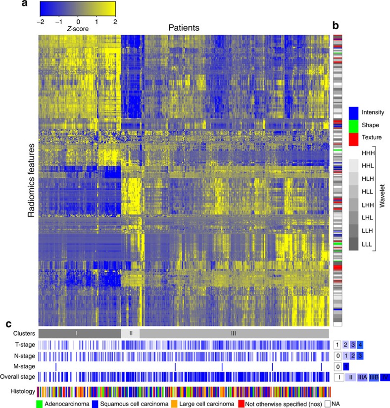
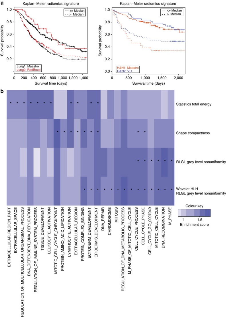
Verification
Claims the paper makes
The claims and statistics of this paper haven’t been checked yet.
Free: it reads the open-access text and checks the first 5 claims that cite a source we can check. Create a free account to check every claim.
Claims other papers make about this one
Each is checked against this paper’s own text: does it say what the citing paper says it does?
The source backs part of the claim, not all of it — or backs it for a different population, condition or comparison.
In the citing paper
“Notably, the signature was comprised of the top features from four classes (size, shape, texture, and wavelets) that were prioritized from a database of 442 features by using test-retest reproducibility and intersubject range.”
From Radiomics: Images Are More than Pictures, They Are Data (2016) · cites this paper as [47]
Experimental — accuracy not yet measuredThe source backs part of the claim, not all of it — or backs it for a different population, condition or comparison.
In the citing paper
“Many studies have shown that imaging features are significantly associated with patterns of gene expression and genetic mutations, demonstrating that radiogenomic analysis can identify different biological mechanisms by means of mathematical and computational devices, enabling the decoding of disease phenotypes by noninvasive methods(48,49).”
From Artificial intelligence, machine learning, computer-aided diagnosis, and radiomics: advances in imaging towards to precision medicine (2019) · cites this paper as [49]
Experimental — accuracy not yet measuredThe cited source says what the claim attributes to it.
In the citing paper
“A study by Aerts et al (47) showed that a radiomic signature could be used to predict outcome in completely independent cohorts of patients with lung cancer from two separate institutions.”
From Radiomics: Images Are More than Pictures, They Are Data (2016) · cites this paper as [47]
The cited source says what the claim attributes to it.
In the citing paper
“Further, this same signature could be applied to cohorts of patients with head and neck cancer with equivalent prognostic power.”
From Radiomics: Images Are More than Pictures, They Are Data (2016) · cites this paper as [47]
In the citing paper
“We used scans from 26 patients from the RIDER dataset19, routinely used for test–retest robustness analysis in tumour imaging19–21.”
From Foundation Model for Cancer Imaging Biomarkers (2024) · cites this paper as [20]
3 more claims about this paper
Create a free account to see every claim other papers make about this one.
Self-check
Percentages, sums and reported differences re-computed from the paper’s own numbers.
The self-check needs the paper’s full text.
Citations
Cites
Subramanian, A., Tamayo, P., Mootha, V. K., Mukherjee, S., Ebert, B. L., Gillette, M. A., Paulovich, A., Pomeroy, S. L., Golub, T. R., Lander, E. S., et al. (2005) · Proceedings of the National Academy of Sciences · doi:10.1073/pnas.0506580102 · Cited by 53,625
Harrell, F. E. (2001) · Springer Series in Statistics · doi:10.1007/978-1-4757-3462-1 · Cited by 9,195
Intratumor Heterogeneity and Branched Evolution Revealed by Multiregion Sequencing
Gerlinger, M., Rowan, A. J., Horswell, S., Larkin, J., Endesfelder, D., Gronroos, E., Martinez, P., Matthews, N., Stewart, A., Tarpey, P., et al. (2012) · New England Journal of Medicine · doi:10.1056/nejmoa1113205 · Cited by 7,774 · cited 2×
“It is known that multiple subclonal populations coexist within tumours, reflecting extensive intratumoral ‘somatic evolution’ 15,16 .”
BackgroundMolecular signatures database (MSigDB) 3.0
Liberzon, A., Subramanian, A., Pinchback, R., Thorvaldsdóttir, H., Tamayo, P., & Mesirov, J. P. (2011) · Bioinformatics · doi:10.1093/bioinformatics/btr260 · Cited by 7,111
Harrell (2015) · Springer Series in Statistics · doi:10.1007/978-3-319-19425-7 · Cited by 6,878
Radiomics: extracting more information from medical images using advanced feature analysis
Lambin, P., Rios-Velazquez, E., Leijenaar, R., Carvalho, S., van Stiphout, R. G. P. M., Granton, P., Zegers, C. M. L., Gillies, R., Boellard, R., Dekker, A., et al. (2012) · European Journal of Cancer · doi:10.1016/j.ejca.2011.11.036 · Cited by 6,084
Self-citationDistant metastasis occurs late during the genetic evolution of pancreatic cancer
Yachida, S., Jones, S., Bozic, I., Antal, T., Leary, R., Fu, B., Kamiyama, M., Hruban, R. H., Eshleman, J. R., Nowak, M. A., et al. (2010) · Nature · doi:10.1038/nature09515 · Cited by 2,502 · cited 1×
“It is known that multiple subclonal populations coexist within tumours, reflecting extensive intratumoral ‘somatic evolution’ 15,16 .”
BackgroundRadiomics: the process and the challenges
Kumar, V., Gu, Y., Basu, S., Berglund, A., Eschrich, S. A., Schabath, M. B., Forster, K., Aerts, H. J. W. L., Dekker, A., Fenstermacher, D., et al. (2012) · Magnetic Resonance Imaging · doi:10.1016/j.mri.2012.06.010 · Cited by 2,258
Self-citationTixier, F., Le Rest, C. C., Hatt, M., Albarghach, N., Pradier, O., Metges, J.-P., Corcos, L., & Visvikis, D. (2011) · Journal of Nuclear Medicine · doi:10.2967/jnumed.110.082404 · Cited by 696
Decoding global gene expression programs in liver cancer by noninvasive imaging
Segal, E., Sirlin, C. B., Ooi, C., Adler, A. S., Gollub, J., Chen, X., Chan, B. K., Matcuk, G. R., Barry, C. T., Chang, H. Y., et al. (2007) · Nature Biotechnology · doi:10.1038/nbt1306 · Cited by 629
Cited by
Items 101–200 of 1,023
Radiogenomics: bridging imaging and genomics
Bodalal, Z., Trebeschi, S., Nguyen-Kim, T. D. L., Schats, W., & Beets-Tan, R. (2019) · Abdominal Radiology · doi:10.1007/s00261-019-02028-w · Cited by 310
“Radiomics is the rapidly growing field of radiological research where routine patient images/scans are converted into mineable quantitative data [4] that can then be leveraged to decode the tumor phenotype for applications ranging from improved diagnostics to prognostication to therapeutic response prediction [5].”
BackgroundMachine Learning–Based Radiomics for Molecular Subtyping of Gliomas
Lu, C.-F., Hsu, F.-T., Hsieh, K. L.-C., Kao, Y.-C. J., Cheng, S.-J., Hsu, J. B.-K., Tsai, P.-H., Chen, R.-J., Huang, C.-C., Yen, Y., et al. (2018) · Clinical Cancer Research · doi:10.1158/1078-0432.ccr-17-3445 · Cited by 306
“The 8 shape and size features were calculated based on the three-dimensional geometry of the tumor volumes (8, 13).”
BackgroundMethodZwanenburg, A. (2019) · European Journal of Nuclear Medicine and Molecular Imaging · doi:10.1007/s00259-019-04391-8 · Cited by 306
“A robustness analysis has been performed in other studies, and has likewise been able to validate models externally; see, for example, references [140, 141].”
MethodKhorrami, M., Prasanna, P., Gupta, A., Patil, P., Velu, P. D., Thawani, R., Corredor, G., Alilou, M., Bera, K., Fu, P., et al. (2020) · Cancer Immunology Research · doi:10.1158/2326-6066.cir-19-0476 · Cited by 297
Choe, J., Lee, S. M., Do, K.-H., Lee, G., Lee, J.-G., Lee, S. M., & Seo, J. B. (2019) · Radiology · doi:10.1148/radiol.2019181960 · Cited by 296
Geras, K. J., Mann, R. M., & Moy, L. (2019) · Radiology · doi:10.1148/radiol.2019182627 · Cited by 295
“Radiomics, an expansion of CAD, is defined as the conversion of images to minable data by means of digital decoding of radiologic images into quantitative features (125).”
BackgroundTrebeschi, S., van Griethuysen, J. J. M., Lambregts, D. M. J., Lahaye, M. J., Parmar, C., Bakers, F. C. H., Peters, N. H. G. M., Beets-Tan, R. G. H., & Aerts, H. J. W. L. (2017) · Scientific Reports · doi:10.1038/s41598-017-05728-9 · Cited by 292
Self-citationRadiogenomics: What It Is and Why It Is Important
Mazurowski, M. A. (2015) · Journal of the American College of Radiology · doi:10.1016/j.jacr.2015.04.019 · Cited by 290
“Finally, ‘ radiogenomics’ has been equated with another approach called ‘ radiomics’ [5], [6], [7].”
BackgroundToward data‐efficient learning: A benchmark for COVID‐19 CT lung and infection segmentation
Ma, J., Wang, Y., An, X., Ge, C., Yu, Z., Chen, J., Zhu, Q., Dong, G., He, J., He, Z., et al. (2021) · Medical Physics · doi:10.1002/mp.14676 · Cited by 261
Park, J. E., Kim, D., Kim, H. S., Park, S. Y., Kim, J. Y., Cho, S. J., Shin, J. H., & Kim, J. H. (2020) · European Radiology · doi:10.1007/s00330-019-06360-z · Cited by 282
“Notably, only five studies [11, 12, 20, 26, 47] conducted imaging at multiple time points and tested feature robustness.”
BackgroundLee, G., Lee, H. Y., Park, H., Schiebler, M. L., van Beek, E. J. R., Ohno, Y., Seo, J. B., & Leung, A. (2017) · European Journal of Radiology · doi:10.1016/j.ejrad.2016.09.005 · Cited by 282
“In general, researchers have shown promising results in using radiomics to identify radiographic tumor phenotypes that favored specific genetic expressions [6,15,86,88,89].”
BackgroundInfluentialJi, G.-W., Zhu, F.-P., Xu, Q., Wang, K., Wu, M.-Y., Tang, W.-W., Li, X.-C., & Wang, X.-H. (2020) · Radiology · doi:10.1148/radiol.2020191470 · Cited by 273
Truhn, D., Schrading, S., Haarburger, C., Schneider, H., Merhof, D., & Kuhl, C. (2019) · Radiology · doi:10.1148/radiol.2018181352 · Cited by 273
Radiomics-based Prognosis Analysis for Non-Small Cell Lung Cancer
Zhang, Y., Oikonomou, A., Wong, A., Haider, M. A., & Khalvati, F. (2017) · Scientific Reports · doi:10.1038/srep46349 · Cited by 269
Kickingereder, P., Götz, M., Muschelli, J., Wick, A., Neuberger, U., Shinohara, R. T., Sill, M., Nowosielski, M., Schlemmer, H.-P., Radbruch, A., et al. (2016) · Clinical Cancer Research · doi:10.1158/1078-0432.ccr-16-0702 · Cited by 267
“Image registration was performed with the FMRIB software library (FSL, http://fsl.fmrib.ox.ac.uk/fsl/fslwiki/FSL).”
BackgroundMethodInfluentialPeerlings, J., Woodruff, H. C., Winfield, J. M., Ibrahim, A., Van Beers, B. E., Heerschap, A., Jackson, A., Wildberger, J. E., Mottaghy, F. M., DeSouza, N. M., et al. (2019) · Scientific Reports · doi:10.1038/s41598-019-41344-5 · Cited by 266
Self-citationIbrahim, A., Primakov, S., Beuque, M., Woodruff, H. C., Halilaj, I., Wu, G., Refaee, T., Granzier, R., Widaatalla, Y., Hustinx, R., et al. (2021) · Methods · doi:10.1016/j.ymeth.2020.05.022 · Cited by 265
Self-citationSun, Q., Lin, X., Zhao, Y., Li, L., Yan, K., Liang, D., Sun, D., & Li, Z.-C. (2020) · Frontiers in Oncology · doi:10.3389/fonc.2020.00053 · Cited by 263
“The detailed definitions of the radiomics features used can be found in two articles (22, 23).”
BackgroundRadiomics and deep learning in lung cancer
Avanzo, M., Stancanello, J., Pirrone, G., & Sartor, G. (2020) · Strahlentherapie und Onkologie · doi:10.1007/s00066-020-01625-9 · Cited by 263
Gray-level invariant Haralick texture features
Löfstedt, T., Brynolfsson, P., Asklund, T., Nyholm, T., & Garpebring, A. (2019) · PLOS ONE · doi:10.1371/journal.pone.0212110 · Cited by 261
“tumor phenotype [27], survival [28, 29] and tumor classification [30].”
BackgroundLarue, R. T. H. M., van Timmeren, J. E., de Jong, E. E. C., Feliciani, G., Leijenaar, R. T. H., Schreurs, W. M. J., Sosef, M. N., Raat, F. H. P. J., van der Zande, F. H. R., Das, M., et al. (2017) · Acta Oncologica · doi:10.1080/0284186x.2017.1351624 · Cited by 262
Self-citationGenova, C., Dellepiane, C., Carrega, P., Sommariva, S., Ferlazzo, G., Pronzato, P., Gangemi, R., Filaci, G., Coco, S., & Croce, M. (2022) · Frontiers in Immunology · doi:10.3389/fimmu.2021.799455 · Cited by 260
“Radiomics is a high-throughput extraction of features from medical images using computer algorithms, aimed at providing quantitative information on tissue composition that otherwise cannot be detected through simple observation (102, 103).”
BackgroundDeep learning models in medical image analysis
Tsuneki, M. (2022) · Journal of Oral Biosciences · doi:10.1016/j.job.2022.03.003 · Cited by 257
Rapid review: radiomics and breast cancer
Valdora, F., Houssami, N., Rossi, F., Calabrese, M., & Tagliafico, A. S. (2018) · Breast Cancer Research and Treatment · doi:10.1007/s10549-018-4675-4 · Cited by 258
“[5], “Images are more than pictures, they are data.”
BackgroundInfluentialMeyer, M., Ronald, J., Vernuccio, F., Nelson, R. C., Ramirez-Giraldo, J. C., Solomon, J., Patel, B. N., Samei, E., & Marin, D. (2019) · Radiology · doi:10.1148/radiol.2019190928 · Cited by 258
Deep learning classification of lung cancer histology using CT images
Chaunzwa, T. L., Hosny, A., Xu, Y., Shafer, A., Diao, N., Lanuti, M., Christiani, D. C., Mak, R. H., & Aerts, H. J. W. L. (2021) · Scientific Reports · doi:10.1038/s41598-021-84630-x · Cited by 257
“A distinct cohort of lung cancer patients treated with surgery (Lung3), which is publicly available at The Cancer Imaging Archive (TCIA) was used as an independent validation dataset 44,45 .”
MethodSelf-citationHe, L., Huang, Y., Ma, Z., Liang, C., Liang, C., & Liu, Z. (2016) · Scientific Reports · doi:10.1038/srep34921 · Cited by 256
Tracking tumor biology with radiomics: a systematic review utilizing a radiomics quality score
Sanduleanu, S., Woodruff, H. C., de Jong, E. E. C., van Timmeren, J. E., Jochems, A., Dubois, L., & Lambin, P. (2018) · Radiotherapy and Oncology · doi:10.1016/j.radonc.2018.03.033 · Cited by 242
“The other 2 studies had an average score 50 and both of these studies [14,16] addressed a potential clinical relevance for the reported imaging biomarker.”
BackgroundSelf-citationImage-Based Cardiac Diagnosis With Machine Learning: A Review
Martin-Isla, C., Campello, V. M., Izquierdo, C., Raisi-Estabragh, Z., Baeßler, B., Petersen, S. E., & Lekadir, K. (2020) · Frontiers in Cardiovascular Medicine · doi:10.3389/fcvm.2020.00001 · Cited by 248
Radiomic machine-learning classifiers for prognostic biomarkers of advanced nasopharyngeal carcinoma
Zhang, B., He, X., Ouyang, F., Gu, D., Dong, Y., Zhang, L., Mo, X., Huang, W., Tian, J., & Zhang, S. (2017) · Cancer Letters · doi:10.1016/j.canlet.2017.06.004 · Cited by 246
Deep learning and radiomics in precision medicine
Parekh, V. S., & Jacobs, M. A. (2019) · Expert Review of Precision Medicine and Drug Development · doi:10.1080/23808993.2019.1585805 · Cited by 246
Liu, X., Li, Y., Qian, Z., Sun, Z., Xu, K., Wang, K., Liu, S., Fan, X., Li, S., Zhang, Z., et al. (2018) · NeuroImage: Clinical · doi:10.1016/j.nicl.2018.10.014 · Cited by 245
“Next, quantitative radiomic features were extracted using the automated approach reported in a previous study which provided a detailed description of each feature in its supplementary material (Aerts et al., 2014).”
BackgroundMethodTexture Analysis of Imaging: What Radiologists Need to Know
Varghese, B. A., Cen, S. Y., Hwang, D. H., & Duddalwar, V. A. (2019) · American Journal of Roentgenology · doi:10.2214/ajr.18.20624 · Cited by 243
“The underlying assumption of the technique is that the gray-scale values creating the image of the tumor and the spatial and temporal interrelationships of these values reflect the phenotypic variations of the tumor, which are indicative of genetic and other molecular variations [7].”
BackgroundPark, H., Lim, Y., Ko, E. S., Cho, H.-H., Lee, J. E., Han, B.-K., Ko, E. Y., Choi, J. S., & Park, K. W. (2018) · Clinical Cancer Research · doi:10.1158/1078-0432.ccr-17-3783 · Cited by 243
“Other pathologic diagnoses in the 17 patients were as follows:mucinous carcinoma (7), medullary carcinoma (2), invasive micropapillary carcinoma (3), tubular carcinoma (1), invasive apocrine carcinoma (3), and adenoid cystic carcinoma (1).”
BackgroundDong, Y., Feng, Q., Yang, W., Lu, Z., Deng, C., Zhang, L., Lian, Z., Liu, J., Luo, X., Pei, S., et al. (2018) · European Radiology · doi:10.1007/s00330-017-5005-7 · Cited by 239
“Radiomics has been a research hotspot in recent years, and it is the process of the conversion medical images into high-dimensional, mineable and quantitative imaging features via high-throughput extraction of datacharacterisation algorithms [13, 14].”
Can CT-based radiomics signature predict KRAS/NRAS/BRAF mutations in colorectal cancer?
Yang, L., Dong, D., Fang, M., Zhu, Y., Zang, Y., Liu, Z., Zhang, H., Ying, J., Zhao, X., & Tian, J. (2018) · European Radiology · doi:10.1007/s00330-017-5146-8 · Cited by 238
“coefficients (ICCs) were used to estimate the robustness of the features [25].”
Continuous Learning AI in Radiology: Implementation Principles and Early Applications
Pianykh, O. S., Langs, G., Dewey, M., Enzmann, D. R., Herold, C. J., Schoenberg, S. O., & Brink, J. A. (2020) · Radiology · doi:10.1148/radiol.2020200038 · Cited by 237
Repeatability of Multiparametric Prostate MRI Radiomics Features
Schwier, M., van Griethuysen, J., Vangel, M. G., Pieper, S., Peled, S., Tempany, C., Aerts, H. J. W. L., Kikinis, R., Fennessy, F. M., & Fedorov, A. (2019) · Scientific Reports · doi:10.1038/s41598-019-45766-z · Cited by 237
Self-citationRadiomics of Brain MRI: Utility in Prediction of Metastatic Tumor Type
Kniep, H. C., Madesta, F., Schneider, T., Hanning, U., Schönfeld, M. H., Schön, G., Fiehler, J., Gauer, T., Werner, R., & Gellissen, S. (2019) · Radiology · doi:10.1148/radiol.2018180946 · Cited by 237
Criteria for the translation of radiomics into clinically useful tests
Huang, E. P., O’Connor, J. P. B., McShane, L. M., Giger, M. L., Lambin, P., Kinahan, P. E., Siegel, E. L., & Shankar, L. K. (2023) · Nature Reviews Clinical Oncology · doi:10.1038/s41571-022-00707-0 · Cited by 230
“A radiomic test might also be useful in multiple target populations; the test based on the model described by Aerts et al.16, for example, might be useful for predicting the outcomes of patients with head and neck cancer or non-small-cell lung cancer receiving standard-of-care therapies.”
MethodSelf-citationNam, D., Chapiro, J., Paradis, V., Seraphin, T. P., & Kather, J. N. (2022) · JHEP Reports · doi:10.1016/j.jhepr.2022.100443 · Cited by 226
“assemble a list of quantitative visual features such as tumour size, roundness, symmetry and intensity on images.(4) These features are subsequently inputted into a classification algorithm, for”
Quantitative imaging of cancer in the postgenomic era: Radio(geno)mics, deep learning, and habitats
Napel, S., Mu, W., Jardim‐Perassi, B. V., Aerts, H. J. W. L., & Gillies, R. J. (2018) · Cancer · doi:10.1002/cncr.31630 · Cited by 229
Self-citationRadiomic-Based Pathological Response Prediction from Primary Tumors and Lymph Nodes in NSCLC
Coroller, T. P., Agrawal, V., Huynh, E., Narayan, V., Lee, S. W., Mak, R. H., & Aerts, H. J. W. L. (2017) · Journal of Thoracic Oncology · doi:10.1016/j.jtho.2016.11.2226 · Cited by 228
“Radiomics may have a critical role in precision medicine, as it can predict clinical end points in patients before treatment.(9,11) Precision medicine is particularly important for patients with stage III NSCLC, as it is known that multiple forms of local therapy (radiation or surgery) are not beneficial for all patie…”
BackgroundSelf-citationThe crucial role of multiomic approach in cancer research and clinically relevant outcomes
Lu, M., & Zhan, X. (2018) · EPMA Journal · doi:10.1007/s13167-018-0128-8 · Cited by 224
“Analysis of radiomics-based features, comprehensive quantification information relating to the tumor phenotypes could be obtained [262, 263].”
BackgroundZhou, Y., He, L., Huang, Y., Chen, S., Wu, P., Ye, W., Liu, Z., & Liang, C. (2017) · Abdominal Radiology · doi:10.1007/s00261-017-1072-0 · Cited by 223
“In contrast to conventional CT-based imaging features, texture analysis enables a greater degree of information reflecting underlying biologic heterogeneity to be derived and qualified at low cost [21, 22].”
ResultFeng, Z., Rong, P., Cao, P., Zhou, Q., Zhu, W., Yan, Z., Liu, Q., & Wang, W. (2018) · European Radiology · doi:10.1007/s00330-017-5118-z · Cited by 224
Kim, J. Y., Park, J. E., Jo, Y., Shim, W. H., Nam, S. J., Kim, J. H., Yoo, R.-E., Choi, S. H., & Kim, H. S. (2019) · Neuro-Oncology · doi:10.1093/neuonc/noy133 · Cited by 224
Ma, X., Wei, J., Gu, D., Zhu, Y., Feng, B., Liang, M., Wang, S., Zhao, X., & Tian, J. (2019) · European Radiology · doi:10.1007/s00330-018-5985-y · Cited by 219
The role of artificial intelligence in medical imaging research
Tang, X. (2020) · BJR|Open · doi:10.1259/bjro.20190031 · Cited by 216
“37 The success of radiomics can potentially avoid undesirable complications caused by biopsy 38,39 and achieve the same or better prediction outcome.”
BackgroundKickingereder, P., Neuberger, U., Bonekamp, D., Piechotta, P. L., Götz, M., Wick, A., Sill, M., Kratz, A., Shinohara, R. T., Jones, D. T. W., et al. (2018) · Neuro-Oncology · doi:10.1093/neuonc/nox188 · Cited by 219
Leger, S., Zwanenburg, A., Pilz, K., Lohaus, F., Linge, A., Zöphel, K., Kotzerke, J., Schreiber, A., Tinhofer, I., Budach, V., et al. (2017) · Scientific Reports · doi:10.1038/s41598-017-13448-3 · Cited by 215
“The signature obtained by the Spearman feature selection method achieved a higher validation performance than the signature by Aerts et al .”
BackgroundMethodInfluentialLeijenaar, R. T. H., Carvalho, S., Hoebers, F. J. P., Aerts, H. J. W. L., van Elmpt, W. J. C., Huang, S. H., Chan, B., Waldron, J. N., O’sullivan, B., & Lambin, P. (2015) · Acta Oncologica · doi:10.3109/0284186x.2015.1061214 · Cited by 217
“[27], can be found in the Supplementary Appendix, to be found online at http://informahealthcare.”
BackgroundMethodInfluentialSelf-citationLu, H., Arshad, M., Thornton, A., Avesani, G., Cunnea, P., Curry, E., Kanavati, F., Liang, J., Nixon, K., Williams, S. T., et al. (2019) · Nature Communications · doi:10.1038/s41467-019-08718-9 · Cited by 212
CT-based radiomic analysis of stereotactic body radiation therapy patients with lung cancer
Huynh, E., Coroller, T. P., Narayan, V., Agrawal, V., Hou, Y., Romano, J., Franco, I., Mak, R. H., & Aerts, H. J. W. L. (2016) · Radiotherapy and Oncology · doi:10.1016/j.radonc.2016.05.024 · Cited by 215
Self-citationUmehara, K., Ota, J., & Ishida, T. (2018) · Journal of Digital Imaging · doi:10.1007/s10278-017-0033-z · Cited by 213
“In all, 89 chest CT images of patients diagnosed with nonsmall cell lung cancer, who were surgically treated [19, 20], were obtained from The Cancer Imaging Archive (TCIA) [21].”
MethodVoxel size and gray level normalization of CT radiomic features in lung cancer
Shafiq-ul-Hassan, M., Latifi, K., Zhang, G., Ullah, G., Gillies, R., & Moros, E. (2018) · Scientific Reports · doi:10.1038/s41598-018-28895-9 · Cited by 215
Self-citationCurrent AI technologies in cancer diagnostics and treatment
Tiwari, A., Mishra, S., & Kuo, T.-R. (2025) · Molecular Cancer · doi:10.1186/s12943-025-02369-9 · Cited by 207
Santos, M. K., Ferreira Júnior, J. R., Wada, D. T., Tenório, A. P. M., Nogueira-Barbosa, M. H., & Marques, P. M. D. A. (2019) · Radiologia Brasileira · doi:10.1590/0100-3984.2019.0049 · Cited by 214
“Many studies have shown that imaging features are significantly associated with patterns of gene expression and genetic mutations, demonstrating that radiogenomic analysis can identify different biological mechanisms by means of mathematical and computational devices, enabling the decoding of disease phenotypes by non…”
BackgroundThe future of personalised radiotherapy for head and neck cancer
Caudell, J. J., Torres-Roca, J. F., Gillies, R. J., Enderling, H., Kim, S., Rishi, A., Moros, E. G., & Harrison, L. B. (2017) · The Lancet Oncology · doi:10.1016/s1470-2045(17)30252-8 · Cited by 214
Self-citationRadiomics and radiogenomics in gliomas: a contemporary update
Singh, G., Manjila, S., Sakla, N., True, A., Wardeh, A. H., Beig, N., Vaysberg, A., Matthews, J., Prasanna, P., & Spektor, V. (2021) · British Journal of Cancer · doi:10.1038/s41416-021-01387-w · Cited by 213
Defining a Radiomic Response Phenotype: A Pilot Study using targeted therapy in NSCLC
Aerts, H. J. W. L., Grossmann, P., Tan, Y., Oxnard, G. R., Rizvi, N., Schwartz, L. H., & Zhao, B. (2016) · Scientific Reports · doi:10.1038/srep33860 · Cited by 213
“Radiomic features have been associated with clinical outcomes 4,8,25–29 , however the predictive capability of radiomics for response to targeted therapies remains largely unknown.”
InfluentialSelf-citationChong, H.-H., Yang, L., Sheng, R.-F., Yu, Y.-L., Wu, D.-J., Rao, S.-X., Yang, C., & Zeng, M.-S. (2021) · European Radiology · doi:10.1007/s00330-020-07601-2 · Cited by 211
“Subsequently, a set of 854 features radiomics features were extracted from the original and three-dimensional wavelet filters images [24], including tumor shape, size, intensity, and texture (Table S2).”
MethodNoninvasive IDH1 mutation estimation based on a quantitative radiomics approach for grade II glioma
Yu, J., Shi, Z., Lian, Y., Li, Z., Liu, T., Gao, Y., Wang, Y., Chen, L., & Mao, Y. (2017) · European Radiology · doi:10.1007/s00330-016-4653-3 · Cited by 211
Deep Learning Techniques to Diagnose Lung Cancer
Wang, L. (2022) · Cancers · doi:10.3390/cancers14225569 · Cited by 209
“[66] Non-small cell lung cancer (NSCLC)-Radiomics database 13,482 CT images from 89 patients”
BackgroundZhu, X., Dong, D., Chen, Z., Fang, M., Zhang, L., Song, J., Yu, D., Zang, Y., Liu, Z., Shi, J., et al. (2018) · European Radiology · doi:10.1007/s00330-017-5221-1 · Cited by 211
Highly accurate model for prediction of lung nodule malignancy with CT scans
Causey, J. L., Zhang, J., Ma, S., Jiang, B., Qualls, J. A., Politte, D. G., Prior, F., Zhang, S., & Huang, X. (2018) · Scientific Reports · doi:10.1038/s41598-018-27569-w · Cited by 209
Mu, W., Jiang, L., Shi, Y., Tunali, I., Gray, J. E., Katsoulakis, E., Tian, J., Gillies, R. J., & Schabath, M. B. (2021) · Journal for ImmunoTherapy of Cancer · doi:10.1136/jitc-2020-002118 · Cited by 207
Self-citationMali, S. A., Ibrahim, A., Woodruff, H. C., Andrearczyk, V., Müller, H., Primakov, S., Salahuddin, Z., Chatterjee, A., & Lambin, P. (2021) · Journal of Personalized Medicine · doi:10.3390/jpm11090842 · Cited by 207
“Studies have shown the effects of image acquisition parameters on the reproducibility of radiomic features [21,24–27]. the image domain (Section 3) are performed on the whole image (raw before feature Many studies [1,28–31] have also explored the discriminative power of radiomic features.…”
BackgroundMethodSelf-citationBladder Cancer Treatment Response Assessment in CT using Radiomics with Deep-Learning
Cha, K. H., Hadjiiski, L., Chan, H.-P., Weizer, A. Z., Alva, A., Cohan, R. H., Caoili, E. M., Paramagul, C., & Samala, R. K. (2017) · Scientific Reports · doi:10.1038/s41598-017-09315-w · Cited by 207
“Previous studies decoded tumor phenotypes 11 using radiomics features, or evaluated the correlation between radiomics features with radiation therapy dose and radiation pneumonitis 12 in lung CT.”
MethodRadiomics and artificial intelligence for precision medicine in lung cancer treatment
Chen, M., Copley, S. J., Viola, P., Lu, H., & Aboagye, E. O. (2023) · Seminars in Cancer Biology · doi:10.1016/j.semcancer.2023.05.004 · Cited by 206
Vial, A., Stirling, D., Field, M., Ros, M., Ritz, C., Carolan, M., Holloway, L., & Miller, A. A. (2018) · Translational Cancer Research · doi:10.21037/tcr.2018.05.02 · Cited by 205
“Deterministic feature extraction is the most common method where a mathematical formula is employed to extract features relating to imaging features such as texture, intensity or shape, this method is employed in the following papers (1,22,23,28,30-34).”
BackgroundMethodInfluentialKann, B. H., Aneja, S., Loganadane, G. V., Kelly, J. R., Smith, S. M., Decker, R. H., Yu, J. B., Park, H. S., Yarbrough, W. G., Malhotra, A., et al. (2018) · Scientific Reports · doi:10.1038/s41598-018-32441-y · Cited by 207
“Machine learning has more recently been applied in medical imaging analysis, when its use is incorporated into radiomics studies, which extract and analyze quantitative image features 17,18 .”
MethodKim, S., Shin, J., Kim, D.-Y., Choi, G. H., Kim, M.-J., & Choi, J.-Y. (2019) · Clinical Cancer Research · doi:10.1158/1078-0432.ccr-18-2861 · Cited by 205
“Radiomics is an emerging field that converts medical imaging into high-dimensional mineable features using a large number of image-characterization algorithms (11).”
BackgroundLou, B., Doken, S., Zhuang, T., Wingerter, D., Gidwani, M., Mistry, N., Ladic, L., Kamen, A., & Abazeed, M. E. (2019) · The Lancet Digital Health · doi:10.1016/s2589-7500(19)30058-5 · Cited by 206
“6,7 Some of the features extracted with radiomics have been shown to capture distinct tumour characteristics and exhibit prognostic power; 8 limitations to handcrafted image features, however, are their manual labelling and their inability to conform to a specific task.”
BackgroundZhang, J., Zhao, X., Zhao, Y., Zhang, J., Zhang, Z., Wang, J., Wang, Y., Dai, M., & Han, J. (2020) · European Journal of Nuclear Medicine and Molecular Imaging · doi:10.1007/s00259-019-04592-1 · Cited by 205
Shiri, I., Rahmim, A., Ghaffarian, P., Geramifar, P., Abdollahi, H., & Bitarafan-Rajabi, A. (2017) · European Radiology · doi:10.1007/s00330-017-4859-z · Cited by 204
Deep learning in head & neck cancer outcome prediction
Diamant, A., Chatterjee, A., Vallières, M., Shenouda, G., & Seuntjens, J. (2019) · Scientific Reports · doi:10.1038/s41598-019-39206-1 · Cited by 200
Foundation Model for Cancer Imaging Biomarkers
Pai, S., Bontempi, D., Hadzic, I., Prudente, V., Sokač, M., Chaunzwa, T. L., Bernatz, S., Hosny, A., Mak, R. H., Birkbak, N. J., et al. (2024) · Nature Machine Intelligence · doi:10.1038/s42256-024-00807-9 · Cited by 199
Self-citationZhang, Y.-P., Zhang, X.-Y., Cheng, Y.-T., Li, B., Teng, X.-Z., Zhang, J., Lam, S., Zhou, T., Ma, Z.-R., Sheng, J.-B., et al. (2023) · Military Medical Research · doi:10.1186/s40779-023-00458-8 · Cited by 199
“Aerts et al. [125] used gene set enrichment analysis to compare radiomics features with gene expression profiles and found that the features were significantly correlated with different biologic gene sets.”
BackgroundJi, G.-W., Zhang, Y.-D., Zhang, H., Zhu, F.-P., Wang, K., Xia, Y.-X., Zhang, Y.-D., Jiang, W.-J., Li, X.-C., & Wang, X.-H. (2019) · Radiology · doi:10.1148/radiol.2018181408 · Cited by 199
Feng, S.-T., Jia, Y., Liao, B., Huang, B., Zhou, Q., Li, X., Wei, K., Chen, L., Li, B., Wang, W., et al. (2019) · European Radiology · doi:10.1007/s00330-018-5935-8 · Cited by 199
Fornacon-Wood, I., Mistry, H., Ackermann, C. J., Blackhall, F., McPartlin, A., Faivre-Finn, C., Price, G. J., & O’Connor, J. P. B. (2020) · European Radiology · doi:10.1007/s00330-020-06957-9 · Cited by 198
Radiomics and Artificial Intelligence for Biomarker and Prediction Model Development in Oncology
Forghani, R., Savadjiev, P., Chatterjee, A., Muthukrishnan, N., Reinhold, C., & Forghani, B. (2019) · Computational and Structural Biotechnology Journal · doi:10.1016/j.csbj.2019.07.001 · Cited by 195
“The image processing pipeline involved 440 3D radiomic features, described in Aerts et al. [10] and extracted from manually delineated tumor regions from each patient's pre-treatment CT images.”
MethodChu, L. C., Park, S., Kawamoto, S., Fouladi, D. F., Shayesteh, S., Zinreich, E. S., Graves, J. S., Horton, K. M., Hruban, R. H., Yuille, A. L., et al. (2019) · American Journal of Roentgenology · doi:10.2214/ajr.18.20901 · Cited by 193
Genomic and immune heterogeneity are associated with differential responses to therapy in melanoma
Reuben, A., Spencer, C. N., Prieto, P. A., Gopalakrishnan, V., Reddy, S. M., Miller, J. P., Mao, X., De Macedo, M. P., Chen, J., Song, X., et al. (2017) · npj Genomic Medicine · doi:10.1038/s41525-017-0013-8 · Cited by 166
“This technique has been employed to capture and evaluate the imaging data of a wide variety of solid tumors, including gliomas and lung, breast, colon, hepatocellular, and renal cell cancers,(15) among others, to predict responses to various cancer treatments, provide prognostic information, 19 identify tumor histolog…”
Machine learning applications in prostate cancer magnetic resonance imaging
Cuocolo, R., Cipullo, M. B., Stanzione, A., Ugga, L., Romeo, V., Radice, L., Brunetti, A., & Imbriaco, M. (2019) · European Radiology Experimental · doi:10.1186/s41747-019-0109-2 · Cited by 193
“These large datasets have been analysed to obtain useful clinical information such as correlation to other biomarkers, patient prognosis or treatment outcome [17 – 19].”
MethodMu, W., Tunali, I., Gray, J. E., Qi, J., Schabath, M. B., & Gillies, R. J. (2020) · European Journal of Nuclear Medicine and Molecular Imaging · doi:10.1007/s00259-019-04625-9 · Cited by 194
Self-citationMokrane, F.-Z., Lu, L., Vavasseur, A., Otal, P., Peron, J.-M., Luk, L., Yang, H., Ammari, S., Saenger, Y., Rousseau, H., et al. (2020) · European Radiology · doi:10.1007/s00330-019-06347-w · Cited by 193
“Third, we identified five robust features regarding segmentation effect able to differentiate HCC from non-HCC [17, 18].”
BackgroundLigero, M., Jordi-Ollero, O., Bernatowicz, K., Garcia-Ruiz, A., Delgado-Muñoz, E., Leiva, D., Mast, R., Suarez, C., Sala-Llonch, R., Calvo, N., et al. (2021) · European Radiology · doi:10.1007/s00330-020-07174-0 · Cited by 191
“Aerts et al considered radiomics variability as a feature selection tool by using test–retest analysis, eliminating radiomics features with high variability based on their cohort results [5, 11].”
BackgroundMethodTest–Retest Data for Radiomics Feature Stability Analysis: Generalizable or Study-Specific?
van Timmeren, J. E., Leijenaar, R. T. H., van Elmpt, W., Wang, J., Zhang, Z., Dekker, A., & Lambin, P. (2016) · Tomography · doi:10.18383/j.tom.2016.00208 · Cited by 191
“Mathematical descriptions of all features are published elsewhere (4).”
BackgroundMethodSelf-citationSong, J., Shi, J., Dong, D., Fang, M., Zhong, W., Wang, K., Wu, N., Huang, Y., Liu, Z., Cheng, Y., et al. (2018) · Clinical Cancer Research · doi:10.1158/1078-0432.ccr-17-2507 · Cited by 191
“These algorithms were partially defined by Aerts and colleagues' study (27) and partially defined by Song and colleagues' study (28).”
MethodClassification of the glioma grading using radiomics analysis
Cho, H.-H., Lee, S.-H., Kim, J., & Park, H. (2018) · PeerJ · doi:10.7717/peerj.5982 · Cited by 192
“Many studies have demonstrated that distinct tumor types in many organs can be quantified by radiomics analysis and the results of the radiomics can be used as imaging biomarkers for supporting clinical decision making (Zacharaki et al., 2009; Aerts et al., 2014; Kickingereder et al., 2016; Li et al., 2016; Bowen et a…”
BackgroundZhou, M., Leung, A., Echegaray, S., Gentles, A., Shrager, J. B., Jensen, K. C., Berry, G. J., Plevritis, S. K., Rubin, D. L., Napel, S., et al. (2018) · Radiology · doi:10.1148/radiol.2017161845 · Cited by 190
“Our radiogenomics analysis has several advantages over previous studies (Fig 1) (14,15).”
Assessing the accuracy of a deep learning method to risk stratify indeterminate pulmonary nodules
Massion, P. P., Antic, S., Ather, S., Arteta, C., Brabec, J., Chen, H., Declerck, J., Dufek, D., Hickes, W., Kadir, T., et al. (2020) · American Journal of Respiratory and Critical Care Medicine · doi:10.1164/rccm.201903-0505oc · Cited by 190
Qian, Z., Li, Y., Wang, Y., Li, L., Li, R., Wang, K., Li, S., Tang, K., Zhang, C., Fan, X., et al. (2019) · Cancer Letters · doi:10.1016/j.canlet.2019.02.054 · Cited by 189
“To further reduce the variations in the manual segmentations between radiologists, the ICCs were calculated for each feature and only stable features were selected for further analysis [33].”
MethodGray-level discretization impacts reproducible MRI radiomics texture features
Duron, L., Balvay, D., Vande Perre, S., Bouchouicha, A., Savatovsky, J., Sadik, J.-C., Thomassin-Naggara, I., Fournier, L., & Lecler, A. (2019) · PLOS ONE · doi:10.1371/journal.pone.0213459 · Cited by 188
“Medical imaging is progressively shifting from conventional visual image analysis to quantitative personalized medicine thanks to the recent development of data-driven analysis methods like radiomics [1].”
BackgroundSong, Y., Zhang, Y., Yan, X., Liu, H., Zhou, M., Hu, B., & Yang, G. (2018) · Journal of Magnetic Resonance Imaging · doi:10.1002/jmri.26047 · Cited by 186
“10 However, studies focused on combining DCNNs and mp-MRI to solve lesion detection and classification problems of the prostate have been limited.”
BackgroundHow to develop a meaningful radiomic signature for clinical use in oncologic patients
Papanikolaou, N., Matos, C., & Koh, D. M. (2020) · Cancer Imaging · doi:10.1186/s40644-020-00311-4 · Cited by 186
MAPS: A Quantitative Radiomics Approach for Prostate Cancer Detection
Cameron, A., Khalvati, F., Haider, M. A., & Wong, A. (2016) · IEEE Transactions on Biomedical Engineering · doi:10.1109/tbme.2015.2485779 · Cited by 185
“Studies on lung and head-and-neck cancer patients have confirmed the prognostic power of radiomics features for patient outcome prediction for personalized medicine [32], [33].”
Backgroundvan Timmeren, J. E., Leijenaar, R. T. H., van Elmpt, W., Reymen, B., Oberije, C., Monshouwer, R., Bussink, J., Brink, C., Hansen, O., & Lambin, P. (2017) · Radiotherapy and Oncology · doi:10.1016/j.radonc.2017.04.016 · Cited by 184
Self-citation
Metadata
- Authors
- Aerts, H. J. W. L., Velazquez, E. R., Leijenaar, R. T. H., Parmar, C., Grossmann, P., Carvalho, S., Bussink, J., Monshouwer, R., Haibe-Kains, B., Rietveld, D., Hoebers, F., Rietbergen, M. M., Leemans, C. R., Dekker, A., Quackenbush, J., Gillies, R. J., & Lambin, P.
- Affiliations
- Brigham and Women's Hospital (US)
- Maastricht University (NL)
- Radboud University Medical Center (NL)
- Amsterdam UMC Location Vrije Universiteit Amsterdam (NL)
- Dana-Farber Cancer Institute (US)
- Moffitt Cancer Center (US)
- Corresponding author
- Aerts, Hugo J. W. L.
- ORCID
- Venue
- Nature Communications
- Published
- 2014-06-03
- Volume
- 5
- Issue
- 1
- Pages
- 4006-4006
- ISSN
- 2041-1723 (ISSN-L)
- PMC
- PMC4059926
- OpenAlex
- W2103004421
- Semantic Scholar
- 3b002068646831a6f59fabb6daeb0209ed54049d
- Cited by
- 5,132
- References
- 29
- Citations by year
- 2026: 415 · 2025: 460 · 2024: 458 · 2023: 510 · 2022: 567 · 2021: 638 · 2020: 587 · 2019: 552 · 2018: 388 · 2017: 283 · 2016: 175 · 2015: 82 · 2014: 11
- FWCI
- 142.84
- Type
- journal article
- Language
- en
- Update notices
- correction (2014-08-07)
- Open access
- hybrid
- doi.org
- Licence
- Locations
- Nature Communications (publishedVersion, cc-by-nc-nd)
- PubMed Central (publishedVersion, cc-by-nc-nd)
- PubMed (publishedVersion)
- Pure Amsterdam UMC (publishedVersion)
- Amsterdam UMC (VU Amsterdam) - Institutional Repository (publishedVersion)
- Radboud Repository (Radboud University) (submittedVersion)
- Digital Access to Scholarship at Harvard (DASH) (Harvard University) (submittedVersion, cc-by)
- RePEc: Research Papers in Economics (submittedVersion)
- Research Publications (Maastricht University) (submittedVersion, cc-by-nc-nd)
- Radboud Repository (Radboud University) (submittedVersion, other-oa)
- Research Publications (Maastricht University) (submittedVersion, other-oa)
- Data Archiving and Networked Services (DANS) (submittedVersion)
- Funders
- European Federation of Pharmaceutical Industries and Associations
- KWF Kankerbestrijding (UM 2009-4454, 2011-5020, KWF UM 2009-4454, KWF UM 2011-5020, 2009-4454)
- Center for Translational Molecular Medicine (grant 030-103, 030-103)
- Innovative Medicines Initiative (115151)
- National Institutes of Health (U01 CA)
- National Cancer Institute (NIH-USA U01CA 143062-01, U01CA143062)
- Interreg
- Topics
- Health Sciences › Medicine › Radiology, Nuclear Medicine and Imaging › Radiomics and Machine Learning in Medical Imaging
- Physical Sciences › Computer Science › Computer Vision and Pattern Recognition › Image Retrieval and Classification Techniques
- Health Sciences › Medicine › Radiology, Nuclear Medicine and Imaging › Medical Imaging Techniques and Applications
- Keywords
- radiomics, tumor phenotype, lung cancer, head and neck cancer, radiogenomics, intratumoral heterogeneity, computed tomography, image texture features, gene expression, non-invasive imaging, treatment decision support
- MeSH terms
- Adenocarcinoma, Carcinoma, Non-Small-Cell Lung, Carcinoma, Squamous Cell, Female, Head and Neck Neoplasms, Humans, Lung Neoplasms, Male, Phenotype, Prognosis, Tomography, X-Ray Computed, Tumor Burden, Positron-Emission Tomography, Multimodal Imaging
About these checks
Registry, retraction, Cites and statistics results are computed, not judged, with one exception: a printed reference that no index lists is read once by a model before it counts as a citation error. Claims are extracted by a model and kept only when their quote appears verbatim in the text; each cited source is then checked, with a model’s help, for whether it says what the claim relies on. Those verdicts can be wrong — read the claim’s quote and the cited source before relying on one.
Statistics and arithmetic are recomputed, not judged.
Confidence bands are shown only for fields calibrated on our annotated test set. None is calibrated yet, so no band is shown.
What counts as a claim ›How we measure confidence ›How we check this ›
Are you an author, or do you see a problem with this page? Request a review
Data sources
Bibliographic data: Semantic Scholar, OpenAlex, Crossref.
Contains information from the Semantic Scholar Academic Graph Datasets, made available under the ODC Attribution License (ODC-BY 1.0).
Semantic Scholar’s data is described in: Kinney, R., et al. (2023). The Semantic Scholar Open Data Platform. arXiv:2301.10140.
