

Decoding tumour phenotype by noninvasive imaging using a quantitative radiomics approach
Open accessThe DOI matches its Crossref record; 29 of the 29 references it cites were found in a registry or an index.
Abstract
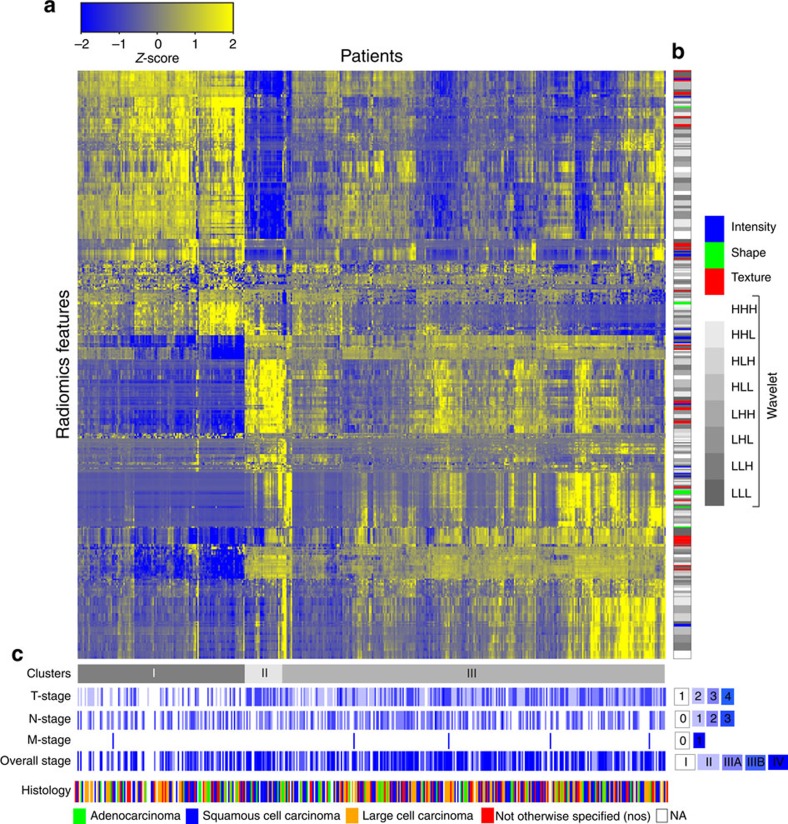
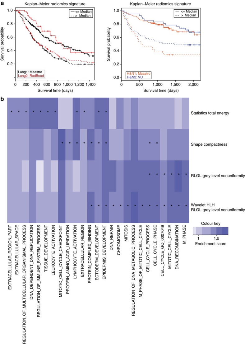
Human cancers exhibit strong phenotypic differences that can be visualized noninvasively by medical imaging. Radiomics refers to the comprehensive quantification of tumour phenotypes by applying a large number of quantitative image features. Here we present a radiomic analysis of 440 features quantifying tumour image intensity, shape and texture, which are extracted from computed tomography data of 1,019 patients with lung or head-and-neck cancer. We find that a large number of radiomic features have prognostic power in independent data sets of lung and head-and-neck cancer patients, many of which were not identified as significant before. Radiogenomics analysis reveals that a prognostic radiomic signature, capturing intratumour heterogeneity, is associated with underlying gene-expression patterns. These data suggest that radiomics identifies a general prognostic phenotype existing in both lung and head-and-neck cancer. This may have a clinical impact as imaging is routinely used in clinical practice, providing an unprecedented opportunity to improve decision-support in cancer treatment at low cost.
Overview
The claims and statistics of this paper haven’t been checked yet.
Free: it reads the open-access text and checks the first 5 claims that cite a source we can check. Create a free account to check every claim.
Integrity signals
Registry
Cites
Statistics
Figures


Verification
Claims the paper makes
The claims and statistics of this paper haven’t been checked yet.
Free: it reads the open-access text and checks the first 5 claims that cite a source we can check. Create a free account to check every claim.
Claims other papers make about this one
Each is checked against this paper’s own text: does it say what the citing paper says it does?
The source backs part of the claim, not all of it — or backs it for a different population, condition or comparison.
In the citing paper
“Notably, the signature was comprised of the top features from four classes (size, shape, texture, and wavelets) that were prioritized from a database of 442 features by using test-retest reproducibility and intersubject range.”
From Radiomics: Images Are More than Pictures, They Are Data (2016) · cites this paper as [47]
Experimental — accuracy not yet measuredThe source backs part of the claim, not all of it — or backs it for a different population, condition or comparison.
In the citing paper
“Many studies have shown that imaging features are significantly associated with patterns of gene expression and genetic mutations, demonstrating that radiogenomic analysis can identify different biological mechanisms by means of mathematical and computational devices, enabling the decoding of disease phenotypes by noninvasive methods(48,49).”
From Artificial intelligence, machine learning, computer-aided diagnosis, and radiomics: advances in imaging towards to precision medicine (2019) · cites this paper as [49]
Experimental — accuracy not yet measuredThe cited source says what the claim attributes to it.
In the citing paper
“A study by Aerts et al (47) showed that a radiomic signature could be used to predict outcome in completely independent cohorts of patients with lung cancer from two separate institutions.”
From Radiomics: Images Are More than Pictures, They Are Data (2016) · cites this paper as [47]
The cited source says what the claim attributes to it.
In the citing paper
“Further, this same signature could be applied to cohorts of patients with head and neck cancer with equivalent prognostic power.”
From Radiomics: Images Are More than Pictures, They Are Data (2016) · cites this paper as [47]
In the citing paper
“We used scans from 26 patients from the RIDER dataset19, routinely used for test–retest robustness analysis in tumour imaging19–21.”
From Foundation Model for Cancer Imaging Biomarkers (2024) · cites this paper as [20]
3 more claims about this paper
Create a free account to see every claim other papers make about this one.
Self-check
Percentages, sums and reported differences re-computed from the paper’s own numbers.
The self-check needs the paper’s full text.
Citations
Cites
Subramanian, A., Tamayo, P., Mootha, V. K., Mukherjee, S., Ebert, B. L., Gillette, M. A., Paulovich, A., Pomeroy, S. L., Golub, T. R., Lander, E. S., et al. (2005) · Proceedings of the National Academy of Sciences · doi:10.1073/pnas.0506580102 · Cited by 53,625
Harrell, F. E. (2001) · Springer Series in Statistics · doi:10.1007/978-1-4757-3462-1 · Cited by 9,195
Intratumor Heterogeneity and Branched Evolution Revealed by Multiregion Sequencing
Gerlinger, M., Rowan, A. J., Horswell, S., Larkin, J., Endesfelder, D., Gronroos, E., Martinez, P., Matthews, N., Stewart, A., Tarpey, P., et al. (2012) · New England Journal of Medicine · doi:10.1056/nejmoa1113205 · Cited by 7,774 · cited 2×
“It is known that multiple subclonal populations coexist within tumours, reflecting extensive intratumoral ‘somatic evolution’ 15,16 .”
BackgroundMolecular signatures database (MSigDB) 3.0
Liberzon, A., Subramanian, A., Pinchback, R., Thorvaldsdóttir, H., Tamayo, P., & Mesirov, J. P. (2011) · Bioinformatics · doi:10.1093/bioinformatics/btr260 · Cited by 7,111
Harrell (2015) · Springer Series in Statistics · doi:10.1007/978-3-319-19425-7 · Cited by 6,878
Radiomics: extracting more information from medical images using advanced feature analysis
Lambin, P., Rios-Velazquez, E., Leijenaar, R., Carvalho, S., van Stiphout, R. G. P. M., Granton, P., Zegers, C. M. L., Gillies, R., Boellard, R., Dekker, A., et al. (2012) · European Journal of Cancer · doi:10.1016/j.ejca.2011.11.036 · Cited by 6,084
Self-citationDistant metastasis occurs late during the genetic evolution of pancreatic cancer
Yachida, S., Jones, S., Bozic, I., Antal, T., Leary, R., Fu, B., Kamiyama, M., Hruban, R. H., Eshleman, J. R., Nowak, M. A., et al. (2010) · Nature · doi:10.1038/nature09515 · Cited by 2,502 · cited 1×
“It is known that multiple subclonal populations coexist within tumours, reflecting extensive intratumoral ‘somatic evolution’ 15,16 .”
BackgroundRadiomics: the process and the challenges
Kumar, V., Gu, Y., Basu, S., Berglund, A., Eschrich, S. A., Schabath, M. B., Forster, K., Aerts, H. J. W. L., Dekker, A., Fenstermacher, D., et al. (2012) · Magnetic Resonance Imaging · doi:10.1016/j.mri.2012.06.010 · Cited by 2,258
Self-citationTixier, F., Le Rest, C. C., Hatt, M., Albarghach, N., Pradier, O., Metges, J.-P., Corcos, L., & Visvikis, D. (2011) · Journal of Nuclear Medicine · doi:10.2967/jnumed.110.082404 · Cited by 696
Decoding global gene expression programs in liver cancer by noninvasive imaging
Segal, E., Sirlin, C. B., Ooi, C., Adler, A. S., Gollub, J., Chen, X., Chan, B. K., Matcuk, G. R., Barry, C. T., Chang, H. Y., et al. (2007) · Nature Biotechnology · doi:10.1038/nbt1306 · Cited by 629
Cited by
Items 501–600 of 1,023
Zhang, J., Yao, K., Liu, P., Liu, Z., Han, T., Zhao, Z., Cao, Y., Zhang, J., Zhang, J., Tian, J., et al. (2020) · EBioMedicine · doi:10.1016/j.ebiom.2020.102933 · Cited by 105
“[14] Aerts HJ, Velazquez ER, Leijenaar RT, Parmar C, Grossmann P, Carvalho S, et al.”
Tong, Y., Li, J., Huang, Y., Zhou, J., Liu, T., Guo, Y., Yu, J., Zhou, S., Wang, Y., & Chang, C. (2020) · Academic Radiology · doi:10.1016/j.acra.2020.07.017 · Cited by 101
Fumagalli, C., & Barberis, M. C. P. (2021) · Diagnostics · doi:10.3390/diagnostics11091555 · Cited by 121
Gote-Schniering, J., Maciukiewicz, M., Gabryś, H. S., Brunner, M., Blüthgen, C., Meier, C., Braga-Lagache, S., Uldry, A., Heller, M., Distler, O., et al. (2021) · European Respiratory Journal · doi:10.1183/13993003.04503-2020 · Cited by 108
Somatic mutations associated with MRI-derived volumetric features in glioblastoma
Gutman, D. A., Dunn, W. D., Grossmann, P., Cooper, L. A. D., Holder, C. A., Ligon, K. L., Alexander, B. M., & Aerts, H. J. W. L. (2015) · Neuroradiology · doi:10.1007/s00234-015-1576-7 · Cited by 96
“Our current work expands upon these results by using a semi-automated digital quantification technique, which recent work has shown to be more objective and lead to more robust findings than qualitative stagingmethods used in the past [14].”
BackgroundResultSelf-citationCozzi, L., Dinapoli, N., Fogliata, A., Hsu, W., Reggiori, G., Lobefalo, F., Kirienko, M., Sollini, M., Franceschini, D., Comito, T., et al. (2017) · BMC Cancer · doi:10.1186/s12885-017-3847-7 · Cited by 96
“In particular, radiomics is an emerging field that converts imaging data into a high dimensional mineable feature space using a large number of automatically extracted data-characterization algorithms [15, 16].”
Wu, S., Meng, J., Yu, Q., Li, P., & Fu, S. (2019) · Journal of Cancer Research and Clinical Oncology · doi:10.1007/s00432-018-2787-1 · Cited by 103
Siegersma, K. R., Leiner, T., Chew, D. P., Appelman, Y., Hofstra, L., & Verjans, J. W. (2019) · Netherlands Heart Journal · doi:10.1007/s12471-019-01311-1 · Cited by 96
Zanfardino, M., Franzese, M., Pane, K., Cavaliere, C., Monti, S., Esposito, G., Salvatore, M., & Aiello, M. (2019) · Journal of Translational Medicine · doi:10.1186/s12967-019-2073-2 · Cited by 96
Cardiac magnetic resonance radiomics: basic principles and clinical perspectives
Raisi‐Estabragh, Z., Izquierdo, C., Campello, V. M., Martín-Isla, C., Jaggi, A., Harvey, N. C., Lekadir, K., & Petersen, S. E. (2020) · European Heart Journal - Cardiovascular Imaging · doi:10.1093/ehjci/jeaa028 · Cited by 100
“Furthermore, the limited CMR radiomics literature and the more extensive work from oncology suggest that decoding image phenotype through radiomics texture analysis may provide additional value in reflecting pathology at the tissue level, with the potential to provide insights into disease pathophysiology.(38) However…”
BackgroundThe effect of feature normalization methods in radiomics
Demircioğlu, A. (2024) · Insights into Imaging · doi:10.1186/s13244-023-01575-7 · Cited by 125
Advancements in AI based healthcare techniques with FOCUS ON diagnostic techniques
Kalra, N., Verma, P., & Verma, S. (2024) · Computers in Biology and Medicine · doi:10.1016/j.compbiomed.2024.108917 · Cited by 95
Preliminary investigation into sources of uncertainty in quantitative imaging features
Fave, X. J., Cook, M. C., Frederick, A., Zhang, L., Yang, J., Fried, D., Stingo, F. C., & Court, L. E. (2015) · Computerized Medical Imaging and Graphics · doi:10.1016/j.compmedimag.2015.04.006 · Cited by 105
Abdollahi, H., Mostafaei, S., Cheraghi, S., Shiri, I., Mahdavi, S. R., & Kazemnejad, A. (2018) · Physica Medica · doi:10.1016/j.ejmp.2017.10.008 · Cited by 94
“In another way, the newer field, radiomics tries to find links between quantified medical images and clinical/biological data [10,11].”
BackgroundHu, T., Wang, S.-P., Huang, L., Wang, J.-Z., Shi, D.-B., Li, Y., Tong, T., & Peng, W. (2018) · European Radiology · doi:10.1007/s00330-018-5539-3 · Cited by 95
“In another study with a large cohort [8], they also found that GLCM texture features showed strong correlations with gene expression in both lung cancer and head and neck cancer.”
BackgroundMethodLinning, E., Lü, L., Li, L., Yang, H., Schwartz, L. H., & Zhao, B. (2018) · Academic Radiology · doi:10.1016/j.acra.2018.10.013 · Cited by 103
Lv, W., Yuan, Q., Wang, Q., Ma, J., Feng, Q., Chen, W., Rahmim, A., & Lu, L. (2019) · Molecular Imaging and Biology · doi:10.1007/s11307-018-01304-3 · Cited by 101
“In order to characterize intratumor heterogeneity more comprehensively, a discrete, one-level, and undecimated 3D wavelet transform BCoiflet 1^ was applied to each PET or CT image [39], by performing low-pass (L) or high-pass (H) filter along x-, y-, or z-directions, resulting in 8 decompositions of original image, no…”
MethodQuantitative imaging for radiotherapy purposes
Gurney‐Champion, O. J., Mahmood, F., van Schie, M. A., Julian, R., George, B., Philippens, M. E. P., van der Heide, U. A., Thorwarth, D., & Redalen, K. R. (2020) · Radiotherapy and Oncology · doi:10.1016/j.radonc.2020.01.026 · Cited by 103
Xie, C., Du, R., Ho, J. W. K., Pang, H., Chiu, K. W.-H., Lee, E. Y. P., & Vardhanabhuti, V. (2020) · European Journal of Nuclear Medicine and Molecular Imaging · doi:10.1007/s00259-020-04756-4 · Cited by 103
Polverari, G., Ceci, F., Bertaglia, V., Reale, M. L., Rampado, O., Gallio, E., Passera, R., Liberini, V., Scapoli, P., Arena, V., et al. (2020) · Cancers · doi:10.3390/cancers12051163 · Cited by 104
Taghavi, M., Trebeschi, S., Simões, R., Meek, D. B., Beckers, R. C. J., Lambregts, D. M. J., Verhoef, C. K., Houwers, J. B., van der Heide, U. A., Beets‐Tan, R. G. H., et al. (2020) · Abdominal Radiology · doi:10.1007/s00261-020-02624-1 · Cited by 104
Benchmarking Feature Selection Methods in Radiomics
Demircioğlu, A. (2022) · Investigative Radiology · doi:10.1097/rli.0000000000000855 · Cited by 103
“This technique, termed radiomics, has become a standardized method and can potentially aid in clinical decisions ranging from diagnosis, choice of therapy strategies to response and survival predictions and allows therefore for patientcentric diagnosis.(1,2) Radiomics is used primarily in oncology, where it has been u…”
MethodHarnessing progress in radiotherapy for global cancer control
Jaffray, D. A., Knaul, F. M., Baumann, M. A., & Gospodarowicz, M. K. (2023) · Nature Cancer · doi:10.1038/s43018-023-00619-7 · Cited by 107
Image biomarkers and explainable AI: handcrafted features versus deep learned features
Rundo, L., & Militello, C. (2024) · European Radiology Experimental · doi:10.1186/s41747-024-00529-y · Cited by 121
Approaches to modernize the combination drug development paradigm
Day, D., & Siu, L. L. (2016) · Genome Medicine · doi:10.1186/s13073-016-0369-x · Cited by 103
“Emerging radiomic techniques, which enable high-throughput extraction of a large number of quantitative features from imaging modalities (computed tomography (CT), positron emission tomography (PET), or magnetic resonance imaging (MRI)), bring hope of providing noninvasive methods to track phenotypic changes in anatom…”
BackgroundRadiomics and radiogenomics for precision radiotherapy
Wu, J., Tha, K. K., Xing, L., & Li, R. (2018) · Journal of Radiation Research · doi:10.1093/jrr/rrx102 · Cited by 101
“Aerts and colleagues proposed a radiomics signature for predicting overall survival in lung cancer patients treated with radiotherapy [37].”
BackgroundLiu, S., He, J., Liu, S., Ji, C., Guan, W., Chen, L., Guan, Y., Yang, X., & Zhou, Z. (2019) · European Radiology · doi:10.1007/s00330-019-06368-5 · Cited by 94
“The detailed explanations and formulas of radiomics features were shown in Supplementarymaterials, as described previously [28].”
MethodPfaehler, E. A. G., van Sluis, J., Merema, B. B. J., van Ooijen, P. M. A., Berendsen, R. C. M., van Velden, F. H. P., & Boellaard, R. (2019) · Journal of Nuclear Medicine · doi:10.2967/jnumed.119.229724 · Cited by 94
“Analyzing the combinations of patient genetics and tumor phenotype in medical images can provide additional information on treatment response and diagnosis and therefore has the potential to help in clinical decision making (1).”
BackgroundDong, D., Zhang, F., Zhong, L.-Z., Fang, M.-J., Huang, C.-L., Yao, J., Sun, Y. H., Tian, J., Ma, J., & Tang, L. (2019) · BMC Medicine · doi:10.1186/s12916-019-1422-6 · Cited by 99
Identifying spatial imaging biomarkers of glioblastoma multiforme for survival group prediction
Zhou, M., Chaudhury, B., Hall, L., Goldgof, D. B., Gillies, R. J., & Gatenby, R. A. (2016) · Journal of Magnetic Resonance Imaging · doi:10.1002/jmri.25497 · Cited by 104
“As seen in Table 2, the accuracies from features at (P,R) 5 (8,1) were highest in two spatial mappings between T1-CE and FLAIR, and FLAIR and T2-w, suggesting the scale of (P,R) 5 (8,1) captured the most discriminative information for subregions.”
MethodSelf-citationWerner, R. A., Ilhan, H., Lehner, S., Papp, L., Zsótér, N., Schatka, I., Muegge, D. O., Javadi, M. S., Higuchi, T., Buck, A. K., et al. (2018) · Molecular Imaging and Biology · doi:10.1007/s11307-018-1252-5 · Cited by 105
Wang, H. J., Hu, D., Yao, H., Chen, M.-D., Li, S., Chen, H., Luo, J., Feng, Y., & Guo, Y. (2019) · European Radiology · doi:10.1007/s00330-019-06222-8 · Cited by 93
Huang, C., Cintra, M. B., Brennan, K., Zhou, M., Colevas, A. D., Fischbein, N. J., Zhu, S., & Gevaert, O. (2019) · EBioMedicine · doi:10.1016/j.ebiom.2019.06.034 · Cited by 107
Jiang, X., Li, J., Kan, Y., Yu, T., Chang, S., Sha, X., Zheng, H., Luo, Y., & Wang, S. (2020) · IEEE Transactions on Computational Biology and Bioinformatics · doi:10.1109/tcbb.2019.2963867 · Cited by 101
“that extracts high-throughput quantitative features from medical images, such as CT [22], ultrasound [23] and MRI [24], and builds discriminative models to assist in clinical decision-making [22].”
BackgroundDevelopment and application of artificial intelligence in cardiac imaging
Jiang, B., Guo, N., Ge, Y., Zhang, L., Oudkerk, M., & Xie, X. (2020) · British Journal of Radiology · doi:10.1259/bjr.20190812 · Cited by 105
Rios Velazquez, E., Meier, R., Dunn Jr, W. D., Alexander, B., Wiest, R., Bauer, S., Gutman, D. A., Reyes, M., & Aerts, H. J. W. L. (2015) · Scientific Reports · doi:10.1038/srep16822 · Cited by 92
Self-citationChoi, E.-R., Lee, H. Y., Jeong, J. Y., Choi, Y.-L., Kim, J., Bae, J., Lee, K. S., & Shim, Y. M. (2016) · Oncotarget · doi:10.18632/oncotarget.11693 · Cited by 92
Predicting Gleason Score of Prostate Cancer Patients Using Radiomic Analysis
Chaddad, A., Niazi, T. M., Probst, S., Bladou, F., Anidjar, M., & Bahoric, B. (2018) · Frontiers in Oncology · doi:10.3389/fonc.2018.00630 · Cited by 101
“Offering a non-invasive and low-cost automated technique for analysis of tumor properties based on MR images, radiomics has recently been used to interrogate the tumor heterogeneity of several types of cancer, such as GBM (21), lung (22, 23), colorectal (24, 25), and PCa (17, 26–28) among others.”
MethodLi, Z., Zhai, G., Zhang, J., Wang, Z., Liu, G., Wu, G., Liang, D., & Zheng, H. (2018) · European Radiology · doi:10.1007/s00330-018-5872-6 · Cited by 98
“The basic hypothesis of radiogenomics is that expression of specific genes or driver mutations could impact the imaging features [21, 22].”
BackgroundMethodInfluentialStandardization of imaging features for radiomics analysis
Haga, A., Takahashi, W., Aoki, S., Nawa, K., Yamashita, H., Abe, O., & Nakagawa, K. (2019) · The Journal of Medical Investigation · doi:10.2152/jmi.66.35 · Cited by 92
Outcome prediction of head and neck squamous cell carcinoma by MRI radiomic signatures
Mes, S. W., van Velden, F. H. P., Peltenburg, B., Peeters, C. F. W., Beest, D. E. T., van de Wiel, M. A., Mekke, J. M., Mulder, D. C., Martens, R. M., Castelijns, J. A., et al. (2020) · European Radiology · doi:10.1007/s00330-020-06962-y · Cited by 101
Self-citationRadiomics in Lung Diseases Imaging: State-of-the-Art for Clinicians
Frix, A.-N., Cousin, F., Refaee, T., Bottari, F., Vaidyanathan, A., Desir, C., Vos, W. G., Walsh, S., Occhipinti, M., Lovinfosse, P., et al. (2021) · Journal of Personalized Medicine · doi:10.3390/jpm11070602 · Cited by 100
Self-citationClassification of low-grade and high-grade glioma using multi-modal image radiomics features
Cho, H.-H., & Park, H. (2017) · Annual International Conference of the IEEE Engineering in Medicine and Biology Society (EMBC) · doi:10.1109/embc.2017.8037508 · Cited by 107
“All features were calculated using the formula described in [4].”
MethodInfluentialComputer-Aided Grading of Gliomas Combining Automatic Segmentation and Radiomics
Chen, W., Liu, B., Peng, S., Sun, J., & Xu, Q. (2018) · International Journal of Biomedical Imaging · doi:10.1155/2018/2512037 · Cited by 102
Radiomics in Pulmonary Lesion Imaging
Hassani, C., Varghese, B. A., Nieva, J. J., & Duddalwar, V. A. (2019) · American Journal of Roentgenology · doi:10.2214/ajr.18.20623 · Cited by 97
Bulens, P., Couwenberg, A. M., Intven, M., Debucquoy, A., Vandecaveye, V., Cutsem, E. V., D’Hoore, A., Wolthuis, A. M., Mukherjee, P., Gevaert, O., et al. (2019) · Radiotherapy and Oncology · doi:10.1016/j.radonc.2019.07.033 · Cited by 100
Wu, Y., Jiang, J., Chen, L., Lu, J., Ge, J., Liu, F., Yu, J., Lin, W., Zuo, C., & Wang, J. (2019) · Annals of Translational Medicine · doi:10.21037/atm.2019.11.26 · Cited by 98
“Although widely used in oncology research (31-34), it has recently been extended to other medical applications such as neurodegenerative diseases.”
MethodRadiomics: a primer on high-throughput image phenotyping
Lafata, K. J., Wang, Y., Konkel, B., Yin, F.-F., & Bashir, M. R. (2021) · Abdominal Radiology · doi:10.1007/s00261-021-03254-x · Cited by 102
“Unsupervised clustering has been particularly useful to imaging applications in radiomics [8, 131, 143–145], segmentation [146, 147], and reconstruction [148].”
BackgroundMethodLiao, H., Zhang, Z., Chen, J., Liao, M., Xu, L., Wu, Z., Yuan, K., Song, B., & Zeng, Y. (2019) · Annals of Surgical Oncology · doi:10.1245/s10434-019-07815-9 · Cited by 95
Zhou, S., Liu, T., Zhou, J., Huang, Y., Guo, Y., Yu, J., Wang, Y., & Chang, C. (2020) · Frontiers in Oncology · doi:10.3389/fonc.2020.01591 · Cited by 92
“It can not only quantitatively extract and analyze the features of US images, but also identify the tumor information from images that can’t be macroscopically recognized (23, 24).”
BackgroundIntegrated Biomarkers for the Management of Indeterminate Pulmonary Nodules
Kammer, M. N., Lakhani, D. A., Balar, A., Antic, S., Kussrow, A. K., Webster, R. L., Mahapatra, S. S., Barad, U., Shah, C., Atwater, T., et al. (2021) · American Journal of Respiratory and Critical Care Medicine · doi:10.1164/rccm.202012-4438oc · Cited by 97
Self-citationVicini, S., Bortolotto, C., Rengo, M., Ballerini, D., Bellini, D., Carbone, I., Preda, L., Laghi, A., Coppola, F., & Faggioni, L. (2022) · La radiologia medica · doi:10.1007/s11547-022-01512-6 · Cited by 104
“In addition to the development of ML and DL algorithms, a rapidly emerging discipline known as radiomics is facilitating the digital encoding of radiological pictures into quantitative features, including size, textural patterns and shape indicators [8, 9].”
BackgroundIllimoottil, M., & Ginat, D. T. (2023) · Cancers · doi:10.3390/cancers15133267 · Cited by 110
“Combining radiomic features extracted from MRI, CT, or PET scans with genomic data, such as gene expression profiles or mutational status, can improve the accuracy of tumor detection, segmentation, and outcome prediction [54].”
BackgroundMethodRobust Intratumor Partitioning to Identify High-Risk Subregions in Lung Cancer: A Pilot Study
Wu, J., Gensheimer, M. F., Dong, X., Rubin, D. L., Napel, S. A., Diehn, M., Loo, B. W., & Li, R. (2016) · International Journal of Radiation Oncology*Biology*Physics · doi:10.1016/j.ijrobp.2016.03.018 · Cited by 98
“A number of studies have focused on extracting advanced, quantitative image features from clinical images to predict lung cancer prognosis (12-17).”
BackgroundZerka, F., Urovi, V., Vaidyanathan, A., Barakat, S., Leijenaar, R. T. H., Walsh, S., Gabrani-Juma, H., Miraglio, B., Woodruff, H. C., Dumontier, M., et al. (2020) · IEEE Access · doi:10.1109/access.2020.3029445 · Cited by 89
“Our objective, using the NSCLC-Radiomics dataset, is not to improve the signature developed by Aerts et al. [21] but rather to prove the feasibility of a blockchain based distributed learning approach and to illustrate that the distribution of data over multiple data centers provides similar…”
MethodSelf-citationTranslational research in oncology—10 years of progress and future prospects
Doroshow, J. H., & Kummar, S. (2014) · Nature Reviews Clinical Oncology · doi:10.1038/nrclinonc.2014.158 · Cited by 99
A cascaded dual-pathway residual network for lung nodule segmentation in CT images
Liu, H. Q., Cao, H., Song, E., Ma, G., Xu, X., Jin, R., Jin, Y., & Hung, C. (2019) · Physica Medica · doi:10.1016/j.ejmp.2019.06.003 · Cited by 95
Hamerla, G., Meyer, H., Schob, S., Ginat, D. T., Altman, A. M., Lim, C. C. T., Gihr, G. A., Horvath-Rizea, D., Hoffmann, K., & Surov, A. (2019) · Magnetic Resonance Imaging · doi:10.1016/j.mri.2019.08.011 · Cited by 90
Joo, L., Park, J. E., Park, S. Y., Nam, S. J., Kim, Y., Kim, J. H., & Kim, H. S. (2020) · Neuro-Oncology · doi:10.1093/neuonc/noaa190 · Cited by 102
Radiomics in medical imaging: pitfalls and challenges in clinical management
Fusco, R., Granata, V., Grazzini, G., Pradella, S., Borgheresi, A., Bruno, A., Palumbo, P., Bruno, F., Grassi, R., Giovagnoni, A., et al. (2022) · Japanese Journal of Radiology · doi:10.1007/s11604-022-01271-4 · Cited by 95
“in 2014 [86] that assessed the prognostic values of over 400 features extracted by CT acquired before any treatment.”
BackgroundPrimary Rectal Cancer: Repeatability of Global and Local-Regional MR Imaging Texture Features
Gourtsoyiannis, N., Doumou, G., Prezzi, D., Taylor, B. P., Stirling, J., Taylor, N. J., Siddique, M. M., Cook, G., Glynne‐Jones, R., & Goh, V. (2017) · Radiology · doi:10.1148/radiol.2017161375 · Cited by 94
Radiomics-based Assessment of Radiation-induced Lung Injury After Stereotactic Body Radiotherapy
Moran, A., Daly, M. E., Yip, S. S. F., & Yamamoto, T. (2017) · Clinical Lung Cancer · doi:10.1016/j.cllc.2017.05.014 · Cited by 88
de Perrot, T., Hofmeister, J., Burgermeister, S., Martin, S. P., Feutry, G., Klein, J., & Montet, X. (2019) · European Radiology · doi:10.1007/s00330-019-6004-7 · Cited by 88
Prognostic Value of CT Radiomic Features in Resectable Pancreatic Ductal Adenocarcinoma
Khalvati, F., Zhang, Y., Baig, S., Lobo-Mueller, E. M., Karanicolas, P., Gallinger, S., & Haider, M. A. (2019) · Scientific Reports · doi:10.1038/s41598-019-41728-7 · Cited by 88
Seidler, M., Forghani, B., Reinhold, C., Lara, A. P., Romero-Sánchez, G. T., Muthukrishnan, N., Wichmann, J. L., Melki, G., Yu, E., & Forghani, R. (2019) · Computational and Structural Biotechnology Journal · doi:10.1016/j.csbj.2019.07.004 · Cited by 92
Zheng, J., Kong, J., Wu, S., Li, Y., Cai, J., Yu, H., Xie, W., Qin, H., Wu, Z., Huang, J., et al. (2019) · Cancer · doi:10.1002/cncr.32490 · Cited by 90
Granzier, R. W. Y., Verbakel, N. M. H., Ibrahim, A., van Timmeren, J. E., van Nijnatten, T. J. A., Leijenaar, R. T. H., Lobbes, M. B. I., Smidt, M. L., & Woodruff, H. C. (2020) · Scientific Reports · doi:10.1038/s41598-020-70940-z · Cited by 99
Self-citationOrlhac, F., Nioche, C., Klyuzhin, I. S., Rahmim, A., & Buvat, I. (2021) · PET Clinics · doi:10.1016/j.cpet.2021.06.007 · Cited by 105
An overview and a roadmap for artificial intelligence in hematology and oncology
Rösler, W., Altenbuchinger, M., Baeßler, B., Beißbarth, T., Beutel, G., Bock, R., von Bubnoff, N. C. C., Eckardt, J., Foersch, S., Loeffler, C. M. L., et al. (2023) · Journal of Cancer Research and Clinical Oncology · doi:10.1007/s00432-023-04667-5 · Cited by 101
“Traditionally, in the 2010s, “Radiomics” machine-learning methods have used sets of expert-defined visual “features”, coupled with simple ML models (Aerts et al. 2014).”
MethodLan, T., Kuang, S., Liang, P., Ning, C., Li, Q., Wang, L., Wang, Y., Lin, Z., Hu, H., Yang, L., et al. (2024) · International Journal of Surgery · doi:10.1097/js9.0000000000001578 · Cited by 108
“[43] revealed that a prognostic radiomic signature, which re fl ects intratumor heterogeneity, is associated with underlying gene expression patterns.”
BackgroundZhang, S., Song, G., Zang, Y., Jia, J., Wang, C., Li, C., Tian, J., Dong, D., & Zhang, Y. (2018) · European Radiology · doi:10.1007/s00330-017-5180-6 · Cited by 88
Convolution Neural Networks for diagnosing colon and lung cancer histopathological imagesPreprint
Mangal, S., Chaurasia, A., & Khajanchi, A. (2020) · arXiv (Cornell University) · doi:10.48550/arxiv.2009.03878 · Cited by 226
Yi, X., Guan, X., Chen, C., Zhang, Y., Zhang, Z., Li, M., Liu, P., Yu, A., Long, X., Liu, L., et al. (2018) · Journal of Cancer · doi:10.7150/jca.26356 · Cited by 87
Big Data and the Future of Radiology Informatics
Kansagra, A. P., Yu, J. J., Chatterjee, A. R., Lenchik, L., Chow, D. S., Prater, A. B., Yeh, J., Doshi, A. M., Hawkins, C. M., Heilbrun, M. E., et al. (2015) · Academic Radiology · doi:10.1016/j.acra.2015.10.004 · Cited by 91
Cheng, F., Hong, H., Yang, S., & Wei, Y. (2016) · Briefings in Bioinformatics · doi:10.1093/bib/bbw051 · Cited by 99
Yin, P., Mao, N., Zhao, C., Wu, J., Chen, L., & Hong, N. (2018) · Journal of Magnetic Resonance Imaging · doi:10.1002/jmri.26238 · Cited by 93
Variation in algorithm implementation across radiomics software
Foy, J. J., Robinson, K. R., Li, H., Giger, M. L., Al-Hallaq, H., & Armato, S. G. (2018) · Journal of Medical Imaging · doi:10.1117/1.jmi.5.4.044505 · Cited by 87
“4 Since the development of several systematic and large-scale texture analy-sis-based computer-aided diagnosis (CAD) schemes began at the University of Chicago in the early 1980s, investigators have used texture analysis over the decades to develop various automated detection, diagnosis, and…”
BackgroundLu, W., Zhong, L., Dong, D., Fang, M., Dai, Q., Leng, S., Zhang, L., Sun, W., Tian, J., Zheng, J., et al. (2019) · European Journal of Radiology · doi:10.1016/j.ejrad.2019.07.018 · Cited by 89
Liu, Y., Shi, H., Huang, S., Chen, X., Zhou, H., Chang, H., Xia, Y., Wang, G., & Yang, X. (2019) · Quantitative Imaging in Medicine and Surgery · doi:10.21037/qims.2019.07.08 · Cited by 87
Song, W., Yu, X., Guo, D.-J., Liu, H., Tang, Z.-Y., Liu, X., Zhou, J., Zhang, H.-P., Liu, Y.-Y., & Liu, X. (2019) · Journal of Magnetic Resonance Imaging · doi:10.1002/jmri.26977 · Cited by 81
Khatua, S., Cooper, L. J. N., Sandberg, D. I., Ketonen, L., Johnson, J. M., Rytting, M., Liu, D. D., Meador, H. B., Trikha, P., Nakkula, R. J., et al. (2020) · Neuro-Oncology · doi:10.1093/neuonc/noaa047 · Cited by 87
Diagnosis of Distant Metastasis of Lung Cancer: Based on Clinical and Radiomic Features
Zhou, H., Dong, D., Chen, B., Fang, M., Cheng, Y., Gan, Y., Zhang, R., Zhang, L., Zang, Y., Liu, Z., et al. (2017) · Translational Oncology · doi:10.1016/j.tranon.2017.10.010 · Cited by 86
“According to a previous study [9], the 485 radiomic features could be divided into four groups based on the tumor's intensity, shape, texture, and wavelet.”
BackgroundMethodInfluentialMRI radiomics analysis of molecular alterations in low-grade gliomas
Shofty, B., Artzi, M., Bashat, D. B., Liberman, G., Haim, O., Kashanian, A., Bokstein, F., Blumenthal, D. T., Ram, Z., & Shahar, T. (2017) · International Journal of Computer Assisted Radiology and Surgery · doi:10.1007/s11548-017-1691-5 · Cited by 88
Liu, T., Ge, X., Yu, J., Guo, Y., Wang, Y., Wang, W., & Cui, L. (2018) · International Journal of Computer Assisted Radiology and Surgery · doi:10.1007/s11548-018-1796-5 · Cited by 86
“All the 682 features in the high-throughput feature sets based on multi-modality images includes histogram features, gray-level co-occurrence matrix (GLCM)-based features, gray-level run-length matrix (GLRLM)-based features, gray-level size zone matrix (GLSZM)-based features, and neighborhood gray-tone difference matr…”
Varghese, B., Chen, F., Hwang, D., Palmer, S. L., De Castro Abreu, A. L., Ukimura, O., Aron, M., Aron, M., Gill, I., Duddalwar, V., et al. (2019) · Scientific Reports · doi:10.1038/s41598-018-38381-x · Cited by 84
The Challenges of Diagnostic Imaging in the Era of Big Data
Aiello, M., Cavaliere, C., d'Albore, A., & Salvatore, M. (2019) · Journal of Clinical Medicine · doi:10.3390/jcm8030316 · Cited by 103
Yuan, C., Wang, Z., Gu, D., Tian, J., Zhao, P., Wei, J., Yang, X., Hao, X., Dong, D., He, N., et al. (2019) · Cancer Imaging · doi:10.1186/s40644-019-0207-7 · Cited by 87
“Image filtering was implemented on original 3-dimension tumor slices with undecimated wavelet transform [27], which decomposed the original image into eight decompositions.”
MethodWu, J., Mayer, A. T., & Li, R. (2020) · Seminars in Cancer Biology · doi:10.1016/j.semcancer.2020.12.005 · Cited by 99
Lemarignier, C., Martineau, A., Teixeira, L., Vercellino, L., Espié, M., Merlet, P., & Groheux, D. (2017) · European Journal of Nuclear Medicine and Molecular Imaging · doi:10.1007/s00259-017-3641-4 · Cited by 86
Assessing radiomic feature robustness to interpolation in 18F-FDG PET imaging
Whybra, P., Parkinson, C., Foley, K., Staffurth, J. N., & Spezi, E. (2019) · Scientific Reports · doi:10.1038/s41598-019-46030-0 · Cited by 90
Hajianfar, G., Shiri, I., Maleki, H., Oveisi, N., Haghparast, A., Abdollahi, H., & Oveisi, M. (2019) · World Neurosurgery · doi:10.1016/j.wneu.2019.08.232 · Cited by 89
“The aforementioned features are used during clinical decision making for patient diagnosis, prognosis, and therapy response prediction/assessment (16-18).”
BackgroundImmunomodulatory effect of mushrooms and their bioactive compounds in cancer: A comprehensive review
Pathak, M. P., Pathak, K., Saikia, R., Gogoi, U., Ahmad, M. Z., Patowary, P., & Das, A. (2022) · Biomedicine & Pharmacotherapy · doi:10.1016/j.biopha.2022.112901 · Cited by 96
Radiomics and Machine Learning in Brain Tumors and Their Habitat: A Systematic Review
Tabassum, M., Suman, A. A., Molina, E. S., Pan, E., Ieva, A. D., & Liu, S. (2023) · Cancers · doi:10.3390/cancers15153845 · Cited by 109
“There is an increasing interest in the use of radiomics to extract reproducible features from images, including complex pa erns not visible to the human eye [8,9].”
BackgroundLi, Y., Lu, L., Xiao, M., Dercle, L., Huang, Y., Zhang, Z., Schwartz, L. H., Li, D., & Zhao, B. (2018) · Scientific Reports · doi:10.1038/s41598-018-36421-0 · Cited by 83
Invasive ductal breast cancer: preoperative predict Ki-67 index based on radiomics of ADC maps
Zhang, Y., Zhu, Y., Zhang, K., Liu, Y., Cui, J., Tao, J., Wang, Y., & Wang, S. (2019) · La radiologia medica · doi:10.1007/s11547-019-01100-1 · Cited by 84
The Application of Radiomics in Breast MRI: A Review
Ye, D., Wang, H., & Yu, T. (2020) · Technology in Cancer Research & Treatment · doi:10.1177/1533033820916191 · Cited by 84
“In recent years, genomics, transcriptomics, proteomics, and metabolomics are used to characterize molecular biology of tumors, which were helpful for personalized therapy.(102,103) Although lots of published studies have evaluated the application for years, the association between other omics technologies and radiomic…”
BackgroundFernando, K. R. M., & Tsokos, C. P. (2022) · Information Fusion · doi:10.1016/j.inffus.2022.12.013 · Cited by 97
“Technological advancements have enabled quantitative assessment of neuropathology facilitating quantification of tumor size, shape, and textual patterns (Aerts et al., 2014; Bi et al., 2019).”
BackgroundRevisiting the Robustness of PET-Based Textural Features in the Context of Multi-Centric Trials
Bailly, C., Bodet-Milin, C., Couespel, S., Necib, H., Kraeber-Bodéré, F., Ansquer, C., & Carlier, T. (2016) · PLOS ONE · doi:10.1371/journal.pone.0159984 · Cited by 84
Shi, Z., Zhu, C., Degnan, A. J., Xia, T., Li, J., Chen, L., Zhang, X., Peng, W., Chen, C., Lu, J., et al. (2018) · European Radiology · doi:10.1007/s00330-018-5395-1 · Cited by 84
“[32] showed that a prognostic radiomic signature, capturing intratumour heterogeneity, was associated with underlying gene-expression patterns in lung and head-and-neck cancer patients.”
InfluentialRadiomics analysis allows for precise prediction of epilepsy in patients with low-grade gliomas
Liu, Z., Wang, Y., Liu, X., Du, Y., Tang, Z., Wang, K., Wei, J., Dong, D., Zang, Y., Dai, J., et al. (2018) · NeuroImage Clinical · doi:10.1016/j.nicl.2018.04.024 · Cited by 88
Metadata
- Authors
- Aerts, H. J. W. L., Velazquez, E. R., Leijenaar, R. T. H., Parmar, C., Grossmann, P., Carvalho, S., Bussink, J., Monshouwer, R., Haibe-Kains, B., Rietveld, D., Hoebers, F., Rietbergen, M. M., Leemans, C. R., Dekker, A., Quackenbush, J., Gillies, R. J., & Lambin, P.
- Affiliations
- Brigham and Women's Hospital (US)
- Maastricht University (NL)
- Radboud University Medical Center (NL)
- Amsterdam UMC Location Vrije Universiteit Amsterdam (NL)
- Dana-Farber Cancer Institute (US)
- Moffitt Cancer Center (US)
- Corresponding author
- Aerts, Hugo J. W. L.
- ORCID
- Venue
- Nature Communications
- Published
- 2014-06-03
- Volume
- 5
- Issue
- 1
- Pages
- 4006-4006
- ISSN
- 2041-1723 (ISSN-L)
- PMC
- PMC4059926
- OpenAlex
- W2103004421
- Semantic Scholar
- 3b002068646831a6f59fabb6daeb0209ed54049d
- Cited by
- 5,132
- References
- 29
- Citations by year
- 2026: 415 · 2025: 460 · 2024: 458 · 2023: 510 · 2022: 567 · 2021: 638 · 2020: 587 · 2019: 552 · 2018: 388 · 2017: 283 · 2016: 175 · 2015: 82 · 2014: 11
- FWCI
- 142.84
- Type
- journal article
- Language
- en
- Update notices
- correction (2014-08-07)
- Open access
- hybrid
- doi.org
- Licence
- Locations
- Nature Communications (publishedVersion, cc-by-nc-nd)
- PubMed Central (publishedVersion, cc-by-nc-nd)
- PubMed (publishedVersion)
- Pure Amsterdam UMC (publishedVersion)
- Amsterdam UMC (VU Amsterdam) - Institutional Repository (publishedVersion)
- Radboud Repository (Radboud University) (submittedVersion)
- Digital Access to Scholarship at Harvard (DASH) (Harvard University) (submittedVersion, cc-by)
- RePEc: Research Papers in Economics (submittedVersion)
- Research Publications (Maastricht University) (submittedVersion, cc-by-nc-nd)
- Radboud Repository (Radboud University) (submittedVersion, other-oa)
- Research Publications (Maastricht University) (submittedVersion, other-oa)
- Data Archiving and Networked Services (DANS) (submittedVersion)
- Funders
- European Federation of Pharmaceutical Industries and Associations
- KWF Kankerbestrijding (UM 2009-4454, 2011-5020, KWF UM 2009-4454, KWF UM 2011-5020, 2009-4454)
- Center for Translational Molecular Medicine (grant 030-103, 030-103)
- Innovative Medicines Initiative (115151)
- National Institutes of Health (U01 CA)
- National Cancer Institute (NIH-USA U01CA 143062-01, U01CA143062)
- Interreg
- Topics
- Health Sciences › Medicine › Radiology, Nuclear Medicine and Imaging › Radiomics and Machine Learning in Medical Imaging
- Physical Sciences › Computer Science › Computer Vision and Pattern Recognition › Image Retrieval and Classification Techniques
- Health Sciences › Medicine › Radiology, Nuclear Medicine and Imaging › Medical Imaging Techniques and Applications
- Keywords
- radiomics, tumor phenotype, lung cancer, head and neck cancer, radiogenomics, intratumoral heterogeneity, computed tomography, image texture features, gene expression, non-invasive imaging, treatment decision support
- MeSH terms
- Adenocarcinoma, Carcinoma, Non-Small-Cell Lung, Carcinoma, Squamous Cell, Female, Head and Neck Neoplasms, Humans, Lung Neoplasms, Male, Phenotype, Prognosis, Tomography, X-Ray Computed, Tumor Burden, Positron-Emission Tomography, Multimodal Imaging
About these checks
Registry, retraction, Cites and statistics results are computed, not judged, with one exception: a printed reference that no index lists is read once by a model before it counts as a citation error. Claims are extracted by a model and kept only when their quote appears verbatim in the text; each cited source is then checked, with a model’s help, for whether it says what the claim relies on. Those verdicts can be wrong — read the claim’s quote and the cited source before relying on one.
Statistics and arithmetic are recomputed, not judged.
Confidence bands are shown only for fields calibrated on our annotated test set. None is calibrated yet, so no band is shown.
What counts as a claim ›How we measure confidence ›How we check this ›
Are you an author, or do you see a problem with this page? Request a review
Data sources
Bibliographic data: Semantic Scholar, OpenAlex, Crossref.
Contains information from the Semantic Scholar Academic Graph Datasets, made available under the ODC Attribution License (ODC-BY 1.0).
Semantic Scholar’s data is described in: Kinney, R., et al. (2023). The Semantic Scholar Open Data Platform. arXiv:2301.10140.
