

Decoding tumour phenotype by noninvasive imaging using a quantitative radiomics approach
Open accessThe DOI matches its Crossref record; 29 of the 29 references it cites were found in a registry or an index.
Abstract
Human cancers exhibit strong phenotypic differences that can be visualized noninvasively by medical imaging. Radiomics refers to the comprehensive quantification of tumour phenotypes by applying a large number of quantitative image features. Here we present a radiomic analysis of 440 features quantifying tumour image intensity, shape and texture, which are extracted from computed tomography data of 1,019 patients with lung or head-and-neck cancer. We find that a large number of radiomic features have prognostic power in independent data sets of lung and head-and-neck cancer patients, many of which were not identified as significant before. Radiogenomics analysis reveals that a prognostic radiomic signature, capturing intratumour heterogeneity, is associated with underlying gene-expression patterns. These data suggest that radiomics identifies a general prognostic phenotype existing in both lung and head-and-neck cancer. This may have a clinical impact as imaging is routinely used in clinical practice, providing an unprecedented opportunity to improve decision-support in cancer treatment at low cost.
Overview
The claims and statistics of this paper haven’t been checked yet.
Free: it reads the open-access text and checks the first 5 claims that cite a source we can check. Create a free account to check every claim.
Integrity signals
Registry
Cites
Statistics
Figures


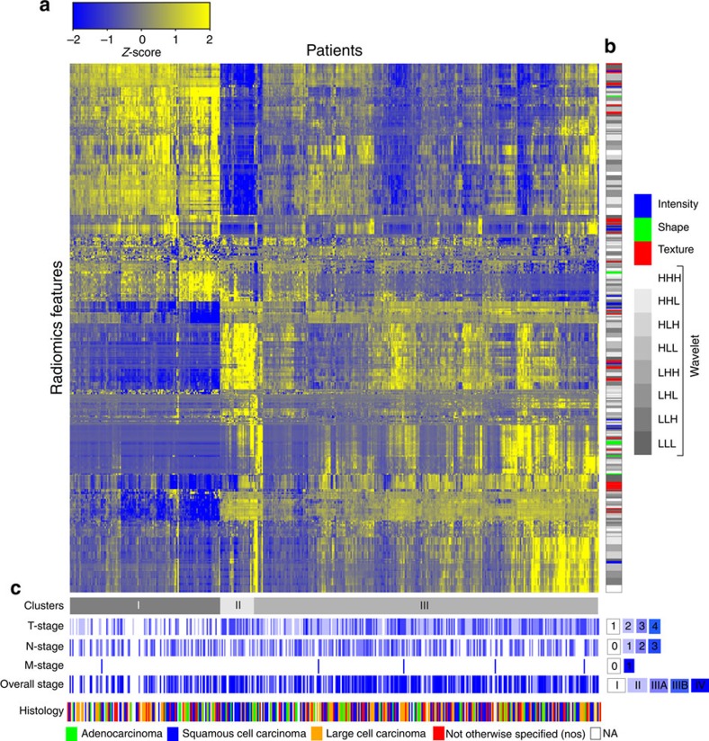
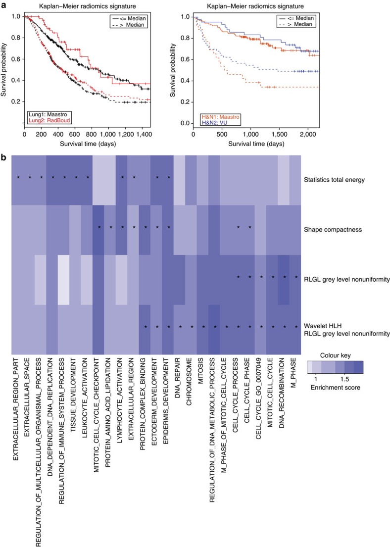
Verification
Claims the paper makes
The claims and statistics of this paper haven’t been checked yet.
Free: it reads the open-access text and checks the first 5 claims that cite a source we can check. Create a free account to check every claim.
Claims other papers make about this one
Each is checked against this paper’s own text: does it say what the citing paper says it does?
The source backs part of the claim, not all of it — or backs it for a different population, condition or comparison.
In the citing paper
“Notably, the signature was comprised of the top features from four classes (size, shape, texture, and wavelets) that were prioritized from a database of 442 features by using test-retest reproducibility and intersubject range.”
From Radiomics: Images Are More than Pictures, They Are Data (2016) · cites this paper as [47]
Experimental — accuracy not yet measuredThe source backs part of the claim, not all of it — or backs it for a different population, condition or comparison.
In the citing paper
“Many studies have shown that imaging features are significantly associated with patterns of gene expression and genetic mutations, demonstrating that radiogenomic analysis can identify different biological mechanisms by means of mathematical and computational devices, enabling the decoding of disease phenotypes by noninvasive methods(48,49).”
From Artificial intelligence, machine learning, computer-aided diagnosis, and radiomics: advances in imaging towards to precision medicine (2019) · cites this paper as [49]
Experimental — accuracy not yet measuredThe cited source says what the claim attributes to it.
In the citing paper
“A study by Aerts et al (47) showed that a radiomic signature could be used to predict outcome in completely independent cohorts of patients with lung cancer from two separate institutions.”
From Radiomics: Images Are More than Pictures, They Are Data (2016) · cites this paper as [47]
The cited source says what the claim attributes to it.
In the citing paper
“Further, this same signature could be applied to cohorts of patients with head and neck cancer with equivalent prognostic power.”
From Radiomics: Images Are More than Pictures, They Are Data (2016) · cites this paper as [47]
In the citing paper
“We used scans from 26 patients from the RIDER dataset19, routinely used for test–retest robustness analysis in tumour imaging19–21.”
From Foundation Model for Cancer Imaging Biomarkers (2024) · cites this paper as [20]
3 more claims about this paper
Create a free account to see every claim other papers make about this one.
Self-check
Percentages, sums and reported differences re-computed from the paper’s own numbers.
The self-check needs the paper’s full text.
Citations
Cites
Subramanian, A., Tamayo, P., Mootha, V. K., Mukherjee, S., Ebert, B. L., Gillette, M. A., Paulovich, A., Pomeroy, S. L., Golub, T. R., Lander, E. S., et al. (2005) · Proceedings of the National Academy of Sciences · doi:10.1073/pnas.0506580102 · Cited by 53,625
Harrell, F. E. (2001) · Springer Series in Statistics · doi:10.1007/978-1-4757-3462-1 · Cited by 9,195
Intratumor Heterogeneity and Branched Evolution Revealed by Multiregion Sequencing
Gerlinger, M., Rowan, A. J., Horswell, S., Larkin, J., Endesfelder, D., Gronroos, E., Martinez, P., Matthews, N., Stewart, A., Tarpey, P., et al. (2012) · New England Journal of Medicine · doi:10.1056/nejmoa1113205 · Cited by 7,774 · cited 2×
“It is known that multiple subclonal populations coexist within tumours, reflecting extensive intratumoral ‘somatic evolution’ 15,16 .”
BackgroundMolecular signatures database (MSigDB) 3.0
Liberzon, A., Subramanian, A., Pinchback, R., Thorvaldsdóttir, H., Tamayo, P., & Mesirov, J. P. (2011) · Bioinformatics · doi:10.1093/bioinformatics/btr260 · Cited by 7,111
Harrell (2015) · Springer Series in Statistics · doi:10.1007/978-3-319-19425-7 · Cited by 6,878
Radiomics: extracting more information from medical images using advanced feature analysis
Lambin, P., Rios-Velazquez, E., Leijenaar, R., Carvalho, S., van Stiphout, R. G. P. M., Granton, P., Zegers, C. M. L., Gillies, R., Boellard, R., Dekker, A., et al. (2012) · European Journal of Cancer · doi:10.1016/j.ejca.2011.11.036 · Cited by 6,084
Self-citationDistant metastasis occurs late during the genetic evolution of pancreatic cancer
Yachida, S., Jones, S., Bozic, I., Antal, T., Leary, R., Fu, B., Kamiyama, M., Hruban, R. H., Eshleman, J. R., Nowak, M. A., et al. (2010) · Nature · doi:10.1038/nature09515 · Cited by 2,502 · cited 1×
“It is known that multiple subclonal populations coexist within tumours, reflecting extensive intratumoral ‘somatic evolution’ 15,16 .”
BackgroundRadiomics: the process and the challenges
Kumar, V., Gu, Y., Basu, S., Berglund, A., Eschrich, S. A., Schabath, M. B., Forster, K., Aerts, H. J. W. L., Dekker, A., Fenstermacher, D., et al. (2012) · Magnetic Resonance Imaging · doi:10.1016/j.mri.2012.06.010 · Cited by 2,258
Self-citationTixier, F., Le Rest, C. C., Hatt, M., Albarghach, N., Pradier, O., Metges, J.-P., Corcos, L., & Visvikis, D. (2011) · Journal of Nuclear Medicine · doi:10.2967/jnumed.110.082404 · Cited by 696
Decoding global gene expression programs in liver cancer by noninvasive imaging
Segal, E., Sirlin, C. B., Ooi, C., Adler, A. S., Gollub, J., Chen, X., Chan, B. K., Matcuk, G. R., Barry, C. T., Chang, H. Y., et al. (2007) · Nature Biotechnology · doi:10.1038/nbt1306 · Cited by 629
Cited by
Items 401–500 of 1,023
Radiomics as a personalized medicine tool in lung cancer: Separating the hope from the hype
Fornacon-Wood, I., Finn, C. F., O’Connor, J. P. B., & Price, G. J. (2020) · Lung Cancer · doi:10.1016/j.lungcan.2020.05.028 · Cited by 117
Liu, J., Sun, D., Chen, L., Zheng, F., Song, W., Guo, D., Ni, T., Liu, C., Lin, F., Xia, Y., et al. (2019) · Frontiers in Oncology · doi:10.3389/fonc.2019.00980 · Cited by 114
Wang, X.-H., Long, L., Cui, Y., Jia, A. Y., Zhu, X., Wang, H., Wang, Z., Zhan, C.-M., Wang, W., & Wang, W. (2020) · British Journal of Cancer · doi:10.1038/s41416-019-0706-0 · Cited by 116
“This research approach utilises high-throughput extraction of feature data from radiographic images,(12) and can potentially develop models to predict lesion phenotypes and prognosis in a non-invasive manner.(13,14) To our knowledge, there are relatively limited radiomics analysis data about prognosis estimation in HC…”
Biology-guided deep learning predicts prognosis and cancer immunotherapy response
Jiang, Y., Zhang, Z., Wang, W., Huang, W., Chen, C., Xi, S., Ahmad, M. U., Ren, Y., Sang, S., Xie, J., et al. (2023) · Nature Communications · doi:10.1038/s41467-023-40890-x · Cited by 132
“Our result is consistent with previous fi ndings that tumor heterogeneity is associated with a worse prognosis 26 .”
Imaging genomics in cancer research: limitations and promises
Bai, H. X., Lee, A. M., Yang, L., Zhang, P., Davatzikos, C., Maris, J. M., & Diskin, S. J. (2016) · The British Journal of Radiology · doi:10.1259/bjr.20151030 · Cited by 113
Genotype prediction of ATRX mutation in lower-grade gliomas using an MRI radiomics signature
Li, Y., Liu, X., Qian, Z., Sun, Z., Xu, K., Wang, K., Fan, X., Zhang, Z., Li, S., Wang, Y., et al. (2018) · European Radiology · doi:10.1007/s00330-017-5267-0 · Cited by 120
Comprehensive Investigation on Controlling for CT Imaging Variabilities in Radiomics Studies
Ger, R. B., Zhou, S., Chi, P.-C. M., Lee, H. J., Layman, R. R., Jones, A. K., Goff, D. L., Fuller, C. D., Howell, R. M., Li, H., et al. (2018) · Scientific Reports · doi:10.1038/s41598-018-31509-z · Cited by 113
Buchlak, Q. D., Esmaili, N., Leveque, J., Bennett, C. C., Farrokhi, F. R., & Piccardi, M. (2021) · Journal of Clinical Neuroscience · doi:10.1016/j.jocn.2021.04.043 · Cited by 123
Demircioğlu, A. (2021) · Insights into Imaging · doi:10.1186/s13244-021-01115-1 · Cited by 132
“In the same spirit, Welch et al. showed that three out of four features of the seminal radiomics model presented by Aerts et al. [1] highly correlated with tumor volume and that tumor volume alone yields the same performance, basically questioning whether radiomics beyond tumor volume has any…”
BackgroundMethodGrossmann, P., Narayan, V., Chang, K., Rahman, R., Abrey, L., Reardon, D. A., Schwartz, L. H., Wen, P. Y., Alexander, B. M., Huang, R., et al. (2017) · Neuro-Oncology · doi:10.1093/neuonc/nox092 · Cited by 112
Self-citationRahmim, A., Huang, P., Shenkov, N., Fotouhi, S., Davoodi‐Bojd, E., Lu, L., Mari, Z. K., Hamid Soltanian-zadeh, A., & Sossi, V. (2017) · NeuroImage Clinical · doi:10.1016/j.nicl.2017.08.021 · Cited by 115
“…a large number of quantitative features from radiological images, aiming to uncover correlates of disease characteristics that are ordinarily not visually observed or quantitatively measured (Aerts et al., 2014; Asselin et al., 2012; Chicklore et al., 2013; Kumar et al., 2012; Lambin et al., 2012).”
Bogowicz, M., Leijenaar, R. T. H., Tanadini‐Lang, S., Riesterer, O., Pruschy, M. N., Studer, G., Unkelbach, J., Gückenberger, M., Konukoğlu, E., & Lambin, P. (2017) · Radiotherapy and Oncology · doi:10.1016/j.radonc.2017.10.023 · Cited by 115
“Most of the radiomics studies have used cross-validation to quantify model performance and so far only one model has been validated in an external and independent dataset [17,18].”
BackgroundSelf-citationTan, X.-Z., Ma, Z., Yan, L., Ye, W., Liu, Z., & Liang, C. (2018) · European Radiology · doi:10.1007/s00330-018-5581-1 · Cited by 122
The complexity of tumor shape, spiculatedness, correlates with tumor radiomic shape features
Limkin, E. J., Reuzé, S., Carré, A., Sun, R., Schernberg, A., Alexis, A., Deutsch, E., Ferté, C., & Robert, C. (2019) · Scientific Reports · doi:10.1038/s41598-019-40437-5 · Cited by 111
Yun, J., Park, J. E., Lee, H., Ham, S., Kim, N., & Kim, H. S. (2019) · Scientific Reports · doi:10.1038/s41598-019-42276-w · Cited by 126
Han, W., Qin, L., Bay, C., Chen, X., Yu, K.-H., Miskin, N., Li, A., Xu, X., & Young, G. (2020) · American Journal of Neuroradiology · doi:10.3174/ajnr.a6365 · Cited by 112
Liu, A., Wang, Z., Yang, Y., Wang, J., Dai, X., Wang, L., Lu, Y., & Xue, F. (2020) · 癌症:英文版 · doi:10.1002/cac2.12002 · Cited by 124
“Previously, a large number of studies have confirmed that radiomics is effective for the diagnosis and prognosis of lung cancer and pulmonary nodules [12, 23, 24].”
BackgroundMethodZhou, H., Jin, Y., Dai, L., Zhang, M., Qiu, Y., wang, K., Tian, J., & Zheng, J. (2020) · European Journal of Radiology · doi:10.1016/j.ejrad.2020.108992 · Cited by 112
Machine Learning in Oncology: Methods, Applications, and Challenges
Bertsimas, D., & Wiberg, H. (2020) · JCO Clinical Cancer Informatics · doi:10.1200/cci.20.00072 · Cited by 137
“characteristics.(35) More recently, deep learning has been applied to directly leverage raw imaging data.”
BackgroundArtificial Intelligence in CT and MR Imaging for Oncological Applications
Paudyal, R., Shah, A. D., Akın, O., Gian, R. K., Konar, A. S., Hatzoglou, V., Mahmood, U. A., Lee, N. Y., Wong, R. J., Banerjee, S., et al. (2023) · Cancers · doi:10.3390/cancers15092573 · Cited by 131
“Radiomics, or the non-invasive extraction of quantitative information from images, is well developed in oncology, with several groups demonstrating its utility for both cancer diagnosis and treatment response prediction of multiple solid cancers [62].”
BackgroundRadiomics in PET/CT: More Than Meets the Eye?
Hatt, M., Tixier, F., Visvikis, D., & Rest, C. C. L. (2016) · Journal of Nuclear Medicine · doi:10.2967/jnumed.116.184655 · Cited by 113
“Within this context, a rigorous process of model development (proper training) and validation (independent large cohorts) is needed, which is still far from being a standard, although some encouraging results have been published (17,18).”
BackgroundMRI features predict p53 status in lower-grade gliomas via a machine-learning approach
Li, Y., Qian, Z., Xu, K., Wang, K., Fan, X., Li, S., Jiang, T., Liu, X., & Wang, Y. (2017) · NeuroImage Clinical · doi:10.1016/j.nicl.2017.10.030 · Cited by 117
“However, many studies have already demonstrated the robustness of radiomics feature extraction in terms of repeatability and reproducibility in test/re-test settings (Aerts et al., 2014; Fried et al., 2014; Parmar et al., 2015).”
InfluentialRadiomics in stratification of pancreatic cystic lesions: Machine learning in action
Dalal, V., Carmicheal, J., Dhaliwal, A., Jain, M., Kaur, S., & Batra, S. K. (2019) · Cancer Letters · doi:10.1016/j.canlet.2019.10.023 · Cited by 113
“This premise is supported by the fact that lesions exhibit measurable differences in shape, texture, and grayness in the radiographic images captured [59].”
BackgroundWang, S., Dong, L.-Y., Wang, X., & Wang, X. (2020) · Open Medicine · doi:10.1515/med-2020-0028 · Cited by 132
Coy, H., Hsieh, K., Wu, W., Nagarajan, M. B., Young, J. R., Douek, M. L., Brown, M. S., Scalzo, F., & Raman, S. S. (2019) · Abdominal Radiology · doi:10.1007/s00261-019-01929-0 · Cited by 110
Imaging Phenotyping Using Radiomics to Predict Micropapillary Pattern within Lung Adenocarcinoma
Song, S. H., Park, H., Lee, G., Lee, H. Y., Sohn, I., Kim, H. S., Lee, S. H., Jeong, J. Y., Kim, J., Lee, K. S., et al. (2016) · Journal of Thoracic Oncology · doi:10.1016/j.jtho.2016.11.2230 · Cited by 120
“We selected statistically significant features from demographic and pathologic factors and selected the radiomics variable with the lowest p value from each radiomics category to remove redundancy within the radiomics information, as in Aerts et al.12 For two groups, a multivariate logistic regression model with the u…”
MethodEffect of tube current on computed tomography radiomic features
Mackin, D., Ger, R., Dodge, C., Fave, X., Chi, P.-C., Zhang, L., Yang, J., Bache, S., Dodge, C., Jones, A. K., et al. (2018) · Scientific Reports · doi:10.1038/s41598-018-20713-6 · Cited by 110
Exploration of PET and MRI radiomic features for decoding breast cancer phenotypes and prognosis
Huang, S.-Y., Franc, B. L., Harnish, R. J., Liu, G., Mitra, D., Copeland, T. P., Arasu, V. A., Kornak, J., Jones, E. F., Behr, S. C., et al. (2018) · npj Breast Cancer · doi:10.1038/s41523-018-0078-2 · Cited by 116
Li, Z., Bai, H., Sun, Q., Zhao, Y., Lv, Y., Zhou, J. H., Liang, C., Chen, Y., Liang, D., & Zheng, H. (2018) · Cancer Medicine · doi:10.1002/cam4.1863 · Cited by 115
Sun, C., Tian, X., Liu, Z., Li, W., Li, P., Chen, J., Zhang, W., Fang, Z., Du, P., Duan, H., et al. (2019) · EBioMedicine · doi:10.1016/j.ebiom.2019.07.049 · Cited by 111
Radiomics in Breast Imaging from Techniques to Clinical Applications: A Review
Lee, S.-H., Park, H., & Ko, E. S. (2020) · Korean Journal of Radiology · doi:10.3348/kjr.2019.0855 · Cited by 115
“Current radiomics features are “hand-crafted” features, which are formed by expert opinions (9).”
BackgroundMethodInfluentialHassan, S. U., Abdulkadir, S. J., Zahid, M. S. M., & Al-Selwi, S. M. (2025) · Computers in Biology and Medicine · doi:10.1016/j.compbiomed.2024.109569 · Cited by 108
Desseroit, M.-C., Visvikis, D., Tixier, F., Majdoub, M., Perdrisot, R., Guillevin, R., Rest, C. C. L., & Hatt, M. (2016) · European Journal of Nuclear Medicine and Molecular Imaging · doi:10.1007/s00259-016-3325-5 · Cited by 116
Machine Learning in Radiation Oncology: Opportunities, Requirements, and Needs
Feng, M. U.-S., Valdés, G., Dixit, N., & Solberg, T. D. (2018) · Frontiers in Oncology · doi:10.3389/fonc.2018.00110 · Cited by 124
“The limited reproducibility of imaging systems both within and across institutions remains a significant challenge for radiomics (65, 66).”
BackgroundNing, Z., Luo, J., Li, Y., Han, S., Feng, Q., Xu, Y., Chen, W., Chen, T., & Zhang, Y. (2019) · IEEE Journal of Biomedical and Health Informatics · doi:10.1109/jbhi.2018.2841992 · Cited by 109
“Many studies proved the applicability of radiomics in risk prediction of tumors, such as soft-tissue sarcomas [14], non-small-cell lung cancer [15], and head-and-neck cancer [16].”
A New Challenge for Radiologists: Radiomics in Breast Cancer
Crivelli, P., Ledda, R. E., Parascandolo, N., Fara, A., Soro, D., & Conti, M. (2018) · BioMed Research International · doi:10.1155/2018/6120703 · Cited by 114
Lang, N., Zhang, Y., Zhang, E., Zhang, J., Chow, D. S., Chang, P., Yu, H. J., Yuan, H., & Su, M. (2019) · Magnetic Resonance Imaging · doi:10.1016/j.mri.2019.02.013 · Cited by 111
Morin, O., Chen, W., Nassiri, F., Susko, M. S., Magill, S. T., Vasudevan, H. N., Wu, A., Vallières, M., Gennatas, E. D., Valdés, G., et al. (2019) · Neuro-Oncology Advances · doi:10.1093/noajnl/vdz011 · Cited by 112
Li, Y., Zhang, Y., Fang, Q., Zhang, X., Hou, P., Wu, H., & Wang, X. (2021) · European Journal of Nuclear Medicine and Molecular Imaging · doi:10.1007/s00259-020-05119-9 · Cited by 120
Giganti, F., Antunes, S., Salerno, A., Ambrosi, A., Marra, P., Nicoletti, R., Orsenigo, E., Chiari, D., Albarello, L., Staudacher, C., et al. (2016) · European Radiology · doi:10.1007/s00330-016-4540-y · Cited by 113
“We used textural features in line with literature [26], ensuring the reproducibility of our study by other centres.”
MethodResultZhang, Q., Xiao, Y., Suo, J., Shi, J., Yu, J., Guo, Y., Wang, Y., & Zheng, H. (2017) · Ultrasound in Medicine & Biology · doi:10.1016/j.ultrasmedbio.2016.12.016 · Cited by 110
Zinn, P. O., Singh, S. K., Kotrotsou, A., Hassan, I., Thomas, G., Luedi, M. M., Elakkad, A., Elshafeey, N., Idris, T., Mosley, J., et al. (2018) · Clinical Cancer Research · doi:10.1158/1078-0432.ccr-17-3420 · Cited by 108
“6 captures microscale information hidden within conventional imaging and beyond what is visible to the naked human eye [5-7].”
BackgroundFord, J. C., Dogan, N., Young, L. A., & Yang, F. (2018) · Contrast Media & Molecular Imaging · doi:10.1155/2018/1729071 · Cited by 111
Niu, J., Zhang, S., Ma, S., Diao, J., Zhou, W., Tian, J., Zang, Y., & Jia, W. (2018) · European Radiology · doi:10.1007/s00330-018-5725-3 · Cited by 108
Li, P., Wang, X., Xu, C., Liu, C., Zheng, C., FULHAM, M. J., Feng, D. D., Wang, L., Song, S., & Huang, G. (2020) · European Journal of Nuclear Medicine and Molecular Imaging · doi:10.1007/s00259-020-04684-3 · Cited by 108
Ligero, M., Garcia-Ruiz, A., Viaplana, C., Villacampa, G., Raciti, M. V., Landa, J., Matos, I., Martin-Liberal, J., Ochoa-de-Olza, M., Hierro, C., et al. (2021) · Radiology · doi:10.1148/radiol.2021200928 · Cited by 107
Shen, C., Liu, Z., Wang, Z., Guo, J., Zhang, H., Wang, Y., Qin, J., Li, H., Fang, M., Tang, Z., et al. (2018) · Translational Oncology · doi:10.1016/j.tranon.2018.04.005 · Cited by 108
Elhalawani, H. M., Kanwar, A., Mohamed, A., White, A. L., Zafereo, J., Wong, A. J., Berends, J. E., Abohashem, S., Williams, B., Aymard, J. M., et al. (2018) · Scientific Reports · doi:10.1038/s41598-017-14687-0 · Cited by 117
Li, Z., Li, H., Wang, S., Dong, D., Yin, F., Chen, A., Wang, S., Zhao, G., Wang, S., Tian, J., et al. (2018) · Journal of Magnetic Resonance Imaging · doi:10.1002/jmri.26531 · Cited by 107
Fan, M., Xia, P., Clarke, R. C., Wang, Y., & Li, L. (2020) · Nature Communications · doi:10.1038/s41467-020-18703-2 · Cited by 116
Kirienko, M., Sollini, M., Corbetta, M., Voulaz, E., Gozzi, N., Interlenghi, M., Gallivanone, F., Castiglioni, I., Asselta, R., Duga, S., et al. (2021) · European Journal of Nuclear Medicine and Molecular Imaging · doi:10.1007/s00259-021-05371-7 · Cited by 125
Wang, Y., Lang, J., Zuo, J. Z., Dong, Y., Hu, Z., Xu, X., Zhang, Y., Wang, Q., Yang, L., Wong, S. T. C., et al. (2022) · European Radiology · doi:10.1007/s00330-022-08887-0 · Cited by 117
“Radiomics could provide a better understanding of cancer pathophysiology for clinical decision-making by using a large number of quantitative image features to characterize the tumor heterogeneity [18].”
BackgroundRadiomics analysis to predict pulmonary nodule malignancy using machine learning approaches
Warkentin, M. T., Al‐Sawaihey, H., Lam, S. C., Liu, G., Diergaarde, B. B., Yuan, J., Wilson, D. O., Atkar-Khattra, S., Grant, B., Brhane, Y., et al. (2024) · Thorax · doi:10.1136/thorax-2023-220226 · Cited by 114
Skripcak, T., Belka, C., Bosch, W. R., Brink, C., Brunner, T. B., Budach, V., Büttner, D., Debus, J., Dekker, A. L. A. J., Grau, C., et al. (2014) · Radiotherapy and Oncology · doi:10.1016/j.radonc.2014.10.001 · Cited by 112
“Resulting data pools represent the primary input for generation of medical knowledge bases with a broad range of applications, including predictive models for decision support systems based on clinical data [2] and discovery of prognostic features in radiomics [3].”
BackgroundSelf-citationStoyanova, R., Pollack, A., Takhar, M., Lynne, C., Parra, N., Lam, L. L. C., Alshalalfa, M., Buerki, C., Castillo, R., Jorda, M., et al. (2016) · Oncotarget · doi:10.18632/oncotarget.10523 · Cited by 106
“We linked radiomic features to existing genomic and clinical information to improve risk stratification, as has been previously described by us [10] and others for different tumor types [11].”
MethodSelf-citationPapp, L., Pötsch, N., Grahovac, M., Schmidbauer, V. U., Wöehrer, A., Preusser, M., Mitterhauser, M., Kiesel, B., Wadsak, W., Beyer, T., et al. (2017) · Journal of Nuclear Medicine · doi:10.2967/jnumed.117.202267 · Cited by 111
“Here, filtering during preprocessing (24,25) or machine learning (ML)–driven approaches to feature selection (26,27) can be incorporated to reduce the number of features for predictive analyses.”
BackgroundTong, H., Sun, J., Fang, J., Zhang, M., Liu, H., Xia, R., Zhou, W., Liu, K., & Chen, X. (2022) · Frontiers in Immunology · doi:10.3389/fimmu.2022.859323 · Cited by 126
“Compared with conventional imaging methods, radiomics can provide a more detailed characterization of tumor heterogeneity beyond the human eye, which re fl ects either macroscopic or cellular and molecular properties of tumor tissues (17 – 20).”
MethodHuynh, E., Coroller, T. P., Narayan, V., Agrawal, V., Romano, J., Franco, I. “., Parmar, C., Hou, Y., Mak, R. H., & Aerts, H. J. W. L. (2017) · PLoS ONE · doi:10.1371/journal.pone.0169172 · Cited by 113
Self-citationPérez-Beteta, J., Molina-García, D., Ortiz-Alhambra, J. A., Fernández-Romero, A., Luque, B., Arregui, E., Calvo, M., Moreno, J. M. B., Meléndez, B., de Lope, Á. R., et al. (2018) · Radiology · doi:10.1148/radiol.2018171051 · Cited by 116
Dinapoli, N., Barbaro, B., Gatta, R., Chiloiro, G., Casà, C., Masciocchi, C., Damiani, A., Boldrini, L., Gambacorta, M. A., Dezio, M., et al. (2018) · International Journal of Radiation Oncology*Biology*Physics · doi:10.1016/j.ijrobp.2018.04.065 · Cited by 110
“Nowadays radiological images can represent a source of data that can be analyzed by using automated computer-based techniques, working on numerical informations coded within the Digital Imaging and Communications in Medicine (DICOM) files (1): this images numerical analysis has been named “radiomics” (2).”
MethodInfluentialSelf-citationXu, X., Zhang, X., Tian, Q., Wang, H. J., Cui, L., Li, S., Tang, X., Baojuan Li, A., Dolz, J., Ayed, I. B., et al. (2018) · Journal of Magnetic Resonance Imaging · doi:10.1002/jmri.26327 · Cited by 104
European Society of Radiology, deSouza, N. M., Achten, E., Alberich-Bayarri, A., Bamberg, F., Boellaard, R., Clément, O., Fournier, L., Gallagher, F., Golay, X., et al. (2019) · Insights into Imaging · doi:10.1186/s13244-019-0764-0 · Cited by 105
“tissue or disease [129, 130]: it depends on the type of imaging data (CT, MRI, PET) and is influenced by image”
BackgroundApplication of radiomics and machine learning in head and neck cancers
Peng, Z., Wang, Y., Wang, Y., Jiang, S., Fan, R., Zhang, H., & Jiang, W. (2021) · International Journal of Biological Sciences · doi:10.7150/ijbs.55716 · Cited by 121
“Automatic image separation does not require human involvement, avoids heterogeneity between and within evaluators, and results are more repeatable, faster, and more suitable for large imaging datasets [34,40].”
BackgroundLu, L., Dercle, L., Zhao, B., & Schwartz, L. H. (2021) · Nature Communications · doi:10.1038/s41467-021-26990-6 · Cited by 104
“These medical image analysis algorithms, also known as radio-mics approaches 12 , allow high-throughput conversion of medical images into mineable quantitative data, which make it feasible to assess tumor morphological changes quantitatively.”
BackgroundEntropy-based consensus clustering for patient stratification
Liu, H., Zhao, R., Fang, H., Cheng, F., Fu, Y., & Liu, Y. (2017) · Bioinformatics · doi:10.1093/bioinformatics/btx167 · Cited by 106
Bickelhaupt, S., Jaeger, P. F., Laun, F. B., Lederer, W., Daniel, H., Kuder, T. A., Wuesthof, L., Paech, D., Bonekamp, D., Radbruch, A., et al. (2018) · Radiology · doi:10.1148/radiol.2017170273 · Cited by 103
An MRI-based Radiomics Classifier for Preoperative Prediction of Ki-67 Status in Breast Cancer
Liang, C., Cheng, Z., Huang, Y., He, L., Chen, X., Ma, Z., Huang, X., Liang, C., & Liu, Z. (2018) · Academic Radiology · doi:10.1016/j.acra.2018.01.006 · Cited by 104
Pfaehler, E. A. G., Zhovannik, I., Wei, L., Boellaard, R., Dekker, A. L. A. J., Monshouwer, R., Naqa, I. E., Bussink, J., Gillies, R. J., Wee, L., et al. (2021) · Physics and Imaging in Radiation Oncology · doi:10.1016/j.phro.2021.10.007 · Cited by 114
Self-citationPulmonary ground-glass opacity: computed tomography features, histopathology and molecular pathology
written on behalf of the AME Lung Cancer Collaborative Group, Gao, J.-W., Rizzo, S., Ma, L.-H., Qiu, X.-Y., Warth, A., Seki, N., Hasegawa, M., Zou, J.-W., Li, Q., et al. (2017) · Translational Lung Cancer Research · doi:10.21037/tlcr.2017.01.02 · Cited by 109
“These imaging features capture distinct phenotypic differences of tumours and may have prognostic power and thus clinical significance across different diseases (57).”
BackgroundYi, X., Pei, Q., Zhang, Y., Zhu, H., Wang, Z., Chen, C., Li, Q., Long, X., Tan, F., Zhou, Z., et al. (2019) · Frontiers in Oncology · doi:10.3389/fonc.2019.00552 · Cited by 103
Zhong, Y., She, Y., Deng, J., Chen, S., Wang, T., Yang, M., Ma, M., Song, Y., Qi, H., Wang, Y., et al. (2021) · Radiology · doi:10.1148/radiol.2021210902 · Cited by 110
Sujit, S. J., Aminu, M., Karpinets, T. V., Chen, P., Saad, M. B., Salehjahromi, M., Boom, J. D., Qayati, M., George, J. M., Allen, H., et al. (2024) · Nature Communications · doi:10.1038/s41467-024-47512-0 · Cited by 125
“Tumor volume stands out as a pivotal predictor in lung cancer prognosis, exerting a substantial in fl uence on disease recurrence and overall survival 52 – 54 .”
Liu, Y., Zhang, Y., Cheng, R.-F., Liu, S., Qu, F., Yin, X., Wang, Q., Xiao, B., & Ye, Z. (2018) · Journal of Magnetic Resonance Imaging · doi:10.1002/jmri.26192 · Cited by 102
“By converting medical images into high-dimensional, mineable, and quantitative imaging features via high-throughput extraction of data characterization algorithms, radiomics analysis of a large imaging dataset provides an unprecedented opportunity to improve decision support in oncology at low cost and noninvasively.(…”
BackgroundArita, H., Kinoshita, M., Kawaguchi, A., Takahashi, M., Narita, Y., Terakawa, Y., Tsuyuguchi, N., Okita, Y., Nonaka, M., Moriuchi, S., et al. (2018) · Scientific Reports · doi:10.1038/s41598-018-30273-4 · Cited by 111
Su, C., Jiang, J., Zhang, S., Shi, J., Xu, K., Shen, N., Zhang, J., Li, L., Zhao, L., Zhang, J., et al. (2018) · European Radiology · doi:10.1007/s00330-018-5704-8 · Cited by 108
Mahon, R. N., Ghita, M., Hugo, G. D., & Weiss, E. (2019) · Physics in Medicine and Biology · doi:10.1088/1361-6560/ab6177 · Cited by 114
“(Aerts et al 2014, Fave et al 2017, Parekh and Jacobs 2017) Radiomics has been applied to various clinical endpoints and anatomical sites with varying levels of success.”
BackgroundQuantification of tumor heterogeneity: from data acquisition to metric generation
Kashyap, A., Rapsomaniki, M. A., Barros, V., Fomitcheva-Khartchenko, A., Martinelli, A. L., Rodriguez, A. F., Gabrani, M., Rosen-Zvi, M., & Kaigala, G. (2022) · Trends in Biotechnology · doi:10.1016/j.tibtech.2021.11.006 · Cited by 98
“A second issue of Esophageal cancer [141] Colorectal cancer [142] Pancreatic cancer [143] Gastric cancer [144] Lung and head-and-neck cancer [150] Clear cell renal cell carcinoma [152] PET Cervix and head and neck cancer [157] Esophageal carcinoma [158] Lung cancer [159] MRI Breast cancer [164 –…”
BackgroundLi, J., Qiu, Z., Zhang, C., Chen, S., Wang, M.-M., Meng, Q., Lu, H., Wei, L., Lv, H., Zhong, W., et al. (2022) · European Radiology · doi:10.1007/s00330-022-09055-0 · Cited by 115
“Clinical characteristics of patient cohorts Patients in LUNG1, R01, RIDER, GDPH-1, and GDPH-2 cohorts were diagnosed with NSCLC, and those in the HN1 cohort were diagnosed with HNC. Specifically, the two GDPH cohorts only contain patients with early-stage lung adenocarcinoma (LUAD) with corresponding pathological clas…”
MethodInfluentialLee, S. E., Han, K., Kwak, J. Y., Lee, E., & Kim, E. (2018) · Scientific Reports · doi:10.1038/s41598-018-31906-4 · Cited by 103
Handcrafted versus deep learning radiomics for prediction of cancer therapy response
Hosny, A., Aerts, H. J. W. L., & Mak, R. H. (2019) · The Lancet Digital Health · doi:10.1016/s2589-7500(19)30062-7 · Cited by 108
Self-citationMukherjee, P., Zhou, M., Lee, E., Schicht, A., Balagurunathan, Y., Napel, S., Gillies, R., Wong, S., Thieme, A., Leung, A., et al. (2020) · Nature Machine Intelligence · doi:10.1038/s42256-020-0173-6 · Cited by 100
Self-citationSparse Representation-Based Radiomics for the Diagnosis of Brain Tumors
Wu, G., Chen, Y., Wang, Y., Yu, J., Lv, X., Ju, X., Shi, Z., Chen, L., & Chen, Z. (2017) · IEEE Transactions on Medical Imaging · doi:10.1109/tmi.2017.2776967 · Cited by 100
“ods, such as p-value comparison [2]–[4] and correlation”
BackgroundMethodInfluentialMetabolic Tumor Volume Metrics in Lymphoma
Kostakoğlu, L., & Chauvie, S. (2017) · Seminars in Nuclear Medicine · doi:10.1053/j.semnuclmed.2017.09.005 · Cited by 108
Qiu, W., Kuang, H., Nair, J. R., Assis, Z., Najm, M., McDougall, C. C., McDougall, B., Chung, K. J., Wilson, A. T., Goyal, M., et al. (2018) · American Journal of Neuroradiology · doi:10.3174/ajnr.a5918 · Cited by 100
“that describe thrombus characteristics were extracted in an automated manner from the above images.(7,13) These features can be”
MethodA PET Radiomics Model to Predict Refractory Mediastinal Hodgkin Lymphoma
Milgrom, S. A., Elhalawani, H. M., Lee, J., Wang, Q., Mohamed, A., Dabaja, B. S., Pinnix, C. C., Gunther, J. R., Court, L. E., Rao, A., et al. (2019) · Scientific Reports · doi:10.1038/s41598-018-37197-z · Cited by 103
Oikonomou, E. K., Siddique, M. M., & Antoniades, C. A. (2020) · Cardiovascular Research · doi:10.1093/cvr/cvaa021 · Cited by 116
“of lung tumours was found to be superior to traditional tumour, node, and metastasis staging in predicting future mortality,(28,31) and also associated with discrete transcriptional changes in tumour biology.(31) 4.”
BackgroundRadiomics feature robustness as measured using an MRI phantom
Lee, J., Steinmann, A., Ding, Y., Lee, H. J., Owens, C. A., Wang, J., Yang, J., Followill, D. S., Ger, R. B., Mackin, D., et al. (2021) · Scientific Reports · doi:10.1038/s41598-021-83593-3 · Cited by 100
Development and clinical application of radiomics in lung cancer
Chen, B., Zhang, R., Gan, Y., Yang, L., & Li, W. (2017) · Radiation Oncology · doi:10.1186/s13014-017-0885-x · Cited by 105
“Wavelets expressed as ‘Grey Level Nonuniformity HLH’ have been used to describe the intratumoral heterogeneity of lung cancer [32].”
Jethanandani, A., Lin, T. A., Volpe, S., Elhalawani, H. M., Mohamed, A., Yang, P., & Fuller, C. D. (2018) · Frontiers in Oncology · doi:10.3389/fonc.2018.00131 · Cited by 107
“Iranian Journal of Radiology (2017) (5):e48035.”
BackgroundInfluentialda Nóbrega, R. V. M., Filho, P. P. R., Rodrigues, M. B., da Silva, S. P. P., Dourado, C. M. J. D. M., & de Albuquerque, V. H. C. (2018) · Neural Computing and Applications · doi:10.1007/s00521-018-3895-1 · Cited by 100
RetractedTargeting acidity in cancer and diabetes
Gillies, R. J., Pilot, C., Marunaka, Y., & Fais, S. (2019) · Biochimica et Biophysica Acta (BBA) - Reviews on Cancer · doi:10.1016/j.bbcan.2019.01.003 · Cited by 108
Self-citationLung Lesion Extraction Using a Toboggan Based Growing Automatic Segmentation Approach
Song, J., Yang, C., Fan, L., Wang, K., Yang, F., Liu, S., & Tian, J. (2015) · IEEE Transactions on Medical Imaging · doi:10.1109/tmi.2015.2474119 · Cited by 99
“With the hypothesis that deep analysis of radiographic images can inform and quantify the microenvironment and the extent of intra-tumoral heterogeneity for personalized medicine [3], [4], analysis of large numbers of image features extracted from computed tomography (CT) with high throughput can capture spatial and t…”
BackgroundMethodRadiomics in radiooncology – challenging the medical physicist
Peeken, J. C., Bernhofer, M., Wiestler, B., Goldberg, T., Cremers, D., Rost, B., Wilkens, J. J., Combs, S. E., & Nüsslin, F. (2018) · Physica Medica · doi:10.1016/j.ejmp.2018.03.012 · Cited by 98
Ou, D., Blanchard, P., Rosellini, S., Lévy, A., Nguyen, F., Leijenaar, R. T. H., Garberis, I., Gorphe, P., Bidault, F., Ferté, C., et al. (2017) · Oral Oncology · doi:10.1016/j.oraloncology.2017.06.015 · Cited by 99
“It is also reported recently that radiomics is able to identify different gene expressions [6,24], and the correlation of radiomics with other potential biomarkers such as PD-1/PD-L1 is not evaluated in our current analysis.”
BackgroundMethodResultSelf-citationGranzier, R. W. Y., van Nijnatten, T. J. A., Woodruff, H. C., Smidt, M. L., & Lobbes, M. B. I. (2019) · European Journal of Radiology · doi:10.1016/j.ejrad.2019.108736 · Cited by 103
Global landscape of SARS-CoV-2 mutations and conserved regions
Abbasian, M. H., Mahmanzar, M., Rahimian, K., Mahdavi, B., Tokhanbigli, S., Moradi, B., Sisakht, M. M., & Deng, Y. (2023) · Journal of Translational Medicine · doi:10.1186/s12967-023-03996-w · Cited by 107
“The region corresponding to the C-terminal domain of SARS-CoV NSP3 was found to be significantly less mutated likely due to its vital role in inducing the formation of [2] double-membrane vesicles [6].”
BackgroundRadiomics for Response and Outcome Assessment for Non-Small Cell Lung Cancer
Shi, L., He, Y., Yuan, Z., Benedict, S., Valicenti, R. K., Qiu, J., & Rong, Y. (2018) · Technology in Cancer Research & Treatment · doi:10.1177/1533033818782788 · Cited by 103
“Overall, in survival studies based on CT images, those radiomics features quantifying shapes, intensity, and texture, conventional features (such as Eastern Cooperative Oncology Group performance status, pleural retraction, and diameter), and delta-radiomics features are predictive, and when they are combined with cli…”
BackgroundMethodInfluentialvan Dijk, L. V., Thor, M., Steenbakkers, R. J. H. M., Apte, A., Zhai, T., Borra, R., Noordzij, W., Estilo, C. L., Lee, N. Y., Langendijk, J. A., et al. (2018) · Radiotherapy and Oncology · doi:10.1016/j.radonc.2018.06.012 · Cited by 94
Robustness of radiomic features in magnetic resonance imaging: review and a phantom study
Cattell, R. F., Chen, S., & Huang, C. (2019) · Visual Computing for Industry Biomedicine and Art · doi:10.1186/s42492-019-0025-6 · Cited by 107
Kurç, T., Bakas, S., Ren, X., Bagari, A., Momeni, A., Huang, Y., Zhang, L., Kumar, A., Thibault, M., Qi, Q., et al. (2020) · Frontiers in Neuroscience · doi:10.3389/fnins.2020.00027 · Cited by 103
“Radiomics deals with the extraction, analysis, and interpretation of large sets of visual and sub-visual image features for organ-level quantification and classification of tumors (Lambin et al., 2012; Gillies, 2013; Aerts et al., 2014; Parmar et al., 2015; Gillies et al., 2016; Zwanenburg et al., 2016).”
Method
Metadata
- Authors
- Aerts, H. J. W. L., Velazquez, E. R., Leijenaar, R. T. H., Parmar, C., Grossmann, P., Carvalho, S., Bussink, J., Monshouwer, R., Haibe-Kains, B., Rietveld, D., Hoebers, F., Rietbergen, M. M., Leemans, C. R., Dekker, A., Quackenbush, J., Gillies, R. J., & Lambin, P.
- Affiliations
- Brigham and Women's Hospital (US)
- Maastricht University (NL)
- Radboud University Medical Center (NL)
- Amsterdam UMC Location Vrije Universiteit Amsterdam (NL)
- Dana-Farber Cancer Institute (US)
- Moffitt Cancer Center (US)
- Corresponding author
- Aerts, Hugo J. W. L.
- ORCID
- Venue
- Nature Communications
- Published
- 2014-06-03
- Volume
- 5
- Issue
- 1
- Pages
- 4006-4006
- ISSN
- 2041-1723 (ISSN-L)
- PMC
- PMC4059926
- OpenAlex
- W2103004421
- Semantic Scholar
- 3b002068646831a6f59fabb6daeb0209ed54049d
- Cited by
- 5,132
- References
- 29
- Citations by year
- 2026: 415 · 2025: 460 · 2024: 458 · 2023: 510 · 2022: 567 · 2021: 638 · 2020: 587 · 2019: 552 · 2018: 388 · 2017: 283 · 2016: 175 · 2015: 82 · 2014: 11
- FWCI
- 142.84
- Type
- journal article
- Language
- en
- Update notices
- correction (2014-08-07)
- Open access
- hybrid
- doi.org
- Licence
- Locations
- Nature Communications (publishedVersion, cc-by-nc-nd)
- PubMed Central (publishedVersion, cc-by-nc-nd)
- PubMed (publishedVersion)
- Pure Amsterdam UMC (publishedVersion)
- Amsterdam UMC (VU Amsterdam) - Institutional Repository (publishedVersion)
- Radboud Repository (Radboud University) (submittedVersion)
- Digital Access to Scholarship at Harvard (DASH) (Harvard University) (submittedVersion, cc-by)
- RePEc: Research Papers in Economics (submittedVersion)
- Research Publications (Maastricht University) (submittedVersion, cc-by-nc-nd)
- Radboud Repository (Radboud University) (submittedVersion, other-oa)
- Research Publications (Maastricht University) (submittedVersion, other-oa)
- Data Archiving and Networked Services (DANS) (submittedVersion)
- Funders
- European Federation of Pharmaceutical Industries and Associations
- KWF Kankerbestrijding (UM 2009-4454, 2011-5020, KWF UM 2009-4454, KWF UM 2011-5020, 2009-4454)
- Center for Translational Molecular Medicine (grant 030-103, 030-103)
- Innovative Medicines Initiative (115151)
- National Institutes of Health (U01 CA)
- National Cancer Institute (NIH-USA U01CA 143062-01, U01CA143062)
- Interreg
- Topics
- Health Sciences › Medicine › Radiology, Nuclear Medicine and Imaging › Radiomics and Machine Learning in Medical Imaging
- Physical Sciences › Computer Science › Computer Vision and Pattern Recognition › Image Retrieval and Classification Techniques
- Health Sciences › Medicine › Radiology, Nuclear Medicine and Imaging › Medical Imaging Techniques and Applications
- Keywords
- radiomics, tumor phenotype, lung cancer, head and neck cancer, radiogenomics, intratumoral heterogeneity, computed tomography, image texture features, gene expression, non-invasive imaging, treatment decision support
- MeSH terms
- Adenocarcinoma, Carcinoma, Non-Small-Cell Lung, Carcinoma, Squamous Cell, Female, Head and Neck Neoplasms, Humans, Lung Neoplasms, Male, Phenotype, Prognosis, Tomography, X-Ray Computed, Tumor Burden, Positron-Emission Tomography, Multimodal Imaging
About these checks
Registry, retraction, Cites and statistics results are computed, not judged, with one exception: a printed reference that no index lists is read once by a model before it counts as a citation error. Claims are extracted by a model and kept only when their quote appears verbatim in the text; each cited source is then checked, with a model’s help, for whether it says what the claim relies on. Those verdicts can be wrong — read the claim’s quote and the cited source before relying on one.
Statistics and arithmetic are recomputed, not judged.
Confidence bands are shown only for fields calibrated on our annotated test set. None is calibrated yet, so no band is shown.
What counts as a claim ›How we measure confidence ›How we check this ›
Are you an author, or do you see a problem with this page? Request a review
Data sources
Bibliographic data: Semantic Scholar, OpenAlex, Crossref.
Contains information from the Semantic Scholar Academic Graph Datasets, made available under the ODC Attribution License (ODC-BY 1.0).
Semantic Scholar’s data is described in: Kinney, R., et al. (2023). The Semantic Scholar Open Data Platform. arXiv:2301.10140.
