

Decoding tumour phenotype by noninvasive imaging using a quantitative radiomics approach
Open accessThe DOI matches its Crossref record; 29 of the 29 references it cites were found in a registry or an index.
Abstract
Human cancers exhibit strong phenotypic differences that can be visualized noninvasively by medical imaging. Radiomics refers to the comprehensive quantification of tumour phenotypes by applying a large number of quantitative image features. Here we present a radiomic analysis of 440 features quantifying tumour image intensity, shape and texture, which are extracted from computed tomography data of 1,019 patients with lung or head-and-neck cancer. We find that a large number of radiomic features have prognostic power in independent data sets of lung and head-and-neck cancer patients, many of which were not identified as significant before. Radiogenomics analysis reveals that a prognostic radiomic signature, capturing intratumour heterogeneity, is associated with underlying gene-expression patterns. These data suggest that radiomics identifies a general prognostic phenotype existing in both lung and head-and-neck cancer. This may have a clinical impact as imaging is routinely used in clinical practice, providing an unprecedented opportunity to improve decision-support in cancer treatment at low cost.
Overview
The claims and statistics of this paper haven’t been checked yet.
Free: it reads the open-access text and checks the first 5 claims that cite a source we can check. Create a free account to check every claim.
Integrity signals
Registry
Cites
Statistics
Figures


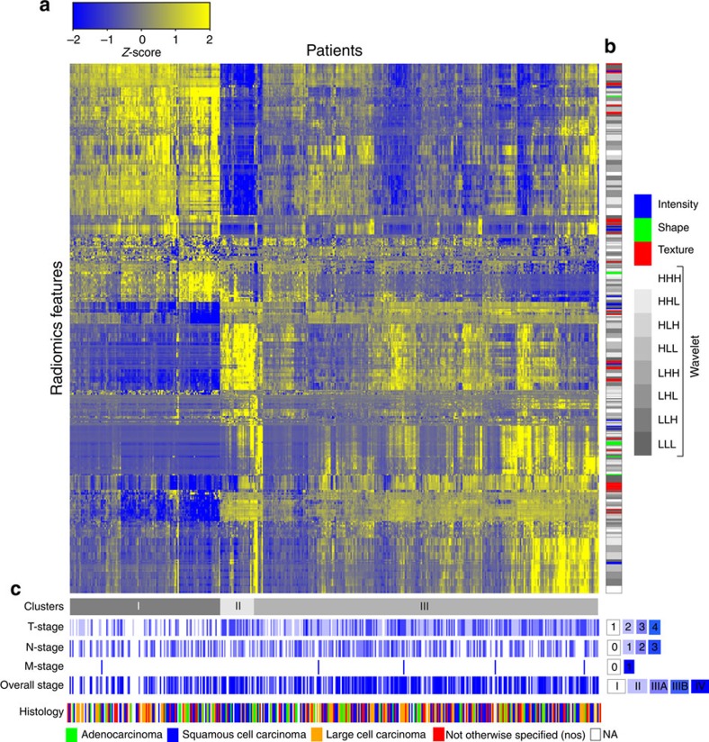
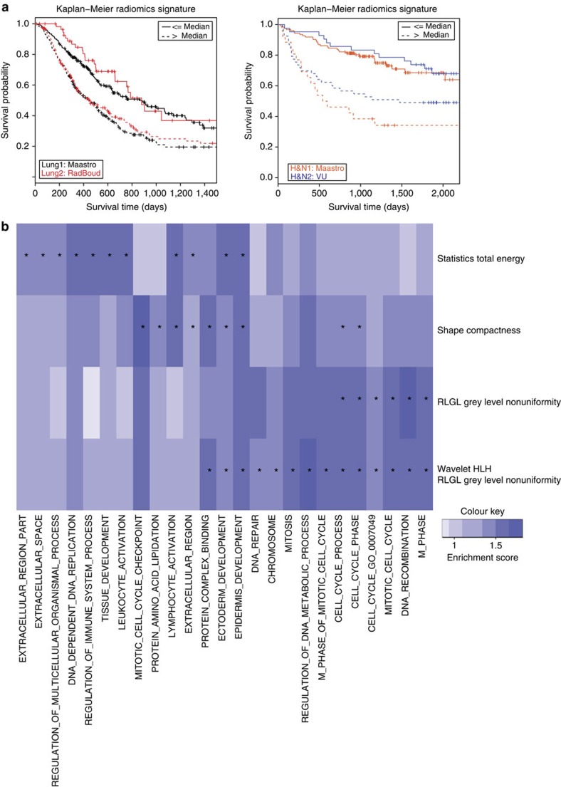
Verification
Claims the paper makes
The claims and statistics of this paper haven’t been checked yet.
Free: it reads the open-access text and checks the first 5 claims that cite a source we can check. Create a free account to check every claim.
Claims other papers make about this one
Each is checked against this paper’s own text: does it say what the citing paper says it does?
The source backs part of the claim, not all of it — or backs it for a different population, condition or comparison.
In the citing paper
“Notably, the signature was comprised of the top features from four classes (size, shape, texture, and wavelets) that were prioritized from a database of 442 features by using test-retest reproducibility and intersubject range.”
From Radiomics: Images Are More than Pictures, They Are Data (2016) · cites this paper as [47]
Experimental — accuracy not yet measuredThe source backs part of the claim, not all of it — or backs it for a different population, condition or comparison.
In the citing paper
“Many studies have shown that imaging features are significantly associated with patterns of gene expression and genetic mutations, demonstrating that radiogenomic analysis can identify different biological mechanisms by means of mathematical and computational devices, enabling the decoding of disease phenotypes by noninvasive methods(48,49).”
From Artificial intelligence, machine learning, computer-aided diagnosis, and radiomics: advances in imaging towards to precision medicine (2019) · cites this paper as [49]
Experimental — accuracy not yet measuredThe cited source says what the claim attributes to it.
In the citing paper
“A study by Aerts et al (47) showed that a radiomic signature could be used to predict outcome in completely independent cohorts of patients with lung cancer from two separate institutions.”
From Radiomics: Images Are More than Pictures, They Are Data (2016) · cites this paper as [47]
The cited source says what the claim attributes to it.
In the citing paper
“Further, this same signature could be applied to cohorts of patients with head and neck cancer with equivalent prognostic power.”
From Radiomics: Images Are More than Pictures, They Are Data (2016) · cites this paper as [47]
In the citing paper
“We used scans from 26 patients from the RIDER dataset19, routinely used for test–retest robustness analysis in tumour imaging19–21.”
From Foundation Model for Cancer Imaging Biomarkers (2024) · cites this paper as [20]
3 more claims about this paper
Create a free account to see every claim other papers make about this one.
Self-check
Percentages, sums and reported differences re-computed from the paper’s own numbers.
The self-check needs the paper’s full text.
Citations
Cites
Subramanian, A., Tamayo, P., Mootha, V. K., Mukherjee, S., Ebert, B. L., Gillette, M. A., Paulovich, A., Pomeroy, S. L., Golub, T. R., Lander, E. S., et al. (2005) · Proceedings of the National Academy of Sciences · doi:10.1073/pnas.0506580102 · Cited by 53,625
Harrell, F. E. (2001) · Springer Series in Statistics · doi:10.1007/978-1-4757-3462-1 · Cited by 9,195
Intratumor Heterogeneity and Branched Evolution Revealed by Multiregion Sequencing
Gerlinger, M., Rowan, A. J., Horswell, S., Larkin, J., Endesfelder, D., Gronroos, E., Martinez, P., Matthews, N., Stewart, A., Tarpey, P., et al. (2012) · New England Journal of Medicine · doi:10.1056/nejmoa1113205 · Cited by 7,774 · cited 2×
“It is known that multiple subclonal populations coexist within tumours, reflecting extensive intratumoral ‘somatic evolution’ 15,16 .”
BackgroundMolecular signatures database (MSigDB) 3.0
Liberzon, A., Subramanian, A., Pinchback, R., Thorvaldsdóttir, H., Tamayo, P., & Mesirov, J. P. (2011) · Bioinformatics · doi:10.1093/bioinformatics/btr260 · Cited by 7,111
Harrell (2015) · Springer Series in Statistics · doi:10.1007/978-3-319-19425-7 · Cited by 6,878
Radiomics: extracting more information from medical images using advanced feature analysis
Lambin, P., Rios-Velazquez, E., Leijenaar, R., Carvalho, S., van Stiphout, R. G. P. M., Granton, P., Zegers, C. M. L., Gillies, R., Boellard, R., Dekker, A., et al. (2012) · European Journal of Cancer · doi:10.1016/j.ejca.2011.11.036 · Cited by 6,084
Self-citationDistant metastasis occurs late during the genetic evolution of pancreatic cancer
Yachida, S., Jones, S., Bozic, I., Antal, T., Leary, R., Fu, B., Kamiyama, M., Hruban, R. H., Eshleman, J. R., Nowak, M. A., et al. (2010) · Nature · doi:10.1038/nature09515 · Cited by 2,502 · cited 1×
“It is known that multiple subclonal populations coexist within tumours, reflecting extensive intratumoral ‘somatic evolution’ 15,16 .”
BackgroundRadiomics: the process and the challenges
Kumar, V., Gu, Y., Basu, S., Berglund, A., Eschrich, S. A., Schabath, M. B., Forster, K., Aerts, H. J. W. L., Dekker, A., Fenstermacher, D., et al. (2012) · Magnetic Resonance Imaging · doi:10.1016/j.mri.2012.06.010 · Cited by 2,258
Self-citationTixier, F., Le Rest, C. C., Hatt, M., Albarghach, N., Pradier, O., Metges, J.-P., Corcos, L., & Visvikis, D. (2011) · Journal of Nuclear Medicine · doi:10.2967/jnumed.110.082404 · Cited by 696
Decoding global gene expression programs in liver cancer by noninvasive imaging
Segal, E., Sirlin, C. B., Ooi, C., Adler, A. S., Gollub, J., Chen, X., Chan, B. K., Matcuk, G. R., Barry, C. T., Chang, H. Y., et al. (2007) · Nature Biotechnology · doi:10.1038/nbt1306 · Cited by 629
Cited by
Items 601–700 of 1,023
Machine Learning and Deep Neural Networks in Thoracic and Cardiovascular Imaging
Retson, T., Besser, A. H., Sall, S., Golden, D. I., & Hsiao, A. (2019) · Journal of Thoracic Imaging · doi:10.1097/rti.0000000000000385 · Cited by 92
van Timmeren, J. E., van Elmpt, W., Leijenaar, R. T. H., Reymen, B. J. T., Monshouwer, R., Bussink, J., Paelinck, L., Bogaert, E., Wagter, C. D., Elamin, E. E., et al. (2019) · Radiotherapy and Oncology · doi:10.1016/j.radonc.2019.03.032 · Cited by 90
“59 for all datasets combined, which was not significantly different compared to the validation performance in other studies [5,10,23].”
MethodResultSelf-citationAntonopoulos, A. S., Angelopoulos, A., Tsioufis, K. P., Antoniades, C. A., & Tousoulis, D. (2021) · European Journal of Preventive Cardiology · doi:10.1093/eurjpc/zwab067 · Cited by 97
McGarry, S. D., Hurrell, S. L., Kaczmarowski, A. L., Cochran, E. J., Connelly, J., Rand, S. D., Schmainda, K. M., & LaViolette, P. S. (2016) · Tomography · doi:10.18383/j.tom.2016.00250 · Cited by 84
“These techniques are broadly defined as radiomics (9, 10, 13-15).”
BackgroundInfluentialLarue, R. T. H. M., Klaassen, R., Jochems, A., Leijenaar, R. T. H., Hulshof, M. C. C. M., Henegouwen, M. I. V. B., Schreurs, W. M. J., Sosef, M. N., van Elmpt, W., van Laarhoven, H. W. M., et al. (2018) · Acta Oncologica · doi:10.1080/0284186x.2018.1486039 · Cited by 84
“This is in line with previous radiomics studies, in which CT imaging features describing tumor heterogeneity also were shown to have prognostic value in lung [11,13], head-and-neck [12] and esophageal cancer [14–16].”
ResultSelf-citationWang, H., Chen, H., Duan, S., Hao, D., & Liu, J. (2019) · Journal of Magnetic Resonance Imaging · doi:10.1002/jmri.26901 · Cited by 82
Biology-guided radiotherapy: redefining the role of radiotherapy in metastatic cancer
Shirvani, S. M., Huntzinger, C. J., Melcher, T., Olcott, P. D., Voronenko, Y., Bartlett-Roberto, J., & Mazin, S. R. (2020) · British Journal of Radiology · doi:10.1259/bjr.20200873 · Cited by 85
Iqbal, A., Sharif, M. I., Yasmin, M., Raza, M., & Aftab, S. (2022) · International Journal of Multimedia Information Retrieval · doi:10.1007/s13735-022-00240-x · Cited by 97
The progress of multimodal imaging combination and subregion based radiomics research of cancers
Zhang, L., Wang, Y., Peng, Z., Weng, Y., Fang, Z., Xiao, F., Zhang, C., Fan, Z., Huang, K., Zhu, Y., et al. (2022) · International Journal of Biological Sciences · doi:10.7150/ijbs.71046 · Cited by 86
“aid to a clinical decision aid in the context of personalized medicine (13).”
BackgroundThorwarth, D. (2015) · British Journal of Radiology · doi:10.1259/bjr.20150056 · Cited by 88
Role and future of MRI in radiation oncology
Das, I. J., McGee, K. P., Tyagi, N., & Wang, H. (2018) · British Journal of Radiology · doi:10.1259/bjr.20180505 · Cited by 82
Ariji, Y., Sugita, Y., Nagao, T., Nakayama, A., Fukuda, M., Kise, Y., Nozawa, M., Nishiyama, M., Katumata, A., & Ariji, E. (2019) · Oral Radiology · doi:10.1007/s11282-019-00391-4 · Cited by 83
Zheng, J., Chakraborty, J., Chapman, W. C., Gerst, S. R., Gönen, M., Pak, L. M., Jarnagin, W. R., DeMatteo, R. P., Do, R. K. G., Simpson, A. L., et al. (2017) · Journal of the American College of Surgeons · doi:10.1016/j.jamcollsurg.2017.09.003 · Cited by 86
Soft Tissue Sarcomas: Preoperative Predictive Histopathological Grading Based on Radiomics of MRI
Zhang, Y., Zhu, Y., Shi, X., Tao, J., Cui, J., Dai, Y., Zheng, M., & Wang, S. (2018) · Academic Radiology · doi:10.1016/j.acra.2018.09.025 · Cited by 81
Guo, D., Gu, D., Wang, H., Wei, J., Wang, Z.-L., Hao, X., Ji, Q., Cao, S., Song, Z., Jiang, J., et al. (2019) · European Journal of Radiology · doi:10.1016/j.ejrad.2019.05.010 · Cited by 81
Li, X., Yin, G., Zhang, Y., Dai, D., Liu, J., Chen, P., Zhu, L., Ma, W., & Xu, W. (2019) · Frontiers in Oncology · doi:10.3389/fonc.2019.01062 · Cited by 83
Yang, Y., Feng, F., Qiu, Y.-J., Zheng, G., Ge, Y., & Wang, Y. (2020) · Abdominal Radiology · doi:10.1007/s00261-020-02733-x · Cited by 82
“As for Rad-score, radiomics represents a comprehensive determination of tumor phenotypes with a plenty of quantitative image features [20].”
BackgroundMethodRadiomic Analysis: Study Design, Statistical Analysis, and Other Bias Mitigation Strategies
Moskowitz, C. S., Welch, M. L., Jacobs, M. A., Kurland, B. F., & Simpson, A. L. (2022) · Radiology · doi:10.1148/radiol.211597 · Cited by 89
Optimize and refine therapeutic index in radiation therapy: Overview of a century
Chargari, C., Magné, N., Guy, J., Rancoule, C., Lévy, A., Goodman, K. A., & Deutsch, É. (2016) · Cancer Treatment Reviews · doi:10.1016/j.ctrv.2016.03.001 · Cited by 98
Peeken, J. C., Bernhofer, M., Spraker, M. B., Pfeiffer, D., Devečka, M., Thamer, A., Shouman, M. A., Ott, A., Nüsslin, F., Mayr, N. A., et al. (2019) · Radiotherapy and Oncology · doi:10.1016/j.radonc.2019.01.004 · Cited by 83
Metabolism and Its Sequelae in Cancer Evolution and Therapy
Gillies, R. J., & Gatenby, R. A. (2015) · The Cancer Journal · doi:10.1097/ppo.0000000000000102 · Cited by 85
Self-citationMulti-scale Modeling in Clinical Oncology: Opportunities and Barriers to Success
Yankeelov, T. E., An, G., Saut, O., Luebeck, E. G., Popel, A. S., Ribba, B., Vicini, P., Zhou, X., Weis, J. A., Ye, K., et al. (2016) · Annals of Biomedical Engineering · doi:10.1007/s10439-016-1691-6 · Cited by 80
Fine-tuning convolutional deep features for MRI based brain tumor classification
Ahmed, K. B., Hall, L., Goldgof, D. B., Liu, R., & Gatenby, R. A. (2017) · Proceedings of SPIE, the International Society for Optical Engineering/Proceedings of SPIE · doi:10.1117/12.2253982 · Cited by 81
“Associating clinical information with these features has enabled the identification of new and reproducible biomarkers, which have been prognostic for clinical outcomes including overall survival ([18], [19]).”
BackgroundSun, W., Jiang, M., Dang, J., Chang, P., & Yin, F.-F. (2018) · Radiation Oncology · doi:10.1186/s13014-018-1140-9 · Cited by 86
MRI-based Radiomics nomogram to detect primary rectal cancer with synchronous liver metastases
Shu, Z., Fang, S., Ding, Z., Mao, D., Cai, R., Yuanjun, C., Pang, P., & Gong, X. (2019) · Scientific Reports · doi:10.1038/s41598-019-39651-y · Cited by 86
Radiomics for predicting hematoma expansion in patients with hypertensive intraparenchymal hematomas
Ma, C., Zhang, Y., Niyazi, T., Wei, J., Guo, G., Liu, J., Liang, S., Liang, F., Yan, P., Wang, K., et al. (2019) · European Journal of Radiology · doi:10.1016/j.ejrad.2019.04.001 · Cited by 81
Integrating imaging and omics data: A review
Antonelli, L., Guarracino, M. R., Maddalena, L., & Sangiovanni, M. (2019) · Biomedical Signal Processing and Control · doi:10.1016/j.bspc.2019.04.032 · Cited by 79
Yang, Z., He, B., Zhuang, X., Gao, X., Wang, D., Li, M., Lin, Z., & Luo, R. (2019) · Journal of Radiation Research · doi:10.1093/jrr/rrz027 · Cited by 79
“Radiomics could be applied to identify tumor phenotype characteristics [6, 7], and to discover prognostic or predictive biomarkers for cancers [8, 9].”
BackgroundRadiomics: A primer for the radiation oncologist
Bibault, J., Xing, L., Giraud, P., Ayachy, R. E., Giraud, N., Decazes, P., Burgun, A., & Giraud, P. (2020) · Cancer/Radiothérapie · doi:10.1016/j.canrad.2020.01.011 · Cited by 80
Wen, Q., Yang, Z., Dai, H., Feng, A., & Li, Q. (2021) · Frontiers in Oncology · doi:10.3389/fonc.2021.620246 · Cited by 95
Artificial Intelligence in Thyroid Field—A Comprehensive Review
Bini, F., Pica, A., Azzimonti, L., Giusti, A., Ruinelli, L., Marinozzi, F., & Trimboli, P. (2021) · Cancers · doi:10.3390/cancers13194740 · Cited by 91
“Typically, radiomics features are divided into [2,6,44]:”
BackgroundMethodInfluentialRen, J., Tian, J., Yuan, Y., Dong, D., Li, X., Shi, Y., & Tao, X. (2018) · European Journal of Radiology · doi:10.1016/j.ejrad.2018.07.002 · Cited by 80
Kaissis, G., Ziegelmayer, S., Lohöfer, F., Algül, H., Eiber, M., Weichert, W., Schmid, R., Friess, H., Rummeny, E., Ankerst, D., et al. (2019) · European Radiology Experimental · doi:10.1186/s41747-019-0119-0 · Cited by 79
“In fact, it leverages data science and machine learning (ML) techniques to exploit non-intuitive image content and integrate it with clinical information to create a generalizable model capable of predicting biological features or the course of disease [5].”
BackgroundMachine learning based brain tumour segmentation on limited data using local texture and abnormality
Bonte, S., Goethals, I., & Holen, R. V. (2018) · Computers in Biology and Medicine · doi:10.1016/j.compbiomed.2018.05.005 · Cited by 91
Palani, D., & Venkatalakshmi, K. (2018) · Journal of Medical Systems · doi:10.1007/s10916-018-1139-7 · Cited by 86
The Dark Side of Radiomics: On the Paramount Importance of Publishing Negative Results
Buvat, I., & Orlhac, F. (2019) · Journal of Nuclear Medicine · doi:10.2967/jnumed.119.235325 · Cited by 90
“Even worse, misleading interpretation of some of the most famous radiomic findings (6) has recently been demonstrated (7), confirming that some radiomic features initially interpreted as biomarkers of tumor heterogeneity were actually surrogates of tumor volume, as shown as early as 2014 (8,9).”
BackgroundA review on radiomics and the future of theranostics for patient selection in precision medicine
Keek, S. A., Leijenaar, R. T. H., Jochems, A., & Woodruff, H. C. (2018) · British Journal of Radiology · doi:10.1259/bjr.20170926 · Cited by 87
Self-citation[18F]FDG PET/CT features for the molecular characterization of primary breast tumors
Antunovic, L., Gallivanone, F., Sollini, M., Sagona, A., Invento, A., Manfrinato, G., Kirienko, M., Tinterri, C., Chiti, A., & Castiglioni, I. (2017) · European Journal of Nuclear Medicine and Molecular Imaging · doi:10.1007/s00259-017-3770-9 · Cited by 78
“Thirteen features described the characteristics of the histogram of the PET voxel intensities of the primary tumor as first-order statistical features (FOSf): energy, entropy, kurtosis, maximum, mean, mean absolute deviation, median, minimum, root mean square, skewness, standard deviation, uniformity and variance [35].”
Bakr, S. H., Echegaray, S., Shah, R. P., Kamaya, A., Louie, J. D., Napel, S. A., Kothary, N., & Gevaert, O. (2017) · Journal of medical imaging · doi:10.1117/1.jmi.4.4.041303 · Cited by 86
“previous work.(26,29,32) Using a single ROI as input, the pipeline computed 464 features, described in previous work(33) for each CT phase that characterized the intensities and textures within the ROIs, the sharpness of lesion boundaries, and the boundary shapes.”
MethodBogowicz, M., Jochems, A., Deist, T. M., Tanadini‐Lang, S., Huang, S. H., Chan, B., Waldron, J. N., Bratman, S. V., O’Sullivan, B., Riesterer, O., et al. (2020) · Scientific Reports · doi:10.1038/s41598-020-61297-4 · Cited by 84
Self-citationHaider, S. P., Burtness, B. A., Yarbrough, W. G., & Payabvash, S. (2020) · Cancers of the Head & Neck · doi:10.1186/s41199-020-00053-7 · Cited by 97
Recent Radiomics Advancements in Breast Cancer: Lessons and Pitfalls for the Next Future
Pesapane, F., Rotili, A., Agazzi, G. M., Botta, F., Raimondi, S., Penco, S., Dominelli, V., Cremonesi, M., Jereczek‐Fossa, B. A., Carrafiello, G., et al. (2021) · Current Oncology · doi:10.3390/curroncol28040217 · Cited by 83
Li, Y., Wei, D., Liu, X., Fan, X., Wang, K., Li, S., Zhang, Z., Ma, K., Qian, T., Jiang, T., et al. (2020) · European Radiology · doi:10.1007/s00330-021-08237-6 · Cited by 87
Calabrese, E., Rudie, J. D., Rauschecker, A. M., Villanueva-Meyer, J. E., Clarke, J. L., Solomon, D. A., & Cha, S. (2022) · Neuro-Oncology Advances · doi:10.1093/noajnl/vdac060 · Cited by 102
Millisecond speed deep learning based proton dose calculation with Monte Carlo accuracy
Pastor‐Serrano, O., & Perkó, Z. (2022) · Physics in Medicine and Biology · doi:10.1088/1361-6560/ac692e · Cited by 98
“Dataset We train DoTA to predict low noise MC dose distributions calculated with MCsquare [60], obtained using a set of 30 CT scans from prostate, lung and head and neck (H&N) cancer patients [61, 62, 63] with 2 mm isotropic grid resolution.”
MethodPET/CT based cross-modal deep learning signature to predict occult nodal metastasis in lung cancer
Zhong, Y., Cai, C., Chen, T., Gui, H., Deng, J., Yang, M., Yu, B., Song, Y., Wang, T., Sun, X., et al. (2023) · Nature Communications · doi:10.1038/s41467-023-42811-4 · Cited by 99
“The deep learning technology which allows the high-dimensional quanti fi cationofradiologicalimagesandgreaterextractionofdetailed characterizations than the human vision, has been proposed as a revolutionary approach for disease diagnoses, prognosis evaluations, and therapeutic decisions 20 – 22 .”
Groheux, D., Majdoub, M., Tixier, F., Rest, C. C. L., Martineau, A., Merlet, P., Espié, M., de Roquancourt, A., Hindié, E., Hatt, M., et al. (2015) · European Journal of Nuclear Medicine and Molecular Imaging · doi:10.1007/s00259-015-3110-x · Cited by 84
“heterogeneity metrics [16,23], with special reference to the comprehensive quantification of tumor phenotypes or popularly denoted “radiomics” [24,25].”
BackgroundHuang, W., Chen, Y., Fedorov, A., Li, X., Jajamovich, G. H., Malyarenko, D. I., Aryal, M. P., LaViolette, P. S., Oborski, M. J., O’Sullivan, F., et al. (2019) · Tomography · doi:10.18383/j.tom.2018.00027 · Cited by 82
Hou, Z., Ren, W., Li, S., Liu, J., Yu, S. Y., Yan, J., & Wan, S. (2017) · Oncotarget · doi:10.18632/oncotarget.22304 · Cited by 83
Park, S., Lee, S. M., Do, K., Lee, J., Bae, W., Park, H., Jung, K.-H., & Seo, J. B. (2019) · Korean Journal of Radiology · doi:10.3348/kjr.2019.0212 · Cited by 88
Clinical Management of Diffuse Low-Grade Gliomas
Lombardi, G., Barresi, V., Castellano, A., Tabouret, É., Pasqualetti, F., Salvalaggio, A., Cerretti, G., Caccese, M., Padovan, M., Zagonel, V., et al. (2020) · Cancers · doi:10.3390/cancers12103008 · Cited by 86
Roy, S. S., Whitehead, T., Li, S., Ademuyiwa, F. O., Wahl, R. L., Dehdashti, F., & Shoghi, K. I. (2021) · European Journal of Nuclear Medicine and Molecular Imaging · doi:10.1007/s00259-021-05489-8 · Cited by 84
Recent advancements in deep learning based lung cancer detection: A systematic review
Dodia, S. J., Annappa, B., & Mahesh, P. A. (2022) · Engineering Applications of Artificial Intelligence · doi:10.1016/j.engappai.2022.105490 · Cited by 89
Chen, M., Lu, H., Copley, S. J., Han, Y., Logan, A., Viola, P., Cortellini, A., Pinato, D. J., Power, D., & Aboagye, E. O. (2023) · Journal of Thoracic Oncology · doi:10.1016/j.jtho.2023.01.089 · Cited by 82
Biomarkers and Imaging of Breast Cancer
Weaver, O. O., & Leung, J. W. T. (2017) · American Journal of Roentgenology · doi:10.2214/ajr.17.18708 · Cited by 77
A new approach to predict lymph node metastasis in solid lung adenocarcinoma: a radiomics nomogram
Yang, X., Pan, X., Liu, H., Gao, D., He, J., Liang, W., & Guan, Y. (2018) · Journal of Thoracic Disease · doi:10.21037/jtd.2018.03.126 · Cited by 83
Han, Y., Xie, Z., Zang, Y., Zhang, S., Gu, D., Zhou, M., Gevaert, O., Wei, J., Li, C., Chen, H., et al. (2018) · Journal of Neuro-Oncology · doi:10.1007/s11060-018-2953-y · Cited by 76
Yamashita, R., Perrin, T., Chakraborty, J., Chou, J. F., Horvat, N., Koszalka, M. A., Midya, A., Gonen, M., Allen, P., Jarnagin, W. R., et al. (2020) · European Radiology · doi:10.1007/s00330-019-06381-8 · Cited by 76
Fukuma, R., Yanagisawa, T., Kinoshita, M., Shinozaki, T., Arita, H., Kawaguchi, A., Takahashi, M., Narita, Y., Terakawa, Y., Tsuyuguchi, N., et al. (2019) · Scientific Reports · doi:10.1038/s41598-019-56767-3 · Cited by 80
Xiao, M., Ma, F., Li, Y., Li, Y., Li, M., Zhang, G., & Qiang, J. (2020) · Journal of Magnetic Resonance Imaging · doi:10.1002/jmri.27101 · Cited by 79
Wu, A., Li, Y., Qi, M., Lu, X., Jia, Q., Guo, F., Dai, Z., Liu, Y., Chen, C., Zhou, L., et al. (2020) · Oral Oncology · doi:10.1016/j.oraloncology.2020.104625 · Cited by 79
Radiomics prognostication model in glioblastoma using diffusion- and perfusion-weighted MRI
Park, J. E., Kim, H. S., Jo, Y., Yoo, R., Choi, S. H., Nam, S. J., & Kim, J. H. (2020) · Scientific Reports · doi:10.1038/s41598-020-61178-w · Cited by 85
Yperman, J., Becker, T., Valkenborg, D., Popescu, V., Hellings, N., Wijmeersch, B. V., & Peeters, L. M. (2020) · BMC Neurology · doi:10.1186/s12883-020-01672-w · Cited by 91
“, radiomics for the analyses of MRI images [50].”
BackgroundLi, J., Chekkoury, A., Prakash, J., Glasl, S., Vetschera, P., Koberstein‐Schwarz, B., Olefir, I., Gujrati, V., Omar, M., & Ntziachristos, V. (2020) · Light Science & Applications · doi:10.1038/s41377-020-0295-y · Cited by 78
McNitt‐Gray, M. F., Napel, S. A., Jaggi, A., Mattonen, S. A., Hadjiiski, L. M., Muzi, M., Goldgof, D. B., Balagurunathan, Y., Pierce, L. A., Kinahan, P. E., et al. (2020) · Tomography · doi:10.18383/j.tom.2019.00031 · Cited by 84
“the eventual goal of being able to accomplish a diagnostic imaging task, such as distinguishing benign from malignant nodules, assessing response to therapy, correlating imaging with genomics, or decoding the object’s imaging phenotype and predicting survival outcomes (5).”
BackgroundPrecision radiotherapy for non-small cell lung cancer
Yang, W.-C., Hsu, F., & Yang, P. (2020) · Journal of Biomedical Science · doi:10.1186/s12929-020-00676-5 · Cited by 85
“[35, 36] used features extracted from cone-beam CT (CBCT) during radiotherapy treatment to validate the previously reported radiomic features for NSCLC outcome prediction [37].”
MethodXu, X., Zhang, J., Yang, K. G., Wang, Q., Chen, X., & Xu, B. (2021) · Brain and Behavior · doi:10.1002/brb3.2085 · Cited by 84
“These features, namely radiom - ics features, have the potential to uncover disease characteristics that fail to be appreciated by the naked eye (Aerts et al., 2014; Wu et al., 2018).”
BackgroundShao, J., Ma, J., Zhang, Q., Li, W., & Wang, C. (2023) · Seminars in Cancer Biology · doi:10.1016/j.semcancer.2023.02.006 · Cited by 90
Feature selection methodology for longitudinal cone-beam CT radiomics
van Timmeren, J. E., Leijenaar, R. T. H., van Elmpt, W., Reymen, B. J. T., & Lambin, P. (2017) · Acta Oncologica · doi:10.1080/0284186x.2017.1350285 · Cited by 76
“Previously, radiomics has been shown to be able to predict outcome for NSCLC patients using CT imaging [6,8,19,29].”
BackgroundSelf-citationThe effect of preprocessing filters on predictive performance in radiomics
Demircioğlu, A. (2022) · European Radiology Experimental · doi:10.1186/s41747-022-00294-w · Cited by 75
“We used seven publicly available datasets (Table 1); six were published as the “WORC” database [9], while one, the head & neck cancer dataset, was published in the seminal paper by Aerts et al. [10].”
MethodLearning from scanners: Bias reduction and feature correction in radiomics
Zhovannik, I., Bussink, J., Traverso, A., Shi, Z., Kalendralis, P., Wee, L., Dekker, A., Fijten, R., & Monshouwer, R. (2019) · Clinical and Translational Radiation Oncology · doi:10.1016/j.ctro.2019.07.003 · Cited by 75
“Subsequently, machine learning algorithms use these radiomic features to predict patient survival time [2,3], treatment toxicity [4], tumor habitat characterization [5].”
MethodSelf-citationOzkan, E., West, A., Dedelow, J. A., Chu, B. F., Zhao, W., Yildiz, V. O., Otterson, G. A., Shilo, K., Ghosh, S., King, M., et al. (2015) · American Journal of Roentgenology · doi:10.2214/ajr.14.14147 · Cited by 75
“and gene expression patterns in lung cancer [7, 8].”
Background4DCT imaging to assess radiomics feature stability: An investigation for thoracic cancers
Larue, R. T. H. M., Van De Voorde, L., van Timmeren, J. E., Leijenaar, R. T. H., Berbée, M., Sosef, M. N., Schreurs, W. M. J., van Elmpt, W., & Lambin, P. (2017) · Radiotherapy and Oncology · doi:10.1016/j.radonc.2017.07.023 · Cited by 75
Self-citationYang, X., He, J., Wang, J., Li, W., Liu, C., Gao, D., & Guan, Y. (2018) · Lung Cancer · doi:10.1016/j.lungcan.2018.09.013 · Cited by 77
Kim, Y. K., Cho, H.-H., Kim, S. T., Park, H., Nam, D., & Kong, D. (2018) · Neuroradiology · doi:10.1007/s00234-018-2091-4 · Cited by 77
Wang, L., Dong, T., Xin, B., Xu, C., Guo, M., Zhang, H., Feng, D. D., Wang, X., & Yu, J. (2019) · European Radiology · doi:10.1007/s00330-018-5949-2 · Cited by 82
Radiomics: Principles and radiotherapy applications
Gardin, I., Grégoire, V., Gibon, D., Kirişli, H. A., Pasquier, D., Thariat, J., & Véra, P. (2019) · Critical Reviews in Oncology/Hematology · doi:10.1016/j.critrevonc.2019.03.015 · Cited by 86
Ger, R. B., Zhou, S., Elgohari, B., Elhalawani, H. M., Mackin, D., Meier, J. G., Nguyen, C. M., Anderson, B., Gay, C., Ning, J., et al. (2019) · PLoS ONE · doi:10.1371/journal.pone.0222509 · Cited by 75
Self-citationFeng, B., Chen, X., Chen, Y., Liu, K., Li, K., Liu, X., Yao, N., Li, Z., Li, R., Zhang, C., et al. (2020) · European Journal of Radiology · doi:10.1016/j.ejrad.2020.109022 · Cited by 81
Zhou, J., Tan, H., Li, W., Liu, Z., Wu, Y., Bai, Y., Fu, F., Jia, X., Feng, A., Liu, H., et al. (2020) · Academic Radiology · doi:10.1016/j.acra.2020.05.040 · Cited by 75
Texture analysis imaging “what a clinical radiologist needs to know”
Corrias, G., Micheletti, G., Barberini, L., Suri, J. S., & Saba, L. (2021) · European Journal of Radiology · doi:10.1016/j.ejrad.2021.110055 · Cited by 82
TSFD-Net: Tissue specific feature distillation network for nuclei segmentation and classification
Ilyas, T., Mannan, Z. I., Khan, A., Azam, S., Kim, H., & Boer, F. D. (2022) · Neural Networks · doi:10.1016/j.neunet.2022.02.020 · Cited by 92
Application of texture analysis to DAT SPECT imaging: Relationship to clinical assessments
Rahmim, A., Salimpour, Y., Jain, S., Blinder, S. A. L., Klyuzhin, I. S., Smith, G. S., Mari, Z. K., & Sossi, V. (2016) · NeuroImage Clinical · doi:10.1016/j.nicl.2016.02.012 · Cited by 79
“There is emerging literature on the use of advanced metrics that quantify tumor uptake heterogeneity and their enhanced prediction of treatment response and survival outcome in different cancers (Aerts et al., 2014; Asselin et al., 2012; Chicklore et al., 2013; Eary et al., 2008; El Naqa et al., 2009; Hatt et al., 201…”
BackgroundProspects of deep learning for medical imaging
Kim, J., Hong, J., & Park, H. (2018) · Precision and Future Medicine · doi:10.23838/pfm.2018.00030 · Cited by 89
Radiomics for the prediction of EGFR mutation subtypes in non‐small cell lung cancer
Li, S., Ding, C., Zhang, H., Song, J., & Wu, L. (2019) · Medical Physics · doi:10.1002/mp.13747 · Cited by 73
Tunali, I., Hall, L. O., Napel, S., Cherezov, D., Guvenis, A., Gillies, R. J., & Schabath, M. B. (2019) · Medical Physics · doi:10.1002/mp.13808 · Cited by 75
Self-citationWu, J., Gensheimer, M. F., Zhang, N., Guo, M., Liang, R., Zhang, C., Fischbein, N. J., Pollom, E. L., Beadle, B. M., Le, Q., et al. (2019) · Journal of Nuclear Medicine · doi:10.2967/jnumed.119.230037 · Cited by 80
Exploring Tumor Heterogeneity Using PET Imaging: The Big Picture
Bailly, C., Bodet‐Milin, C., Bourgeois, M., Gouard, S., Ansquer, C., Barbaud, M., Sébille, J.-C., Chérel, M., Kraeber‐Bodéré, F., & Carlier, T. (2019) · Cancers · doi:10.3390/cancers11091282 · Cited by 81
“Radiomics—the high throughput extraction of large amounts of imaging elements from radiographic images—tackles this challenge and is one of the most promising strategies [3,4].”
BackgroundCNN-based survival model for pancreatic ductal adenocarcinoma in medical imaging
Zhang, Y., Lobo-Mueller, E. M., Karanicolas, P., Gallinger, S., Haider, M. A., & Khalvati, F. (2020) · BMC Medical Imaging · doi:10.1186/s12880-020-0418-1 · Cited by 74
Multicenter evaluation of MRI‐based radiomic features: A phantom study
Rai, R., Holloway, L. C., Brink, C., Field, M., Christiansen, R. L., Yu, S. Y., Barton, M. B., & Liney, G. P. (2020) · Medical Physics · doi:10.1002/mp.14173 · Cited by 87
Shang, J., Ma, S., Guo, Y., Yang, L., Zhang, Q., Xie, F., Ma, Y., Ma, Q., Dang, Y., Zhou, K., et al. (2021) · European Radiology · doi:10.1007/s00330-021-08109-z · Cited by 81
Radiomics and machine learning applications in rectal cancer: Current update and future perspectives
Stanzione, A., Verde, F., Romeo, V., Boccadifuoco, F., Mainenti, P. P., & Maurea, S. (2021) · World Journal of Gastroenterology · doi:10.3748/wjg.v27.i32.5306 · Cited by 80
Peeken, J. C., Asadpour, R., Specht, K., Chen, E. Y., Klymenko, O., Akinkuoroye, V., Hippe, D. S., Spraker, M. B., Schaub, S. K., Dapper, H., et al. (2021) · Radiotherapy and Oncology · doi:10.1016/j.radonc.2021.08.023 · Cited by 81
“Radiomics has been shown to predict various clinical and biological endpoints including pathological characteristics, prognosis, tumor progression, spatial infiltrations, and molecular aberrations in multiple cancer types [16-22].”
BackgroundSelf-citationArtificial Intelligence in Lung Cancer Imaging: Unfolding the Future
Cellina, M., Cè, M., Irmici, G., Ascenti, V., Khenkina, N., Toto-Brocchi, M., Martinenghi, C., Papa, S., & Carrafiello, G. (2022) · Diagnostics · doi:10.3390/diagnostics12112644 · Cited by 74
“[104] USA CT 47 Prospective Prognostic radiomics signature Yes Radiomic data could define a response phenotype for NSCLC patients treated with Gefitinib therapy Strong associations - Limited sample size - Only 11 independent radiomic features”
BackgroundHow Radiomics Can Improve Breast Cancer Diagnosis and Treatment
Pesapane, F., Marco, P. D., Rapino, A., Lombardo, E., Nicosia, L., Tantrige, P. M., Rotili, A., Bozzini, A. C., Penco, S., Dominelli, V., et al. (2023) · Journal of Clinical Medicine · doi:10.3390/jcm12041372 · Cited by 87
Radiomics in colorectal cancer patients
Inchingolo, R., Maino, C., Cannella, R., Vernuccio, F., Cortese, F., Dezio, M., Pisani, A. R., Giandola, T. P., Gatti, M., Giannini, V., et al. (2023) · World Journal of Gastroenterology · doi:10.3748/wjg.v29.i19.2888 · Cited by 86
Kwan, J. Y. Y., Su, J. S., Huang, S. H., Ghoraie, L. S., Xu, W., Chan, B., Yip, K. W., Giuliani, M., Bayley, A., Kim, J., et al. (2018) · International Journal of Radiation Oncology*Biology*Physics · doi:10.1016/j.ijrobp.2018.01.057 · Cited by 83
“Second, patients who underwent tonsillectomy or multiple GTVs were excluded from the study, thereby limiting relevance to this population of patients.”
BackgroundMethodInfluentialSelf-citationOno, T., Hirashima, H., Iramina, H., Mukumoto, N., Miyabe, Y., Nakamura, M., & Mizowaki, T. (2019) · Medical Physics · doi:10.1002/mp.13669 · Cited by 78
Towards Efficient COVID-19 CT Annotation: A Benchmark for Lung and Infection SegmentationPreprint
Ma, J., Wang, Y., An, X., Ge, C., Yu, Z., Chen, J., Zhu, Q., Dong, G., He, J., He, Z., et al. (2020) · arXiv (Cornell University) · Cited by 74
Reproducibility with repeat CT in radiomics study for rectal cancer
Hu, P., Wang, J., Zhong, H., Zhou, Z., Shen, L., Hu, W., & Zhang, Z. (2016) · Oncotarget · doi:10.18632/oncotarget.12199 · Cited by 74
“Nowadays, radiomics become a novel approach because it can utilize medical imaging to quantify the tumor phenotype non-invasively for further study, such as patients’ survival, treatment monitoring and outcome prediction [2].”
Method
Metadata
- Authors
- Aerts, H. J. W. L., Velazquez, E. R., Leijenaar, R. T. H., Parmar, C., Grossmann, P., Carvalho, S., Bussink, J., Monshouwer, R., Haibe-Kains, B., Rietveld, D., Hoebers, F., Rietbergen, M. M., Leemans, C. R., Dekker, A., Quackenbush, J., Gillies, R. J., & Lambin, P.
- Affiliations
- Brigham and Women's Hospital (US)
- Maastricht University (NL)
- Radboud University Medical Center (NL)
- Amsterdam UMC Location Vrije Universiteit Amsterdam (NL)
- Dana-Farber Cancer Institute (US)
- Moffitt Cancer Center (US)
- Corresponding author
- Aerts, Hugo J. W. L.
- ORCID
- Venue
- Nature Communications
- Published
- 2014-06-03
- Volume
- 5
- Issue
- 1
- Pages
- 4006-4006
- ISSN
- 2041-1723 (ISSN-L)
- PMC
- PMC4059926
- OpenAlex
- W2103004421
- Semantic Scholar
- 3b002068646831a6f59fabb6daeb0209ed54049d
- Cited by
- 5,132
- References
- 29
- Citations by year
- 2026: 415 · 2025: 460 · 2024: 458 · 2023: 510 · 2022: 567 · 2021: 638 · 2020: 587 · 2019: 552 · 2018: 388 · 2017: 283 · 2016: 175 · 2015: 82 · 2014: 11
- FWCI
- 142.84
- Type
- journal article
- Language
- en
- Update notices
- correction (2014-08-07)
- Open access
- hybrid
- doi.org
- Licence
- Locations
- Nature Communications (publishedVersion, cc-by-nc-nd)
- PubMed Central (publishedVersion, cc-by-nc-nd)
- PubMed (publishedVersion)
- Pure Amsterdam UMC (publishedVersion)
- Amsterdam UMC (VU Amsterdam) - Institutional Repository (publishedVersion)
- Radboud Repository (Radboud University) (submittedVersion)
- Digital Access to Scholarship at Harvard (DASH) (Harvard University) (submittedVersion, cc-by)
- RePEc: Research Papers in Economics (submittedVersion)
- Research Publications (Maastricht University) (submittedVersion, cc-by-nc-nd)
- Radboud Repository (Radboud University) (submittedVersion, other-oa)
- Research Publications (Maastricht University) (submittedVersion, other-oa)
- Data Archiving and Networked Services (DANS) (submittedVersion)
- Funders
- European Federation of Pharmaceutical Industries and Associations
- KWF Kankerbestrijding (UM 2009-4454, 2011-5020, KWF UM 2009-4454, KWF UM 2011-5020, 2009-4454)
- Center for Translational Molecular Medicine (grant 030-103, 030-103)
- Innovative Medicines Initiative (115151)
- National Institutes of Health (U01 CA)
- National Cancer Institute (NIH-USA U01CA 143062-01, U01CA143062)
- Interreg
- Topics
- Health Sciences › Medicine › Radiology, Nuclear Medicine and Imaging › Radiomics and Machine Learning in Medical Imaging
- Physical Sciences › Computer Science › Computer Vision and Pattern Recognition › Image Retrieval and Classification Techniques
- Health Sciences › Medicine › Radiology, Nuclear Medicine and Imaging › Medical Imaging Techniques and Applications
- Keywords
- radiomics, tumor phenotype, lung cancer, head and neck cancer, radiogenomics, intratumoral heterogeneity, computed tomography, image texture features, gene expression, non-invasive imaging, treatment decision support
- MeSH terms
- Adenocarcinoma, Carcinoma, Non-Small-Cell Lung, Carcinoma, Squamous Cell, Female, Head and Neck Neoplasms, Humans, Lung Neoplasms, Male, Phenotype, Prognosis, Tomography, X-Ray Computed, Tumor Burden, Positron-Emission Tomography, Multimodal Imaging
About these checks
Registry, retraction, Cites and statistics results are computed, not judged, with one exception: a printed reference that no index lists is read once by a model before it counts as a citation error. Claims are extracted by a model and kept only when their quote appears verbatim in the text; each cited source is then checked, with a model’s help, for whether it says what the claim relies on. Those verdicts can be wrong — read the claim’s quote and the cited source before relying on one.
Statistics and arithmetic are recomputed, not judged.
Confidence bands are shown only for fields calibrated on our annotated test set. None is calibrated yet, so no band is shown.
What counts as a claim ›How we measure confidence ›How we check this ›
Are you an author, or do you see a problem with this page? Request a review
Data sources
Bibliographic data: Semantic Scholar, OpenAlex, Crossref.
Contains information from the Semantic Scholar Academic Graph Datasets, made available under the ODC Attribution License (ODC-BY 1.0).
Semantic Scholar’s data is described in: Kinney, R., et al. (2023). The Semantic Scholar Open Data Platform. arXiv:2301.10140.
