

Decoding tumour phenotype by noninvasive imaging using a quantitative radiomics approach
Open accessThe DOI matches its Crossref record; 29 of the 29 references it cites were found in a registry or an index.
Abstract
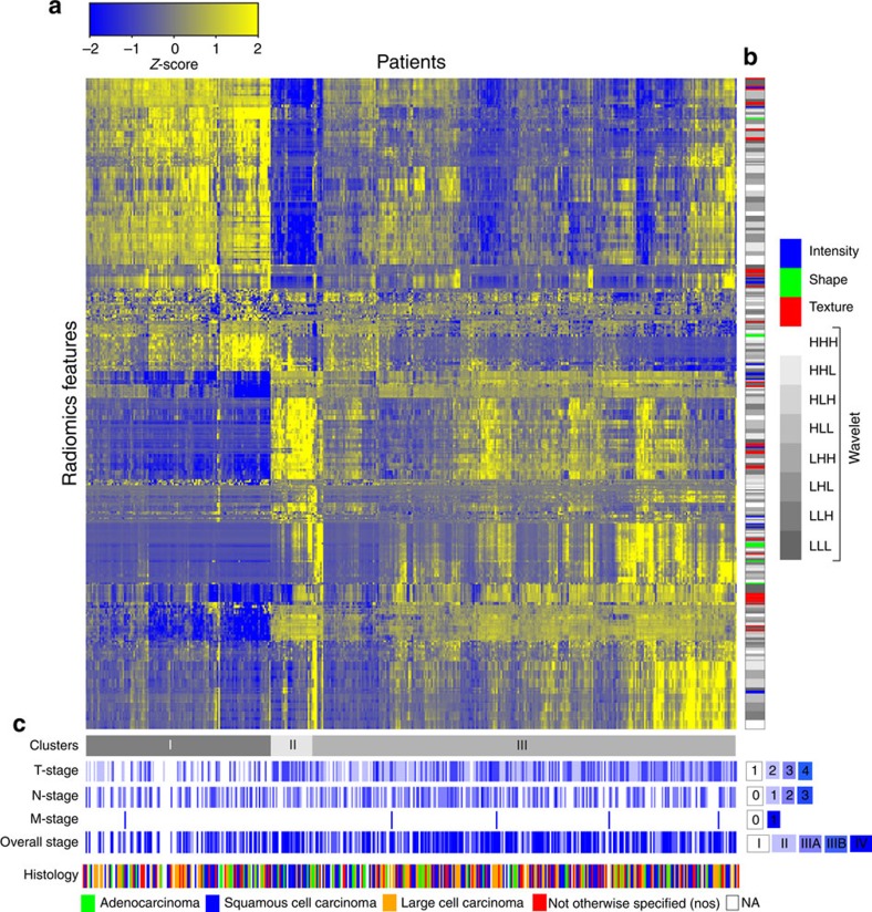
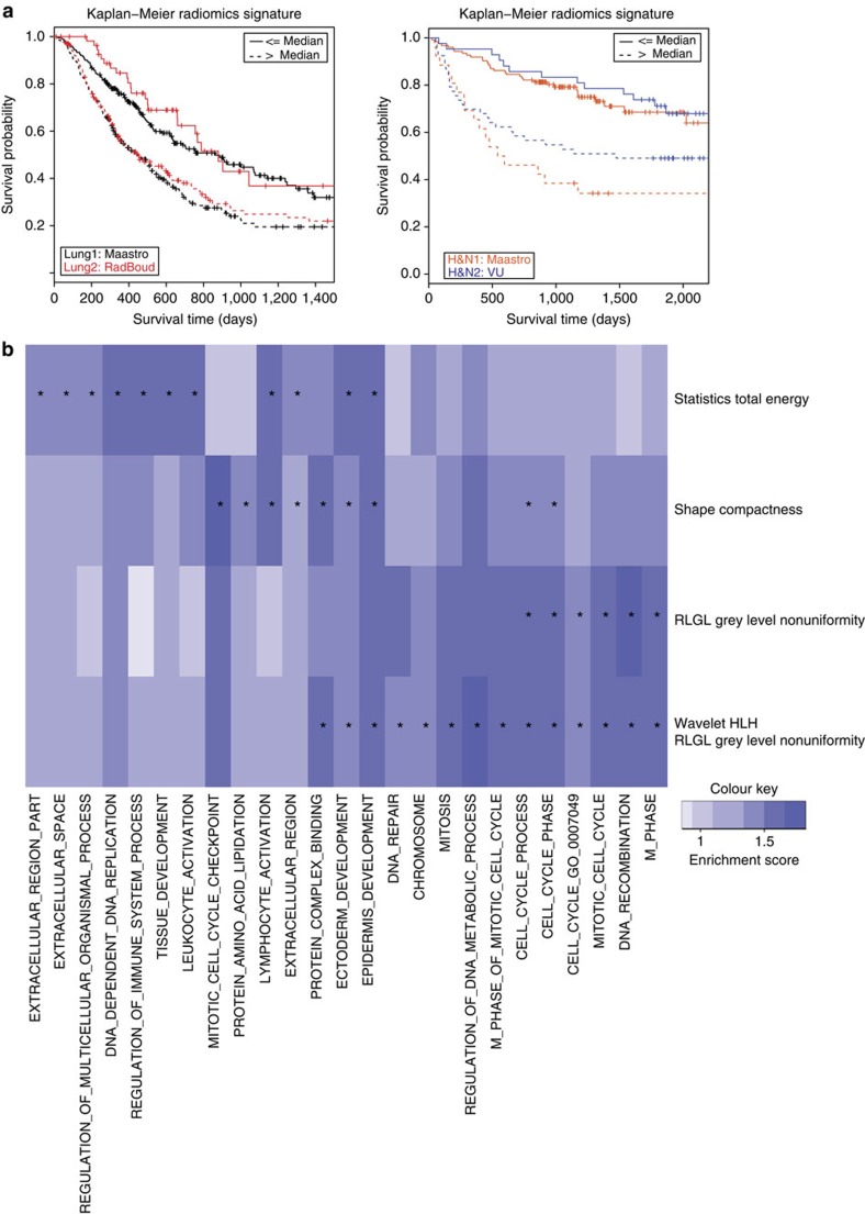
Human cancers exhibit strong phenotypic differences that can be visualized noninvasively by medical imaging. Radiomics refers to the comprehensive quantification of tumour phenotypes by applying a large number of quantitative image features. Here we present a radiomic analysis of 440 features quantifying tumour image intensity, shape and texture, which are extracted from computed tomography data of 1,019 patients with lung or head-and-neck cancer. We find that a large number of radiomic features have prognostic power in independent data sets of lung and head-and-neck cancer patients, many of which were not identified as significant before. Radiogenomics analysis reveals that a prognostic radiomic signature, capturing intratumour heterogeneity, is associated with underlying gene-expression patterns. These data suggest that radiomics identifies a general prognostic phenotype existing in both lung and head-and-neck cancer. This may have a clinical impact as imaging is routinely used in clinical practice, providing an unprecedented opportunity to improve decision-support in cancer treatment at low cost.
Overview
The claims and statistics of this paper haven’t been checked yet.
Free: it reads the open-access text and checks the first 5 claims that cite a source we can check. Create a free account to check every claim.
Integrity signals
Registry
Cites
Statistics
Figures


Verification
Claims the paper makes
The claims and statistics of this paper haven’t been checked yet.
Free: it reads the open-access text and checks the first 5 claims that cite a source we can check. Create a free account to check every claim.
Claims other papers make about this one
Each is checked against this paper’s own text: does it say what the citing paper says it does?
The source backs part of the claim, not all of it — or backs it for a different population, condition or comparison.
In the citing paper
“Notably, the signature was comprised of the top features from four classes (size, shape, texture, and wavelets) that were prioritized from a database of 442 features by using test-retest reproducibility and intersubject range.”
From Radiomics: Images Are More than Pictures, They Are Data (2016) · cites this paper as [47]
Experimental — accuracy not yet measuredThe source backs part of the claim, not all of it — or backs it for a different population, condition or comparison.
In the citing paper
“Many studies have shown that imaging features are significantly associated with patterns of gene expression and genetic mutations, demonstrating that radiogenomic analysis can identify different biological mechanisms by means of mathematical and computational devices, enabling the decoding of disease phenotypes by noninvasive methods(48,49).”
From Artificial intelligence, machine learning, computer-aided diagnosis, and radiomics: advances in imaging towards to precision medicine (2019) · cites this paper as [49]
Experimental — accuracy not yet measuredThe cited source says what the claim attributes to it.
In the citing paper
“A study by Aerts et al (47) showed that a radiomic signature could be used to predict outcome in completely independent cohorts of patients with lung cancer from two separate institutions.”
From Radiomics: Images Are More than Pictures, They Are Data (2016) · cites this paper as [47]
The cited source says what the claim attributes to it.
In the citing paper
“Further, this same signature could be applied to cohorts of patients with head and neck cancer with equivalent prognostic power.”
From Radiomics: Images Are More than Pictures, They Are Data (2016) · cites this paper as [47]
In the citing paper
“We used scans from 26 patients from the RIDER dataset19, routinely used for test–retest robustness analysis in tumour imaging19–21.”
From Foundation Model for Cancer Imaging Biomarkers (2024) · cites this paper as [20]
3 more claims about this paper
Create a free account to see every claim other papers make about this one.
Self-check
Percentages, sums and reported differences re-computed from the paper’s own numbers.
The self-check needs the paper’s full text.
Citations
Cites
Subramanian, A., Tamayo, P., Mootha, V. K., Mukherjee, S., Ebert, B. L., Gillette, M. A., Paulovich, A., Pomeroy, S. L., Golub, T. R., Lander, E. S., et al. (2005) · Proceedings of the National Academy of Sciences · doi:10.1073/pnas.0506580102 · Cited by 53,625
Harrell, F. E. (2001) · Springer Series in Statistics · doi:10.1007/978-1-4757-3462-1 · Cited by 9,195
Intratumor Heterogeneity and Branched Evolution Revealed by Multiregion Sequencing
Gerlinger, M., Rowan, A. J., Horswell, S., Larkin, J., Endesfelder, D., Gronroos, E., Martinez, P., Matthews, N., Stewart, A., Tarpey, P., et al. (2012) · New England Journal of Medicine · doi:10.1056/nejmoa1113205 · Cited by 7,774 · cited 2×
“It is known that multiple subclonal populations coexist within tumours, reflecting extensive intratumoral ‘somatic evolution’ 15,16 .”
BackgroundMolecular signatures database (MSigDB) 3.0
Liberzon, A., Subramanian, A., Pinchback, R., Thorvaldsdóttir, H., Tamayo, P., & Mesirov, J. P. (2011) · Bioinformatics · doi:10.1093/bioinformatics/btr260 · Cited by 7,111
Harrell (2015) · Springer Series in Statistics · doi:10.1007/978-3-319-19425-7 · Cited by 6,878
Radiomics: extracting more information from medical images using advanced feature analysis
Lambin, P., Rios-Velazquez, E., Leijenaar, R., Carvalho, S., van Stiphout, R. G. P. M., Granton, P., Zegers, C. M. L., Gillies, R., Boellard, R., Dekker, A., et al. (2012) · European Journal of Cancer · doi:10.1016/j.ejca.2011.11.036 · Cited by 6,084
Self-citationDistant metastasis occurs late during the genetic evolution of pancreatic cancer
Yachida, S., Jones, S., Bozic, I., Antal, T., Leary, R., Fu, B., Kamiyama, M., Hruban, R. H., Eshleman, J. R., Nowak, M. A., et al. (2010) · Nature · doi:10.1038/nature09515 · Cited by 2,502 · cited 1×
“It is known that multiple subclonal populations coexist within tumours, reflecting extensive intratumoral ‘somatic evolution’ 15,16 .”
BackgroundRadiomics: the process and the challenges
Kumar, V., Gu, Y., Basu, S., Berglund, A., Eschrich, S. A., Schabath, M. B., Forster, K., Aerts, H. J. W. L., Dekker, A., Fenstermacher, D., et al. (2012) · Magnetic Resonance Imaging · doi:10.1016/j.mri.2012.06.010 · Cited by 2,258
Self-citationTixier, F., Le Rest, C. C., Hatt, M., Albarghach, N., Pradier, O., Metges, J.-P., Corcos, L., & Visvikis, D. (2011) · Journal of Nuclear Medicine · doi:10.2967/jnumed.110.082404 · Cited by 696
Decoding global gene expression programs in liver cancer by noninvasive imaging
Segal, E., Sirlin, C. B., Ooi, C., Adler, A. S., Gollub, J., Chen, X., Chan, B. K., Matcuk, G. R., Barry, C. T., Chang, H. Y., et al. (2007) · Nature Biotechnology · doi:10.1038/nbt1306 · Cited by 629
Cited by
Items 301–400 of 1,023
Tunali, I., Gray, J. E., Qi, J., Abdalah, M., Jeong, D. K., Guvenis, A., Gillies, R. J., & Schabath, M. B. (2019) · Lung Cancer · doi:10.1016/j.lungcan.2019.01.010 · Cited by 144
“As radiomic features have been shown to capture biological information and genomic pathways [12] and correlate with genomic mutations [26], we speculate that these features associated with rapid disease progression could be capturing one or more of these characteristics.”
BackgroundSelf-citationChen, S., Qin, J., Ji, X., Lei, B., Wang, T., Ni, D., & Cheng, J.-Z. (2017) · IEEE Transactions on Medical Imaging · doi:10.1109/tmi.2016.2629462 · Cited by 142
Apte, A., Iyer, A., Crispin‐Ortuzar, M., Pandya, R., van Dijk, L. V., Spezi, E., Thor, M., Um, H., Veeraraghavan, H., Oh, J. H., et al. (2018) · Medical Physics · doi:10.1002/mp.13046 · Cited by 143
“Radiomics is hypothesized to be related to the underlying tumor biology and response to treatment 50 depending on the timing of image acquisition (1, 2).”
Zhang, J., Wu, J., Zhou, X. S., Shi, F., & Shen, D. G. (2023) · Seminars in Cancer Biology · doi:10.1016/j.semcancer.2023.09.001 · Cited by 174
Mattonen, S. A., Palma, D. A., Johnson, C., Louie, A. V., Landis, M. S., Rodrigues, G., Chan, I. Y. M., Etemad‐Rezai, R., Yeung, T. P. C., Senan, S., et al. (2015) · International Journal of Radiation Oncology*Biology*Physics · doi:10.1016/j.ijrobp.2015.12.369 · Cited by 151
“These features can be used to build models for clinical outcomes, including diagnostic, prognostic, or predictive information (14, 15).”
BackgroundGiannini, V., Mazzetti, S., Bertotto, I., Chiarenza, C., Cauda, S., Delmastro, E., Bracco, C., Dia, A. D., Leone, F., Médico, E., et al. (2019) · European Journal of Nuclear Medicine and Molecular Imaging · doi:10.1007/s00259-018-4250-6 · Cited by 141
Min, X., Li, M., Dong, D., Feng, Z., Zhang, P., Ke, Z., You, H., Han, F., Ma, H., Tian, J., et al. (2019) · European Journal of Radiology · doi:10.1016/j.ejrad.2019.03.010 · Cited by 139
Shui, L., Ren, H., Yang, X., Li, J., Chen, Z., Cheng, Y., Zhu, H., & Shui, P. (2021) · Frontiers in Oncology · doi:10.3389/fonc.2020.570465 · Cited by 173
“The huge diversity of phenotypes can be demonstrated by noninvasive radiological imaging (9), which reveals many characteristics of tumors in both subjective and qualitative ways (10).”
BackgroundRobust Histopathology Image Analysis: To Label or to Synthesize?
Hou, L., Agarwal, A., Samaras, D., Kurc, T. M., Gupta, R. R., & Saltz, J. H. (2019) · 2019 IEEE/CVF Conference on Computer Vision and Pattern Recognition (CVPR) · doi:10.1109/cvpr.2019.00873 · Cited by 132
“It is the first step in extracting interpretable features that provide valuable diagnostic and prognostic cancer indicators [14, 15, 1, 43, 20].”
BackgroundHa, S., Park, S., Bang, J.-I., Kim, E., & Lee, H. (2017) · Scientific Reports · doi:10.1038/s41598-017-01524-7 · Cited by 149
Su, G.-H., Xiao, Y., You, C., Zheng, R., Zhao, S., Sun, S., Zhou, J.-Y., Lin, L., Wang, H., Shao, Z., et al. (2023) · Science Advances · doi:10.1126/sciadv.adf0837 · Cited by 162
Radiomics feature reproducibility under inter-rater variability in segmentations of CT images
Haarburger, C., Müller-Franzes, G., Weninger, L., Kuhl, C., Truhn, D., & Merhof, D. (2020) · Scientific Reports · doi:10.1038/s41598-020-69534-6 · Cited by 139
“In order to assess feature robustness across segmentations, we evaluated the intraclass correlation coefficient ICC(1,1)33, 34 based on a one-way random model.”
MethodInfluentialNyflot, M. J., Yang, F., Byrd, D. W., Bowen, S. R., Sandison, G. A., & Kinahan, P. E. (2015) · Journal of medical imaging · doi:10.1117/1.jmi.2.4.041002 · Cited by 142
Radiogenomic Analysis of Oncological Data: A Technical Survey
Incoronato, M., Aiello, M., Infante, T., Cavaliere, C., Grimaldi, A., Mirabelli, P., Monti, S., & Salvatore, M. (2017) · International Journal of Molecular Sciences · doi:10.3390/ijms18040805 · Cited by 149
Radiomics with artificial intelligence for precision medicine in radiation therapy
Arimura, H., Soufi, M., Kamezawa, H., Ninomiya, K., & Yamada, M. (2018) · Journal of Radiation Research · doi:10.1093/jrr/rry077 · Cited by 138
“[9] showed the prognostic powers of image features (statistical features and texture features) that have been derived solely from medical (CT) images of lung cancer patients treated with radiation therapy or radiochemotherapy, and the correlations of the image features with gene mutations.”
BackgroundLung cancer histology classification from CT images based on radiomics and deep learning models
Marentakis, P., Karaiskos, P., Kouloulias, V., Kelekis, N., Argentos, S., Oikonomopoulos, N., & Loukas, C. (2021) · Medical & Biological Engineering & Computing · doi:10.1007/s11517-020-02302-w · Cited by 162
Eertink, J. J., van de Brug, T., Wiegers, S. E., Zwezerijnen, G. J. C., Pfaehler, E. A. G., Lugtenburg, P. J., van der Holt, B., de Vet, H. C. W., Hoekstra, O. S., Boellaard, R., et al. (2021) · European Journal of Nuclear Medicine and Molecular Imaging · doi:10.1007/s00259-021-05480-3 · Cited by 151
Dohan, A. J. J., Gallix, B., Guiu, B., Malicot, K. L., Reinhold, C., Soyer, P. A., Bennouna, J., Ghiringhelli, F., Barbier, E., Boige, V., et al. (2019) · Gut · doi:10.1136/gutjnl-2018-316407 · Cited by 138
Hu, Y., Xie, C., Yang, H., Ho, J. W. K., Wen, J., Han, L., Lam, K. O., Wong, Y., Law, S. Y.-K., Chiu, K. W.-H., et al. (2020) · Radiotherapy and Oncology · doi:10.1016/j.radonc.2020.09.014 · Cited by 145
Wang, J., Kato, F., Oyama-Manabe, N., Li, R., Cui, Y., Tha, K. K., Yamashita, H., Kudo, K., & Shirato, H. (2015) · PLOS ONE · doi:10.1371/journal.pone.0143308 · Cited by 134
Bickelhaupt, S., Paech, D., Kickingereder, P., Steudle, F., Lederer, W., Daniel, H., Götz, M., Gählert, N., Tichy, D., Wiesenfarth, M., et al. (2017) · Journal of Magnetic Resonance Imaging · doi:10.1002/jmri.25606 · Cited by 135
“The radiomic parameters currently in use are based on mathematical definitions that describe descriptive statistics, shape, and texture information about the tissue under investigation.(15,16) Prior studies have reported promising performance of texture features and radiomics with and without the use of machine learni…”
MethodThe Rise of Radiomics and Implications for Oncologic Management
Verma, V., Simone, C. B., Krishnan, S., Lin, S. H., Yang, J., & Hahn, S. M. (2017) · JNCI: Journal of the National Cancer Institute · doi:10.1093/jnci/djx055 · Cited by 135
“The largest study to date, examining 440 radiomic features in 1019 lung and head/neck cancer patients, substantiated several issues discussed above (15).”
BackgroundIntroduction to radiomics for a clinical audience
McCague, C., Ramlee, S., Reinius, M., SELBY, I. A., Hulse, D., Piyatissa, P., Bura, V., Crispin‐Ortuzar, M., Sala, E., & Woitek, R. (2023) · Clinical Radiology · doi:10.1016/j.crad.2022.08.149 · Cited by 154
Panth, K. M., Leijenaar, R. T. H., Carvalho, S., Lieuwes, N. G., Yaromina, A., Dubois, L., & Lambin, P. (2015) · Radiotherapy and Oncology · doi:10.1016/j.radonc.2015.06.013 · Cited by 133
“Radiomics uses mathematical algorithms to extract a large number of quantitative features from medical images, describing tumor image intensity, shape and texture, capturing intra-tumoral heterogeneity [7] (http://www.”
BackgroundSelf-citationHwang, W. L., Marciscano, A. E. E., Niemierko, A., Kim, D. W., Stemmer-Rachamimov, A. O., Curry, W. T., Barker, F. G., Martuza, R. L., Loeffler, J. S., Oh, K. S., et al. (2015) · Neuro-Oncology · doi:10.1093/neuonc/nov285 · Cited by 133
“Multiple patient and baseline tumor factors were evaluated for potential association with histopathological grade and P/R. Age at diagnosis was defined as age at the time of radiologically suspected meningioma that had been subsequently confirmed by histopathology.”
BackgroundInfluentialKang, D., Park, J. E., Kim, Y.-H., Kim, J. H., Oh, J. Y., Kim, J., Kim, Y., Kim, S. T., & Kim, H. S. (2018) · Neuro-Oncology · doi:10.1093/neuonc/noy021 · Cited by 133
Deep Learning Applications in Chest Radiography and Computed Tomography: Current State of the Art
Lee, S. M., Seo, J. B., Yun, J., Cho, Y.-H., Vogel‐Claussen, J., Schiebler, M. L., Gefter, W. B., van Beek, E. J. R., Goo, J. M., Lee, K. S., et al. (2019) · Journal of Thoracic Imaging · doi:10.1097/rti.0000000000000387 · Cited by 149
Radiomics in hepatocellular carcinoma: a quantitative review
Wakabayashi, T., Ouhmich, F., Gonzalez-Cabrera, C., Felli, E., Saviano, A., Agnus, V., Savadjiev, P., Baumert, T. F., Pessaux, P., Marescaux, J., et al. (2019) · Hepatology International · doi:10.1007/s12072-019-09973-0 · Cited by 151
“The details of every stage of the radiomics pipeline, their implementation details and limitations have been discussed at length elsewhere [29-32].”
BackgroundZhang, H., Mao, Y., Chen, X., Wu, G., Liu, X., Zhang, P., Bai, Y., Lu, P., Yao, W., Wang, Y., et al. (2019) · European Radiology · doi:10.1007/s00330-019-06124-9 · Cited by 131
Zhao, K., Ding, Y., Han, Y., Fan, Y., Alexander-Bloch, A. F., Han, T., Jin, D., Liu, B., Lu, J., Song, C., et al. (2020) · Science Bulletin · doi:10.1016/j.scib.2020.04.003 · Cited by 132
Deep learning guided stroke management: a review of clinical applications
Feng, R., Badgeley, M., Mocco, J., & Oermann, E. K. (2018) · Journal of NeuroInterventional Surgery · doi:10.1136/neurintsurg-2017-013355 · Cited by 131
“14 43 44 Notably, featurization of target tissues depends on a sufficient signal-to-noise, which can be improved by target identification via segmentation to select regions of interest.”
BackgroundLi, Q., Bai, H., Chen, Y., Sun, Q., Liu, L., Zhou, S., Wang, G., Liang, C., & Li, Z. (2017) · Scientific Reports · doi:10.1038/s41598-017-14753-7 · Cited by 141
“The inclusion criteria were: (1) newly diagnosed and treatment-naive GBM, (2) availability of overall survival information, (3) availability of pre-treatment MR imaging including T1-weighted, T1-weighted Gadolinium contrast-enhanced, T2-weighted, and T2-weighted FLAIR images (T1, T1C, T2, and FLAIR), (4) MR images wit…”
BackgroundKim, H., Goo, J. M., Lee, K. H., Kim, Y. T., & Park, C. M. (2020) · Radiology · doi:10.1148/radiol.2020192764 · Cited by 130
Sun, P., Wang, D., Mok, V., & Shi, L. (2019) · IEEE Access · doi:10.1109/access.2019.2928975 · Cited by 141
“Radiomics is one of the analysis methodologies which converts imaging data into high-throughput mineable features [7], [8].”
MethodLu, G., Nishio, N., van den Berg, N. S., Martin, B. A., Fakurnejad, S., van Keulen, S., Colevas, A. D., Thurber, G. M., & Rosenthal, E. L. (2020) · Nature Communications · doi:10.1038/s41467-020-19498-y · Cited by 136
Radiomics Analysis of Gadoxetic Acid–enhanced MRI for Staging Liver Fibrosis
Park, H. J., Lee, S. S., Park, B., Yun, J., Sung, Y. S., Shim, W. H., Shin, Y. M., Kim, S. Y., Lee, S. J., & Lee, M.-G. (2018) · Radiology · doi:10.1148/radiol.2018181197 · Cited by 134
Prostate cancer radiomics and the promise of radiogenomics
Stoyanova, R. S., Takhar, M. M., Tschudi, Y., Ford, J. C., Solórzano, G. A. A., Erho, N., Balagurunathan, Y., Punnen, S. P., Davicioni, E., Gillies, R. J., et al. (2016) · Translational Cancer Research · doi:10.21037/tcr.2016.06.20 · Cited by 139
Self-citationAn ensemble learning approach for brain cancer detection exploiting radiomic features
Brunese, L., Mercaldo, F., Reginelli, A., & Santone, A. (2019) · Computer Methods and Programs in Biomedicine · doi:10.1016/j.cmpb.2019.105134 · Cited by 139
Lin, P., Yang, P., Chen, S., Shao, Y., Xu, L., Wu, Y., Teng, W., Zhou, X., Li, B., Luo, C., et al. (2020) · Cancer Imaging · doi:10.1186/s40644-019-0283-8 · Cited by 134
Li, C., Song, L., & Yin, J. (2021) · Journal of Magnetic Resonance Imaging · doi:10.1002/jmri.27651 · Cited by 133
“Radiomics involves high throughput extraction and analysis of large amounts of advanced quantitative features from medical images and can be used to determine the relationships between such features and underlying pathophysiology.(11) Previous studies have reported that radiomics analysis based on DCE-MRI can be used…”
BackgroundMethodInfluentialPermuth, J. B., Collaborative, T. F. P., Choi, J., Balarunathan, Y., Kim, J., Chen, D.-T., Chen, L., Orcutt, S., Doepker, M. P., Gage, K., et al. (2016) · Oncotarget · doi:10.18632/oncotarget.11768 · Cited by 127
“In previous analyses of lung lesions and other cancers [21, 27], we evaluated reproducibility of CT-based image features subjected to typical patient level variability; known stable features were used to show the translational potential for multi-institutional application of radiomics [27].”
MethodSelf-citationEilaghi, A., Baig, S., Zhang, Y., Zhang, J., Karanicolas, P., Gallinger, S., Khalvati, F., & Haider, M. A. (2017) · BMC Medical Imaging · doi:10.1186/s12880-017-0209-5 · Cited by 127
“The in-depth feature analysis of tumor sites has been brought into the “omics” terminology, called radiomics, which is defined as the high-throughput extraction of image features from radiographic images [1, 4, 5].”
BackgroundCusumano, D., Dinapoli, N., Boldrini, L., Chiloiro, G., Gatta, R., Masciocchi, C., Lenkowicz, J., Casà, C., Damiani, A., Azario, L., et al. (2017) · La radiologia medica · doi:10.1007/s11547-017-0838-3 · Cited by 132
“The high number of parameters extractable from digital images, together with clinical and pathological patient data, can be correlated with clinical outcomes to elaborate clinical decision support tools [9].”
BackgroundSelf-citationRadiomics in Oncological PET/CT: a Methodological Overview
Ha, S., Choi, H., Paeng, J. C., & Cheon, G. J. (2019) · Nuclear Medicine and Molecular Imaging · doi:10.1007/s13139-019-00571-4 · Cited by 137
Radiomics and Machine Learning for Radiotherapy in Head and Neck Cancers
Giraud, P., Giraud, P., Gasnier, A., Ayachy, R. E., Kreps, S., Foy, J., Durdux, C., Huguet, F., Burgun, A., & Bibault, J. (2019) · Frontiers in Oncology · doi:10.3389/fonc.2019.00174 · Cited by 136
“(6) previously described study were used.”
BackgroundResultInfluentialHaubold, J., Demircioğlu, A., Gratz, M., Glas, M., Wrede, K. H., Sure, U., Antoch, G., Keyvani, K., Nittka, M., Kannengießer, S., et al. (2019) · European Journal of Nuclear Medicine and Molecular Imaging · doi:10.1007/s00259-019-04602-2 · Cited by 127
“invasive tissue characterization to investigations on the potential of imaging-based, non-invasive tissue characterization algorithms [12, 21].”
BackgroundQuantitative imaging to evaluate malignant potential of IPMNs
Hanania, A. N., Bantis, L. E., Feng, Z., Wang, H., Tamm, E. P., Katz, M. H., Maitra, A., & Koay, E. J. (2016) · Oncotarget · doi:10.18632/oncotarget.11769 · Cited by 143
“Further, feature-based imaging approaches similar to this one have also proven fruitful in other solid tumors, such as glioblastoma [18] and lung cancer [19].”
BackgroundMRI features can predict EGFR expression in lower grade gliomas: A voxel-based radiomic analysis
Li, Y., Liu, X., Xu, K., Qian, Z., Wang, K., Fan, X., Li, S., Wang, Y., & Jiang, T. (2017) · European Radiology · doi:10.1007/s00330-017-4964-z · Cited by 126
Zhao, W., Yang, J., Ni, B., Bi, D., Sun, Y., Xu, M., Zhu, X., Li, C., Jin, L., Gao, P., et al. (2019) · Cancer Medicine · doi:10.1002/cam4.2233 · Cited by 133
“There have been studies investigating the phenotype‐genotype associations to identify associated genomic changes, 18,19 based on tumor morphology on computed tomography (CT) 20,21 and magnetic resonance imaging (MRI).”
BackgroundMethodChen, M., Cao, J., Hu, J., Topatana, W., Li, S., Juengpanich, S., Lin, J., Tong, C., Shen, J., Zhang, B., et al. (2021) · Liver Cancer · doi:10.1159/000512028 · Cited by 134
van Dijk, L. V., Brouwer, C. L., van der Schaaf, A., Burgerhof, J. G. M., Beukinga, R. J., Langendijk, J. A., Sijtsema, N. M., & Steenbakkers, R. J. H. M. (2017) · Radiotherapy and Oncology · doi:10.1016/j.radonc.2016.07.007 · Cited by 123
Reuzé, S., Orlhac, F., Chargari, C., Nioche, C., Limkin, E., Riet, F., Escande, A., Haie-Meder, C., Dercle, L., Gouy, S., et al. (2017) · Oncotarget · doi:10.18632/oncotarget.17856 · Cited by 125
“In the literature, unlike CT studies including multicenter data [38], validation of PET radiomic signatures were mostly performed on subsets of patients from the initial cohort, acquired on the same device with the same acquisition parameters.”
BackgroundTexture analysis of 3D dose distributions for predictive modelling of toxicity rates in radiotherapy
Rossi, L., Bijman, R., Schillemans, W., Aluwini, S., Cavedon, C., Witte, M. G., Incrocci, L., & Heijmen, B. J. M. (2018) · Radiotherapy and Oncology · doi:10.1016/j.radonc.2018.07.027 · Cited by 139
Jiang, Y., Yuan, Q., Lv, W., Xi, S., Huang, W., Sun, Z., Chen, H., Zhao, L., Liu, W., Hu, Y., et al. (2018) · Theranostics · doi:10.7150/thno.28018 · Cited by 125
Meng, L., Dong, D., Chen, X., Fang, M., Wang, R., Li, J., Liu, Z., & Tian, J. (2020) · IEEE Journal of Biomedical and Health Informatics · doi:10.1109/jbhi.2020.3002805 · Cited by 128
“The features are defined in compliance with the Imaging Biomarker Standardization Initiative (IBSI) [22], according to the widely accepted studies [24], [25].”
MethodSANet: A Slice-Aware Network for Pulmonary Nodule Detection
Mei, J., Cheng, M., Xu, G., Wan, L.-R., & Zhang, H. (2021) · IEEE Transactions on Pattern Analysis and Machine Intelligence · doi:10.1109/tpami.2021.3065086 · Cited by 167
“There are also some works for pulmonary nodule classification [28], [45], [46].”
BackgroundApplication of Radiomics and Artificial Intelligence for Lung Cancer Precision Medicine
Tunali, I., Gillies, R. J., & Schabath, M. B. (2021) · Cold Spring Harbor Perspectives in Medicine · doi:10.1101/cshperspect.a039537 · Cited by 139
“Although studies have linked radiomics to biology (Aerts et al. 2014; Grossmann et al. 2017; Morales et al. 2019; Tunali et al. 2019c), consistent efforts in this domain are needed to expedite the transition of radiomics into clinical practice.”
InfluentialSelf-citationRecent progress in transformer-based medical image analysis
Liu, Z., Lv, Q., Yang, Z., Li, Y., Lee, C. H., & Shen, L. (2023) · Computers in Biology and Medicine · doi:10.1016/j.compbiomed.2023.107268 · Cited by 153
“MSHT [275] 2021 CT liver, kidney kidney tumor [266], liver tumor [253] RCSHT [276] 2022 CT chest design-flexible transformer [277] 2022 CT liver, spine liver tumor [222], [278] MAPTransNet [279] 2022 CT lung lung tumor [106, 280] CTUNet [281] 2022 CT pancreas [282]”
BackgroundKarolak, A. M., Markov, D. A., McCawley, L. J., & Rejniak, K. A. (2018) · Journal of The Royal Society Interface · doi:10.1098/rsif.2017.0703 · Cited by 127
Tumor grading of soft tissue sarcomas using MRI-based radiomics
Peeken, J. C., Spraker, M. B., Knebel, C., Dapper, H., Pfeiffer, D., Devečka, M., Thamer, A., Shouman, M. A., Ott, A., von Eisenhart‐Rothe, R., et al. (2019) · EBioMedicine · doi:10.1016/j.ebiom.2019.08.059 · Cited by 130
“Such extracted radiomics features have been shown to predict different clinical endpoints such as patient prognosis or molecular aberrations [11,12].”
BackgroundZhou, W., Zhang, L., Wang, K., Chen, S., Wang, G., Liu, Z., & Liang, C. (2016) · Journal of Magnetic Resonance Imaging · doi:10.1002/jmri.25454 · Cited by 122
“However, as the GLN parameter in this study may not be insensitive to motion blurring, rigorous experiments would be necessary to clarify the effect of motion artifact as well as imaging protocol on the texture quantization for future work on texture-based research, including radiomics.(35,43) In this study the cutoff…”
MethodVargas, H. A., Veeraraghavan, H., Micco, M., Nougaret, S., Lakhman, Y., Meier, A. A., Sosa, R., Soslow, R. A., Levine, D. A., Weigelt, B., et al. (2017) · European Radiology · doi:10.1007/s00330-017-4779-y · Cited by 121
“To date, radiomics methods have been applied to evaluate intra-tumour heterogeneity based on a single site of disease per patient [9, 10].”
Altazi, B. A., Zhang, G. G., Fernandez, D. C., Montejo, M. E., Hunt, D., Werner, J., Biagioli, M. C., & Moros, E. G. (2017) · Journal of Applied Clinical Medical Physics · doi:10.1002/acm2.12170 · Cited by 122
“Therefore, these features are characterized by high sensitivity to the presence of adjacent diagonal elements in the GLCM.(65,66) These characteristics might lead to their remarkable insensitivity toward variation of the studied parameters.”
BackgroundZhang, L., Chen, B., Liu, X., Song, J., Fang, M., Hu, C. Y., Dong, D., Li, W., & Tian, J. (2017) · Translational Oncology · doi:10.1016/j.tranon.2017.10.012 · Cited by 121
She, Y., Zhang, L., Zhu, H., Dai, C., Xie, D., Xie, H., Zhang, W., Zhao, L., Zou, L., Fei, K., et al. (2018) · European Radiology · doi:10.1007/s00330-018-5509-9 · Cited by 129
Saad, M. B., Hong, L., Aminu, M., Vokes, N. I., Chen, P., Salehjahromi, M., Qin, K., Sujit, S. J., Lu, X., Young, E., et al. (2023) · The Lancet Digital Health · doi:10.1016/s2589-7500(23)00082-1 · Cited by 121
“To understand how the high-risk and low-risk groups from Deep-CT mapped onto known clinicopathological and radiographical factors, we assessed whether there were any correlations between the risk groups and these factors; we did not observe any correlations (appendix pp 21–23).”
Fave, X., Zhang, L., Yang, J., Mackin, D., Balter, P., Gomez, D., Followill, D., Jones, A. K., Stingo, F., & Court, L. E. (2016) · Translational Cancer Research · doi:10.21037/tcr.2016.07.11 · Cited by 120
“Features extracted from computed tomography (CT) images have been claimed to be correlated with overall survival (1,2,4,5), gene expression patterns (1,4,6), pathologic findings (7), and stage (3).”
BackgroundSurvival Prediction in High-grade Osteosarcoma Using Radiomics of Diagnostic Computed Tomography
Wu, Y., Xu, L., Yang, P., Lin, N., Huang, X., Pan, W., Li, H., Lin, P., Li, B., Bunpetch, V., et al. (2018) · EBioMedicine · doi:10.1016/j.ebiom.2018.07.006 · Cited by 121
“The heatmap showing the distribution of these radiomics features is shown in Supplementary Data V [19].”
BackgroundPediatric Brain Tumor Genetics: What Radiologists Need to Know
AlRayahi, J., Zápotocký, M., Ramaswamy, V., Hanagandi, P. B., Branson, H. M., Mubarak, W. M., Raybaud, C., & Laughlin, S. (2018) · Radiographics · doi:10.1148/rg.2018180109 · Cited by 131
T2w‐MRI signal normalization affects radiomics features reproducibility
Scalco, E., Belfatto, A., Mastropietro, A., Rancati, T., Avuzzi, B., Messina, A., Valdagni, R., & Rizzo, G. (2020) · Medical Physics · doi:10.1002/mp.14038 · Cited by 127
Radiomics to predict immunotherapy-induced pneumonitis: proof of concept
Colen, R. R., Fujii, T., Bilen, M. A., Kotrotsou, A., Abrol, S., Hess, K. R., Hajjar, J., Suarez-Almazor, M. E., Alshawa, A., Hong, D. S., et al. (2018) · Investigational New Drugs · doi:10.1007/s10637-017-0524-2 · Cited by 120
Yoon, H. J., Sohn, I., Cho, J. H., Lee, H. Y., Kim, J.-H., Choi, Y.-L., Kim, H., Lee, G., Lee, K. S., & Kim, J. (2015) · Medicine · doi:10.1097/md.0000000000001753 · Cited by 120
Saudemont, P., Quanico, J. P., Robin, Y. M., Baud, A., Balog, J., Fatou, B., Tierny, D., Pascal, Q., Minier, K., Pottier, M., et al. (2018) · Cancer Cell · doi:10.1016/j.ccell.2018.09.009 · Cited by 124
Um, H., Tixier, F., Bermudez, D., Deasy, J. O., Young, R. J., & Veeraraghavan, H. (2019) · Physics in Medicine & Biology · doi:10.1088/1361-6560/ab2f44 · Cited by 120
“Edge features were calculated through the use of four different types edge filters: Sobel, Laplacian of Gaussian (LoG), Gabor, and Wavelet. The filters were first convolved with those slices of the MRI containing the regions of interest (tumor or lateral ventricles) and four first order statistics (mean, standard devi…”
InfluentialYip, S. S. F., Liu, Y., Parmar, C., Li, Q., Liu, S., Qu, F., Ye, Z., Gillies, R. J., & Aerts, H. J. W. L. (2017) · Scientific Reports · doi:10.1038/s41598-017-02425-5 · Cited by 118
“Many groups have reported that radiomic features may predict overall survival 19, 25–27 , distant metastasis 28–30 , treatment response 31–33 , and somatic mutations 34–36 in lung cancer patients, as well as other malignancies.”
BackgroundMethodSelf-citationAttiyeh, M. A., Chakraborty, J., Doussot, A., Langdon-Embry, L., Mainarich, S., Gönen, M., Balachandran, V. P., D’Angelica, M. I., DeMatteo, R. P., Jarnagin, W. R., et al. (2018) · Annals of Surgical Oncology · doi:10.1245/s10434-017-6323-3 · Cited by 119
Feng, F., Wang, P., Zhao, K., Zhou, B., Yao, H., Meng, Q., Wang, L., Zhang, Z., Ding, Y., Wang, L., et al. (2018) · Frontiers in Aging Neuroscience · doi:10.3389/fnagi.2018.00290 · Cited by 126
“Quantitative radiomic features were calculated using in-house MATLAB script as previously reported (Aerts et al., 2014; Huynh et al., 2016), and detailed descriptions of each feature are listed in the supplemental information to maintain the scientific integrity of the present study.”
MethodMultiparametric ultrasomics of significant liver fibrosis: A machine learning-based analysis
Li, W., Huang, Y., Zhuang, B., Liu, G., Hu, H.-T., Li, X., Liang, J., Wang, Z., Huang, X.-W., Zhang, C.-Q., et al. (2018) · European Radiology · doi:10.1007/s00330-018-5680-z · Cited by 120
“Mathematical definitions of all radiomic features were previously described and are detailed in the Supplementary Materials [18].”
MethodThe Contribution of Evolutionary Game Theory to Understanding and Treating Cancer
Wölfl, B., Rietmole, H. T., Salvioli, M., Kaznatcheev, A., Thuijsman, F., Brown, J. S., Burgering, B. M. T., & Staňková, K. (2021) · Dynamic Games and Applications · doi:10.1007/s13235-021-00397-w · Cited by 139
“Quantitative imaging features, such as shape, edge, texture or environment can be extracted and used to build descriptive and predictive models relating image features to tumor cell types [3, 70].”
MethodEmerging technologies and their impact on regulatory science
Anklam, E., Bahl, M. I., Ball, R., Beger, R. D., Cohen, J. E., Fitzpatrick, S., Girard, P., Halamoda‐Kenzaoui, B., Hinton, D., Hirose, A., et al. (2021) · Experimental Biology and Medicine · doi:10.1177/15353702211052280 · Cited by 148
Bowen, S. R., Yuh, W. T. C., Hippe, D. S., Wu, W., Partridge, S. C., Elias, S. N., Jia, G., Huang, Z., Sandison, G. A., Nelson, D. P., et al. (2017) · Journal of Magnetic Resonance Imaging · doi:10.1002/jmri.25874 · Cited by 127
“Radiomics features of tumor heterogeneity show promise as predictive correlates of clinical outcomes in a variety of cancer patients(9) and have also been linked to oncogenic expression.(10) The majority of published investigations on tumor radiomics have focused on a single imaging modality, predominantly computed to…”
Novel Nomogram for Preoperative Prediction of Early Recurrence in Intrahepatic Cholangiocarcinoma
Liang, W., Xu, L., Yang, P., Zhang, L., Wan, D., Huang, Q., Niu, T., & Chen, F. (2018) · Frontiers in Oncology · doi:10.3389/fonc.2018.00360 · Cited by 126
“contoured by the two radiologists (11).”
Kim, J., Choi, S. J., Lee, S.-H., Lee, H. Y., & Park, H. (2018) · American Journal of Roentgenology · doi:10.2214/ajr.18.19507 · Cited by 123
Abdollahi, H., Mofid, B., Shiri, I., Razzaghdoust, A., Saadipoor, A., Mahdavi, A., Galandooz, H. M., & Mahdavi, S. R. (2019) · La radiologia medica · doi:10.1007/s11547-018-0966-4 · Cited by 127
Lefebvre, T. L., Ueno, Y. R., Dohan, A. J. J., Chatterjee, A., Vallières, M., Winter-Reinhold, E., Saif, S., Levesque, I. R., Zeng, X. Z., Forghani, R., et al. (2022) · Radiology · doi:10.1148/radiol.212873 · Cited by 128
Lu, L., Lv, W., Jiang, J., Ma, J., Feng, Q., Rahmim, A., & Chen, W. (2016) · Molecular Imaging and Biology · doi:10.1007/s11307-016-0973-6 · Cited by 117
Radiomics in head and neck cancer: from exploration to application
Wong, A. J., Kanwar, A., Mohamed, A., & Fuller, C. D. (2016) · Translational Cancer Research · doi:10.21037/tcr.2016.07.18 · Cited by 138
“In addition, recent investigations revealing that radiomic signatures have translational capacity between cancer types, yet retain cancer-specific cluster features, are highly promising towards the effort to standardize and refine radiomic signatures (34,35).”
BackgroundLiu, L., Liu, Y., Xu, L., Li, Z., Lv, H., Dong, N., Li, W., Yang, Z., Wang, Z., & Jin, E. (2016) · Journal of Magnetic Resonance Imaging · doi:10.1002/jmri.25460 · Cited by 118
Pfaehler, E., Beukinga, R. J., de Jong, J. R., Slart, R. H. J. A., Slump, C. H., Dierckx, R. A. J. O., & Boellaard, R. (2019) · Medical Physics · doi:10.1002/mp.13322 · Cited by 116
Forghani, R., Chatterjee, A., Reinhold, C., Lara, A. P., Romero-Sánchez, G. T., Ueno, Y. R., Bayat, M., Alexander, J. W. M., Kadi, L., Chankowsky, J., et al. (2019) · European Radiology · doi:10.1007/s00330-019-06159-y · Cited by 125
Radiomics: Data Are Also Images
Hatt, M., Rest, C. C. L., Tixier, F., Badic, B., Schick, U., & Visvikis, D. (2019) · Journal of Nuclear Medicine · doi:10.2967/jnumed.118.220582 · Cited by 126
“Similarly, the CT-derived radiomics signature for lung and head and neck cancers (64) was demonstrated to actually reflect mostly tumor volume rather than actual tumor heterogeneity and shape, as the shape (compactness) and textural (GLNUGLRLM) parameters selected for this signature were later shown to be highly corre…”
BackgroundBasler, L., Gabryś, H. S., Hogan, S. A., Pavic, M., Bogowicz, M., Vuong, D., Tanadini‐Lang, S., Forster, R. E., Kudura, K., Huellner, M. W., et al. (2020) · Clinical Cancer Research · doi:10.1158/1078-0432.ccr-20-0020 · Cited by 125
Radiomics in radiation oncology—basics, methods, and limitations
Lohmann, P., Bousabarah, K., Hoevels, M., & Treuer, H. (2020) · Strahlentherapie und Onkologie · doi:10.1007/s00066-020-01663-3 · Cited by 135
Influence of CT acquisition and reconstruction parameters on radiomic feature reproducibility
Midya, A., Chakraborty, J., Gönen, M., Gian, R. K., & Simpson, A. L. (2018) · Journal of medical imaging · doi:10.1117/1.jmi.5.1.011020 · Cited by 136
“5(1) Journal of Medical Imaging 5(1), 011020 (Jan–Mar 2018)”
Morin, O., Vallières, M., Jochems, A., Woodruff, H. C., Valdés, G., Braunstein, S. E., Wildberger, J. E., Villanueva-Meyer, J. E., Kearney, V. P., Yom, S. S., et al. (2018) · International Journal of Radiation Oncology*Biology*Physics · doi:10.1016/j.ijrobp.2018.08.032 · Cited by 125
“Specific quantitative imaging modeling studies were performed on a comprehensive list of tumor sites, including head and neck, (4, 8-13) lung, (3, 4, 14-18) breast, (19-29) liver, (30, 31) cervix, (32-36) prostate, (37, 38) extremities (sarcoma), (39, 40) and brain.”
BackgroundMethodSelf-citationMultiparametric MRI and radiomics in prostate cancer: a review
Sun, Y., Reynolds, H. M., Parameswaran, B., Wraith, D., Finnegan, M. E., Williams, S., & Haworth, A. (2019) · Australasian Physical & Engineering Sciences in Medicine · doi:10.1007/s13246-019-00730-z · Cited by 116
“This is based on the hypothesis that a distinct tumour phenotypic differences can be captured by imaging features [97].”
BackgroundDevelopment of an Immune-Pathology Informed Radiomics Model for Non-Small Cell Lung Cancer
Tang, C., Hobbs, B., Amer, A., Li, X., Behrens, C., Canales, J. R., Cuentas, E. P., Villalobos, P., Fried, D., Chang, J. Y., et al. (2018) · Scientific Reports · doi:10.1038/s41598-018-20471-5 · Cited by 154
Meignan, M., Barrington, S., Itti, E., Gallamini, A., Haioun, C., & Polliack, A. (2014) · Leukemia & Lymphoma · doi:10.3109/10428194.2013.802784 · Cited by 115
Wu, J., Cui, Y., Sun, X., Cao, G., Li, B., Ikeda, D. M., Kurian, A. W., & Li, R. (2017) · Clinical Cancer Research · doi:10.1158/1078-0432.ccr-16-2415 · Cited by 115
Ma, Z., Fang, M., Huang, Y., He, L., Chen, X., Liang, C., Huang, X., Cheng, Z., Dong, D., Liang, C., et al. (2017) · European Journal of Radiology · doi:10.1016/j.ejrad.2017.04.007 · Cited by 116
“[11] and 54 additional modified phenotypic features designed and obtained by our group.”
Background
Metadata
- Authors
- Aerts, H. J. W. L., Velazquez, E. R., Leijenaar, R. T. H., Parmar, C., Grossmann, P., Carvalho, S., Bussink, J., Monshouwer, R., Haibe-Kains, B., Rietveld, D., Hoebers, F., Rietbergen, M. M., Leemans, C. R., Dekker, A., Quackenbush, J., Gillies, R. J., & Lambin, P.
- Affiliations
- Brigham and Women's Hospital (US)
- Maastricht University (NL)
- Radboud University Medical Center (NL)
- Amsterdam UMC Location Vrije Universiteit Amsterdam (NL)
- Dana-Farber Cancer Institute (US)
- Moffitt Cancer Center (US)
- Corresponding author
- Aerts, Hugo J. W. L.
- ORCID
- Venue
- Nature Communications
- Published
- 2014-06-03
- Volume
- 5
- Issue
- 1
- Pages
- 4006-4006
- ISSN
- 2041-1723 (ISSN-L)
- PMC
- PMC4059926
- OpenAlex
- W2103004421
- Semantic Scholar
- 3b002068646831a6f59fabb6daeb0209ed54049d
- Cited by
- 5,132
- References
- 29
- Citations by year
- 2026: 415 · 2025: 460 · 2024: 458 · 2023: 510 · 2022: 567 · 2021: 638 · 2020: 587 · 2019: 552 · 2018: 388 · 2017: 283 · 2016: 175 · 2015: 82 · 2014: 11
- FWCI
- 142.84
- Type
- journal article
- Language
- en
- Update notices
- correction (2014-08-07)
- Open access
- hybrid
- doi.org
- Licence
- Locations
- Nature Communications (publishedVersion, cc-by-nc-nd)
- PubMed Central (publishedVersion, cc-by-nc-nd)
- PubMed (publishedVersion)
- Pure Amsterdam UMC (publishedVersion)
- Amsterdam UMC (VU Amsterdam) - Institutional Repository (publishedVersion)
- Radboud Repository (Radboud University) (submittedVersion)
- Digital Access to Scholarship at Harvard (DASH) (Harvard University) (submittedVersion, cc-by)
- RePEc: Research Papers in Economics (submittedVersion)
- Research Publications (Maastricht University) (submittedVersion, cc-by-nc-nd)
- Radboud Repository (Radboud University) (submittedVersion, other-oa)
- Research Publications (Maastricht University) (submittedVersion, other-oa)
- Data Archiving and Networked Services (DANS) (submittedVersion)
- Funders
- European Federation of Pharmaceutical Industries and Associations
- KWF Kankerbestrijding (UM 2009-4454, 2011-5020, KWF UM 2009-4454, KWF UM 2011-5020, 2009-4454)
- Center for Translational Molecular Medicine (grant 030-103, 030-103)
- Innovative Medicines Initiative (115151)
- National Institutes of Health (U01 CA)
- National Cancer Institute (NIH-USA U01CA 143062-01, U01CA143062)
- Interreg
- Topics
- Health Sciences › Medicine › Radiology, Nuclear Medicine and Imaging › Radiomics and Machine Learning in Medical Imaging
- Physical Sciences › Computer Science › Computer Vision and Pattern Recognition › Image Retrieval and Classification Techniques
- Health Sciences › Medicine › Radiology, Nuclear Medicine and Imaging › Medical Imaging Techniques and Applications
- Keywords
- radiomics, tumor phenotype, lung cancer, head and neck cancer, radiogenomics, intratumoral heterogeneity, computed tomography, image texture features, gene expression, non-invasive imaging, treatment decision support
- MeSH terms
- Adenocarcinoma, Carcinoma, Non-Small-Cell Lung, Carcinoma, Squamous Cell, Female, Head and Neck Neoplasms, Humans, Lung Neoplasms, Male, Phenotype, Prognosis, Tomography, X-Ray Computed, Tumor Burden, Positron-Emission Tomography, Multimodal Imaging
About these checks
Registry, retraction, Cites and statistics results are computed, not judged, with one exception: a printed reference that no index lists is read once by a model before it counts as a citation error. Claims are extracted by a model and kept only when their quote appears verbatim in the text; each cited source is then checked, with a model’s help, for whether it says what the claim relies on. Those verdicts can be wrong — read the claim’s quote and the cited source before relying on one.
Statistics and arithmetic are recomputed, not judged.
Confidence bands are shown only for fields calibrated on our annotated test set. None is calibrated yet, so no band is shown.
What counts as a claim ›How we measure confidence ›How we check this ›
Are you an author, or do you see a problem with this page? Request a review
Data sources
Bibliographic data: Semantic Scholar, OpenAlex, Crossref.
Contains information from the Semantic Scholar Academic Graph Datasets, made available under the ODC Attribution License (ODC-BY 1.0).
Semantic Scholar’s data is described in: Kinney, R., et al. (2023). The Semantic Scholar Open Data Platform. arXiv:2301.10140.
