

The DOI matches its Crossref record; 85 of the 85 references it cites were found in a registry or an index; 12 of the 110 sources its claims cite were checked for support.
Paracrine roles of cellular senescence in promoting tumourigenesis
Open accessAbstract
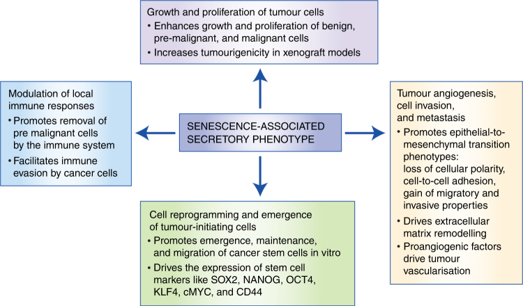
Senescent cells activate genetic programmes that irreversibly inhibit cellular proliferation, but also endow these cells with distinctive metabolic and signalling phenotypes. Although senescence has historically been considered a protective mechanism against tumourigenesis, the activities of senescent cells are increasingly being associated with age-related diseases, including cancer. An important feature of senescent cells is the secretion of a vast array of pro-inflammatory cytokines, chemokines, and growth factors collectively known as the senescence-associated secretory phenotype (SASP). Recent research has shown that SASP paracrine signalling can mediate several pro-tumourigenic effects, such as enhancing malignant phenotypes and promoting tumour initiation. In this review, we summarise the paracrine activities of senescent cells and their role in tumourigenesis through direct effects on growth and proliferation of tumour cells, tumour angiogenesis, invasion and metastasis, cellular reprogramming and emergence of tumour-initiating cells, and tumour interactions with the local immune environment. The evidence described here suggests cellular senescence acts as a double-edged sword in cancer pathogenesis, which demands further attention in order to support the use of senolytic or SASP-modulating compounds for cancer treatment.
Overview
AI snapshot
Written from the paper’s full text.
TL;DR
This review argues that senescent cells, through the senescence-associated secretory phenotype (SASP), can act in a paracrine manner to promote multiple pro-tumourigenic processes, challenging the view of senescence as purely tumour-suppressive. It concludes that senescence is a double-edged sword in cancer and highlights senolytic or SASP-modulating compounds as potential therapeutic strategies.
Key findings
- SASP factors can directly promote tumour cell growth and proliferation: co-culture or co-injection of senescent fibroblasts increased tumourigenicity in xenograft models, including primary breast cancer tissues; AREG blockade reduced senescent-fibroblast-conditioned-media-induced growth of benign prostate epithelial cells; and deletion of IL1β reduced IL6 and CXCL1 expression and the number and size of liver tumours in an obesity-associated liver cancer model.
- Senescent cells can promote angiogenesis, invasion, and metastasis: conditioned media from senescent cells induced EMT in non-aggressive breast cancer, mesothelioma, and melanoma cell lines; MMP2 and MMP3 secreted by senescent cells enhanced invasive properties; IL6 promoted tumour-supportive angiogenesis in a Ras-driven tumour model; and co-injection of senescent fibroblasts or peritoneal mesothelial cells with cancer cells increased tumour angiogenesis in xenografts.
- SASP can inhibit differentiation and promote tumour-initiating/stem-like phenotypes: keratinocytes exposed to senescent-cell medium expressed tumour stem cell marker CD44; co-culture with senescent myeloma cells promoted cancer stem-like cells; mesothelioma SASP produced a highly clonogenic subpopulation with enhanced tumour formation in mice; and IL6 or CCL2 induced tumourigenic potential in melanoma cells in a STAT3-dependent manner with NANOG, SOX2, and OCT4 expression.
- Senescent cells modulate local immunity in opposing ways: they can promote immune clearance via CCL2-mediated NK-T cell attraction, and p53 restoration in murine liver carcinomas triggered innate immune infiltration and complete tumour regression; however, senescent stromal cells and IL6 can increase suppressive myeloid cells, inhibit anti-tumour T-cell responses, and enhance tumour growth.
Methods
- Narrative review of published literature, not a primary experimental study.
- Synthesizes in vitro and in vivo evidence on paracrine effects of senescent cells and SASP components across tumour growth, angiogenesis/invasion/metastasis, cellular reprogramming/tumour-initiating cells, and immune interactions.
- Main analysis is conceptual organization into four paracrine activity domains, with a table of example senolytic and SASP-modulating compounds; no systematic search strategy or quantitative meta-analysis is stated.
Limitations
- The paper states that the relationship between senescence, tumourigenesis, and the immune system is complex and remains incompletely understood, and that further research is required to clarify factors controlling pro- and anti-tumour surveillance activities.
- The paper states that its subdivisions of senescence-associated activities are mainly conceptual, because senescent cells exert compounded effects and current experimental approaches cannot easily distinguish these activities, especially in vivo.
- Evident from the text: as a narrative review, it does not report a systematic literature search, quantitative effect sizes, or formal risk-of-bias assessment.
Model-written summary. Check it against the paper before relying on it.
Integrity signals
Registry
Cites
Statistics
Figures
Verification
Claims the paper makes
43 Not checked yet · 6 Own interpretation · 2 Partly supported · 2 No basis given · 1 Supported · 1 Supported so far · accuracy not yet measured
The sources with a verdict support this claim; the rest weren’t checked yet, have no full text we could read, or their verdicts aren’t rated yet.
In the paper
“Similarly, co-injection of senescent fibroblasts or peritoneal mesothelial cells with cancer cells in xenograft models results in significantly greater tumour angiogenesis.62,63”
§ Paracrine roles of cellular senescence in promoting tumourigenesis > Introduction > Tumour angiogenesis, invasion, and metastasis
What the sources say
- Not checked yetcites [62] Coppe, J. P., Kauser, K., Campisi, J. & Beausejour, C. M. Secretion of vascular endothelial growth factor by primary human fibroblasts at senescence. J. Biol. Chem. 281, 29568–29574 (2006). source
- Supportedcites [63] Mikula-Pietrasik, J. et al. Senescent peritoneal mesothelium induces a pro-angiogenic phenotype in ovarian cancer cells in vitro and in a mouse xenograft model in vivo. Clin. Exp. Metastasis 33, 15–27 (2016). source
The cited paper supports the peritoneal mesothelial-cell part of the sentence. In Results and discussion (Fig. 5), it reports significantly greater CD31/CD34 staining, a measure of tumour angiogenesis, in xenografts formed after co-injection with senescent HPMCs than in the comparison groups. (LLM, conf=0.92)
plaindirect- Asserted: passes — The authors put it forward as true.
- Checkable: passes — It can be checked against evidence.
- Load-bearing: passes — The paper’s conclusions depend on it.
- Based: passes — The paper gives a reason why it holds.
Every source the paper cites for this claim was checked and supports it.
In the paper
“In addition, induction of senescence and SASP in mesothelioma cells led to the emergence of a subpopulation of highly clonogenic cells with enhanced ability to form tumours when xenografted in mice.55”
§ Paracrine roles of cellular senescence in promoting tumourigenesis > Introduction > Cellular reprogramming of cells and emergence of tumour-initiating cells in culture
What the sources say
- Supportedcites [55] Canino, C. et al. SASP mediates chemoresistance and tumor-initiating-activity of mesothelioma cells. Oncogene 31, 3148–3163 (2012). source
The Abstract reports that conditioned media from senescent mesothelioma cells induced clonogenic subpopulations. In “Senescent MPM cells secrete cytokines and pro-mitogenic factors,” cells pretreated with that media formed tumours more frequently and with shorter latency after xenografting in mice. (LLM, conf=0.93)
plaindirect- Asserted: passes — The authors put it forward as true.
- Checkable: passes — It can be checked against evidence.
- Load-bearing: passes — The paper’s conclusions depend on it.
- Based: passes — The paper gives a reason why it holds.
- Supported
A conclusion the paper draws from its own results — it cites no other source for it, so there is none to check. The quote shows it is in the paper.
In the paper
“The evidence described here suggests cellular senescence acts as a double-edged sword in cancer pathogenesis, which demands further attention in order to support the use of senolytic or SASP-modulating compounds for cancer treatment.”
§ Paracrine roles of cellular senescence in promoting tumourigenesis > Abstract
hedgeddirect- Asserted: passes — The authors put it forward as true.
- Checkable: passes — It can be checked against evidence.
- Load-bearing: passes — The paper’s conclusions depend on it.
- Based: passes — The paper gives a reason why it holds.
- Rests on an assumption: The pro-tumourigenic paracrine effects of senescent cells observed in in vitro and mouse models are representative of human cancer pathogenesis.
None of the sources cited for this claim have been checked yet.
In the paper
“Co-culture of senescent fibroblasts, induced by various stimuli (e.g. radiation, DNA damage, replicative exhaustion), can promote the growth and proliferation of benign, pre-malignant, and malignant cells from a range of tumour types.9,12,26,41–44”
§ Paracrine roles of cellular senescence in promoting tumourigenesis > Introduction > Growth and proliferation of tumour cells
What the sources say
- Not checked yetcites [9] Takasugi, M. et al. Small extracellular vesicles secreted from senescent cells promote cancer cell proliferation through EphA2. Nat. Commun. 8, 15729 (2017). source
- Not checked yetcites [12] Coppe, J. P. et al. Senescence-associated secretory phenotypes reveal cell-nonautonomous functions of oncogenic RAS and the p53 tumor suppressor. PLoS Biol. 6, 2853–2868 (2008). source
- Not checked yetcites [26] Krtolica, A., Parrinello, S., Lockett, S., Desprez, P. Y. & Campisi, J. Senescent fibroblasts promote epithelial cell growth and tumorigenesis: a link between cancer and aging. Proc. Natl Acad. Sci. USA 98, 12072–12077 (2001). source
- Not checked yetcites [41] Parrinello, S., Coppe, J. P., Krtolica, A. & Campisi, J. Stromal-epithelial interactions in aging and cancer: senescent fibroblasts alter epithelial cell differentiation. J. Cell Sci. 118(Pt 3), 485–496 (2005). source
- Not checked yetcites [42] Dilley, T. K., Bowden, G. T. & Chen, Q. M. Novel mechanisms of sublethal oxidant toxicity: induction of premature senescence in human fibroblasts confers tumor promoter activity. Exp. Cell Res. 290, 38–48 (2003). source
- Not checked yetcites [43] Liu, D. & Hornsby, P. J. Senescent human fibroblasts increase the early growth of xenograft tumors via matrix metalloproteinase secretion. Cancer Res. 67, 3117–3126 (2007). source
- Not checked yetcites [44] Tsai, K. K., Chuang, E. Y., Little, J. B. & Yuan, Z. M. Cellular mechanisms for low-dose ionizing radiation-induced perturbation of the breast tissue microenvironment. Cancer Res. 65, 6734–6744 (2005). source
plaindirect- Asserted: passes — The authors put it forward as true.
- Checkable: passes — It can be checked against evidence.
- Load-bearing: passes — The paper’s conclusions depend on it.
- Based: passes — The paper gives a reason why it holds.
A conclusion the paper draws from its own results — it cites no other source for it, so there is none to check. The quote shows it is in the paper.
In the paper
“Recent research has shown that SASP paracrine signalling can mediate several pro-tumourigenic effects, such as enhancing malignant phenotypes and promoting tumour initiation.”
§ Paracrine roles of cellular senescence in promoting tumourigenesis > Abstract
plaindirect- Asserted: passes — The authors put it forward as true.
- Checkable: passes — It can be checked against evidence.
- Load-bearing: passes — The paper’s conclusions depend on it.
- Based: passes — The paper gives a reason why it holds.
None of the sources cited for this claim have been checked yet.
In the paper
“However, the deleterious consequences that result from ineffective clearance of senescent cells and their over-accumulation in tissues can promote age-related diseases and cancer.2,26–30”
§ Paracrine roles of cellular senescence in promoting tumourigenesis > Introduction
What the sources say
- Not checked yetcites [2] Munoz-Espin, D. & Serrano, M. Cellular senescence: from physiology to pathology. Nat. Rev. Mol. Cell Biol. 15, 482–496 (2014). source
- Not checked yetcites [26] Krtolica, A., Parrinello, S., Lockett, S., Desprez, P. Y. & Campisi, J. Senescent fibroblasts promote epithelial cell growth and tumorigenesis: a link between cancer and aging. Proc. Natl Acad. Sci. USA 98, 12072–12077 (2001). source
- Not checked yetcites [27] Campisi, J. Aging, cellular senescence, and cancer. Annu Rev. Physiol. 75, 685–705 (2013). source
- Not checked yetcites [28] Lecot, P., Alimirah, F., Desprez, P. Y., Campisi, J. & Wiley, C. Context-dependent effects of cellular senescence in cancer development. Br. J. Cancer 114, 1180–1184 (2016). source
- Not checked yetcites [29] Kuilman, T. et al. Oncogene-induced senescence relayed by an interleukin-dependent inflammatory network. Cell 133, 1019–1031 (2008). source
- Not checked yetcites [30] Perez-Mancera, P. A., Young, A. R. & Narita, M. Inside and out: the activities of senescence in cancer. Nat. Rev. Cancer 14, 547–558 (2014). source
plaindirect- Asserted: passes — The authors put it forward as true.
- Checkable: passes — It can be checked against evidence.
- Load-bearing: passes — The paper’s conclusions depend on it.
- Based: passes — The paper gives a reason why it holds.
None of the sources cited for this claim have been checked yet.
In the paper
“There is mounting evidence that dysregulation or inappropriate activation of senescence contributes to tumour progression and malignancy.5,7,17”
§ Paracrine roles of cellular senescence in promoting tumourigenesis > Introduction
What the sources say
- Not checked yetcites [5] Rodier, F. & Campisi, J. Four faces of cellular senescence. J. Cell Biol. 192, 547–556 (2011). source
- Not checked yetcites [7] Coppe, J. P., Desprez, P. Y., Krtolica, A. & Campisi, J. The senescence-associated secretory phenotype: the dark side of tumor suppression. Annu Rev. Pathol. 5, 99–118 (2010). source
- Not checked yetcites [17] Tchkonia, T., Zhu, Y., van Deursen, J., Campisi, J. & Kirkland, J. L. Cellular senescence and the senescent secretory phenotype: therapeutic opportunities. J. Clin. Invest. 123, 966–972 (2013). source
plaindirect- Asserted: passes — The authors put it forward as true.
- Checkable: passes — It can be checked against evidence.
- Load-bearing: passes — The paper’s conclusions depend on it.
- Based: passes — The paper gives a reason why it holds.
None of the sources cited for this claim have been checked yet.
In the paper
“Cells present in the tumour microenvironment, such as fibroblasts, can become senescent and promote the growth and proliferation of tumour cells.7,39,40”
§ Paracrine roles of cellular senescence in promoting tumourigenesis > Introduction > Growth and proliferation of tumour cells
What the sources say
- Not checked yetcites [7] Coppe, J. P., Desprez, P. Y., Krtolica, A. & Campisi, J. The senescence-associated secretory phenotype: the dark side of tumor suppression. Annu Rev. Pathol. 5, 99–118 (2010). source
- Not checked yetcites [39] Alspach, E., Fu, Y. & Stewart, S. A. Senescence and the pro-tumorigenic stroma. Crit. Rev. Oncog. 18, 549–558 (2013). source
- Not checked yetcites [40] Liu, S. et al. Simvastatin suppresses breast cancer cell proliferation induced by senescent cells. Sci. Rep. 5, 17895 (2015).
plaindirect- Asserted: passes — The authors put it forward as true.
- Checkable: passes — It can be checked against evidence.
- Load-bearing: passes — The paper’s conclusions depend on it.
- Based: passes — The paper gives a reason why it holds.
The paper states this without a source or data behind it here, so there is nothing to check it against. The quote shows it is in the paper.
In the paper
“SASP effects can be beneficial or deleterious for normal physiology depending on its composition, intensity, and the local tissue microenvironment.”
§ Paracrine roles of cellular senescence in promoting tumourigenesis > Introduction
plainconditional- Asserted: passes — The authors put it forward as true.
- Checkable: passes — It can be checked against evidence.
- Load-bearing: unclear — It is unclear whether the paper’s conclusions depend on it.
- Based: passes — The paper gives a reason why it holds.
In the paper
“Supporting this notion, the burden of senescent cells in tissues increases significantly with age in mice, primates, and humans,27 and they can be found in both benign and malignant tumours.31–35”
§ Paracrine roles of cellular senescence in promoting tumourigenesis > Introduction
What the sources say
- Not checked yetcites [27] Campisi, J. Aging, cellular senescence, and cancer. Annu Rev. Physiol. 75, 685–705 (2013). source
plaindirect- Asserted: passes — The authors put it forward as true.
- Checkable: passes — It can be checked against evidence.
- Load-bearing: unclear — It is unclear whether the paper’s conclusions depend on it.
- Based: passes — The paper gives a reason why it holds.
- Probably not a substantial claim. 3
- It may state two separate claims as one. 19
The cited sources support only part of this claim — read each source’s verdict below.
In the paper
“Furthermore, the SASP is involved in valuable physiological processes such as promoting tissue repair,18–20 fine-tuning the development of embryonic structures,21–23 and stimulating immune surveillance.24,25”
§ Paracrine roles of cellular senescence in promoting tumourigenesis > Introduction
What the sources say
- Supportedcites [18] Jun, J. I. & Lau, L. F. The matricellular protein CCN1 induces fibroblast senescence and restricts fibrosis in cutaneous wound healing. Nat. Cell Biol. 12, 676–685 (2010). source
This citation is attached to the tissue-repair clause, not the separate claims about embryonic development or immune surveillance. The Results report that CCN1-induced senescence produces an antifibrotic program in cutaneous wounds, and the Introduction identifies secreted proteins involved in extracellular-matrix metabolism as part of the SASP. Together, these support a beneficial role for the senescent-cell secretory response in wound repair. (LLM, conf=0.88)
- Supportedcites [19] Demaria, M. et al. An essential role for senescent cells in optimal wound healing through secretion of PDGF-AA. Dev. Cell. 31, 722–733 (2014). source
This source is cited for the tissue-repair clause, not the separately cited claims about embryonic development or immune surveillance. The Introduction states that wound-associated senescent cells promote healing by secreting PDGF-A, a SASP factor; the Discussion explicitly describes a positive role for the SASP in tissue repair. Page numbers are not available in our copy. (LLM, conf=0.94)
- Supportedcites [20] Krizhanovsky, V. et al. Senescence of activated stellate cells limits liver fibrosis. Cell 134, 657–667 (2008). source
The citation to this paper is attached to the immune-surveillance clause, not the tissue-repair or embryonic-development clauses. In the Results section, the paper reports that senescent activated stellate cells upregulate genes predicted to enhance immune surveillance; the Discussion links those factors to immune clearance. (LLM, conf=0.93)
- Supportedcites [21] Storer, M. et al. Senescence is a developmental mechanism that contributes to embryonic growth and patterning. Cell 155, 1119–1130 (2013). source
This source is cited for the embryonic-development part of the sentence, not the separate tissue-repair or immune-surveillance claims. The Discussion (p. 9) says that embryonic signaling centers instruct growth and patterning through secreted factors that are also SASP components; the Figure 7 caption (p. 8) describes senescence as instructing local tissue patterning. (LLM, conf=0.90)
- Partly supportedcites [22] Munoz-Espin, D. et al. Programmed cell senescence during mammalian embryonic development. Cell 155, 1104–1118 (2013). sourceExperimental — accuracy not yet measured
The Clearance of Senescent Cells and Compensatory Mechanisms section reports that developmental senescence helps eliminate mesonephric tubules and establish the correct balance of cells in the embryonic endolymphatic sac. The Introduction describes how the SASP can promote tissue remodeling, but the paper does not establish that the SASP itself fine-tunes those embryonic structures. The tissue-repair and immune-surveillance clauses have separate citations and are not assessed here. (LLM, conf=0.82)
- Partly supportedcites [23] Davaapil, H., Brockes, J. P. & Yun, M. H. Conserved and novel functions of programmed cellular senescence during vertebrate development. Development 144, 106–114 (2017). sourceExperimental — accuracy not yet measured
The citation to this paper is attached to the embryonic-development part of the sentence, not the tissue-repair or immune-surveillance parts. In the Discussion, the authors suggest that programmed senescence contributes to cement-gland organogenesis and adjacent tissue remodelling, but say it is only possible that a TGFβ-dependent secretory phenotype mediates those effects. The claim presents the SASP's developmental role more firmly than the paper does. (LLM, conf=0.87)
- Supportedcites [24] Xue, W. et al. Senescence and tumour clearance is triggered by p53 restoration in murine liver carcinomas. Nature 445, 656–660 (2007). source
The citation to this paper is attached to “stimulating immune surveillance,” not to tissue repair or embryonic development. The Abstract reports that senescent tumour cells upregulated immune-modulating signals and triggered an innate immune response that contributed to tumour clearance. (LLM, conf=0.90)
- Not checked yetcites [25] Lujambio, A. To clear, or not to clear (senescent cells)? That is the question. Bioessays 38(Suppl. 1), S56–S64 (2016). source
plaindirect- Asserted: passes — The authors put it forward as true.
- Checkable: passes — It can be checked against evidence.
- Load-bearing: fails — The paper’s conclusions may not depend on it.
- Based: passes — The paper gives a reason why it holds.
Probably not a substantial claim. The paper’s conclusions may not depend on it. It may state two separate claims as one.
- Supported
The cited sources support only part of this claim — read each source’s verdict below.
In the paper
“Importantly, genetic or chemical ablation of senescent cells in mouse models delays the onset of age-related disorders, including cancer, leading to increased life-spans and promoting tissue rejuvenation in late life.36–38”
§ Paracrine roles of cellular senescence in promoting tumourigenesis > Introduction
What the sources say
- Partly supportedcites [36] Chang, J. et al. Clearance of senescent cells by ABT263 rejuvenates aged hematopoietic stem cells in mice. Nat. Med. 22, 78–83 (2016). sourceExperimental — accuracy not yet measured
The Abstract reports that genetic clearance delayed several age-associated disorders in a progeroid mouse model, and that ABT263 cleared senescent cells and rejuvenated aged stem cells in mice. It does not report delayed cancer onset or increased lifespan; it describes extending health span as a prospect, not a demonstrated result. (LLM, conf=0.89)
- Partly supportedcites [37] Baker, D. J. et al. Clearance of p16Ink4a-positive senescent cells delays ageing-associated disorders. Nature 479, 232–236 (2011). sourceExperimental — accuracy not yet measured
The main text (Fig. 2) reports that drug-triggered clearance of p16Ink4a-positive cells in progeroid mice delayed selected age-related phenotypes. It does not report delayed cancer onset and explicitly says overall survival was not substantially extended (Supplementary Fig. 5d). The late-life analysis (Fig. 4) describes attenuated decline in muscle and fat rather than a reversal of ageing, so “tissue rejuvenation” overstates that result. (LLM, conf=0.90)
- Partly supportedcites [38] Baker, D. J. et al. Naturallyoccurringp16(Ink4a)-positive cells shorten healthy lifespan. Nature 530, 184–189 (2016). sourceExperimental — accuracy not yet measured
The Abstract and Discussion (page unavailable) report that clearing p16Ink4a-positive cells in mice extended median lifespan, delayed tumor progression, and slowed age-related deterioration in several organs. But this study used a drug to activate a genetically engineered cell-ablation system; it does not establish that chemical ablation alone has those effects. It reports preservation of tissue function after treatment beginning at midlife, not rejuvenation of already deteriorated tissue in late life. (LLM, conf=0.86)
plaindirect- Asserted: passes — The authors put it forward as true.
- Checkable: passes — It can be checked against evidence.
- Load-bearing: passes — The paper’s conclusions depend on it.
- Based: passes — The paper gives a reason why it holds.
It may state two separate claims as one.
- Partly supported
None of the sources cited for this claim have been checked yet.
In the paper
“The composition and intensity of the SASP response can be affected by several factors including the senescence-inducing mechanism, cell type, and the amount of time passed since senescence initiation, indicating that there is no singular SASP.12–17”
§ Paracrine roles of cellular senescence in promoting tumourigenesis > Introduction
What the sources say
- Not checked yetcites [12] Coppe, J. P. et al. Senescence-associated secretory phenotypes reveal cell-nonautonomous functions of oncogenic RAS and the p53 tumor suppressor. PLoS Biol. 6, 2853–2868 (2008). source
- Not checked yetcites [13] Maciel-Baron, L. A. et al. Senescence associated secretory phenotype profile from primary lung mice fibroblasts depends on the senescence induction stimuli. Age 38, 26 (2016). source
- Not checked yetcites [14] Coppe, J. P. et al. Tumor suppressor and agingbiomarkerp16(INK4a) induces cellular senescence without the associated inflammatory secretory phenotype. J. Biol. Chem. 286, 36396–36403 (2011). source
- Not checked yetcites [15] Kipling, D. et al. A transcriptomic analysis of the EK1.Br strain of human fibroblastoid keratocytes: the effects of growth, quiescence and senescence. Exp. Eye Res. 88, 277–285 (2009). source
- Not checked yetcites [16] Hoare, M. et al. NOTCH1 mediates a switch between two distinct secretomes during senescence. Nat. Cell Biol. 18, 979–992 (2016). source
- Not checked yetcites [17] Tchkonia, T., Zhu, Y., van Deursen, J., Campisi, J. & Kirkland, J. L. Cellular senescence and the senescent secretory phenotype: therapeutic opportunities. J. Clin. Invest. 123, 966–972 (2013). source
hedgeddirect- Asserted: passes — The authors put it forward as true.
- Checkable: passes — It can be checked against evidence.
- Load-bearing: unclear — It is unclear whether the paper’s conclusions depend on it.
- Based: passes — The paper gives a reason why it holds.
It may state two separate claims as one.
None of the sources cited for this claim have been checked yet.
In the paper
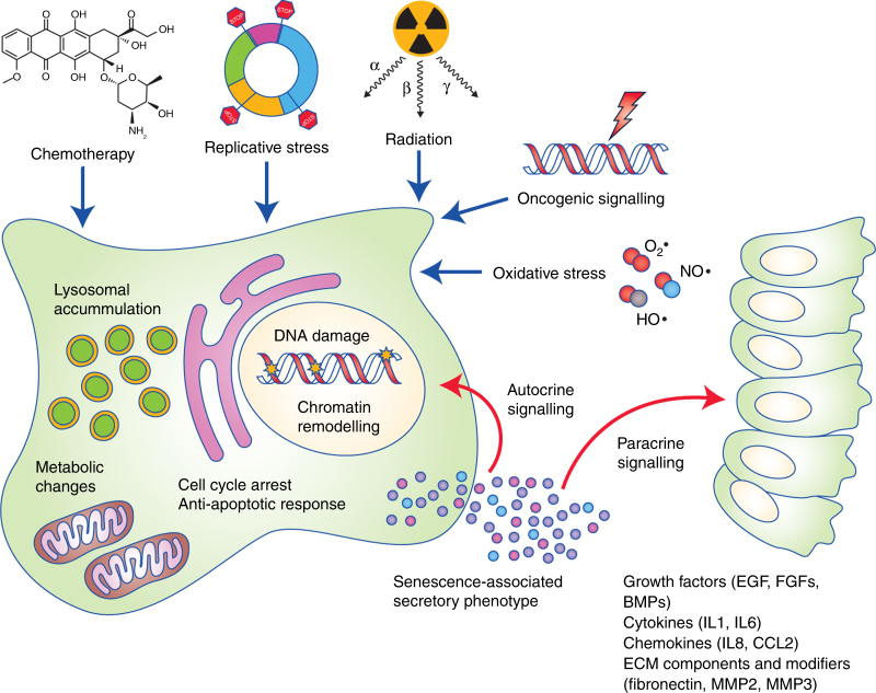
“Activation of the senescence programme leads to cellular and molecular changes such as proliferation arrest, chromatin remodelling, elevated expression of cell cycle inhibitors (such as p16INK4A or p21CIP1), activation of a DNA damage response, enlargement of the lysosomal compartment, and activation of a senescence-associated secretory phenotype (SASP).5,6”
§ Paracrine roles of cellular senescence in promoting tumourigenesis > Introduction
What the sources say
plaindirect- Asserted: passes — The authors put it forward as true.
- Checkable: passes — It can be checked against evidence.
- Load-bearing: passes — The paper’s conclusions depend on it.
- Based: passes — The paper gives a reason why it holds.
It may state two separate claims as one.
The paper states this without a source or data behind it here, so there is nothing to check it against. The quote shows it is in the paper.
In the paper
“Senescence serves as a response to stress, and several inducing stimuli have now been identified including chemotherapeutic, radiation and oxidative stress, amongst others (Fig. 1).”
§ Paracrine roles of cellular senescence in promoting tumourigenesis > Introduction
plaindirect- Asserted: passes — The authors put it forward as true.
- Checkable: passes — It can be checked against evidence.
- Load-bearing: fails — The paper’s conclusions may not depend on it.
- Based: passes — The paper gives a reason why it holds.
Probably not a substantial claim. The paper’s conclusions may not depend on it. It may state two separate claims as one.
None of the sources cited for this claim have been checked yet.
In the paper
“Supporting this notion, the burden of senescent cells in tissues increases significantly with age in mice, primates, and humans,27 and they can be found in both benign and malignant tumours.31–35”
§ Paracrine roles of cellular senescence in promoting tumourigenesis > Introduction
What the sources say
- Not checked yetcites [31] Collado, M. et al. Tumour biology: senescence in premalignant tumours. Nature 436, 642 (2005). source
- Not checked yetcites [32] Haugstetter, A. M. et al. Cellular senescence predicts treatment outcome in metastasised colorectal cancer. Br. J. Cancer 103, 505–509 (2010). source
- Not checked yetcites [33] Vizioli, M. G. et al. Evidence of oncogene-induced senescence in thyroid carcinogenesis. Endocr. Relat. Cancer 18, 743–757 (2011). source
- Not checked yetcites [34] Reimann, M. et al. Tumor stroma-derived TGF-beta limits myc-driven lymphomagenesis via Suv39h1-dependent senescence. Cancer Cell. 17, 262–272 (2010). source
- Not checked yetcites [35] Roberson, R. S., Kussick, S. J., Vallieres, E., Chen, S. Y. & Wu, D. Y. Escape from therapy-induced accelerated cellular senescence in p53-null lung cancer cells and in human lung cancers. Cancer Res. 65, 2795–2803 (2005). source
plain- Asserted: passes — The authors put it forward as true.
- Checkable: passes — It can be checked against evidence.
- Load-bearing: fails — The paper’s conclusions may not depend on it.
- Based: passes — The paper gives a reason why it holds.
Probably not a substantial claim. The paper’s conclusions may not depend on it.
None of the sources cited for this claim have been checked yet.
In the paper
“Senescence was traditionally considered an innate anti-cancer mechanism as it can serve to eliminate damaged cells,3,5 whereby activation of the senescence programme in cells harbouring oncogenic mutations serves as a tumour suppressor mechanism, preventing the expansion of these mutated cells and progression into malignancies.3,5”
§ Paracrine roles of cellular senescence in promoting tumourigenesis > Introduction
What the sources say
plaindirect- Asserted: passes — The authors put it forward as true.
- Checkable: passes — It can be checked against evidence.
- Load-bearing: unclear — It is unclear whether the paper’s conclusions depend on it.
- Based: passes — The paper gives a reason why it holds.
It may state two separate claims as one.
None of the sources cited for this claim have been checked yet.
In the paper
“Senescent cells can contribute to the acquisition of invasive and metastatic properties of cancer cells, as well as the induction of tumour-associated angiogenesis.7,39”
§ Paracrine roles of cellular senescence in promoting tumourigenesis > Introduction > Tumour angiogenesis, invasion, and metastasis
What the sources say
- Not checked yetcites [7] Coppe, J. P., Desprez, P. Y., Krtolica, A. & Campisi, J. The senescence-associated secretory phenotype: the dark side of tumor suppression. Annu Rev. Pathol. 5, 99–118 (2010). source
- Not checked yetcites [39] Alspach, E., Fu, Y. & Stewart, S. A. Senescence and the pro-tumorigenic stroma. Crit. Rev. Oncog. 18, 549–558 (2013). source
plainconditional- Asserted: passes — The authors put it forward as true.
- Checkable: passes — It can be checked against evidence.
- Load-bearing: passes — The paper’s conclusions depend on it.
- Based: passes — The paper gives a reason why it holds.
It may state two separate claims as one.
None of the sources cited for this claim have been checked yet.
In the paper
“Interestingly, the SASP is able to inhibit differentiation both in vitro and in vivo, while in some cases leads to acquisition of stem cell characteristics.41,44,54,55,64–66”
§ Paracrine roles of cellular senescence in promoting tumourigenesis > Introduction > Cellular reprogramming of cells and emergence of tumour-initiating cells in culture
What the sources say
- Not checked yetcites [41] Parrinello, S., Coppe, J. P., Krtolica, A. & Campisi, J. Stromal-epithelial interactions in aging and cancer: senescent fibroblasts alter epithelial cell differentiation. J. Cell Sci. 118(Pt 3), 485–496 (2005). source
- Not checked yetcites [44] Tsai, K. K., Chuang, E. Y., Little, J. B. & Yuan, Z. M. Cellular mechanisms for low-dose ionizing radiation-induced perturbation of the breast tissue microenvironment. Cancer Res. 65, 6734–6744 (2005). source
- Not checked yetcites [54] Ohanna, M. et al. Secretome from senescent melanoma engages the STAT3 pathway to favor reprogramming of naive melanoma towards a tumor-initiating cell phenotype. Oncotarget 4, 2212–2224 (2013). source
- Not checked yetcites [55] Canino, C. et al. SASP mediates chemoresistance and tumor-initiating-activity of mesothelioma cells. Oncogene 31, 3148–3163 (2012). source
- Not checked yetcites [64] Cahu, J., Bustany, S. & Sola, B. Senescence-associated secretory phenotype favors the emergence of cancer stem-like cells. Cell Death Dis. 3, e446 (2012). source
- Not checked yetcites [65] Ritschka, B. et al. The senescence-associated secretory phenotype induces cellular plasticity and tissue regeneration. Genes Dev. 31, 172–183 (2017). source
- Not checked yetcites [66] Mosteiro, L. et al. Tissue damage and senescence provide critical signals for cellular reprogramming in vivo. Science 354, (2016). source
plaindirect- Asserted: passes — The authors put it forward as true.
- Checkable: passes — It can be checked against evidence.
- Load-bearing: passes — The paper’s conclusions depend on it.
- Based: passes — The paper gives a reason why it holds.
It may state two separate claims as one.
In the paper
“Exposure of keratinocytes to the culture medium from senescent cells promotes expression of tumour stem cell markers, such as CD44, and leads to a greater regenerative capacity in vivo. 65”
§ Paracrine roles of cellular senescence in promoting tumourigenesis > Introduction > Cellular reprogramming of cells and emergence of tumour-initiating cells in culture
What the sources say
- Not checked yetcites [65] Ritschka, B. et al. The senescence-associated secretory phenotype induces cellular plasticity and tissue regeneration. Genes Dev. 31, 172–183 (2017). source
plaindirect- Asserted: passes — The authors put it forward as true.
- Checkable: passes — It can be checked against evidence.
- Load-bearing: passes — The paper’s conclusions depend on it.
- Based: passes — The paper gives a reason why it holds.
It may state two separate claims as one.
In the paper
“In addition, individual SASP components can contribute to induce EMT phenotypes; IL6, for example, has been shown to have cell-adhesion disrupting actions, which is an important component of invasion.56”
§ Paracrine roles of cellular senescence in promoting tumourigenesis > Introduction > Tumour angiogenesis, invasion, and metastasis
What the sources say
- Not checked yetcites [56] Tamm, I., Kikuchi, T., Cardinale, I. & Krueger, J. G. Cell-adhesion-disrupting action of interleukin 6 in human ductal breast carcinoma cells. Proc. Natl Acad. Sci. USA 91, 3329–3333 (1994). source
plaindirect- Asserted: passes — The authors put it forward as true.
- Checkable: passes — It can be checked against evidence.
- Load-bearing: unclear — It is unclear whether the paper’s conclusions depend on it.
- Based: passes — The paper gives a reason why it holds.
It may state two separate claims as one.
None of the sources cited for this claim have been checked yet.
In the paper
“A large number of proangiogenic factors are also known to be secreted by senescent cells, whereas angiostatic molecules have not been found to be secreted.27,60”
§ Paracrine roles of cellular senescence in promoting tumourigenesis > Introduction > Tumour angiogenesis, invasion, and metastasis
What the sources say
- Not checked yetcites [27] Campisi, J. Aging, cellular senescence, and cancer. Annu Rev. Physiol. 75, 685–705 (2013). source
- Not checked yetcites [60] Oubaha, M. et al. Senescence-associated secretory phenotype contributes to pathological angiogenesis in retinopathy. Sci. Transl. Med. 8, 362ra144 (2016). source
boosted- Asserted: passes — The authors put it forward as true.
- Checkable: passes — It can be checked against evidence.
- Load-bearing: passes — The paper’s conclusions depend on it.
- Based: passes — The paper gives a reason why it holds.
It may state two separate claims as one.
A conclusion the paper draws from its own results — it cites no other source for it, so there is none to check. The quote shows it is in the paper.
In the paper
“These experiments collectively demonstrate that the SASP can promote cancer cell growth, challenging the view that senescence is primordially a beneficial process involved in preventing cancer progression.”
§ Paracrine roles of cellular senescence in promoting tumourigenesis > Introduction > Growth and proliferation of tumour cells
boosteddirect- Asserted: passes — The authors put it forward as true.
- Checkable: passes — It can be checked against evidence.
- Load-bearing: passes — The paper’s conclusions depend on it.
- Based: passes — The paper gives a reason why it holds.
It may state two separate claims as one.
None of the sources cited for this claim have been checked yet.
In the paper
“Furthermore, cellular reprogramming, which is the process by which adult differentiated cells can be induced to become functionally equivalent to embryonic stem cells, can be induced in vivo by senescent cells through SASP activation, and this can be stimulated in different models of tissue damage.”
§ Paracrine roles of cellular senescence in promoting tumourigenesis > Introduction > Cellular reprogramming of cells and emergence of tumour-initiating cells in culture
plaindirect- Asserted: passes — The authors put it forward as true.
- Checkable: passes — It can be checked against evidence.
- Load-bearing: passes — The paper’s conclusions depend on it.
- Based: passes — The paper gives a reason why it holds.
It may state two separate claims as one.
None of the sources cited for this claim have been checked yet.
In the paper
“Cells undergoing damage-induced senescence are often cleared by the immune system, as several SASP factors are cytokines and chemokines that can modulate the local immune environment.2,3,5,70,71”
§ Paracrine roles of cellular senescence in promoting tumourigenesis > Introduction > Modulation of local immune response and immune evasion by senescent cells
What the sources say
- Not checked yetcites [2] Munoz-Espin, D. & Serrano, M. Cellular senescence: from physiology to pathology. Nat. Rev. Mol. Cell Biol. 15, 482–496 (2014). source
- Not checked yetcites [3] He, S. & Sharpless, N. E. Senescence in health and disease. Cell 169, 1000–1011 (2017). source
- Not checked yetcites [5] Rodier, F. & Campisi, J. Four faces of cellular senescence. J. Cell Biol. 192, 547–556 (2011). source
- Not checked yetcites [70] Vicente, R., Mausset-Bonnefont, A. L., Jorgensen, C., Louis-Plence, P. & Brondello, J. M. Cellular senescence impact on immune cell fate and function. Aging Cell 15, 400–16 (2016). source
- Not checked yetcites [71] Velarde, M. C., Demaria, M. & Campisi, J. Senescent cells and their secretory phenotype as targets for cancer therapy. Interdiscip. Top. Gerontol. 38, 17–27 (2013). source
plaindirect- Asserted: passes — The authors put it forward as true.
- Checkable: passes — It can be checked against evidence.
- Load-bearing: unclear — It is unclear whether the paper’s conclusions depend on it.
- Based: passes — The paper gives a reason why it holds.
It may state two separate claims as one.
In the paper
“Furthermore, it was shown that this leads to the inhibition of anti-tumour T-cell responses and enhanced tumour growth.77”
§ Paracrine roles of cellular senescence in promoting tumourigenesis > Introduction > Modulation of local immune response and immune evasion by senescent cells
What the sources say
- Not checked yetcites [77] Ruhland, M. K. et al. Stromal senescence establishes an immunosuppressive microenvironment that drives tumorigenesis. Nat. Commun. 7, 11762 (2016). source
plaindirect- Asserted: passes — The authors put it forward as true.
- Checkable: passes — It can be checked against evidence.
- Load-bearing: passes — The paper’s conclusions depend on it.
- Based: passes — The paper gives a reason why it holds.
It may state two separate claims as one.
In the paper
“In this sense, a crucial role for the mechanistic target of rapamycin (mTOR) complex has recently been unveiled, whereby it can either counteract or facilitate reprogramming by cell-intrinsic and cell-extrinsic mechanisms, respectively.69”
§ Paracrine roles of cellular senescence in promoting tumourigenesis > Introduction > Cellular reprogramming of cells and emergence of tumour-initiating cells in culture
What the sources say
- Not checked yetcites [69] Aarts, M. et al. Coupling shRNA screens with single-cell RNA-seq identifies a dual role for mTOR in reprogramming-induced senescence. Genes Dev. 31, 2085–2098 (2017). source
plaindirect- Asserted: passes — The authors put it forward as true.
- Checkable: passes — It can be checked against evidence.
- Load-bearing: unclear — It is unclear whether the paper’s conclusions depend on it.
- Based: passes — The paper gives a reason why it holds.
It may state two separate claims as one.
None of the sources cited for this claim have been checked yet.
In the paper
“While senescence is a barrier to reprogramming in vitro, the paracrine activities of senescent cells can promote the expression of stem cell markers and proliferation of neighbouring cells in vivo, 66–68 and IL6 is a key player in driving this process.”
§ Paracrine roles of cellular senescence in promoting tumourigenesis > Introduction > Cellular reprogramming of cells and emergence of tumour-initiating cells in culture
What the sources say
- Not checked yetcites [66] Mosteiro, L. et al. Tissue damage and senescence provide critical signals for cellular reprogramming in vivo. Science 354, (2016). source
- Not checked yetcites [67] Chiche, A. et al. Injury-induced senescence enables in vivo reprogramming in skeletal muscle. Cell. Stem Cell. 20, 407–14.e4 (2017). source
- Not checked yetcites [68] Mosteiro, L., Pantoja, C., de Martino, A. & Serrano, M. Senescence promotes in vivo reprogramming through p16(INK)(4a) and IL-6. Aging Cell (2017). PubMed PMID: 29280266. Epub 2017/12/28. eng. https://doi.org/10.1111/acel.12711 . source
plaindirect- Asserted: passes — The authors put it forward as true.
- Checkable: passes — It can be checked against evidence.
- Load-bearing: passes — The paper’s conclusions depend on it.
- Based: passes — The paper gives a reason why it holds.
It may state two separate claims as one.
A conclusion the paper draws from its own results — it cites no other source for it, so there is none to check. The quote shows it is in the paper.
In the paper
“Together, these data suggest that senescent cells, through their SASP, can induce undifferentiated cellular states; depending on the context, this can be beneficial (e.g. tissue regeneration) or harmful (e.g. promotion of tumour-initiating cells).”
§ Paracrine roles of cellular senescence in promoting tumourigenesis > Introduction > Cellular reprogramming of cells and emergence of tumour-initiating cells in culture
hedgeddirect- Asserted: passes — The authors put it forward as true.
- Checkable: passes — It can be checked against evidence.
- Load-bearing: passes — The paper’s conclusions depend on it.
- Based: passes — The paper gives a reason why it holds.
It may state two separate claims as one.
None of the sources cited for this claim have been checked yet.
In the paper
“Since senescent cells are generally abundant in benign tumours and also present at low numbers in several malignancies,31–34,57 their paracrine activities could contribute to tumour progression and cancer metastasis.”
§ Paracrine roles of cellular senescence in promoting tumourigenesis > Conclusion
What the sources say
- Not checked yetcites [31] Collado, M. et al. Tumour biology: senescence in premalignant tumours. Nature 436, 642 (2005). source
- Not checked yetcites [32] Haugstetter, A. M. et al. Cellular senescence predicts treatment outcome in metastasised colorectal cancer. Br. J. Cancer 103, 505–509 (2010). source
- Not checked yetcites [33] Vizioli, M. G. et al. Evidence of oncogene-induced senescence in thyroid carcinogenesis. Endocr. Relat. Cancer 18, 743–757 (2011). source
- Not checked yetcites [34] Reimann, M. et al. Tumor stroma-derived TGF-beta limits myc-driven lymphomagenesis via Suv39h1-dependent senescence. Cancer Cell. 17, 262–272 (2010). source
- Not checked yetcites [57] Kim, Y. H. et al. Senescent tumor cells lead the collective invasion in thyroid cancer. Nat. Commun. 8, 15208 (2017).
hedgedconditional- Asserted: passes — The authors put it forward as true.
- Checkable: passes — It can be checked against evidence.
- Load-bearing: passes — The paper’s conclusions depend on it.
- Based: passes — The paper gives a reason why it holds.
It may state two separate claims as one.
The paper relies on this without support.
The pro-tumourigenic paracrine effects of senescent cells observed in in vitro and mouse models are representative of human cancer pathogenesis.
“The evidence described here suggests cellular senescence acts as a double-edged sword in cancer pathogenesis, which demands further attention in order to support the use of senolytic or SASP-modulating compounds for cancer treatment.”
Claims other papers make about this one
Each is checked against this paper’s own text: does it say what the citing paper says it does?
The cited source says what the claim attributes to it.
In the citing paper
“Senescence has the potential to spread damage to surrounding tissue via paracrine signaling with SASP factors, while simultaneously using autocrine reinforcement to maintain their senescent phenotype [48].”
From DNA repair deficiency and senescence in concussed professional athletes involved in contact sports (2019) · cites this paper as [48]
In the citing paper
“In particular, cellular senescence can cause significant effects on brain health and function by affecting various cell types and spreading its effects to surrounding tissue [48].”
From DNA repair deficiency and senescence in concussed professional athletes involved in contact sports (2019) · cites this paper as [48]
Citations
Cites
The serial cultivation of human diploid cell strains
Hayflick, L., & Moorhead, P. S. (1961) · Experimental Cell Research · doi:10.1016/0014-4827(61)90192-6 · Cited by 8,508
The senescence-associated secretory phenotype: the dark side of tumor suppression
Coppé, J.-P., Desprez, P.-Y., Krtolica, A., & Campisi, J. (2010) · Annual Review of Pathology: Mechanisms of Disease · doi:10.1146/annurev-pathol-121808-102144 · Cited by 5,206
Clearance of p16Ink4a-positive senescent cells delays ageing-associated disorders
Baker, D. J., Wijshake, T., Tchkonia, T., LeBrasseur, N. K., Childs, B. G., van de Sluis, B., Kirkland, J. L., & van Deursen, J. M. (2011) · Nature · doi:10.1038/nature10600 · Cited by 3,670
Aging, Cellular Senescence, and Cancer
Campisi, J. (2013) · Annual Review of Physiology · doi:10.1146/annurev-physiol-030212-183653 · Cited by 2,954 · cited 1×
“A large number of proangiogenic factors are also known to be secreted by senescent cells, whereas angiostatic molecules have not been found to be secreted.(27,60) In particular, IL6 has been reported to promote tumour-supportive angiogenesis in a Rasdriven tumour model.”
Naturally occurring p16Ink4a-positive cells shorten healthy lifespan
Baker, D. J., Childs, B. G., Durik, M., Wijers, M. E., Sieben, C. J., Zhong, J., A. Saltness, R., Jeganathan, K. B., Verzosa, G. C., Pezeshki, A., et al. (2016) · Nature · doi:10.1038/nature16932 · Cited by 2,876
Cellular senescence: from physiology to pathology
Muñoz-Espín, D., & Serrano, M. (2014) · Nature Reviews Molecular Cell Biology · doi:10.1038/nrm3823 · Cited by 2,818
The role of senescent cells in ageing
van Deursen, J. M. (2014) · Nature · doi:10.1038/nature13193 · Cited by 2,623
The Achilles' heel of senescent cells: from transcriptome to senolytic drugs
Zhu, Y., Tchkonia, T., Pirtskhalava, T., Gower, A. C., Ding, H., Giorgadze, N., Palmer, A. K., Ikeno, Y., Hubbard, G. B., Lenburg, M., et al. (2015) · Aging Cell · doi:10.1111/acel.12344 · Cited by 2,584
Senescence and tumour clearance is triggered by p53 restoration in murine liver carcinomas
Xue, W., Zender, L., Miething, C., Dickins, R. A., Hernando, E., Krizhanovsky, V., Cordon-Cardo, C., & Lowe, S. W. (2007) · Nature · doi:10.1038/nature05529 · Cited by 2,473
Obesity-induced gut microbial metabolite promotes liver cancer through senescence secretome
Yoshimoto, S., Loo, T. M., Atarashi, K., Kanda, H., Sato, S., Oyadomari, S., Iwakura, Y., Oshima, K., Morita, H., Hattori, M., et al. (2013) · Nature · doi:10.1038/nature12347 · Cited by 2,219
Cited by
Sorted by citations
A guide to assessing cellular senescence in vitro and in vivo
González‐Gualda, E., Baker, A. G., Fruk, L., & Muñoz‐Espín, D. (2021) · The FEBS Journal · doi:10.1111/febs.15570 · Cited by 645
Role of p53 in the Regulation of Cellular Senescence
Mijit, M., Caracciolo, V., Melillo, A., Amicarelli, F., & Giordano, A. (2020) · Biomolecules · doi:10.3390/biom10030420 · Cited by 639
Identification and characterization of Cardiac Glycosides as senolytic compounds
Triana-Martínez, F., Picallos-Rabina, P., Da Silva-Álvarez, S., Pietrocola, F., Llanos, S., Rodilla, V., Soprano, E., Pedrosa, P., Ferreirós, A., Barradas, M., et al. (2019) · Nature Communications · doi:10.1038/s41467-019-12888-x · Cited by 384
González‐Gualda, E., Pàez‐Ribes, M., Lozano‐Torres, B., Macias, D., Wilson, J. R., González‐López, C., Ou, H., Mirón‐Barroso, S., Zhang, Z., Lérida‐Viso, A., et al. (2020) · Aging Cell · doi:10.1111/acel.13142 · Cited by 303
Cellular senescence in development, regeneration and disease
Rhinn, M., Ritschka, B., & Keyes, W. M. (2019) · Development · doi:10.1242/dev.151837 · Cited by 279
Targeting senescent cells in translational medicine
Paez‐Ribes, M., González‐Gualda, E., Doherty, G. J., & Muñoz‐Espín, D. (2019) · EMBO Molecular Medicine · doi:10.15252/emmm.201810234 · Cited by 293
Aging of the cells: Insight into cellular senescence and detection Methods.
Mohamad Kamal, N. S., Safuan, S., Shamsuddin, S., & Foroozandeh, P. (2020) · European Journal of Cell Biology · doi:10.1016/j.ejcb.2020.151108 · Cited by 287
Clearance of senescent macrophages ameliorates tumorigenesis in KRAS-driven lung cancer
Haston, S., Gonzalez-Gualda, E., Morsli, S., Ge, J., Reen, V., Calderwood, A., Moutsopoulos, I., Panousopoulos, L., Deletic, P., Carreno, G., et al. (2023) · Cancer Cell · doi:10.1016/j.ccell.2023.05.004 · Cited by 251
Connective tissue and fibroblast senescence in skin aging
Wlaschek, M., Maity, P., Makrantonaki, E., & Scharffetter-Kochanek, K. (2021) · Journal of Investigative Dermatology · doi:10.1016/j.jid.2020.11.010 · Cited by 245
Cellular senescence in cancer: from mechanisms to detection
Ou, H., Hoffmann, R. J., González‐López, C., Doherty, G. J., Korkola, J. E., & Muñoz‐Espín, D. (2020) · Molecular Oncology · doi:10.1002/1878-0261.12807 · Cited by 208
On the evolution of cellular senescence
Kowald, A., Passos, J. F., & Kirkwood, T. B. L. (2020) · Aging Cell · doi:10.1111/acel.13270 · Cited by 189
Galactose‐modified duocarmycin prodrugs as senolytics
Guerrero, A., Guiho, R., Herranz, N., Uren, A., Withers, D. J., Martínez‐Barbera, J. P., Tietze, L. F., & Gil, J. (2020) · Aging Cell · doi:10.1111/acel.13133 · Cited by 172
Paul, R., Dorsey, J. F., & Fan, Y. (2021) · Pharmacology & Therapeutics · doi:10.1016/j.pharmthera.2021.107985 · Cited by 170
Senescence and senotherapeutics: a new field in cancer therapy
Myrianthopoulos, V., Evangelou, K., Vasileiou, P. V. S., Cooks, T., Vassilakopoulos, T. P., Pangalis, G. A., Kouloukoussa, M., Kittas, C., Georgakilas, A. G., & Gorgoulis, V. G. (2019) · Pharmacology & Therapeutics · doi:10.1016/j.pharmthera.2018.08.006 · Cited by 160
Senescence and the tumor-immune landscape: Implications for cancer immunotherapy
Chibaya, L., Snyder, J. T., & Ruscetti, M. A. (2022) · Seminars in Cancer Biology · doi:10.1016/j.semcancer.2022.02.005 · Cited by 146
Wang, Y., Xiong, H., Liu, D., Hill, C. N., Ertay, A., Li, J., Zou, Y., Miller, P., White, E., Downward, J., et al. (2019) · Autophagy · doi:10.1080/15548627.2019.1569912 · Cited by 134
Targeting Cardiac Stem Cell Senescence to Treat Cardiac Aging and Disease
Cianflone, E., Torella, M., Biamonte, F., Angelis, A. D., Urbanek, K., Costanzo, F. S., Rota, M., Ellison, G. M., & Torella, D. (2020) · Cells · doi:10.3390/cells9061558 · Cited by 137
Boyajian, J. L., Ghebretatios, M., Schaly, S., Islam, P., & Prakash, S. (2021) · Nutrients · doi:10.3390/nu13124550 · Cited by 115
Cellular senescence in lung cancer: Molecular mechanisms and therapeutic interventions
Jha, S. K., Rubis, G. D., Devkota, S. R., Zhang, Y., Adhikari, R., Jha, L. A., Bhattacharya, K., Mehndiratta, S., Gupta, G., Singh, S. K., et al. (2024) · Ageing Research Reviews · doi:10.1016/j.arr.2024.102315 · Cited by 138
Paramos-de-Carvalho, D., Jacinto, A., & Saúde, L. (2021) · eLife · doi:10.7554/elife.72449 · Cited by 111
Senescent cells and macrophages: key players for regeneration?
Elder, S. S., & Emmerson, E. (2020) · Open Biology · doi:10.1098/rsob.200309 · Cited by 114
Jeong, E., Brady, O. A., Martina, J. A., Pirooznia, M., Tunc, I., & Puertollano, R. (2018) · eLife · doi:10.7554/elife.40856 · Cited by 100
Wang, H., Fu, H., Zhu, R., Wu, X., Ji, X., Li, X., Jiang, H., Lin, Z., Tang, X., Sun, S., et al. (2020) · Aging · doi:10.18632/aging.103200 · Cited by 96
Biological mechanisms of aging predict age-related disease co-occurrence in patients
Fraser, H. C., Kuan, V., Johnen, R., Zwierzyna, M., Hingorani, A. D., Beyer, A., & Partridge, L. (2022) · Aging Cell · doi:10.1111/acel.13524 · Cited by 90
Self-citationCellular senescence in cancer: clinical detection and prognostic implications
Domen, A., Deben, C., Verswyvel, J., Flieswasser, T., Prenen, H., Peeters, M., Lardon, F., & Wouters, A. (2022) · Journal of Experimental & Clinical Cancer Research · doi:10.1186/s13046-022-02555-3 · Cited by 94
Senescent Tumor Cells Build a Cytokine Shield in Colorectal Cancer
Choi, Y. W., Kim, Y. H., Oh, S. Y., Suh, K. W., Kim, Y., Lee, G., Yoon, J. E., Park, S. S., Lee, Y., Park, Y. J., et al. (2021) · Advanced Science · doi:10.1002/advs.202002497 · Cited by 92
Potential Regulators of the Senescence-Associated Secretory Phenotype During Senescence and Aging
Han, X., Lei, Q., Xie, J., Liu, H., Li, J., Zhang, X., Zhang, T., & Gou, X. (2022) · The Journals of Gerontology Series A · doi:10.1093/gerona/glac097 · Cited by 100
Contemporary Biological Insights and Clinical Management of Craniopharyngioma
Apps, J. R., Muller, H. L., Hankinson, T. C., Yock, T. I., & Martı́nez-Barberá, J. P. (2022) · Endocrine Reviews · doi:10.1210/endrev/bnac035 · Cited by 93
Self-citationMazzoni, M., Mauro, G., Erreni, M., Romeo, P., Minna, E., Vizioli, M. G., Belgiovine, C., Rizzetti, M. G., Pagliardini, S., Avigni, R., et al. (2019) · Journal of Experimental & Clinical Cancer Research · doi:10.1186/s13046-019-1198-8 · Cited by 86
DNA Damage-Induced Inflammatory Microenvironment and Adult Stem Cell Response
Cinat, D., Coppes, R. P., & Barazzuol, L. (2021) · Frontiers in Cell and Developmental Biology · doi:10.3389/fcell.2021.729136 · Cited by 79
Hu, Q., Peng, J., Jiang, L., Li, W., Su, Q., Zhang, J., Li, H., Song, M., Cheng, B., Xia, J., et al. (2020) · Cell Death and Disease · doi:10.1038/s41419-020-03126-0 · Cited by 78
Cell senescence in neuropathology: A focus on neurodegeneration and tumours
Carreno, G., Guiho, R., & Martínez-Barberá, J. P. (2020) · Neuropathology and Applied Neurobiology · doi:10.1111/nan.12689 · Cited by 83
TGF-β in the microenvironment induces a physiologically occurring immune-suppressive senescent state
Matsuda, S., Revandkar, A., Dubash, T. D., Ravi, A., Wittner, B. S., Lin, M., Morris, R. T., Burr, R., Guo, H., Seeger, K., et al. (2023) · Cell Reports · doi:10.1016/j.celrep.2023.112129 · Cited by 76
Lee, S. Y., Ju, M. K., Jeon, H. M., Lee, Y. J., Kim, C. H., Park, H. G., Han, S. I., & Kang, H. S. (2018) · Oxidative Medicine and Cellular Longevity · doi:10.1155/2018/1027453 · Cited by 69
Zhao, B., Wu, B. C., Feng, N., Zhang, X., Zhang, X., Wei, Y., & Zhang, W. (2023) · Journal of Hematology & Oncology · doi:10.1186/s13045-023-01426-4 · Cited by 68
Khalil, R., Diab‐Assaf, M., & Lemaître, J.-M. (2023) · Cells · doi:10.3390/cells12060915 · Cited by 68
The controversial role of senescence-associated secretory phenotype (SASP) in cancer therapy
Cao, L., Li, K., Li, Q., Tong, Q., Wang, Y., & Huang, L. (2025) · Molecular Cancer · doi:10.1186/s12943-025-02475-8 · Cited by 79
Selective Vulnerability of Senescent Glioblastoma Cells to BCL-XL Inhibition
Rahman, M., Olson, I., Mansour, M. A., Carlstrom, L. P., Sutiwisesak, R., Saber, R., Rajani, K. R., Warrington, A. E., Howard, A., Schroeder, M. A., et al. (2022) · Molecular Cancer Research · doi:10.1158/1541-7786.mcr-21-0029 · Cited by 67
Bientinesi, E., Lulli, M., Becatti, M., Ristori, S., Margheri, F., & Monti, D. (2022) · Mechanisms of Ageing and Development · doi:10.1016/j.mad.2022.111689 · Cited by 61
Schwab, N., Taskina, D., Leung, E., Innes, B. T., Bader, G. D., & Hazrati, L. (2022) · Frontiers in Neuroscience · doi:10.3389/fnins.2022.1027116 · Cited by 56
Are immunosenescent T cells really senescent?
Slaets, H., Veeningen, N., de Keizer, P. L. J., Hellings, N., & Hendrix, S. (2024) · Aging Cell · doi:10.1111/acel.14300 · Cited by 54
Walters, H., & Cox, L. S. (2021) · Oxidative Medicine and Cellular Longevity · doi:10.1155/2021/6697861 · Cited by 51
Diverse Roles of Cellular Senescence in Skeletal Muscle Inflammation, Regeneration, and Therapeutics
Saito, Y., & Chikenji, T. (2021) · Frontiers in Pharmacology · doi:10.3389/fphar.2021.739510 · Cited by 53
DNA repair deficiency and senescence in concussed professional athletes involved in contact sports
Schwab, N., Grenier, K., & Hazrati, L.-N. (2019) · Acta Neuropathologica Communications · doi:10.1186/s40478-019-0822-3 · Cited by 47
Ezure, T., Sugahara, M., & Amano, S. (2019) · BioFactors · doi:10.1002/biof.1512 · Cited by 47
González-Meljem, J. M., & Martínez-Barberá, J. P. (2021) · Cellular and Molecular Life Sciences · doi:10.1007/s00018-021-03798-7 · Cited by 48
Yamada, Z., Nishio, J., Motomura, K., Mizutani, S., Yamada, S., Mikami, T., & Nanki, T. (2022) · Frontiers in Immunology · doi:10.3389/fimmu.2022.935114 · Cited by 46
The roles of bone remodeling in normal hematopoiesis and age-related hematological malignancies
Zhang, H., Liesveld, J. L., Calvi, L. M., Lipe, B. C., Xing, L., Becker, M. W., Schwarz, E. M., & Yeh, S.-C. A. (2023) · Bone Research · doi:10.1038/s41413-023-00249-w · Cited by 48
Wang, L. Y., Xiong, B., Lü, W., Cheng, Y., Zhu, J., Ai, G., Zhang, X., Liu, X., & Cheng, Z. (2024) · Biomedicine & Pharmacotherapy · doi:10.1016/j.biopha.2024.116474 · Cited by 48
Biological Behaviour of Craniopharyngiomas
Martı́nez-Barberá, J. P., & Andoniadou, C. L. (2020) · Neuroendocrinology · doi:10.1159/000506904 · Cited by 43
Senescence and Immunoregulation in the Tumor Microenvironment
Ruhland, M. K., & Alspach, E. (2021) · Frontiers in Cell and Developmental Biology · doi:10.3389/fcell.2021.754069 · Cited by 45
Activatable senoprobes and senolytics: Novel strategies to detect and target senescent cells
Morsli, S., Doherty, G. J., & Muñoz‐Espín, D. (2022) · Mechanisms of Ageing and Development · doi:10.1016/j.mad.2021.111618 · Cited by 41
Insights from In Vivo Studies of Cellular Senescence
Prieto, L. I., Graves, S. I., & Baker, D. J. (2020) · Cells · doi:10.3390/cells9040954 · Cited by 41
Prognostic implications of cellular senescence in resected non-small cell lung cancer
Domen, A., Deben, C., Pauw, I. D., Hermans, C., Lambrechts, H. A. J., Verswyvel, J., Siozopoulou, V., Pauwels, P., Demaria, M., van de Wiel, M., et al. (2022) · Translational Lung Cancer Research · doi:10.21037/tlcr-22-192 · Cited by 39
Effects of senescence on the tumour microenvironment and response to therapy
Reynolds, L. E., Maallin, S., Haston, S., Martínez-Barberá, J. P., Hodivala‐Dilke, K., & Pedrosa, A. (2023) · FEBS Journal · doi:10.1111/febs.16984 · Cited by 42
Prince, E. W., Apps, J. R., Jeang, J., Chee, K., Medlin, S., Jackson, E. M., Dudley, R., Limbrick, D. D., Naftel, R. P., Johnston, J. M., et al. (2024) · Neuro-Oncology · doi:10.1093/neuonc/noae015 · Cited by 39
Self-citationAiniwan, Y., Chen, Y., Mao, C., Peng, J., Chen, S., Wei, S., Qi, S., & Pan, J. (2022) · Journal of Neuroinflammation · doi:10.1186/s12974-022-02470-6 · Cited by 37
Cellular senescence signaling in cancer: A novel therapeutic target to combat human malignancies
Fakhri, S., Moradi, S. Z., DeLiberto, L. K., & Bishayee, A. (2022) · Biochemical Pharmacology · doi:10.1016/j.bcp.2022.114989 · Cited by 39
Ramadhiani, R., Ikeda, K., Miyagawa, K., Ryanto, G. R. T., Tamada, N., Suzuki, Y., Kirita, Y., Matoba, S., Hirata, K., & Emoto, N. (2023) · iScience · doi:10.1016/j.isci.2023.106662 · Cited by 41
Nutrition and cellular senescence in obesity-related disorders
Rubio‐Tomás, T., Rueda‐Robles, A., Plaza‐Díaz, J., & Álvarez‐Mercado, A. I. (2021) · The Journal of Nutritional Biochemistry · doi:10.1016/j.jnutbio.2021.108861 · Cited by 40
Past and Future Directions for Research on Cellular SenescencePreprint
Zhu, Y. Z., Anastasiadis, Z. P., Espíndola‐Netto, J. M., Evans, T. S., Tchkonia, T., & Kirkland, J. L. (2023) · Cold Spring Harbor Perspectives in Medicine · doi:10.1101/cshperspect.a041205 · Cited by 37
Chaturvedi, P., George, V., Shrestha, N., Wang, M., Dee, M. J., Zhu, X., Liu, B., Egan, J. O., D’Eramo, F., Spanoudis, C. M., et al. (2022) · Molecular Therapy · doi:10.1016/j.ymthe.2022.01.025 · Cited by 34
Han, X., Zhang, T., Zhang, X., Zhang, R., Lao, K., Mi, Y., & Gou, X. (2020) · Mechanisms of Ageing and Development · doi:10.1016/j.mad.2020.111347 · Cited by 33
Endothelial Senescence and Its Impact on Angiogenesis in Alzheimer’s Disease
Georgieva, I., Tchekalarova, J. D., Iliev, D. B., & Tzoneva, R. (2023) · International Journal of Molecular Sciences · doi:10.3390/ijms241411344 · Cited by 29
Crosstalk between exosomes signaling pathway and autophagy flux in senescent human endothelial cells
Mahbubfam, S., Rezaie, J., & Nejati, V. (2022) · Tissue and Cell · doi:10.1016/j.tice.2022.101803 · Cited by 28
Hou, J., Yun, Y. J., Xue, J., Sun, M., & Kim, S. (2019) · Molecular Medicine Reports · doi:10.3892/mmr.2019.10677 · Cited by 27
Mechanisms of radioresistance and the underlying signaling pathways in colorectal cancer cells
Tessmann, J. W., Rocha, M. R., & Morgado‐Díaz, J. A. (2022) · Journal of Cellular Biochemistry · doi:10.1002/jcb.30361 · Cited by 29
An Appraisal on the Value of Using Nutraceutical Based Senolytics and Senostatics in Aging
Kaur, A., Macip, S., & Stover, C. M. (2020) · Frontiers in Cell and Developmental Biology · doi:10.3389/fcell.2020.00218 · Cited by 25
Admasu, T. D., & Yu, J. S. (2025) · Aging Cell · doi:10.1111/acel.70055 · Cited by 30
Senescent Cells in Cancer: Wanted or Unwanted Citizens
Niklander, S. E., Lambert, D., & Hunter, K. D. (2021) · Cells · doi:10.3390/cells10123315 · Cited by 28
Remodeling of tumor microenvironment by cellular senescence and immunosenescence in cervical cancer
He, Y., Qiu, Y., Yang, X., Lu, G., & Zhao, S. (2024) · Seminars in Cancer Biology · doi:10.1016/j.semcancer.2024.11.002 · Cited by 25
Senotherapeutics in Cancer and HIV
Sánchez-Díaz, L., Espinosa-Sánchez, A., Blanco, J. R., & Carnero, A. (2022) · Cells · doi:10.3390/cells11071222 · Cited by 27
Bagge, J., Berg, L. C., Janes, J. G., & MacLeod, J. N. (2022) · BMC Veterinary Research · doi:10.1186/s12917-022-03475-2 · Cited by 24
Tissue factor links inflammation, thrombosis, and senescence in COVID-19
Nguyen, D., Jeon, H.-M., & Lee, J. (2022) · Scientific Reports · doi:10.1038/s41598-022-23950-y · Cited by 25
Obesity, Senescence, and Senolytics
Chaib, S., Tchkonia, T., & Kirkland, J. L. (2021) · Handbook of Experimental Pharmacology · doi:10.1007/164_2021_555 · Cited by 22
Nho, R. S., Rice, C., Prasad, J. M. N., Bone, H., Farkas, L., Rojas, M. M., & Horowitz, J. C. (2023) · Physiological Reports · doi:10.14814/phy2.15759 · Cited by 23
Giatromanolaki, A. N., Kouroupi, M., Balaska, K., & Koukourakis, M. I. (2020) · In Vivo · doi:10.21873/invivo.12154 · Cited by 22
Propagation of senescent phenotypes by extracellular HMGB1 is dependent on its redox state
Shin, J.-W., Jang, D., Kim, S. Y., Lee, J., Gil, T., Shim, E., Kim, J. Y., Kim, H. S., Conboy, M. J., Conboy, I. M., et al. (2025) · Metabolism · doi:10.1016/j.metabol.2025.156259 · Cited by 21
The context-dependent effect of cellular senescence: From embryogenesis and wound healing to aging
Lavarti, R., Alvarez-Diaz, T., Marti, K., Kar, P., & Raju, R. P. (2025) · Ageing Research Reviews · doi:10.1016/j.arr.2025.102760 · Cited by 21
Extracellular vesicles in age-related diseases: disease pathogenesis, intervention, and biomarker
Putri, P. H. L., Alamudi, S. H., Dong, X., & Fu, Y. L. (2025) · Stem Cell Research & Therapy · doi:10.1186/s13287-025-04374-7 · Cited by 25
Nagaraj, K., Sarfstein, R., Laron, Z., & Werner, H. (2022) · Cells · doi:10.3390/cells11203260 · Cited by 20
Tanaka, T., Aoki, R., & Terasaki, M. (2023) · Pharmaceuticals · doi:10.3390/ph16111591 · Cited by 20
Choubey, D. (2022) · Ageing Research Reviews · doi:10.1016/j.arr.2022.101765 · Cited by 19
Sorokina, A. G., Orlova, Y. A., Grigorieva, O., Novoseletskaya, E. S., Basalova, N. A., Alexandrushkina, N. A., Vigovskiy, M. A., Kirillova, K. I., Balatsky, A. V., Samokhodskaya, L. M., et al. (2023) · Experimental Gerontology · doi:10.1016/j.exger.2023.112176 · Cited by 21
Yang, L. J., Wu, X., Bian, S., Zhao, D., Fang, S., & Yuan, H. T. (2024) · Atherosclerosis · doi:10.1016/j.atherosclerosis.2024.117483 · Cited by 18
Loss of p16: A Bouncer of the Immunological Surveillance?
Leon, K. E., Tangudu, N. K., Aird, K. M., & Buj, R. (2021) · Life · doi:10.3390/life11040309 · Cited by 19
Senolysis Reduces Senescence in Veins and Cancer Cell Migration
Estepa‐Fernández, A., Alfonso, M., Morellá‐Aucejo, Á., García‐Fernández, A., Lérida‐Viso, A., Lozano‐Torres, B., Galiana, I., Soriano‐Teruel, P. M., Sancenón, F., Orzáez, M., et al. (2021) · Advanced Therapeutics · doi:10.1002/adtp.202100149 · Cited by 19
Senescence-associated exosomes transfer miRNA-induced fibrosis to neighboring cells
Lee, A. H., Ghosh, D., Koh, I. L., & Dawson, M. (2023) · Aging · doi:10.18632/aging.204539 · Cited by 20
Kirsch‐Volders, M., & Fenech, M. F. (2021) · Mutation Research/Reviews in Mutation Research · doi:10.1016/j.mrrev.2021.108395 · Cited by 20
González‐Gualda, E., Reinius, M., Macías, D., Morsli, S., Ge, J., Olan, I., Martín, J. E., Ou, H., Hartono, M., Puerto-Camacho, M. P., et al. (2026) · Nature Aging · doi:10.1038/s43587-025-01054-2 · Cited by 19
Han, Y., Micklem, G., & Kim, S. Y. (2023) · Ageing Research Reviews · doi:10.1016/j.arr.2023.101849 · Cited by 16
The Uremic Toxin Indoxyl Sulfate Accelerates Senescence in Kidney Proximal Tubule Cells
Yang, Y., Mihajlovic, M., Janssen, M. J., & Masereeuw, R. (2023) · Toxins · doi:10.3390/toxins15040242 · Cited by 17
Therapy-induced normal tissue damage promotes breast cancer metastasis
Perkins, D. W., Steiner, I., Haider, S., Robertson, D. M., Buus, R., O’Leary, L., & Isacke, C. M. (2023) · iScience · doi:10.1016/j.isci.2023.108503 · Cited by 19
At the nucleus of cancer: how the nuclear envelope controls tumor progression
Paganelli, F., Poli, A., Truocchio, S., Martelli, A. M., Palumbo, C., Lattanzi, G., & Chiarini, F. (2025) · MedComm · doi:10.1002/mco2.70073 · Cited by 16
Physical Forces and Transient Nuclear Envelope Rupture during Metastasis: The Key for Success?
Gauthier, B. R., Lorenzo, P. I., & Comaills, V. (2021) · Cancers · doi:10.3390/cancers14010083 · Cited by 15
Sarandy, M. M., Gonçalves, R. V., & Valacchi, G. (2024) · Biomedicines · doi:10.3390/biomedicines12020348 · Cited by 16
Human cytomegalovirus infection triggers a paracrine senescence loop in renal epithelial cells
Raviola, S., Griffante, G., Iannucci, A., Chandel, S., Cigno, I. L., Lacarbonara, D., Caneparo, V., Pasquero, S., Favero, F., Corà, D., et al. (2024) · Communications Biology · doi:10.1038/s42003-024-05957-5 · Cited by 18
Uzhachenko, R. V., Shimamoto, A., Chirwa, S., Ivanov, S. V., Ivanova, A. V., & Shanker, A. (2022) · Cancer Gene Therapy · doi:10.1038/s41417-022-00434-9 · Cited by 14
Role of Senescence in Tumorigenesis and Anticancer Therapy
Stoczyńska-Fidelus, E., Węgierska, M., Kierasińska, A., Ciunowicz, D., & Rieske, P. (2022) · Journal of Oncology · doi:10.1155/2022/5969536 · Cited by 15
Immunohistochemical detection of senescence markers in human sarcomas
Giatromanolaki, A. N., Kouroupi, M., Balaska, K., & Koukourakis, M. I. (2019) · Pathology - Research and Practice · doi:10.1016/j.prp.2019.152800 · Cited by 14
Metadata
- Authors
- Gonzalez-Meljem, J. M., Apps, J. R., Fraser, H. C., & Martinez-Barbera, J. P.
- Affiliations
- University College London (GB)
- Venue
- British Journal of Cancer
- Published
- 2018-04-19
- Volume
- 118
- Issue
- 10
- Pages
- 1283-1288
- ISSN
- 0007-0920 (ISSN-L)
- 1532-1827
- PMC
- PMC5959857
- OpenAlex
- W2800736744
- Semantic Scholar
- 3b072afd7ed2cc52c4b72a5870ba6ae122914888
- Cited by
- 183
- References
- 85
- Citations by year
- 2026: 15 · 2025: 24 · 2024: 17 · 2023: 26 · 2022: 38 · 2021: 27 · 2020: 21 · 2019: 9 · 2018: 6
- FWCI
- 2.06
- Type
- journal article
- Language
- en
- Open access
- hybrid
- doi.org
- Licence
- Locations
- British Journal of Cancer (publishedVersion, cc-by)
- PubMed Central (publishedVersion, cc-by)
- PubMed (publishedVersion)
- UCL Discovery (University College London) (submittedVersion)
- Funders
- NIHR Biomedical Research Centre, Royal Marsden NHS Foundation Trust/Institute of Cancer Research
- Cancer Research UK (S_1731)
- National Institute for Health and Care Research
- University College London
- CHILDREN with CANCER UK (15/190, W1055)
- Brain Tumour Charity (GN-000382)
- Consejo Nacional de Ciencia y Tecnología
- Great Ormond Street Hospital for Children
- Medical Research Council (MR/M000125/1, 1764958)
- Topics
- Life Sciences › Biochemistry, Genetics and Molecular Biology › Cell Biology › Telomeres, Telomerase, and Senescence
- Life Sciences › Biochemistry, Genetics and Molecular Biology › Aging › Genetics, Aging, and Longevity in Model Organisms
- Life Sciences › Biochemistry, Genetics and Molecular Biology › Molecular Biology › TGF-β signaling in diseases
- Keywords
- cellular senescence, senescence-associated secretory phenotype, paracrine signaling, tumourigenesis, tumor angiogenesis, tumor invasion and metastasis, cellular reprogramming, cancer stem cells, tumor microenvironment, senolytics, tumor immunology
- MeSH terms
- Cell Movement, Humans, Neoplasm Invasiveness, Neoplasm Metastasis, Neoplasms, Cellular Senescence, Paracrine Communication, Cell Proliferation, Carcinogenesis, Cellular Reprogramming
About these checks
Registry, retraction, Cites and statistics results are computed, not judged, with one exception: a printed reference that no index lists is read once by a model before it counts as a citation error. Claims are extracted by a model and kept only when their quote appears verbatim in the text; each cited source is then checked, with a model’s help, for whether it says what the claim relies on. Those verdicts can be wrong — read the claim’s quote and the cited source before relying on one.
A language model drew the claims from the paper on 2026-10-10, under version 3 of the claim definition (model deepseek-v4.1-flash, prompt extract-v3).
A second model labelled each claim on 2026-10-10 (model jev-1.13-20260917, definition revision gold-guideline-v0.2).
Cited sources are checked with a language model’s help (gpt-6-sol).
Statistics and arithmetic are recomputed, not judged.
Confidence bands are shown only for fields calibrated on our annotated test set. None is calibrated yet, so no band is shown.
What counts as a claim ›How we measure confidence ›How we check this ›
Are you an author, or do you see a problem with this page? Request a review
