

The DOI matches its Crossref record; 57 of the 57 references it cites were found in a registry or an index.
Spatially informed clustering, integration, and deconvolution of spatial transcriptomics with GraphST
Open accessPublished version of a preprint
Abstract
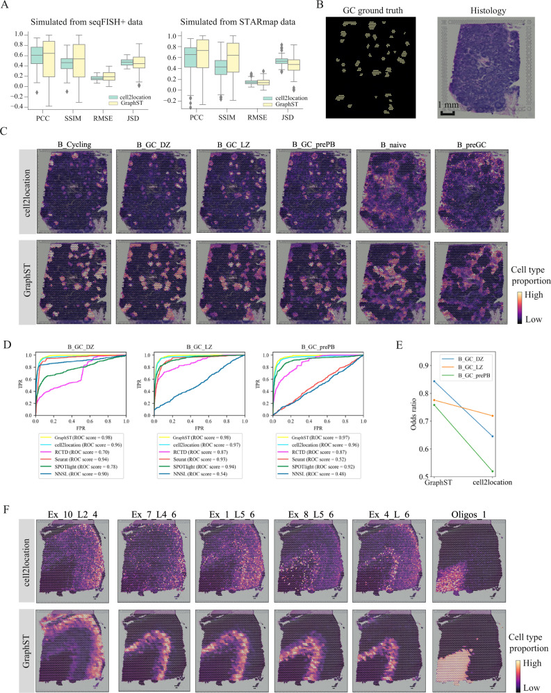
Spatial transcriptomics technologies generate gene expression profiles with spatial context, requiring spatially informed analysis tools for three key tasks, spatial clustering, multisample integration, and cell-type deconvolution. We present GraphST, a graph self-supervised contrastive learning method that fully exploits spatial transcriptomics data to outperform existing methods. It combines graph neural networks with self-supervised contrastive learning to learn informative and discriminative spot representations by minimizing the embedding distance between spatially adjacent spots and vice versa. We demonstrated GraphST on multiple tissue types and technology platforms. GraphST achieved 10% higher clustering accuracy and better delineated fine-grained tissue structures in brain and embryo tissues. GraphST is also the only method that can jointly analyze multiple tissue slices in vertical or horizontal integration while correcting batch effects. Lastly, GraphST demonstrated superior cell-type deconvolution to capture spatial niches like lymph node germinal centers and exhausted tumor infiltrating T cells in breast tumor tissue.
Overview
The claims and statistics of this paper haven’t been checked yet.
Run the check — free, no account needed. It reads the open-access text and checks the sources it cites.
Integrity signals
Registry
Cites
Statistics
Figures
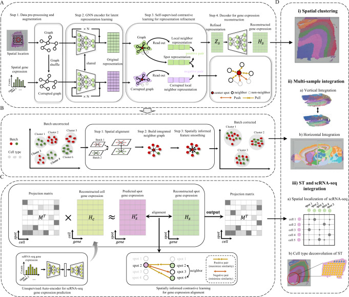
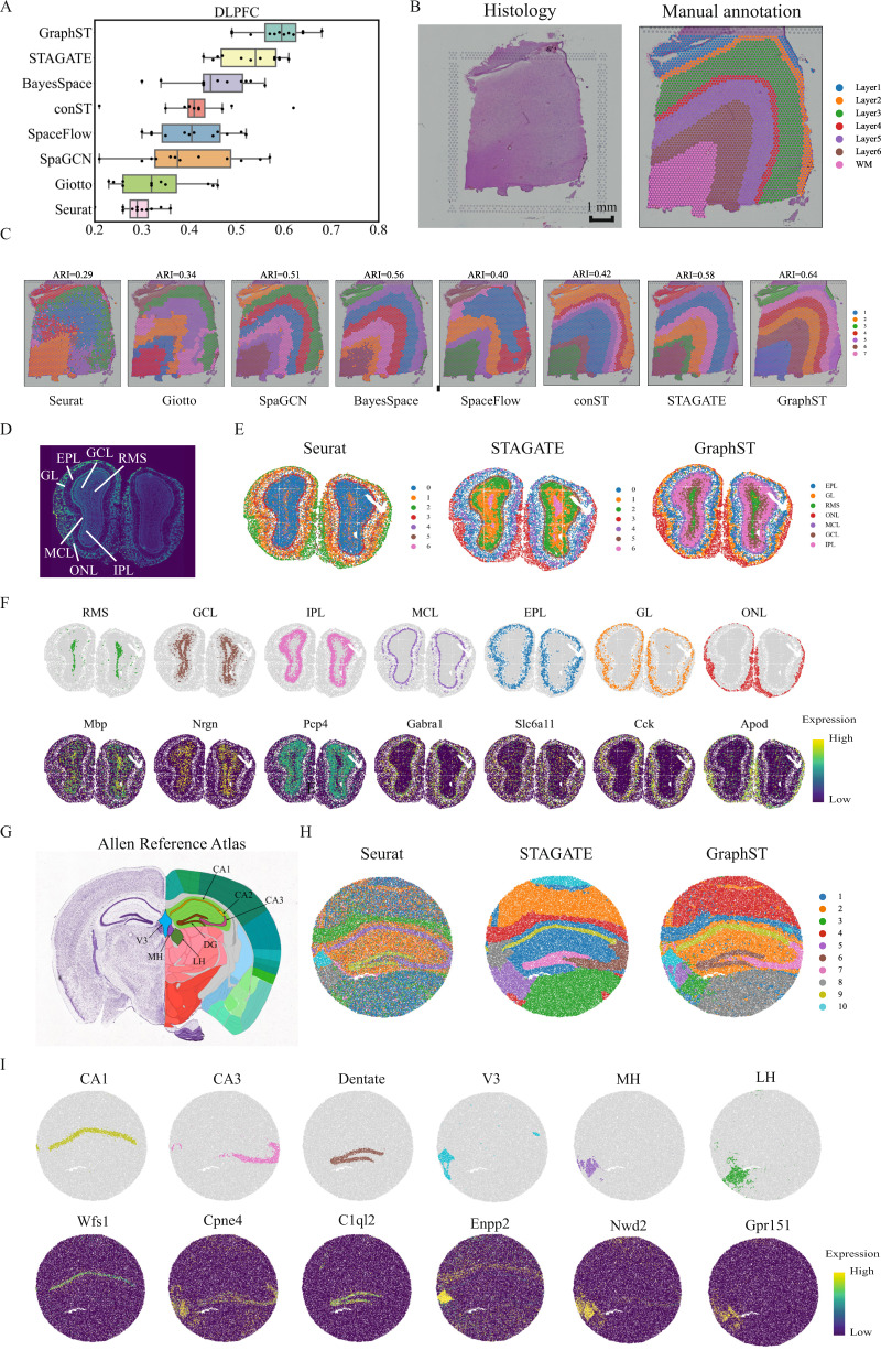
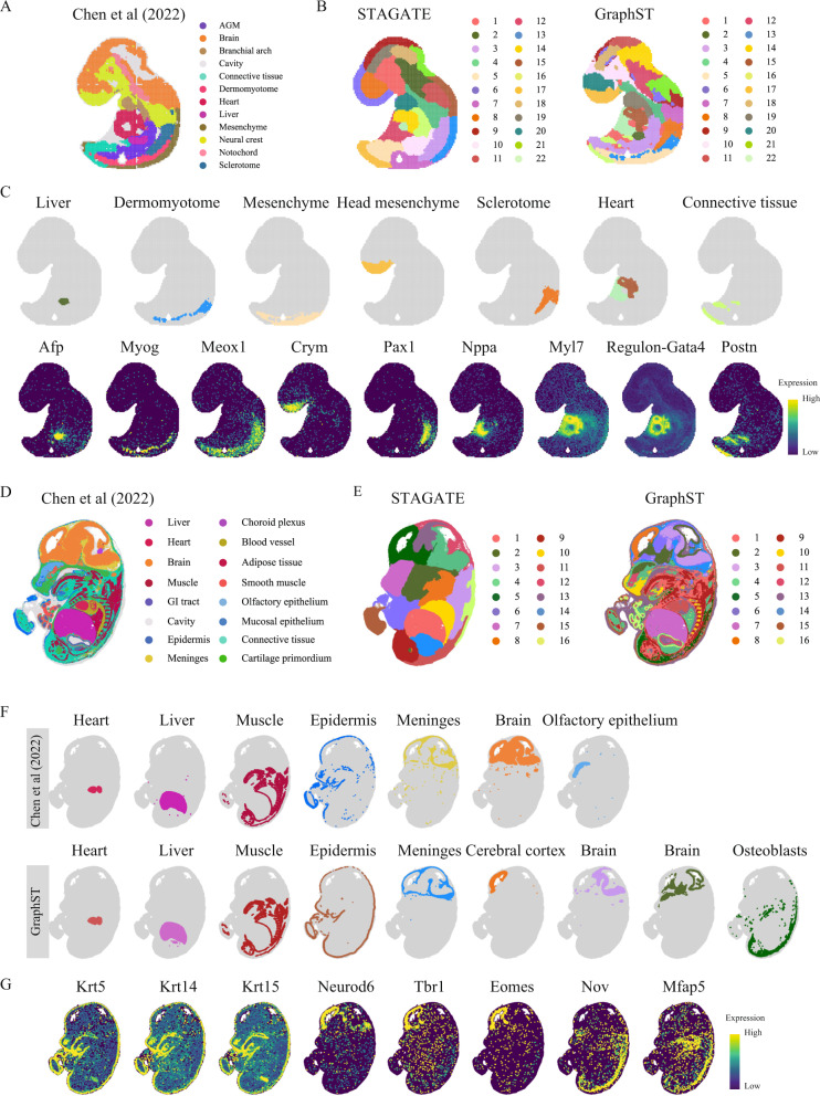
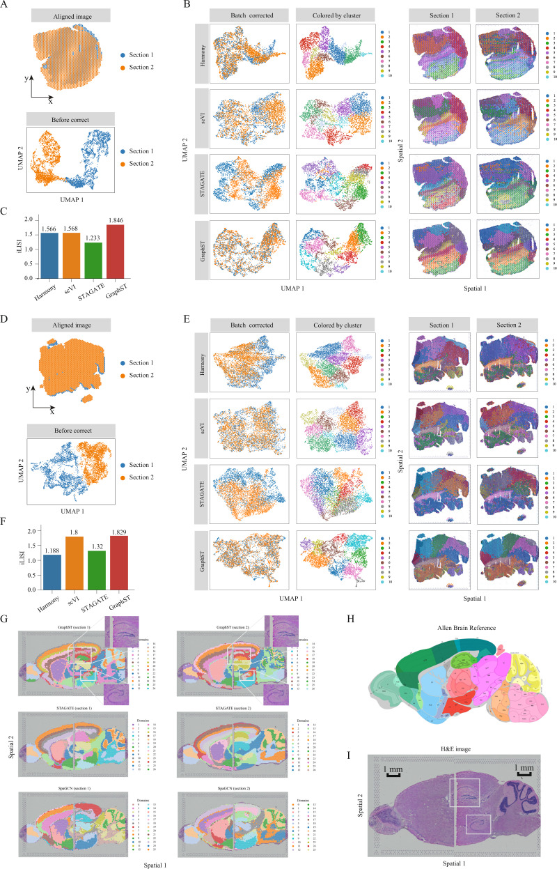
Verification
Claims the paper makes
The claims and statistics of this paper haven’t been checked yet.
Run the check — free, no account needed. It reads the open-access text and checks the sources it cites.
Claims other papers make about this one
Each is checked against this paper’s own text: does it say what the citing paper says it does?
The cited source says what the claim attributes to it.
In the citing paper
“Long et al. introduced the GraphST framework, which combined GNN and contrastive learning to integrate information within spatial transcriptomic datasets, across tissue sections and scRNA-seq datasets [127].”
From Challenges in AI-driven Biomedical Multimodal Data Fusion and Analysis (2025) · cites this paper as [127]
Self-check
Percentages, sums and reported differences re-computed from the paper’s own numbers.
The self-check needs the paper’s full text.
Citations
Cites
Adam: A Method for Stochastic Optimization
Kingma, D. P., & Ba, J. (2014) · arXiv · doi:10.48550/arxiv.1412.6980 · Cited by 98,946
Semi-supervised classification with graph convolutional networks
Kipf, T. N., & Welling, M. (2016) · arXiv · doi:10.48550/arxiv.1609.02907 · Cited by 24,853
Fast unfolding of communities in large networks
Blondel, V. D., Guillaume, J.-L., Lambiotte, R., & Lefebvre, E. (2008) · Journal of Statistical Mechanics: Theory and Experiment · doi:10.1088/1742-5468/2008/10/p10008 · Cited by 21,424
Silhouettes: A graphical aid to the interpretation and validation of cluster analysis
Rousseeuw, P. J. (1987) · Journal of Computational and Applied Mathematics · doi:10.1016/0377-0427(87)90125-7 · Cited by 21,392
Enrichr: a comprehensive gene set enrichment analysis web server 2016 update
Kuleshov, M. V., Jones, M. R., Rouillard, A. D., Fernandez, N. F., Duan, Q., Wang, Z., Koplev, S., Jenkins, S. L., Jagodnik, K. M., Lachmann, A., et al. (2016) · Nucleic Acids Research · doi:10.1093/nar/gkw377 · Cited by 10,799
Fast, sensitive and accurate integration of single-cell data with Harmony
Korsunsky, I., Millard, N., Fan, J., Slowikowski, K., Zhang, F., Wei, K., Baglaenko, Y., Brenner, M., Loh, P.-R., & Raychaudhuri, S. (2019) · Nature Methods · doi:10.1038/s41592-019-0619-0 · Cited by 10,488
SCANPY: large-scale single-cell gene expression data analysis
Wolf, F. A., Angerer, P., & Theis, F. J. (2018) · Genome Biology · doi:10.1186/s13059-017-1382-0 · Cited by 8,881
Solving Least Squares Problems
Lawson, C. L., & Hanson, R. J. (1995) · doi:10.1137/1.9781611971217 · Cited by 7,103
Spatial reconstruction of single-cell gene expression data
Satija, R., Farrell, J. A., Gennert, D., Schier, A. F., & Regev, A. (2015) · Nature Biotechnology · doi:10.1038/nbt.3192 · Cited by 7,055
Deep generative modeling for single-cell transcriptomics
Lopez, R., Regier, J., Cole, M. B., Jordan, M. I., & Yosef, N. (2018) · Nature Methods · doi:10.1038/s41592-018-0229-2 · Cited by 2,530
Slide-seq: A scalable technology for measuring genome-wide expression at high spatial resolution
Rodriques, S. G., Stickels, R. R., Goeva, A., Martin, C. A., Murray, E., Vanderburg, C. R., Welch, J., Chen, L. M., Chen, F., & Macosko, E. Z. (2019) · Science · doi:10.1126/science.aaw1219 · Cited by 2,424
Batch effects in single-cell RNA-sequencing data are corrected by matching mutual nearest neighbors
Haghverdi, L., Lun, A. T. L., Morgan, M. D., & Marioni, J. C. (2018) · Nature Biotechnology · doi:10.1038/nbt.4091 · Cited by 2,416
Spatiotemporal transcriptomic atlas of mouse organogenesis using DNA nanoball-patterned arrays
Chen, A., Liao, S., Cheng, M., Ma, K., Wu, L., Lai, Y., Qiu, X., Yang, J., Xu, J., Hao, S., et al. (2022) · Cell · doi:10.1016/j.cell.2022.04.003 · Cited by 1,784
Self-citationTranscriptome-scale super-resolved imaging in tissues by RNA seqFISH
Eng, C.-H. L., Lawson, M., Zhu, Q., Dries, R., Koulena, N., Takei, Y., Yun, J., Cronin, C., Karp, C., Yuan, G.-C., et al. (2019) · Nature · doi:10.1038/s41586-019-1049-y · Cited by 1,754
Exploring tissue architecture using spatial transcriptomics
Rao, A., Barkley, D., França, G. S., & Yanai, I. (2021) · Nature · doi:10.1038/s41586-021-03634-9 · Cited by 1,700
Robust decomposition of cell type mixtures in spatial transcriptomics
Cable, D. M., Murray, E., Zou, L. S., Goeva, A., Macosko, E. Z., Chen, F., & Irizarry, R. A. (2022) · Nature Biotechnology · doi:10.1038/s41587-021-00830-w · Cited by 1,487
Cell2location maps fine-grained cell types in spatial transcriptomics
Kleshchevnikov, V., Shmatko, A., Dann, E., Aivazidis, A., King, H. W., Li, T., Elmentaite, R., Lomakin, A., Kedlian, V., Gayoso, A., et al. (2022) · Nature Biotechnology · doi:10.1038/s41587-021-01139-4 · Cited by 1,279
Deciphering cell–cell interactions and communication from gene expression
Armingol, E., Officer, A., Harismendy, O., & Lewis, N. E. (2021) · Nature Reviews Genetics · doi:10.1038/s41576-020-00292-x · Cited by 1,267
Transcriptome-scale spatial gene expression in the human dorsolateral prefrontal cortex
Maynard, K. R., Collado-Torres, L., Weber, L. M., Uytingco, C., Barry, B. K., Williams, S. R., Catallini, J. L., Tran, M. N., Besich, Z., Tippani, M., et al. (2021) · Nature Neuroscience · doi:10.1038/s41593-020-00787-0 · Cited by 1,222
Hu, J., Li, X., Coleman, K., Schroeder, A., Ma, N., Irwin, D. J., Lee, E. B., Shinohara, R. T., & Li, M. (2021) · Nature Methods · doi:10.1038/s41592-021-01255-8 · Cited by 1,113
Self-citationSpatial transcriptomics at subspot resolution with BayesSpace
Zhao, E. J., Stone, M. R., Ren, X., Guenthoer, J., Smythe, K. S., Pulliam, T., Williams, S. R., Uytingco, C. R., Taylor, S. E. B., Nghiem, P., et al. (2021) · Nature Biotechnology · doi:10.1038/s41587-021-00935-2 · Cited by 1,032
Giotto: a toolbox for integrative analysis and visualization of spatial expression data
Dries, R., Zhu, Q., Dong, R., Eng, C.-H. L., Li, H., Liu, K., Fu, Y., Zhao, T., Sarkar, A., Bao, F., et al. (2021) · Genome biology · doi:10.1186/s13059-021-02286-2 · Cited by 966
High-Spatial-Resolution Multi-Omics Sequencing via Deterministic Barcoding in Tissue
Liu, Y., Yang, M., Deng, Y., Su, G., Enninful, A., Guo, C. C., Tebaldi, T., Zhang, D., Kim, D., Bai, Z., et al. (2020) · Cell · doi:10.1016/j.cell.2020.10.026 · Cited by 966
Spatial Transcriptomics and In Situ Sequencing to Study Alzheimer’s Disease
Chen, W.-T., Lu, A., Craessaerts, K., Pavie, B., Sala Frigerio, C., Corthout, N., Qian, X., Laláková, J., Kühnemund, M., Voytyuk, I., et al. (2020) · Cell · doi:10.1016/j.cell.2020.06.038 · Cited by 949 · cited 1×
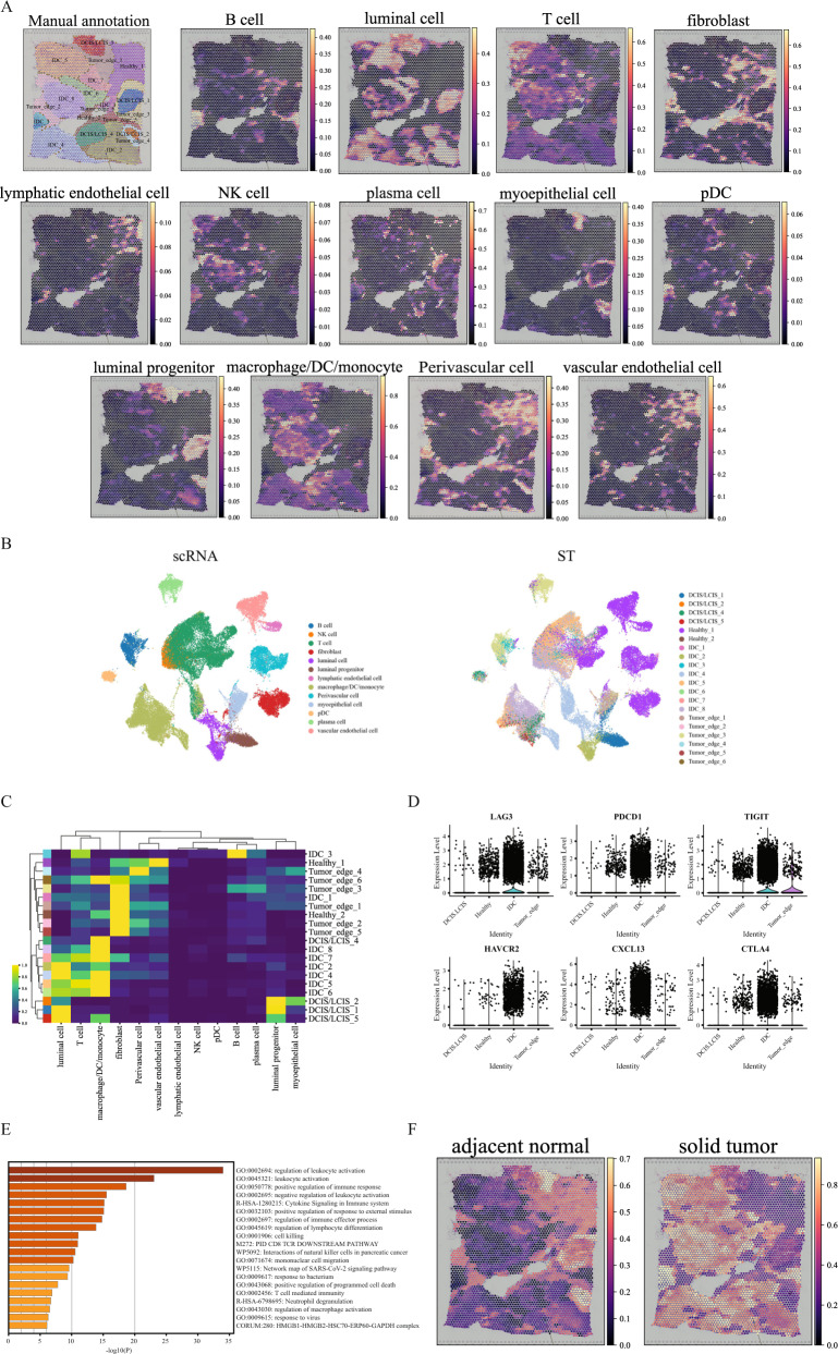
“Of the DCIS/LCIS regions, 4 and 5 only had macrophage/DC/monocytes present, while 1 and 2 had no immune cells mapped onto them.”
Deep learning and alignment of spatially resolved single-cell transcriptomes with Tangram
Biancalani, T., Scalia, G., Buffoni, L., Avasthi, R., Lu, Z., Sanger, A., Tokcan, N., Vanderburg, C. R., Segerstolpe, Å., Zhang, M., et al. (2021) · Nature Methods · doi:10.1038/s41592-021-01264-7 · Cited by 931
A single-cell map of intratumoral changes during anti-PD1 treatment of patients with breast cancer
Bassez, A., Vos, H., Van Dyck, L., Floris, G., Arijs, I., Desmedt, C., Boeckx, B., Vanden Bempt, M., Nevelsteen, I., Lambein, K., et al. (2021) · Nature Medicine · doi:10.1038/s41591-021-01323-8 · Cited by 867 · cited 1×
“Such T cells were also observed by Zhang et al. 50 and Bassez et al. 51 in other human breast cancer samples, and both studies demonstrated their reactivation with anti-PD-L1 therapy to harness their cytotoxicity against the tumor cells.”
Zhang, Y., Chen, H., Mo, H., Hu, X., Gao, R., Zhao, Y., Liu, B., Niu, L., Sun, X., Yu, X., et al. (2021) · Cancer Cell · doi:10.1016/j.ccell.2021.09.010 · Cited by 783
Dong, K., & Zhang, S. (2022) · Nature Communications · doi:10.1038/s41467-022-29439-6 · Cited by 761
A cell atlas of human thymic development defines T cell repertoire formation
Park, J.-E., Botting, R. A., Conde, C. D., Popescu, D.-M., Lavaert, M., Kunz, D. J., Goh, I., Stephenson, E., Ragazzini, R., Tuck, E. J., et al. (2020) · Science · doi:10.1126/science.aay3224 · Cited by 758
Pham, D., Tan, X., Balderson, B., Xu, J., Grice, L. F., Yoon, S., Willis, E. F., Tran, M., Lam, P. Y., Raghubar, A. M., et al. (2023) · Nature Communications · doi:10.1038/s41467-023-43120-6 · Cited by 757
Elosua-Bayes, M., Nieto, P., Mereu, E., Gut, I., & Heyn, H. (2021) · Nucleic Acids Research · doi:10.1093/nar/gkab043 · Cited by 732
Deep Graph InfomaxPreprint
Veličković, P., Fedus, W., Hamilton, W. L., Lió, P., Bengio, Y., & Hjelm, R. D. (2018) · arXiv (Cornell University) · doi:10.48550/arxiv.1809.10341 · Cited by 693
Spatially resolved transcriptomes—next generation tools for tissue exploration
Asp, M., Bergenstråhle, J., & Lundeberg, J. (2020) · BioEssays · doi:10.1002/bies.201900221 · Cited by 666
Spatially informed cell-type deconvolution for spatial transcriptomics
Ma, Y., & Zhou, X. (2022) · Nature Biotechnology · doi:10.1038/s41587-022-01273-7 · Cited by 525
Microscopic examination of spatial transcriptome using Seq-Scope
Cho, C.-S., Xi, J., Si, Y., Park, S.-R., Hsu, J.-E., Kim, M., Jun, G., Kang, H. M., & Lee, J. H. (2021) · Cell · doi:10.1016/j.cell.2021.05.010 · Cited by 520
Chamber Formation and Morphogenesis in the Developing Mammalian Heart
Christoffels, V. M., Habets, P. E. M. H., Franco, D., Campione, M., de Jong, F., Lamers, W. H., Bao, Z., Palmer, S., Biben, C., Harvey, R. P., et al. (2000) · Developmental Biology · doi:10.1006/dbio.2000.9753 · Cited by 502
Li, B., Zhang, W., Guo, C., Xu, H., Li, L., Fang, M., Hu, Y., Zhang, X., Yao, X., Tang, M., et al. (2022) · Nature Methods · doi:10.1038/s41592-022-01480-9 · Cited by 459
Self-citationUnsupervised spatially embedded deep representation of spatial transcriptomics
Xu, H., Fu, H., Long, Y., Ang, K. S., Sethi, R., Chong, K., Li, M., Uddamvathanak, R., Lee, H. K., Ling, J., et al. (2024) · Genome Medicine · doi:10.1186/s13073-024-01283-x · Cited by 413
Self-citationPham, D., Tan, X., Xu, J., et al. (2020) · bioRxiv (Cold Spring Harbor Laboratory) · doi:10.1101/2020.05.31.125658 · Cited by 377
Distinct microbial and immune niches of the human colon
James, K. R., Gomes, T., Elmentaite, R., Kumar, N., Gulliver, E. L., King, H. W., Stares, M., Bareham, B. R., Ferdinand, J. R., Petrova, V. N., et al. (2020) · Nature Immunology · doi:10.1038/s41590-020-0602-z · Cited by 312
SpatialDWLS: accurate deconvolution of spatial transcriptomic data
Dong, R., & Yuan, G. (2021) · Genome biology · doi:10.1186/s13059-021-02362-7 · Cited by 301
Alignment and integration of spatial transcriptomics data
Zeira, R., Land, M., Strzalkowski, A., & Raphael, B. J. (2022) · Nature Methods · doi:10.1038/s41592-022-01459-6 · Cited by 298
Spatially resolved transcriptomics reveals the architecture of the tumor-microenvironment interface
Hunter, M. V., Moncada, R. M., Weiss, J. M., Yanai, I., & White, R. M. (2021) · Nature Communications · doi:10.1038/s41467-021-26614-z · Cited by 295
Liao, J., Lu, X., Shao, X., Zhu, L., & Fan, X. (2020) · Trends in biotechnology · doi:10.1016/j.tibtech.2020.05.006 · Cited by 282
Identifying multicellular spatiotemporal organization of cells with SpaceFlow
Ren, H., Walker, B. L., Cang, Z., & Nie, Q. H. (2022) · Nature Communications · doi:10.1038/s41467-022-31739-w · Cited by 265
King, H. W., Orban, N. T., Riches, J. C., Clear, A. J., Warnes, G., Teichmann, S. A., & James, L. K. (2021) · Science Immunology · doi:10.1126/sciimmunol.abe6291 · Cited by 262
Cell clustering for spatial transcriptomics data with graph neural networks
Li, J., Chen, S., Pan, X., Yuan, Y., & Shen, H. (2022) · Nature Computational Science · doi:10.1038/s43588-022-00266-5 · Cited by 236
DISCO: A database of Deeply Integrated human Single-Cell Omics data
Li, M., Zhang, X., Ang, K. S., Ling, J., Sethi, R., Lee, N. Y. S., Ginhoux, F., & Chen, J. (2022) · Nucleic Acids Research · doi:10.1093/nar/gkab1020 · Cited by 205
Self-citationHu, Q., Hong, Y., Qi, P., Lu, G., Mai, X., Xu, S., He, X., Guo, Y., Gao, L., Jing, Z., et al. (2021) · Nature Communications · doi:10.1038/s41467-021-22300-2 · Cited by 201
conST: an interpretable multi-modal contrastive learning framework for spatial transcriptomicsPreprint
Zong, Y., Yu, T., Wang, X., Wang, Y., Hu, Z., & Li, Y. (2022) · bioRxiv (Cold Spring Harbor Laboratory) · doi:10.1101/2022.01.14.476408 · Cited by 110
AutoGeneS: Automatic gene selection using multi-objective optimization for RNA-seq deconvolution
Aliee, H., & Theis, F. J. (2021) · Cell Systems · doi:10.1016/j.cels.2021.05.006 · Cited by 103
Unsupervised Spatially Embedded Deep Representation of Spatial TranscriptomicsPreprint
Fu, H., Xu, H., Chong, K., Li, M., Ang, K. S., Lee, H. K., Ling, J., Chen, A., Shao, L., Liu, L., et al. (2021) · bioRxiv (Cold Spring Harbor Laboratory) · doi:10.1101/2021.06.15.448542 · Cited by 98
Self-citationCellDART: cell type inference by domain adaptation of single-cell and spatial transcriptomic data
Bae, S., Na, K. J., Koh, J., Lee, D. S., Choi, H., & Kim, Y. T. (2022) · Nucleic Acids Research · doi:10.1093/nar/gkac084 · Cited by 76
Macrophages promote the progression of premalignant mammary lesions to invasive cancer
Carron, E. C., Homra, S., Rosenberg, J. N., Coffelt, S. B., Kittrell, F. S., Zhang, Y., Creighton, C. J., Fuqua, S. A. W., Medina, D., & Machado, H. L. (2017) · Oncotarget · doi:10.18632/oncotarget.14913 · Cited by 74
Continuous Polony Gels for Tissue Mapping with High Resolution and RNA Capture Efficiency
Fu, X., Sun, L., Chen, J. Y., Dong, R., Lin, Y., Palmiter, R. D., Lin, S., & Gu, L. (2021) · doi:10.1101/2021.03.17.435795 · Cited by 64
Self-citationSpatial mapping of cell types by integration of transcriptomics dataPreprint
Andersson, A., Bergenstråhle, J., Asp, M., Bergenstråhle, L., Jurek, A., Navarro, J. F., & Lundeberg, J. (2019) · bioRxiv (Cold Spring Harbor Laboratory) · doi:10.1101/2019.12.13.874495 · Cited by 13
Chen, T., Wang, Z., Li, L., Li, D., Li, Z., Chang, X., Bian, P., Liang, G., Wang, Q., & Xie, T. (2025) · Schloss Dagstuhl – Leibniz-Zentrum für Informatik · doi:10.4230/lipics.ecoop.2025.6
Cited by
Sorted by citations
Pham, D., Tan, X., Balderson, B., Xu, J., Grice, L. F., Yoon, S., Willis, E. F., Tran, M., Lam, P. Y., Raghubar, A. M., et al. (2023) · Nature Communications · doi:10.1038/s41467-023-43120-6 · Cited by 757
Profiling cell identity and tissue architecture with single-cell and spatial transcriptomics
Gulati, G. S., D'Silva, J. P., Liu, Y., Wang, L., & Newman, A. M. (2024) · Nature Reviews Molecular Cell Biology · doi:10.1038/s41580-024-00768-2 · Cited by 347
BANKSY unifies cell typing and tissue domain segmentation for scalable spatial omics data analysis
Singhal, V., Chou, N., Lee, J., Yue, Y., Liu, J., Chock, W. K., Lin, L., Chang, Y.-C., Teo, E. M. L., Aow, J., et al. (2024) · Nature Genetics · doi:10.1038/s41588-024-01664-3 · Cited by 282
Spatiotemporal omics for biology and medicine
Liu, L., Chen, A., Li, Y., Mulder, J., Heyn, H., & Xu, X. (2024) · Cell · doi:10.1016/j.cell.2024.07.040 · Cited by 252
Self-citationDeciphering spatial domains from spatial multi-omics with SpatialGlue
Long, Y., Ang, K. S., Sethi, R., Liao, S., Heng, Y., van Olst, L., Ye, S., Zhong, C., Xu, H., Zhang, D., et al. (2024) · Nature Methods · doi:10.1038/s41592-024-02316-4 · Cited by 223
Self-citation
Metadata
- Authors
- Long, Y., Ang, K. S., Li, M., Chong, K. L. K., Sethi, R., Zhong, C., Xu, H., Ong, Z., Sachaphibulkij, K., Chen, A., Zeng, L., Fu, H., Wu, M., Lim, L. H. K., Liu, L., & Chen, J.
- Affiliations
- Agency for Science, Technology and Research (SG)
- National Neuroscience Institute (SG)
- National University of Singapore (SG)
- BGI Group (China) (CN)
- Duke-NUS Medical School (SG)
- Corresponding author
- Chen, Ao
- Chen, Jinmiao
- ORCID
- Venue
- Nature Communications
- Published
- 2023-03-01
- Volume
- 14
- Issue
- 1
- Pages
- 1155-1155
- ISSN
- 2041-1723 (ISSN-L)
- PMC
- PMC9977836
- OpenAlex
- W4322738920
- Cited by
- 700
- References
- 55
- Citations by year
- 2026: 284 · 2025: 233 · 2024: 156 · 2023: 27
- FWCI
- 62.55
- Type
- journal article
- Language
- en
- Open access
- gold
- doi.org
- Locations
- Nature Communications (publishedVersion, cc-by)
- bioRxiv (Cold Spring Harbor Laboratory) (acceptedVersion, cc-by-nc-nd)
- Research Square (acceptedVersion, cc-by)
- PubMed Central (publishedVersion, cc-by)
- PubMed (publishedVersion)
- RePEc: Research Papers in Economics (submittedVersion)
- DOAJ (DOAJ: Directory of Open Access Journals) (submittedVersion, cc-by-sa)
- Funders
- National Research Foundation
- National Research Foundation Singapore (2017-04, NRF-CRP19-2017-04, MOH-OFLCG18May-0003)
- Medical Research Council
- National Medical Research Council (MOH-OFLCG18May-0003, NMRC /OFLCG/003/2018, NMRC-OFIRG18nov-2013)
- Topics
- Life Sciences › Biochemistry, Genetics and Molecular Biology › Molecular Biology › Single-cell and spatial transcriptomics
- Life Sciences › Biochemistry, Genetics and Molecular Biology › Molecular Biology › Gene expression and cancer classification
- Life Sciences › Biochemistry, Genetics and Molecular Biology › Biophysics › Cell Image Analysis Techniques
- Keywords
- spatial transcriptomics, graph neural networks, self-supervised contrastive learning, spatial clustering, cell type deconvolution, batch effect correction, tumor-infiltrating T cells, breast tumors, brain tissue
- MeSH terms
- Brain, Cluster Analysis, Germinal Center, Gene Expression Profiling, Transcriptome
About these checks
Registry, retraction, Cites and statistics results are computed, not judged, with one exception: a printed reference that no index lists is read once by a model before it counts as a citation error. Claims are extracted by a model and kept only when their quote appears verbatim in the text; each cited source is then checked, with a model’s help, for whether it says what the claim relies on. Those verdicts can be wrong — read the claim’s quote and the cited source before relying on one.
Statistics and arithmetic are recomputed, not judged.
Confidence bands are shown only for fields calibrated on our annotated test set. None is calibrated yet, so no band is shown.
What counts as a claim ›How we measure confidence ›How we check this ›
Are you an author, or do you see a problem with this page? Request a review
