

The DOI matches its Crossref record; 26 of the 26 references it cites were found in a registry or an index.
Architecture of the dynamic fungal cell wall
Open accessAbstract
The fungal cell wall is essential for growth and survival, and is a key target for antifungal drugs and the immune system. The cell wall must be robust but flexible, protective and shielding yet porous to nutrients and membrane vesicles and receptive to exogenous signals. Most fungi have a common inner wall skeleton of chitin and β-glucans that functions as a flexible viscoelastic frame to which a more diverse set of outer cell wall polymers and glycosylated proteins are attached. Whereas the inner wall largely determines shape and strength, the outer wall confers properties of hydrophobicity, adhesiveness, and chemical and immunological heterogeneity. The spatial organization and dynamic regulation of the wall in response to prevailing growth conditions enable fungi to thrive within changing, diverse and often hostile environments. Understanding this architecture provides opportunities to develop diagnostics and drugs to combat life-threatening fungal infections. In this Review, Gow and Lenardon describe how fungal cell walls are organized, focusing on the underlying architectural and mechanical principles that are required to deliver differing and bespoke biochemical and biophysical attributes.
Overview
The claims and statistics of this paper haven’t been checked yet.
Run the check — free, no account needed. It reads the open-access text and checks the sources it cites.
Integrity signals
Registry
Cites
Statistics
Figures
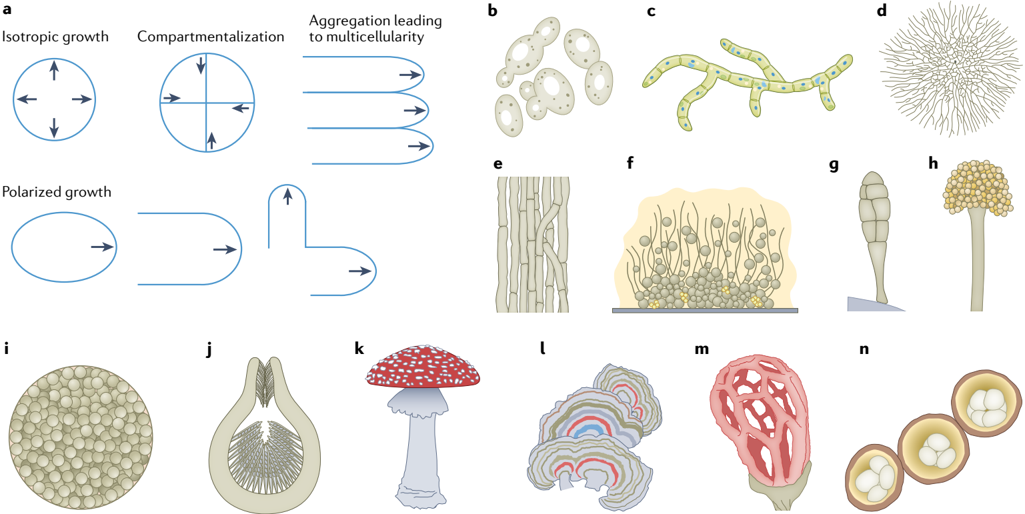
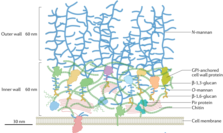
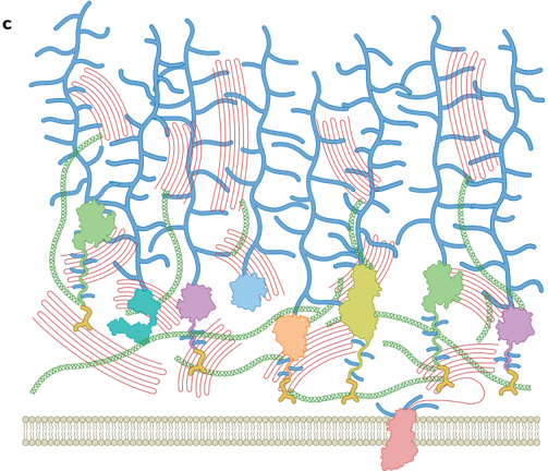
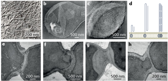
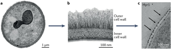
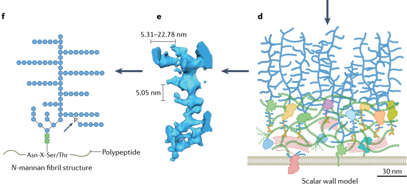
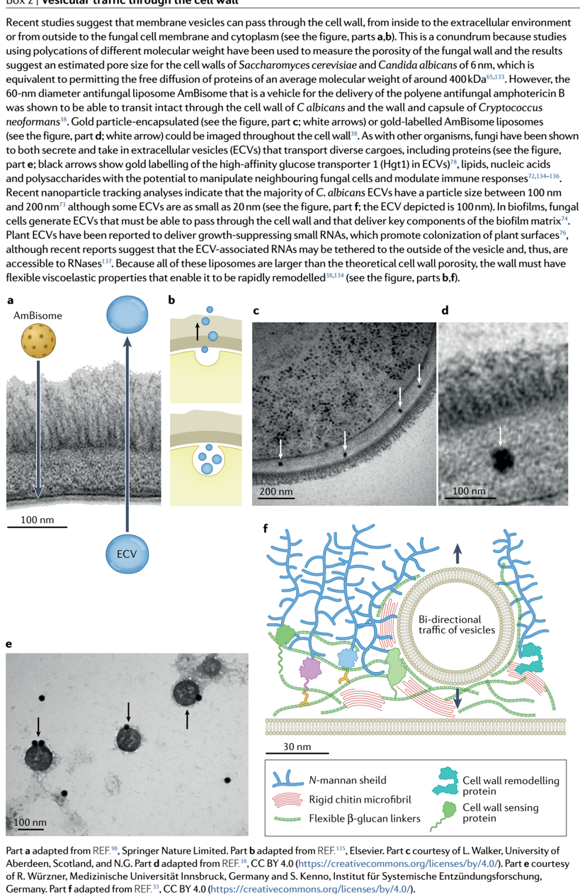
Verification
Claims the paper makes
The claims and statistics of this paper haven’t been checked yet.
Run the check — free, no account needed. It reads the open-access text and checks the sources it cites.
Claims other papers make about this one
Each is checked against this paper’s own text: does it say what the citing paper says it does?
In the citing paper
“The cell walls of C. auris, like C. albicans, contain a higher abundance of N-linked mannans compared to O-linked mannans (23).”
From Dual function of Candida auris mannosyltransferase, MNT5, in biofilm community protection from antifungal therapy and the host (2025) · cites this paper as [23]
In the citing paper
“C. albicans has a well-characterized cell wall composed of an inner layer of chitin, β−1,3-glucan, and β−1,6-glucan, and an outer layer composed of proteins modified with N- and O-linked mannans and phosphomannans, called mannoproteins34,35.”
From Distinct echinocandin responses of Candida albicans and Candida auris cell walls revealed by solid-state NMR (2025) · cites this paper as [35]
In the citing paper
“These α-1,2-mannoses can be connected to the α-1,6-mannan backbone, as well as to β-1,2-mannose, α-1,3-mannose, and phosphate groups found in the sidechains27,35.”
From Distinct echinocandin responses of Candida albicans and Candida auris cell walls revealed by solid-state NMR (2025) · cites this paper as [35]
Self-check
Percentages, sums and reported differences re-computed from the paper’s own numbers.
The self-check needs the paper’s full text.
Citations
Cites
Growth of Candida albicans hyphae
Sudbery, P. E. (2011) · Nature Reviews Microbiology · doi:10.1038/nrmicro2636 · Cited by 1,110
An integrated model of the recognition of Candida albicans by the innate immune system
Netea, M. G., Brown, G. D., Kullberg, B. J., & Gow, N. A. R. (2008) · Nature Reviews Microbiology · doi:10.1038/nrmicro1815 · Cited by 946
Self-citationCandida albicans morphogenesis and host defence: discriminating invasion from colonization
Gow, N. A. R., van de Veerdonk, F. L., Brown, A. J. P., & Netea, M. G. (2012) · Nature Reviews Microbiology · doi:10.1038/nrmicro2711 · Cited by 888
Self-citationCandida Biofilms: Threats, Challenges, and Promising Strategies
Cavalheiro, M., & Teixeira, M. C. (2018) · Frontiers in Medicine · doi:10.3389/fmed.2018.00028 · Cited by 690
Innate Immune Training of Granulopoiesis Promotes Anti-tumor Activity
Kalafati, L., Kourtzelis, I., Schulte-Schrepping, J., Li, X., Hatzioannou, A., Grinenko, T., Hagag, E., Sinha, A., Has, C., Dietz, S., et al. (2020) · Cell · doi:10.1016/j.cell.2020.09.058 · Cited by 648
Stimulation of Chitin Synthesis Rescues Candida albicans from Echinocandins
Walker, L. A., Munro, C. A., de Bruijn, I., Lenardon, M. D., McKinnon, A., & Gow, N. A. R. (2008) · PLoS Pathogens · doi:10.1371/journal.ppat.1000040 · Cited by 445
Self-citationFunction of Candida albicans Adhesin Hwp1 in Biofilm Formation
Nobile, C. J., Nett, J. E., Andes, D. R., & Mitchell, A. P. (2006) · Eukaryotic Cell · doi:10.1128/ec.00194-06 · Cited by 395
Harnessing calcineurin as a novel anti-infective agent against invasive fungal infections.
Steinbach, W. J., Reedy, J. L., Cramer, R. A., Perfect, J. R., & Heitman, J. (2007) · Nature Reviews Microbiology · doi:10.1038/nrmicro1680 · Cited by 313
Ene, I. V., Adya, A. K., Wehmeier, S., Brand, A. C., MacCallum, D. M., Gow, N. A. R., & Brown, A. J. P. (2012) · Cellular Microbiology · doi:10.1111/j.1462-5822.2012.01813.x · Cited by 313
Self-citationDynamic, Morphotype-Specific Candida albicans β-Glucan Exposure during Infection and Drug Treatment
Wheeler, R. T., Kombe, D., Agarwala, S. D., & Fink, G. R. (2008) · PLoS Pathogens · doi:10.1371/journal.ppat.1000227 · Cited by 309
Candida albicans biofilm–induced vesicles confer drug resistance through matrix biogenesis
Zarnowski, R., Sanchez, H., Covelli, A. S., Dominguez, E., Jaromin, A., Bernhardt, J., Mitchell, K. F., Heiss, C., Azadi, P., Mitchell, A., et al. (2018) · PLOS Biology · doi:10.1371/journal.pbio.2006872 · Cited by 260
Plaine, A., Walker, L., Da Costa, G., Mora-Montes, H. M., McKinnon, A., Gow, N. A. R., Gaillardin, C., Munro, C. A., & Richard, M. L. (2008) · Fungal Genetics and Biology · doi:10.1016/j.fgb.2008.08.003 · Cited by 238
Self-citationSpellberg, B. J., Ibrahim, A. S., Avanesian, V., Fu, Y., Myers, C., Phan, Q. T., Filler, S. G., Yeaman, M. R., & Edwards, Jr. (2006) · The Journal of Infectious Diseases · doi:10.1086/504691 · Cited by 180
Ene, I. V., Heilmann, C. J., Sorgo, A. G., Walker, L. A., de Koster, C. G., Munro, C. A., Klis, F. M., & Brown, A. J. P. (2012) · Proteomics · doi:10.1002/pmic.201200228 · Cited by 159
Lionakis, M. S., Iliev, I. D., & Hohl, T. M. (2017) · JCI Insight · doi:10.1172/jci.insight.93156 · Cited by 153
The yeast cell-wall salvage pathway
Popolo, L., Gualtieri, T., & Ragni, E. (2001) · Medical Mycology · doi:10.1080/mmy.39.1.111.121 · Cited by 142
Vaccines for human fungal diseases: close but still a long way to go
Oliveira, L. V. N., Wang, R., Specht, C. A., & Levitz, S. M. (2021) · npj Vaccines · doi:10.1038/s41541-021-00294-8 · Cited by 135
Pradhan, A., Avelar, G. M., Bain, J. M., Childers, D. S., Larcombe, D. E., Netea, M. G., Shekhova, E., Munro, C. A., Brown, G. D., Erwig, L. P., et al. (2018) · mBio · doi:10.1128/mbio.01318-18 · Cited by 133
Self-citationMora-Montes, H. M., Bates, S., Netea, M. G., Castillo, L., Brand, A., Buurman, E. T., Díaz-Jiménez, D. F., Jan Kullberg, B., Brown, A. J. P., Odds, F. C., et al. (2010) · Journal of Biological Chemistry · doi:10.1074/jbc.m109.081513 · Cited by 129
Self-citationCandida albicans Hyphae: From Growth Initiation to Invasion
Desai, J. (2018) · Journal of Fungi · doi:10.3390/jof4010010 · Cited by 117
Lenardon, M. D., Whitton, R. K., Munro, C. A., Marshall, D., & Gow, N. A. R. (2007) · Molecular Microbiology · doi:10.1111/j.1365-2958.2007.05990.x · Cited by 103
Self-citationRezafungin: a novel antifungal for the treatment of invasive candidiasis
Ham, Y. Y., Lewis, J. S., & Thompson, G. R. (2021) · Future Microbiology · doi:10.2217/fmb-2020-0217 · Cited by 66
Eradication of Candida albicans persister cell biofilm by the membranotropic peptide gH625
Galdiero, E., de Alteriis, E., De Natale, A., D’Alterio, A., Siciliano, A., Guida, M., Lombardi, L., Falanga, A., & Galdiero, S. (2020) · Scientific Reports · doi:10.1038/s41598-020-62746-w · Cited by 65
Shivarathri, R., Jenull, S., Stoiber, A., Chauhan, M., Mazumdar, R., Singh, A., Nogueira, F., Kuchler, K., Chowdhary, A., & Chauhan, N. (2020) · mSphere · doi:10.1128/msphere.00973-20 · Cited by 59
Cell wall stress induces alternative fungal cytokinesis and septation strategies
Walker, L. A., Lenardon, M. D., Preechasuth, K., Munro, C. A., & Gow, N. A. R. (2013) · Journal of Cell Science · doi:10.1242/jcs.118885 · Cited by 42
Self-citationMazáň, M., Blanco, N., Kováčová, K., Firáková, Z., Řehulka, P., Farkaš, V., & Arroyo, J. (2013) · Biochemical Journal · doi:10.1042/bj20130354 · Cited by 26
Research works printed but not linked: 110
- Verstrepen, K. J., Reynolds, T. B. & Fink, G. R. Origins of variation in the fungal cell surface. Nat. Rev. Microbiol. 2, 533–540 (2004). · DOI
- de Groot, P. W. et al. A genomic approach for the identification and classification of genes involved in cell wall formation and its regulation in Saccharomyces cerevisiae. Comp. Funct. Genomics 2, 124–142 (2001). · DOI
- Polizeli, M. L., Pietro, R. C., Jorge, J. A. & Terenzi, H. F. Effects of cell wall deficiency on the synthesis of polysaccharide-degrading exoenzymes: a study on mycelial and wall-less phenotypes of the fz; sg; os-1 (‘slime’) triple mutant of Neurospora crassa. J. Gen. Microbiol. 136, 1463–1468 (1990). · DOI
- Ene, I. V. et al. Cell wall remodeling enzymes modulate fungal cell wall elasticity and osmotic stress resistance. mBio 6, e00986 (2015). · DOI
- Money, N. P. & Fischer, M. W. F. in Plant Relationships Vol. 5 (ed. Deising, H. B.) 115–133 (Springer, 2009).
- Davì, V. et al. Mechanosensation dynamically coordinates polar growth and cell wall assembly to promote cell survival. Dev. Cell 45, 170–182.e7 (2018). This report demonstrates that cell wall thickness fluctuates during cell growth and is regulated by a mechanosensitive homeostatic mechanism. · DOI
- Veneault-Fourrey, C., Barooah, M., Egan, M., Wakley, G. & Talbot, N. J. Autophagic fungal cell death is necessary for infection by the rice blast fungus. Science 312, 580–583 (2006). · DOI
- Howard, R. J., Ferrari, M. A., Roach, D. H. & Money, N. P. Penetration of hard substrates by a fungus employing enormous turgor pressures. Proc. Natl Acad. Sci. USA 88, 11281–11284 (1991). · DOI
- Talbot, N. J. Appressoria. Curr. Biol. 29, R144–R146 (2019). · DOI
- Berger, B. W. & Sallada, N. D. Hydrophobins: multifunctional biosurfactants for interface engineering. J. Biol. Eng. 13, 10 (2019). · DOI
- Wösten, H. A. B. Hydrophobins: multipurpose proteins. Annu. Rev. Microbiol. 55, 625–646 (2001). · DOI
- Winandy, L., Schlebusch, O. & Fischer, R. Fungal hydrophobins render stones impermeable for water but keep them permeable for vapor. Sci. Rep. 9, 6264 (2019). This publication explores the biophysical properties of fungal cell wall hydrophobin layers and shows that they have remarkable breathable yet waterproof properties. · DOI
- Nehls, U. & Dietz, S. Fungal aquaporins: cellular functions and ecophysiological perspectives. Appl. Microbiol. Biotechnol. 98, 8835–8851 (2014). · DOI
- Casadevall, A. et al. The capsule of Cryptococcus neoformans. Virulence 10, 822–831 (2019). · DOI
- Tlalka, M., Fricker, M. & Watkinson, S. Imaging of long-distance α-aminoisobutyric acid translocation dynamics during resource capture by Serpula lacrymans. Appl. Environ. Microbiol. 74, 2700–2708 (2008). · DOI
- Aguilar-Trigueros, C. A., Boddy, L., Rillig, M. C. & Fricker, M. D. Network traits predict ecological strategies in fungi. ISME Commun. 2, 2 (2022). · DOI
- Lew, R. R. How does a hypha grow? The biophysics of pressurized growth in fungi. Nat. Rev. Microbiol. 9, 509–518 (2011). · DOI
- Read, N. D. in Oxford Textbook of Medical Mycology Ch. 4 (eds Kibbler, K. C. et al.) 23–34 (Oxford Univ. Press, 2018). · DOI
- Grün, C. H. et al. The structure of cell wall α-glucan from fission yeast. Glycobiology 15, 245–257 (2005). · DOI
- Ma, L. et al. Genome analysis of three Pneumocystis species reveals adaptation mechanisms to life exclusively in mammalian hosts. Nat. Commun. 7, 10740 (2016). · DOI
- Muzzarelli, C. J. & Gooday, G. W. (eds). Chitin in Nature and Technology (Springer, 1986). · DOI
- Kanagawa, M. et al. Structural insights into recognition of triple-helical β-glucans by an insect fungal receptor. J. Biol. Chem. 286, 29158–29165 (2011). · DOI
- Liu, Y. et al. Triple helix conformation of β-d-glucan from Ganoderma lucidum and effect of molecular weight on its immunostimulatory activity. Int. J. Biol. Macromol. 114, 1064–1070 (2018). · DOI
- Fernando, L. D. et al. Structural polymorphism of chitin and chitosan in fungal cell walls from solid-state NMR and principal component analysis. Front. Mol. Biosci. 8, 727053 (2021). · DOI
- Latgé, J. P. & Wang, T. Modern biophysics redefines our understanding of fungal cell wall structure, complexity, and dynamics. mBio 13, e0114522 (2022). · DOI · Paper page
- Orlean, P. & Funai, D. Priming and elongation of chitin chains: implications for chitin synthase mechanism. Cell Surf. 5, 100017 (2019). · DOI
- da Silva Dantas, A. et al. Crosstalk between the calcineurin and cell wall integrity pathways prevents chitin overexpression in Candida albicans. J. Cell Sci. 134, jcs258889 (2021). · DOI · Paper page
- Gow, N. A. R. & Gooday, G. W. Ultrastructure of chitin in hyphae of Candida albicans and other dimorphic and mycelial fungi. Protoplasma 115, 52–58 (1983). · DOI
- Vermeulen, C. A. & Wessels, J. G. H. Ultrastructural differences between wall apices of growing and non-growing hyphae of Schizophyllum commune. Protoplasma 120, 123–131 (1984). · DOI
- Vermeulen, C. A. & Wessels, J. G. Chitin biosynthesis by a fungal membrane preparation. Evidence for a transient non-crystalline state of chitin. Eur. J. Biochem. 158, 411–415 (1986). · DOI
- Lenardon, M. D., Sood, P., Dorfmueller, H. C., Brown, A. J. P. & Gow, N. A. R. Scalar nanostructure of the Candida albicans cell wall; a molecular, cellular and ultrastructural analysis and interpretation. Cell Surf. 6, 100047 (2020). This publication provides the first to-scale model of the C. albicans cell wall and includes a menu of icons which can be used to construct other bespoke cell wall models. · DOI · Paper page
- Kang, X. et al. Molecular architecture of fungal cell walls revealed by solid-state NMR. Nat. Commun. 9, 2747 (2018). This report utilizes solid-state nuclear magnetic resonance to demonstrate that chitin and α-1,3-glucan in the Aspergillus cell wall forms a hydrophobic scaffold which is surrounded by β-glucans and capped by glycoproteins and α-1,3-glucan. · DOI · Paper page
- Krol, P. Synthesis methods, chemical structures and phase structures of linear polyurethanes. Properties and applications of linear polyurethanes in polyurethane elastomers, copolymers and ionomers. Prog. Mater. Sci. 52, 915–1015 (2007). · DOI
- Bates, S. et al. Outer chain N-glycans are required for cell wall integrity and virulence of Candida albicans. J. Biol. Chem. 281, 90–98 (2006). · DOI
- Walker, L. et al. The viscoelastic properties of the fungal cell wall allow traffic of AmBisome as intact liposome vesicles. mBio 9, e02383-17 (2018). This study demonstrates that intact AmBisome vesicles can carry non-elastic gold particle cargoes through the fungal cell wall. · DOI
- Ballou, E. R. et al. Lactate signalling regulates fungal β-glucan masking and immune evasion. Nat. Microbiol. 2, 16238 (2016). This study makes key observations that Candida cells ‘mask’ β-1,3-glucan when grown on lactate, demonstrating that metabolic adaptability makes Candida cells an immunological moving target. · DOI · Paper page
- Pradhan, A. et al. Non-canonical signalling mediates changes in fungal cell wall PAMPs that drive immune evasion. Nat. Commun. 10, 5315 (2019). · DOI · Paper page
- Lopes-Bezerra, L. M. et al. Cell walls of the dimorphic fungal pathogens Sporothrix schenckii and Sporothrix brasiliensis exhibit bilaminate structures and sloughing of extensive and intact layers. PLoS Negl. Trop. Dis. 12, e0006169 (2018). · DOI
- Hobson, R. P. et al. Loss of cell wall mannosylphosphate in Candida albicans does not influence macrophage recognition. J. Biol. Chem. 279, 39628–39635 (2004). · DOI
- Trinel, P. A. et al. Candida albicans serotype B strains synthesize a serotype-specific phospholipomannan overexpressing a β-1,2-linked mannotriose. Mol. Microbiol. 58, 984–998 (2005). · DOI
- Singleton, D. R., Masuoka, J. & Hazen, K. C. Surface hydrophobicity changes of two Candida albicans serotype B mnn4delta mutants. Eukaryot. Cell 4, 639–648 (2005). · DOI
- Silva-Dias, A. et al. Adhesion, biofilm formation, cell surface hydrophobicity, and antifungal planktonic susceptibility: relationship among Candida spp. Front. Microbiol. 6, 205 (2015). · DOI
- Wessels, J. G. H. Hydrophobins, unique fungal proteins. Mycologist 14, 153–159 (2000). · DOI
- Aimanianda, V. et al. Surface hydrophobin prevents immune recognition of airborne fungal spores. Nature 460, 1117–1121 (2009). · DOI · Paper page
- Carrion, S. J. et al. The RodA hydrophobin on Aspergillus fumigatus spores masks dectin-1- and dectin-2-dependent responses and enhances fungal survival in vivo. J. Immunol. 191, 2581–2588 (2013). · DOI
- Willaert, R. G. Adhesins of yeasts: protein structure and interactions. J. Fungi 4, 119 (2018). · DOI
- Staab, J. F., Bradway, S. D., Fidel, P. L. & Sundstrom, P. Adhesive and mammalian transglutaminase substrate properties of Candida albicans Hwp1. Science 283, 1535–1538 (1999). · DOI
- Vendele, I. et al. Mannan detecting C-type lectin receptor probes recognise immune epitopes with diverse chemical, spatial and phylogenetic heterogeneity in fungal cell walls. PLoS Pathog. 16, e1007927 (2020). · DOI
- Kumar, R., Breindel, C., Saraswat, D., Cullen, P. J. & Edgerton, M. Candida albicans Sap6 amyloid regions function in cellular aggregation and zinc binding, and contribute to zinc acquisition. Sci. Rep. 7, 2908 (2017). · DOI
- Lipke, P. N., Klotz, S. A., Dufrene, Y. F., Jackson, D. N. & Garcia-Sherman, M. C. Amyloid-like β-aggregates as force-sensitive switches in fungal biofilms and infections. Microbiol. Mol. Biol. Rev. 82, e00035-17 (2018). · DOI
- Speth, C., Rambach, G., Lass-Flörl, C., Howell, P. L. & Sheppard, D. C. Galactosaminogalactan (GAG) and its multiple roles in Aspergillus pathogenesis. Virulence 10, 976–983 (2019). · DOI
- Gravelat, F. N. et al. Aspergillus galactosaminogalactan mediates adherence to host constituents and conceals hyphal β-glucan from the immune system. PLoS Pathog. 9, e1003575 (2013). This paper demonstrates that an epimerase is required for GAG synthesis which mediates adhesion of Aspergillus to a range of surfaces and is essential for virulence. · DOI · Paper page
- Nosanchuk, J. D., Stark, R. E. & Casadevall, A. Fungal melanin: what do we know about structure? Front. Microbiol. 6, 1463 (2015). · DOI · Paper page
- Liu, S., Youngchim, S., Zamith-Miranda, D. & Nosanchuk, J. D. Fungal melanin and the mammalian immune system. J. Fungi 7, 264 (2021). · DOI
- Casadevall, A., Cordero, R. J. B., Bryan, R., Nosanchuk, J. & Dadachova, E. Melanin, radiation, and energy transduction in fungi. Microbiol. Spectr. 5, 5.2.05 (2017). This work proposes that fungal cell wall melanin can harness electromagnetic radiation as an energy source to promote survival. · DOI · Paper page
- De Nobel, J. G., Dijkers, C., Hooijberg, E. & Klis, F. M. Increased cell wall porosity in Saccharomyces cerevisiae after treatment with dithiothreitol or EDTA. Microbiology 135, 2077–2084 (1989). · DOI
- Yadav, B. et al. Differences in fungal immune recognition by monocytes and macrophages: N-mannan can be a shield or activator of immune recognition. Cell Surf. 6, 100042 (2020). · DOI
- Nazik, H. et al. Pseudomonas phage inhibition of Candida albicans. Microbiology 163, 1568–1577 (2017). · DOI
- Nuss, D. L. Hypovirulence: mycoviruses at the fungal–plant interface. Nat. Rev. Microbiol. 3, 632–642 (2005). · DOI
- Kim, Y. & Mylonakis, E. Killing of Candida albicans filaments by Salmonella enterica serovar Typhimurium is mediated by sopB effectors, parts of a type III secretion system. Eukaryot. Cell 10, 782–790 (2011). · DOI
- Trunk, K. et al. The type VI secretion system deploys antifungal effectors against microbial competitors. Nat. Microbiol. 3, 920–931 (2018). · DOI
- Cuskin, F. et al. Human gut Bacteroidetes can utilize yeast mannan through a selfish mechanism. Nature 517, 165–169 (2015). · DOI
- Zhao, K. et al. Extracellular vesicles secreted by Saccharomyces cerevisiae are involved in cell wall remodelling. Commun. Biol. 2, 305 (2019). · DOI
- Dawson, C. S. et al. Protein markers for Candida albicans EVs include claudin-like Sur7 family proteins. J. Extracell. Vesicles 9, 1750810 (2020). · DOI
- Zhao, M. et al. Turbinmicin inhibits Candida biofilm growth by disrupting fungal vesicle-mediated trafficking. J. Clin. Invest. 131, e145123 (2021). · DOI · Paper page
- He, B. et al. RNA-binding proteins contribute to small RNA loading in plant extracellular vesicles. Nat. Plants 7, 342–352 (2021). · DOI
- Sorgo, A. G., Heilmann, C. J., Brul, S., de Koster, C. G. & Klis, F. M. Beyond the wall: Candida albicans secret(e)s to survive. FEMS Microbiol. Lett. 338, 10–17 (2013). · DOI
- Kenno, S. et al. Candida albicans factor H binding molecule Hgt1p — a low glucose-induced transmembrane protein is trafficked to the cell wall and impairs phagocytosis and killing by human neutrophils. Front. Microbiol. 9, 3319 (2019). · DOI
- Urban, C. et al. The moonlighting protein Tsa1p is implicated in oxidative stress response and in cell wall biogenesis in Candida albicans. Mol. Microbiol. 57, 1318–1341 (2005). · DOI
- Satala, D., Karkowska-Kuleta, J., Zelazna, A., Rapala-Kozik, M. & Kozik, A. Moonlighting proteins at the Candidal cell surface. Microorganisms 8, 1046 (2020). · DOI
- Gow, N. A. R., Latge, J. P. & Munro, C. A. The fungal cell wall: structure, biosynthesis, and function. Microbiol. Spectr. 5, 5.3.01 (2017). · DOI · Paper page
- Riquelme, M. et al. Fungal morphogenesis, from the polarized growth of hyphae to complex reproduction and infection structures. Microbiol. Mol. Biol. Rev. 82, e00068-17 (2018). · DOI
- Arkowitz, R. A. & Bassilana, M. Recent advances in understanding Candida albicans hyphal growth. F1000Res 8, 700 (2019). · DOI · Paper page
- Schuster, M. et al. Co-delivery of cell-wall-forming enzymes in the same vesicle for coordinated fungal cell wall formation. Nat. Microbiol. 1, 16149 (2016). This study shows that the Ustilago class V and class VII chitin synthases (chitin synthases with an N-terminal myosin motor-like domain) and β-1,3-glucan synthases are transported and co-secreted in the same vesicles and that the myosin motor-like domains play a key role in the secretory process. · DOI
- Klis, F. M., de Groot, P. & Hellingwerf, K. Molecular organization of the cell wall of Candida albicans. Med. Mycol. 39, 1–8 (2001). · DOI · Paper page
- Garcia-Rubio, R., de Oliveira, H. C., Rivera, J. & Trevijano-Contador, N. The fungal cell wall: Candida, Cryptococcus, and Aspergillus species. Front. Microbiol. 10, 2993 (2019). · DOI · Paper page
- Yoshimi, A., Miyazawa, K. & Abe, K. Cell wall structure and biogenesis in Aspergillus species. Biosci. Biotechnol. Biochem. 80, 1700–1711 (2016). · DOI
- Knafler, H. C. et al. AP-2-dependent endocytic recycling of the chitin synthase Chs3 regulates polarized growth in Candida albicans. mBio 10, e02421-18 (2019). · DOI
- Munro, C. A. et al. The PKC, HOG and Ca2+ signalling pathways co-ordinately regulate chitin synthesis in Candida albicans. Mol. Microbiol. 63, 1399–1413 (2007). · DOI · Paper page
- Dichtl, K., Samantaray, S. & Wagener, J. Cell wall integrity signalling in human pathogenic fungi. Cell. Microbiol. 18, 1228–1238 (2016). · DOI · Paper page
- Ibe, C. & Munro, C. A. Fungal cell wall: an underexploited target for antifungal therapies. PLoS Pathog. 17, e1009470 (2021). · DOI
- González-Rubio, G., Fernández-Acero, T., Martín, H. & Molina, M. Mitogen-activated protein kinase phosphatases (MKPs) in fungal signaling: conservation, function, and regulation. Int. J. Mol. Sci. 20, 1709 (2019). · DOI
- Geißel, B. et al. Azole-induced cell wall carbohydrate patches kill Aspergillus fumigatus. Nat. Commun. 9, 3098 (2018). This work shows that azole antifungals lead to the formation of carbohydrate patches that penetrate and rupture the cell membrane, leading to cidal effects. · DOI · Paper page
- Erwig, L. P. & Gow, N. A. R. Interactions of fungal pathogens with phagocytes. Nat. Rev. Microbiol. 14, 163–176 (2016). · DOI · Paper page
- Brown, G. D., Willment, J. A. & Whitehead, L. C-Type lectins in immunity and homeostasis. Nat. Rev. Immunol. 18, 374–389 (2018). · DOI
- Casadevall, A. Immunity to invasive fungal diseases. Annu. Rev. Immunol. 40, 121–141 (2022). · DOI
- Rappleye, C. A. & Goldman, W. E. Fungal stealth technology. Trends Immunol. 29, 18–24 (2008). · DOI
- Graus, M. S. et al. Mannan molecular substructures control nanoscale glucan exposure in. Candida. Cell Rep. 24, 2432–2442 (2018). · DOI
- Wheeler, R. T. & Fink, G. R. A drug-sensitive genetic network masks fungi from the immune system. PLoS Pathog. 2, e35 (2006). · DOI
- Cottier, F. et al. Remasking of Candida albicans β-glucan in response to environmental pH is regulated by quorum sensing. mBio 10, e02347-19 (2019). · DOI
- Chen, T., Wagner, A. S. & Reynolds, T. B. When is it appropriate to take off the mask? Signaling pathways that regulate β(1,3)-glucan exposure in Candida albicans. Front. Fungal Biol. 3, 842501 (2022). · DOI
- Lopes, J. P. et al. Evasion of immune surveillance in low oxygen environments enhances Candida albicans virulence. mBio 9, e02120-18 (2018). · DOI · Paper page
- Childers, D. S. et al. Epitope shaving promotes fungal immune evasion. mBio 11, e00984-20 (2020). This work shows that the removal of superficial strands of β-1,3-glucan from the Candida cell surface by the β-glucanase Xog1 contributes to the immunological disguise of cells by preventing glucan recognition by the dectin 1 receptor. · DOI
- Hole, C. R., Lam, W. C., Upadhya, R. & Lodge, J. K. Cryptococcus neoformans chitin synthase 3 plays a critical role in dampening host inflammatory responses. mBio 11, e03373-19 (2020). · DOI
- Lam, W. C. et al. Chitosan biosynthesis and virulence in the human fungal pathogen Cryptococcus gattii. mSphere 4, e00644-19 (2019). · DOI
- Davis, M. R., Donnelley, M. A. & Thompson, G. R. Ibrexafungerp: a novel oral glucan synthase inhibitor. Med. Mycol. 58, 579–592 (2020). · DOI · Paper page
- Pfaller, M. A., Huband, M. D., Flamm, R. K., Bien, P. A. & Castanheira, M. Antimicrobial activity of manogepix, a first-in-class antifungal, and comparator agents tested against contemporary invasive fungal isolates from an international surveillance programme (2018–2019). J. Glob. Antimicrob. Resist. 26, 117–127 (2021). · DOI
- Steinbach, W. J. et al. Calcineurin inhibition or mutation enhances cell wall inhibitors against Aspergillus fumigatus. Antimicrob. Agents Chemother. 51, 2979–2981 (2007). · DOI · Paper page
- Hussain, K. K. et al. Biosensors and diagnostics for fungal detection. J. Fungi 6, 349 (2020). · DOI
- Edwards, J. E. Jr. et al. A fungal immunotherapeutic vaccine (NDV-3A) for treatment of recurrent vulvovaginal candidiasis — a phase 2 randomized, double-blind, placebo-controlled trial. Clin. Infect. Dis. 66, 1928–1936 (2018). · DOI · Paper page
- Datta, K., Lees, A. & Pirofski, L. A. Therapeutic efficacy of a conjugate vaccine containing a peptide mimotope of cryptococcal capsular polysaccharide glucuronoxylomannan. Clin. Vaccin. Immunol. 15, 1176–1187 (2008). · DOI
- Devi, S. J. Preclinical efficacy of a glucuronoxylomannan–tetanus toxoid conjugate vaccine of Cryptococcus neoformans in a murine model. Vaccine 14, 841–844 (1996). · DOI
- Meagher, R. B., Lewis, Z. A., Ambati, S. & Lin, X. Aiming for a bull’s-eye: targeting antifungals to fungi with dectin-decorated liposomes. PLoS Pathog. 17, e1009699 (2021). · DOI
- Rudkin, F. M. et al. Single human B cell-derived monoclonal anti-Candida antibodies enhance phagocytosis and protect against disseminated candidiasis. Nat. Commun. 9, 5288 (2018). · DOI
- Palliyil, S. et al. Monoclonal antibodies targeting surface-exposed epitopes of Candida albicans cell wall proteins confer in vivo protection in an infection model. Antimicrob. Agents Chemother. 66, e0195721 (2022). · DOI
- De Nobel, J. G., Klis, F. M., Munnik, T., Priem, J. & Van Den Ende, H. An assay of relative cell wall porosity in Saccharomyces cerevisiae, Kluyveromyces lactis and Schizosaccharomyces pombe. Yeast 6, 483–490 (1990). · DOI
- Bleackley, M. R., Dawson, C. S. & Anderson, M. A. Fungal extracellular vesicles with a focus on proteomic analysis. Proteomics 19, e1800232 (2019). · DOI
- Casadevall, A., Nosanchuk, J. D., Williamson, P. & Rodrigues, M. L. Vesicular transport across the fungal cell wall. Trends Microbiol. 17, 158–162 (2009). · DOI
- Rodrigues, M. L. & Casadevall, A. A two-way road: novel roles for fungal extracellular vesicles. Mol. Microbiol. 110, 11–15 (2018). · DOI
- Zand Karimi, H. et al. Arabidopsis apoplastic fluid contains sRNA– and circular RNA–protein complexes that are located outside extracellular vesicles. Plant Cell 34, 1863–1881 (2022). · DOI
Cited by
Sorted by citations
Candida albicans and Candida glabrata : global priority pathogens
Katsipoulaki, M., Stappers, M. H. T., Malavia-Jones, D., Brunke, S., Hube, B., & Gow, N. A. R. (2024) · Microbiology and Molecular Biology Reviews · doi:10.1128/mmbr.00021-23 · Cited by 180
Self-citationThe pathobiology of human fungal infections
Brown, G. D., Ballou, E. R., Bates, S., Bignell, E., Borman, A. M., Brand, A., Brown, A. J. P., Coelho, C., Cook, P. C., Farrer, R. A., et al. (2024) · Nature Reviews Microbiology · doi:10.1038/s41579-024-01062-w · Cited by 169
Self-citationThe mechanics of plant morphogenesis
Coen, E., & Cosgrove, D. J. (2023) · Science · doi:10.1126/science.ade8055 · Cited by 151
“To what extent can the principles of plant morphogenesis be extended to microbial and animal development? Like plants, bacteria and fungi have cell walls with fibers that confer mechanical strength but that correspond to peptidoglycans, glycans, or chitin rather than cellulose (100, 101).”
BackgroundExtracellular vesicles: cross-organismal RNA trafficking in plants, microbes, and mammalian cells
Cai, Q., Halilovic, L., Shi, T., Chen, A., He, B., Wu, H., & Jin, H. (2023) · Extracellular Vesicles and Circulating Nucleic Acids · doi:10.20517/evcna.2023.10 · Cited by 99
A polyene macrolide targeting phospholipids in the fungal cell membrane
Deng, Q., Li, Y., He, W., Chen, T., Liu, N., Ma, L., Qiu, Z., Shang, Z., & Wang, Z. (2025) · Nature · doi:10.1038/s41586-025-08678-9 · Cited by 98
“Phospholip-ids, the essential lipids found in fungal cell membranes, have a crucial role in maintaining membrane integrity and intracellular homeostasis 30 .”
Metadata
- Authors
- Gow, N. A. R., & Lenardon, M. D.
- Affiliations
- University of Exeter (GB)
- UNSW Sydney (AU)
- Corresponding author
- Gow, Neil A. R.
- Lenardon, Megan D.
- Venue
- Nature Reviews Microbiology
- Published
- 2022-10-20
- Volume
- 21
- Issue
- 4
- Pages
- 248-259
- ISSN
- 1740-1526 (ISSN-L)
- 1740-1534
- OpenAlex
- W4306878780
- Cited by
- 447
- References
- 138
- Citations by year
- 2026: 146 · 2025: 157 · 2024: 93 · 2023: 48 · 2022: 3
- FWCI
- 7.89
- Type
- journal article
- Language
- en
- Open access
- hybrid
- doi.org
- Licence
- Locations
- Nature Reviews Microbiology (publishedVersion, cc-by)
- PubMed (publishedVersion)
- UNSWorks (University of New South Wales, Sydney, Australia) (submittedVersion, cc-by-nc-nd)
- Funders
- Wellcome Trust (215599/Z/19/Z, 200208/A/15/Z, 101873/Z/13/Z)
- Medical Research Council (MR/N006364, MR/N006364/2, MR/J008230/1, MR/M026663/2, MR/V033417/1)
- Topics
- Life Sciences › Biochemistry, Genetics and Molecular Biology › Molecular Biology › Fungal and yeast genetics research
- Health Sciences › Medicine › Infectious Diseases › Antifungal resistance and susceptibility
- Health Sciences › Medicine › Infectious Diseases › Fungal Infections and Studies
- Keywords
- fungal cell wall, chitin, β-glucan, cell wall architecture, cell wall mechanics, viscoelasticity, glycosylated proteins, cell wall hydrophobicity, cell adhesion, cell wall regulation, antifungal drug targets, fungal infection
- MeSH terms
- Antifungal Agents, Cell Wall, Chitin, Fungal Proteins, Glucans, beta-Glucans
About these checks
Registry, retraction, Cites and statistics results are computed, not judged, with one exception: a printed reference that no index lists is read once by a model before it counts as a citation error. Claims are extracted by a model and kept only when their quote appears verbatim in the text; each cited source is then checked, with a model’s help, for whether it says what the claim relies on. Those verdicts can be wrong — read the claim’s quote and the cited source before relying on one.
Statistics and arithmetic are recomputed, not judged.
Confidence bands are shown only for fields calibrated on our annotated test set. None is calibrated yet, so no band is shown.
What counts as a claim ›How we measure confidence ›How we check this ›
Are you an author, or do you see a problem with this page? Request a review
