

The DOI matches its Crossref record; 50 of the 50 references it cites were found in a registry or an index; 35 of the 56 sources its claims cite were checked for support.
Design and testing of a humanized porcine donor for xenotransplantation
Open accessAbstract
Recent human decedent model studies 1,2 and compassionate xenograft use 3 have explored the promise of porcine organs for human transplantation. To proceed to human studies, a clinically ready porcine donor must be engineered and its xenograft successfully tested in nonhuman primates. Here we describe the design, creation and long-term life-supporting function of kidney grafts from a genetically engineered porcine donor transplanted into a cynomolgus monkey model. The porcine donor was engineered to carry 69 genomic edits, eliminating glycan antigens, overexpressing human transgenes and inactivating porcine endogenous retroviruses. In vitro functional analyses showed that the edited kidney endothelial cells modulated inflammation to an extent that was indistinguishable from that of human endothelial cells, suggesting that these edited cells acquired a high level of human immune compatibility. When transplanted into cynomolgus monkeys, the kidneys with three glycan antigen knockouts alone experienced poor graft survival, whereas those with glycan antigen knockouts and human transgene expression demonstrated significantly longer survival time, suggesting the benefit of human transgene expression in vivo. These results show that preclinical studies of renal xenotransplantation could be successfully conducted in nonhuman primates and bring us closer to clinical trials of genetically engineered porcine renal grafts.
Overview
AI snapshot
Written from the paper’s full text.
TL;DR
The paper reports a Yucatan miniature pig donor engineered with 69 genomic edits—three glycan antigen knockouts, seven human transgenes, and PERV inactivation—and tests its kidneys in cynomolgus monkeys. Renal grafts with human transgenes survived significantly longer than 3KO-only grafts, with life-supporting function up to 758 days.
Key findings
- The porcine donor carried 69 edits: knockout of GGTA1, CMAH and B4GALNT2/B4GALNT2L (3KO); hemizygous insertion of PL15S carrying seven human transgenes (CD46, CD55, THBD, PROCR, CD47, TNFAIP3 and HMOX1) at AAVS1; and inactivation of 59 PERV copies. All seven transgenic proteins were detected in the contralateral kidney and maintained in the graft at necropsy on post-transplantation day 176.
- In vitro, 3KO.7TG ± RI kidney endothelial cells showed reduced C3b deposition versus 3KO cells, were protected from human and cynomolgus monkey serum complement-dependent cytotoxicity, generated activated protein C, reduced thrombin–antithrombin formation, signalled through SIRPα via human CD47, and reduced human TNF-induced caspase 3/7 activation.
- In vivo, 3KO ± RI kidney recipients (n = 6) had short survival (days 4, 6, 21, 26, 35 and 50; median 24 days), whereas 3KO.7TG ± RI recipients (n = 15) had significantly longer graft survival (median 176 days versus 24 days; P = 0.026, log-rank test), with the longest survival at 758 days and ongoing recipients at >673, >429 and >247 days.
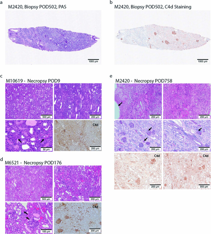
- The single transplanted porcine kidney supported metabolite filtration; measured glomerular filtration rate in 3KO.7TG donors was not different from age-matched WT Yucatan pigs, and a protocol biopsy at post-operative day 502 showed mostly normal kidney histology with patchy fibrosis and glomerular C4d staining.
Methods
- Donor engineering used Yucatan miniature pigs with OO blood type; CRISPR–Cas9-mediated nonhomologous end joining and homology-directed repair plus recombinase-mediated cassette exchange, followed by somatic cell nuclear transfer cloning. Edits were verified by sequencing, droplet digital PCR, long-read whole-genome sequencing and direct RNA sequencing.
- In vitro assays used primary porcine kidney endothelial cells and aortic-derived endothelial cells, with human or cynomolgus monkey serum for antibody binding, C3b deposition, complement-dependent cytotoxicity, activated protein C, thrombin–antithrombin, SIRPα reporter and caspase 3/7 assays; IHC, RNA-seq and single-cell RNA-seq assessed transgene expression.
- In vivo, cynomolgus monkeys were screened for low anti-porcine IgG/IgM and received porcine kidney transplants with bilateral native nephrectomy in most recipients. Immunosuppression included induction B and T lymphocyte depletion, maintenance anti-CD154 antibody and mycophenolate mofetil, and a brief post-transplant course of tacrolimus and steroids; survival was analysed by log-rank test, and histopathology used Banff criteria with xenotransplantation-adapted modifications.
Limitations
- Stated: nonhuman primate constraints meant the experiment was not statistically powered; the number of transplants per genotype was empirically determined, donor–recipient pairing was dictated by availability, and the study was not blind for all involved. The authors also note that Banff scoring may not fully apply to xenotransplantation and that CMAH knockout complications may be specific to Old World monkeys rather than human recipients.
- Stated/evident: residual xenoantigens and antibody binding remained after 3KO, cynomolgus monkey serum showed stronger anti-porcine cytotoxicity than human serum, de novo donor-specific antibody was detected in 8 of 18 transplants, and TMA/AMR in long-term survivors were often associated with infections or biopsy procedures.
- Evident from the text and Table 1: group sizes were small and non-randomized, and protocol variations occurred—some animals received TNX-1500 co-stimulation blockade, some did not receive mycophenolate mofetil, and two recipients had only unilateral native nephrectomy at transplant—so immunosuppression and surgical differences may confound survival comparisons.
Model-written summary. Check it against the paper before relying on it.
Integrity signals
Registry
Cites
Statistics
Figures
Verification
Claims the paper makes
69 Own result · 18 Own interpretation · 12 Own method · 10 Partly supported · 7 No full text of the sources · 6 Supported · 6 Supported so far · 4 No basis given · 1 Sources not found · 1 Not checked yet · accuracy not yet measured
The cited sources support only part of this claim — read each source’s verdict below.
In the paper
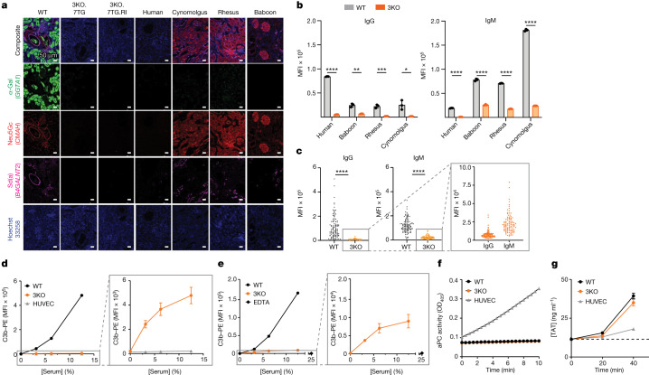
“Porcine cells display three major glycan antigens on their cell surface — α-Gal15, Neu5Gc16 and Sd(a)17 (Fig. 1a) — which are the products of the corresponding glycan synthesis genes, glycoprotein α-galactosyltransferase 1 (GGTA1), cytidine monophospho-N-acetylneuraminic acid hydroxylase (CMAH) and β-1,4-N-acetyl-galactosaminyltransferase 2 (B4GALNT2)/B4GALNT2-like (B4GALNT2L).”
§ Design and testing of a humanized porcine donor for xenotransplantation > Porcine molecular incompatibilities
What the sources say
- Not checked — no full text of the sourcecites [15] Galili, U., Shohet, S. B., Kobrin, E., Stults, C. L. & Macher, B. A. Man, apes, and Old World monkeys differ from other mammals in the expression of α-galactosyl epitopes on nucleated cells. J. Biol. Chem. 263, 17755–17762 (1988). source
- Not checked — no full text of the sourcecites [16] Bouhours, D., Pourcel, C. & Bouhours, J.-F. Simultaneous expression by porcine aorta endothelial cells of glycosphingolipids bearing the major epitope for human xenoreactive antibodies (Galα1–3Gal), blood group H determinant and N-glycolylneuraminic acid. Glycoconj. J. 13, 947–953 (1996). source
- Partly supportedcites [17] Byrne, G. W., Stalboerger, P. G., Du, Z., Davis, T. R. & McGregor, C. G. A. Identification of new carbohydrate and membrane protein antigens in cardiac xenotransplantation. Transplantation 91, 287–292 (2011). sourceExperimental — accuracy not yet measured
The Discussion identifies a porcine B4GALNT2-like gene and links its product to a possible Sd(a)-like glycan on porcine vascular cells. It does not establish that those cells display Sd(a) itself: the authors say the glycan’s precise structure is unknown, and the anti-Sd(a) antibody did not bind the porcine aortic endothelial cells tested. The paper also does not identify B4GALNT2L. (LLM, conf=0.87)
plaindirect- Asserted: passes — The authors put it forward as true.
- Checkable: passes — It can be checked against evidence.
- Load-bearing: passes — The paper’s conclusions depend on it.
- Based: passes — The paper gives a reason why it holds.
The sources with a verdict support this claim; the rest weren’t checked yet, have no full text we could read, or their verdicts aren’t rated yet.
In the paper
“Under physiological conditions, thrombomodulin and endothelial protein C receptor (EPCR) are expressed on the endothelial cell surface and inhibit coagulation by enabling activated protein C (aPC)-mediated regulation26,27.”
§ Design and testing of a humanized porcine donor for xenotransplantation > Porcine molecular incompatibilities
What the sources say
- Supportedcites [26] Mohan Rao, L. V., Esmon, C. T. & Pendurthi, U. R. Endothelial cell protein C receptor: a multiliganded and multifunctional receptor. Blood 124, 1553–1562 (2014). source
The Introduction identifies EPCR as an endothelial-cell transmembrane receptor and says it promotes protein C activation by the thrombin–thrombomodulin complex. Its ‘Regulation of coagulation by EPCR’ passage explains that activated protein C inactivates coagulation cofactors and that endothelial EPCR contributes to this pathway in vivo. Together, these passages support the cited physiological anticoagulant mechanism. (LLM, conf=0.88)
- Not checked — no full text of the sourcecites [27] Esmon, C. Do-all receptor takes on coagulation, inflammation. Nat. Med. 11, 475–477 (2005). source
plaindirect- Asserted: passes — The authors put it forward as true.
- Checkable: passes — It can be checked against evidence.
- Load-bearing: passes — The paper’s conclusions depend on it.
- Based: passes — The paper gives a reason why it holds.
- Supported
Every source the paper cites for this claim was checked and supports it.
In the paper
“This is consistent with other reports that 3KO porcine cells possess additional xenoantigens recognized by OWM serum8,9.”
§ Design and testing of a humanized porcine donor for xenotransplantation > Porcine molecular incompatibilities
What the sources say
- Supportedcites [8] Yamamoto, T. et al. Old World monkeys are less than ideal transplantation models for testing pig organs lacking three carbohydrate antigens (triple-knockout). Sci. Rep. 10, 9771 (2020). source
The Discussion identifies a “4th xenoantigen” in triple-knockout pig cells and says Old World monkeys have antibodies against it. It also reports stronger serum IgM binding and cytotoxicity to those cells in Old World monkeys than in humans. (LLM, conf=0.93)
- Supportedcites [9] Estrada, J. L. et al. Evaluation of human and non-human primate antibody binding to pig cells lacking GGTA1/CMAH/β4GalNT2 genes. Xenotransplantation 22, 194–202 (2015). source
The Results (page unavailable) compare baboon and rhesus macaque serum binding to triple-knockout (KO-3) pig cells and report reduced, rather than eliminated, antibody binding. This supports the claim that these cells retain xenoantigens recognized by old-world monkey serum; the paper does not identify the remaining antigens. (LLM, conf=0.84)
plaincorrelational- Asserted: passes — The authors put it forward as true.
- Checkable: passes — It can be checked against evidence.
- Load-bearing: unclear — It is unclear whether the paper’s conclusions depend on it.
- Based: passes — The paper gives a reason why it holds.
- Supported
The sources with a verdict support this claim; the rest weren’t checked yet, have no full text we could read, or their verdicts aren’t rated yet.
In the paper
“Substantial molecular incompatibilities exist between pigs and humans.”
§ Design and testing of a humanized porcine donor for xenotransplantation > Porcine molecular incompatibilities
What the sources say
- Not checked — no full text of the sourcecites [13] Robson, S. C., Am Esch, J. S. & Bach, F. H. Factors in xenograft rejection. Ann. N. Y. Acad. Sci. 875, 261–276 (1999). source
- Supportedcites [14] Wolf, E., Kemter, E., Klymiuk, N. & Reichart, B. Genetically modified pigs as donors of cells, tissues, and organs for xenotransplantation. Anim. Front. 9, 13–20 (2019). source
The Introduction section on coagulation and inflammation explicitly discusses molecular incompatibilities between porcine and primate coagulation regulators. It also gives a pig–human example: porcine thrombomodulin is a poor co-factor for activating human protein C. The sections on rejection describe further pig–human immune barriers. (LLM, conf=0.90)
plain- Asserted: passes — The authors put it forward as true.
- Checkable: passes — It can be checked against evidence.
- Load-bearing: passes — The paper’s conclusions depend on it.
- Based: passes — The paper gives a reason why it holds.
The sources with a verdict support this claim; the rest weren’t checked yet, have no full text we could read, or their verdicts aren’t rated yet.
In the paper
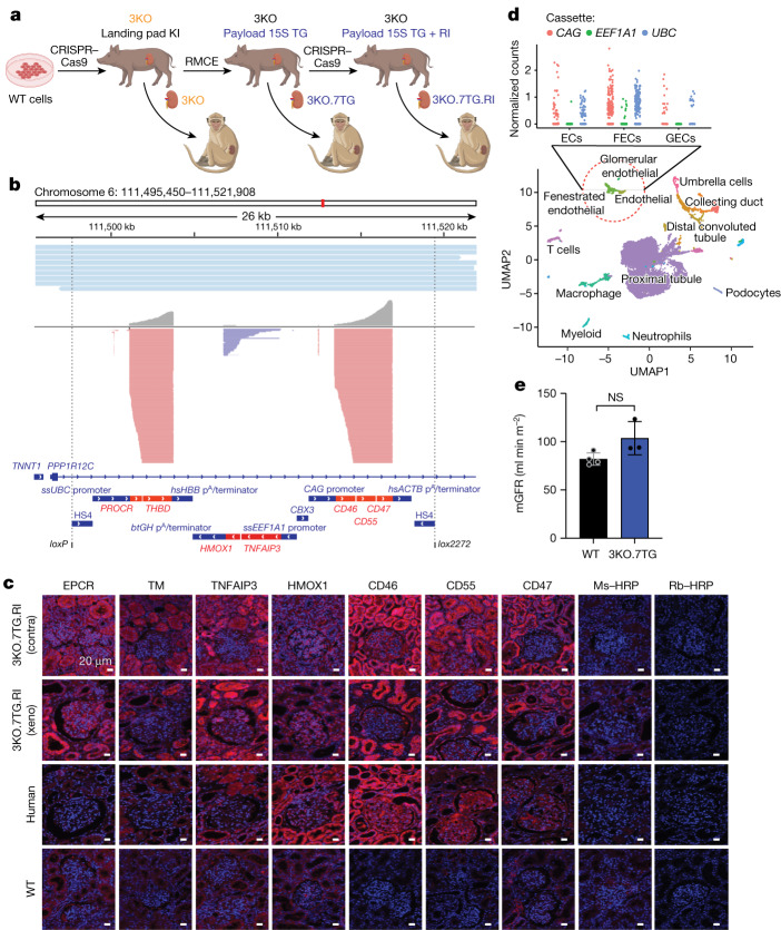
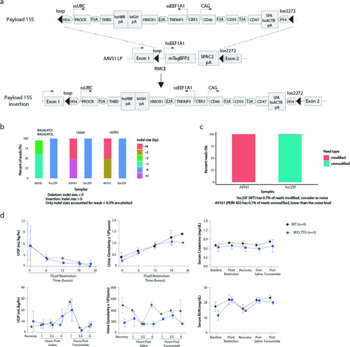
“The pigs were engineered to carry 69 genomic edits, using the clustered regularly interspaced short palindromic repeats (CRISPR) and CRISPR-associated protein 9 (Cas9)-mediated nonhomologous end joining and homology-directed repair30,31, and recombinase-mediated cassette exchange32 (Fig. 2a, Extended Data Fig. 1a and Supplementary Table 2).”
§ Design and testing of a humanized porcine donor for xenotransplantation > A humanized porcine donor
What the sources say
- Supportedcites [30] Cong, L. et al. Multiplex genome engineering using CRISPR/Cas systems. Science 339, 819–823 (2013). source
The citation is attached to the CRISPR-Cas9-mediated NHEJ and HDR methods, not to the citing study’s 69 edits in pigs. The passages labeled Abstract describe Cas9-induced breaks repaired through NHEJ or HDR and demonstrate Cas9-mediated HDR (passage 6); passage 8 describes NHEJ-mediated repair. The separate citation 32 is attached to recombinase-mediated cassette exchange. (LLM, conf=0.91)
- Supportedcites [31] Mali, P. et al. RNA-guided human genome engineering via Cas9. Science 339, 823–826 (2013). source
The paper reports Cas9-mediated nonhomologous end joining at a human genomic locus and homology-directed repair using a donor (main text, Fig. 2 and Fig. 1 discussions). This supports the CRISPR-Cas9 editing methods to which the citation is attached; the pigs and 69 edits describe the citing authors’ work, not a result attributed to this source. (LLM, conf=0.91)
- Not checked — no full text of the sourcecites [32] Schlake, T. & Bode, J. Use of mutated FLP recognition target (FRT) sites for the exchange of expression cassettes at defined chromosomal loci. Biochemistry 33, 12746–12751 (1994). source
plaindirect- Asserted: passes — The authors put it forward as true.
- Checkable: passes — It can be checked against evidence.
- Load-bearing: passes — The paper’s conclusions depend on it.
- Based: passes — The paper gives a reason why it holds.
- Supported
The cited sources support only part of this claim — read each source’s verdict below.
In the paper
“However, in vitro analysis suggests that a human-compatible porcine donor should ideally have all three glycans eliminated to match with the absence of the three glycans in humans8,9.”
§ Design and testing of a humanized porcine donor for xenotransplantation > Main
What the sources say
- Supportedcites [8] Yamamoto, T. et al. Old World monkeys are less than ideal transplantation models for testing pig organs lacking three carbohydrate antigens (triple-knockout). Sci. Rep. 10, 9771 (2020). source
The Results (Expression of Gal, Neu5Gc, and Sda on primate PBMCs; page not supplied) report that human PBMCs express none of the three antigens, while the in vitro Results report that triple-knockout pig PBMCs lack all three. The Discussion says increasing in vitro data indicate that triple-knockout pig organs will be a major advance over single-knockout organs for transplantation into humans, supporting the claim’s hedged recommendation. (LLM, conf=0.91)
- Partly supportedcites [9] Estrada, J. L. et al. Evaluation of human and non-human primate antibody binding to pig cells lacking GGTA1/CMAH/β4GalNT2 genes. Xenotransplantation 22, 194–202 (2015). sourceExperimental — accuracy not yet measured
The Results report that deleting all three genes further reduced human antibody binding, supporting the in vitro rationale for a triple-knockout donor. But the Discussion says only approximately 5% of humans have inactive β4GalNT2; the paper does not support the claim that all three corresponding glycans are absent in humans. (LLM, conf=0.88)
hedged- Asserted: passes — The authors put it forward as true.
- Checkable: passes — It can be checked against evidence.
- Load-bearing: passes — The paper’s conclusions depend on it.
- Based: passes — The paper gives a reason why it holds.
- Supported
The cited sources support only part of this claim — read each source’s verdict below.
In the paper
“After birth, humans develop antibodies to α-Gal and Neu5Gc, referred to as preformed antibodies, upon exposure to molecular mimics of these two antigens16,20,21.”
§ Design and testing of a humanized porcine donor for xenotransplantation > Porcine molecular incompatibilities
What the sources say
- Not checked — no full text of the sourcecites [16] Bouhours, D., Pourcel, C. & Bouhours, J.-F. Simultaneous expression by porcine aorta endothelial cells of glycosphingolipids bearing the major epitope for human xenoreactive antibodies (Galα1–3Gal), blood group H determinant and N-glycolylneuraminic acid. Glycoconj. J. 13, 947–953 (1996). source
- Partly supportedcites [20] Galili, U., Rachmilewitz, E. A., Peleg, A. & Flechner, I. A unique natural human IgG antibody with anti-alpha-galactosyl specificity. J. Exp. Med. 160, 1519–1531 (1984). sourceExperimental — accuracy not yet measured
The age-group results (page not supplied) show anti-Gal titers rising after infancy, and the discussion (page not supplied) suggests intestinal bacteria may provide antigenic stimulation. The paper does not address Neu5Gc, and it proposes rather than establishes exposure to bacterial α-Gal as the cause of antibody development. (LLM, conf=0.87)
- Partly supportedcites [21] Kappler, K. & Hennet, T. Emergence and significance of carbohydrate-specific antibodies. Genes Immun. 21, 224–239 (2020). sourceExperimental — accuracy not yet measured
The ‘Commonly recognized carbohydrate antigens’ section says α-Gal antibodies develop after birth following exposure to intestinal bacteria bearing the α-Gal epitope. It also says contact with NeuGc-containing glycoproteins stimulates anti-NeuGc antibodies, but does not say these arise after birth through exposure to molecular mimics. The copy does not use ‘preformed antibodies’ for this pair. (LLM, conf=0.87)
plaindirect- Asserted: passes — The authors put it forward as true.
- Checkable: passes — It can be checked against evidence.
- Load-bearing: passes — The paper’s conclusions depend on it.
- Based: passes — The paper gives a reason why it holds.
Every source the paper cites for this claim was checked and supports it.
In the paper
“Porcine CD47 does not engage the human SIRPα receptor effectively37.”
§ Design and testing of a humanized porcine donor for xenotransplantation > Genomic edits confer protection
What the sources say
- Supportedcites [37] Ide, K. et al. Role for CD47-SIRPα signaling in xenograft rejection by macrophages. Proc. Natl Acad. Sci. USA 104, 5062–5066 (2007). source
The Discussion reports that porcine CD47 fails to trigger SIRPα phosphorylation in human macrophage-like cells or inhibit engulfment by human macrophages. It also notes that binding to recombinant human SIRPα has been reported, so the support is for ineffective functional engagement, not an absence of physical binding. (LLM, conf=0.90)
plain- Asserted: passes — The authors put it forward as true.
- Checkable: passes — It can be checked against evidence.
- Load-bearing: passes — The paper’s conclusions depend on it.
- Based: passes — The paper gives a reason why it holds.
- Supported
A result the paper reports from its own data — it cites no other source for it, so there is none to check. The quote shows it is in the paper.
In the paper
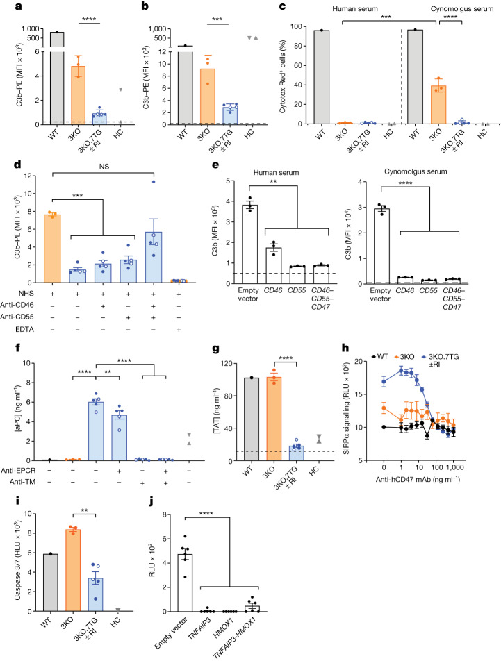
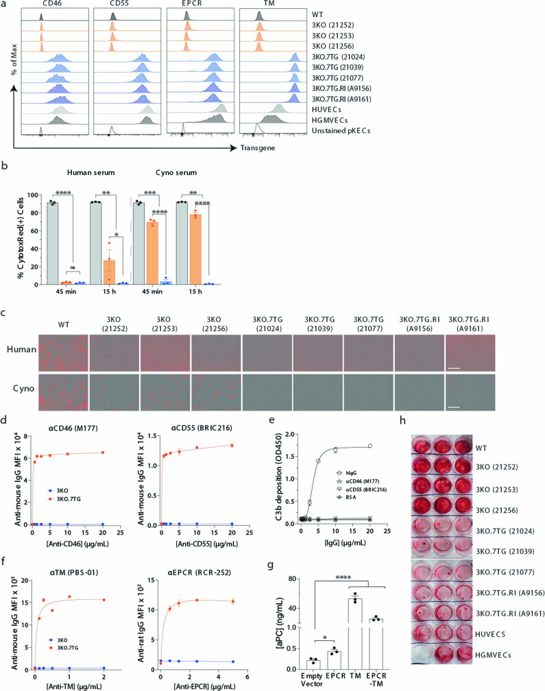
“When incubated in human or cynomolgus monkey serum, the 3KO.7TG ± RI KECs exhibited significantly less C3b deposition than 3KO cells, suggesting that the transgene (or transgenes) impeded complement activation (Fig. 3a,b).”
§ Design and testing of a humanized porcine donor for xenotransplantation > Genomic edits confer protection
hedgeddirect- Asserted: passes — The authors put it forward as true.
- Checkable: passes — It can be checked against evidence.
- Load-bearing: passes — The paper’s conclusions depend on it.
- Based: passes — The paper gives a reason why it holds.
A result the paper reports from its own data — it cites no other source for it, so there is none to check. The quote shows it is in the paper.
In the paper
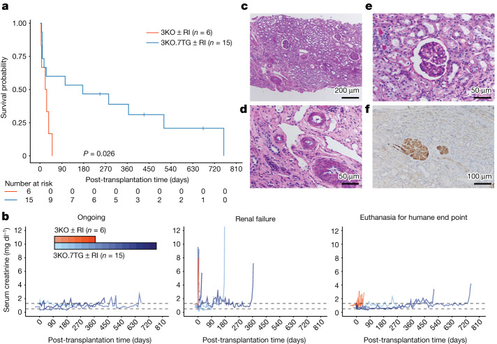
“We show that a humanized porcine renal graft, combined with a clinically relevant immunosuppressive regimen, supported long-term NHP survival for up to 2 years (758 days).”
§ Design and testing of a humanized porcine donor for xenotransplantation > Main
boosteddirect- Asserted: passes — The authors put it forward as true.
- Checkable: passes — It can be checked against evidence.
- Load-bearing: passes — The paper’s conclusions depend on it.
- Based: passes — The paper gives a reason why it holds.
- Rests on an assumption: The immunosuppressive regimen used in the NHP transplants is clinically relevant.
- It may state two separate claims as one. 52
- Probably not a substantial claim. 16
Every source the paper cites for this claim was checked and supports it.
In the paper
“Over the years, various genetically engineered porcine donors have been created and their kidneys transplanted into Old World monkeys (OWMs)4–6.”
§ Design and testing of a humanized porcine donor for xenotransplantation > Main
What the sources say
- Supportedcites [4] Adams, A. B. et al. Anti-C5 antibody tesidolumab reduces early antibody-mediated rejection and prolongs survival in renal xenotransplantation. Ann. Surg. 274, 473–480 (2021). source
The Materials and Methods (Animals, “Pig-to-Rhesus Renal Transplantation”) describe two genetically engineered pig donor types and state that their kidneys were transplanted into rhesus macaques, which are Old World monkeys. This supports the claim attributed to the paper. (LLM, conf=0.94)
- Supportedcites [5] Iwase, H. et al. Immunological and physiological observations in baboons with life-supporting genetically engineered pig kidney grafts. Xenotransplantation 24, e12293 (2017). source
The Methods (Animals) describe two genetically engineered pig donor types, with six or three genetic modifications, whose kidneys were transplanted into baboons. The Introduction also reports earlier transplantation of kidneys from genetically engineered pigs into rhesus monkeys. Together, these passages support the cited background statement. (LLM, conf=0.92)
- Supportedcites [6] Kim, S. C. et al. Long‐term survival of pig‐to‐rhesus macaque renal xenografts is dependent on CD4 T cell depletion. Am. J. Transplant. 19, 2174–2185 (2019). source
The Introduction describes several genetic modifications to porcine donors and discusses pig-to-monkey kidney transplantation. Methods > Animals documents this study’s transplantation of kidneys from αGal-knockout/CD55-transgenic pigs into rhesus macaques; the References also identify genetically engineered pig-kidney transplants into baboons. No page numbers are available in our copy. (LLM, conf=0.91)
plain- Asserted: passes — The authors put it forward as true.
- Checkable: passes — It can be checked against evidence.
- Load-bearing: passes — The paper’s conclusions depend on it.
- Based: passes — The paper gives a reason why it holds.
It may state two separate claims as one.
- Supported
The cited sources support only part of this claim — read each source’s verdict below.
In the paper
“Although renal grafts derived from the porcine donors lacking these three glycans and carrying various human transgenes have been tested in OWMs, graft survival was short8 or not all human transgenes were expressed10.”
§ Design and testing of a humanized porcine donor for xenotransplantation > Main
What the sources say
- Partly supportedcites [8] Yamamoto, T. et al. Old World monkeys are less than ideal transplantation models for testing pig organs lacking three carbohydrate antigens (triple-knockout). Sci. Rep. 10, 9771 (2020). sourceExperimental — accuracy not yet measured
The Results (In vivo study; page unavailable) report that TKO pig kidney grafts were tested in baboons and that recipient survival was 1, 4, and 61 days, significantly shorter than in the GTKO group. The Materials and Methods table lists six human transgenes for each TKO donor, while the Discussion says transgene expression was uniformly good in the great majority of cases. The paper does not establish that some of those transgenes were unexpressed in the TKO grafts. (LLM, conf=0.85)
- Not checked — no full text of the sourcecites [10] Ma, D. et al. Successful long-term TMA- and rejection-free survival of a kidney xenograft with triple xenoantigen knockout plus insertion of multiple human transgenes. Transplantation https://doi.org/10.1097/01.tp.0000698660.82982.ca (2020). source
plain- Asserted: passes — The authors put it forward as true.
- Checkable: passes — It can be checked against evidence.
- Load-bearing: passes — The paper’s conclusions depend on it.
- Based: passes — The paper gives a reason why it holds.
It may state two separate claims as one.
- Partly supported
The cited sources support only part of this claim — read each source’s verdict below.
In the paper
“Xenotransplantation may offer a transformative solution to the worldwide organ shortage crisis1–3.”
§ Design and testing of a humanized porcine donor for xenotransplantation > Main
What the sources say
- Supportedcites [1] Montgomery, R. A. et al. Results of two cases of pig-to-human kidney xenotransplantation. N. Engl. J. Med. 386, 1889–1898 (2022). source
The paper’s opening passage (section E; page not provided) describes a shortage of transplant organs and says xenotransplantation could provide an unlimited, renewable source of lifesaving organs. That supports the citing sentence’s hedged description of a potential solution to organ shortages. (LLM, conf=0.90)
- Supportedcites [2] Porrett, P. M. et al. First clinical‐grade porcine kidney xenotransplant using a human decedent model. Am. J. Transplant. 22, 1037–1053 (2022). source
The Discussion calls xenotransplantation 'arguably the most pragmatic solution to the organ shortage crisis.' The Abstract likewise describes pigs as a promising organ source while acknowledging that further work is needed. These passages support the citing sentence’s qualified statement that xenotransplantation may offer a solution. (LLM, conf=0.92)
- Partly supportedcites [3] Griffith, B. P. et al. Genetically modified porcine-to-human cardiac xenotransplantation. N. Engl. J. Med. 387, 35–44 (2022). sourceExperimental — accuracy not yet measured
The Abstract reports that a patient ineligible for a human heart transplant received a pig heart that functioned normally for several weeks, supporting xenotransplantation as a possible alternative. The paper does not address the worldwide organ shortage or establish that xenotransplantation could solve it; the graft failed on day 49. (LLM, conf=0.78)
hedgedconditional- Asserted: passes — The authors put it forward as true.
- Checkable: fails — It may be too vague to check against evidence.
- Load-bearing: passes — The paper’s conclusions depend on it.
- Based: passes — The paper gives a reason why it holds.
Probably not a substantial claim. It may be too vague to check against evidence.
- Supported
The sources with a verdict support this claim; the rest weren’t checked yet, have no full text we could read, or their verdicts aren’t rated yet.
In the paper
“In humans, GGTA1 (ref. 18) and CMAH19 evolved into pseudogenes and the α-Gal and the Neu5Gc epitopes are not expressed (Fig. 1a).”
§ Design and testing of a humanized porcine donor for xenotransplantation > Porcine molecular incompatibilities
What the sources say
- Not checked — no full text of the sourcecites [18] Shaper, N. L., Lin, S. P., Joziasse, D. H., Kim, D. Y. & Yang-Feng, T. L. Assignment of two human alpha-1,3-galactosyltransferase gene sequences (GGTA1 and GGTA1P) to chromosomes 9q33-q34 and 12q14-q15. Genomics 12, 613–615 (1992). source
- Supportedcites [19] Irie, A., Koyama, S., Kozutsumi, Y., Kawasaki, T. & Suzuki, A. The molecular basis for the absence of N-glycolylneuraminic acid in humans. J. Biol. Chem. 273, 15866–15871 (1998). source
The citation to this paper is attached to CMAH, not GGTA1. The Abstract reports that a deletion in the human CMP-NeuAc hydroxylase gene makes its protein inactive and accounts for the absence of NeuGc in human glycoconjugates. It does not use the word “pseudogene,” but the reported loss of gene function supports that paraphrase. (LLM, conf=0.85)
plaindirect- Asserted: passes — The authors put it forward as true.
- Checkable: passes — It can be checked against evidence.
- Load-bearing: passes — The paper’s conclusions depend on it.
- Based: passes — The paper gives a reason why it holds.
It may state two separate claims as one.
The cited sources support only part of this claim — read each source’s verdict below.
In the paper
“Although elimination of growth hormone receptor gene expression could reduce organ sizes2,3, it comes with other undesired biological consequences7.”
§ Design and testing of a humanized porcine donor for xenotransplantation > Main
What the sources say
- Partly supportedcites [2] Porrett, P. M. et al. First clinical‐grade porcine kidney xenotransplant using a human decedent model. Am. J. Transplant. 22, 1037–1053 (2022). sourceExperimental — accuracy not yet measured
Section 2.3.1 reports that growth hormone receptor knockout was accompanied by reduced serum IGF-1 levels and body weight, establishing a biological effect beyond organ size. It does not characterize the lower IGF-1 level as undesired. The Discussion attributes altered renal structural integrity to the pigs’ genetic modifications collectively, not specifically to growth hormone receptor knockout. (LLM, conf=0.75)
- Could not verifycites [3] Griffith, B. P. et al. Genetically modified porcine-to-human cardiac xenotransplantation. N. Engl. J. Med. 387, 35–44 (2022). sourceExperimental — accuracy not yet measured
Citation 7 is attached to the claim that eliminating growth hormone receptor expression has undesired biological consequences. The Methods section says the receptor was knocked out to reduce xenograft growth, and the Results section reports later graft injury, but neither attributes that injury or another adverse consequence to the knockout. The Results say the mechanism of the injury was still under investigation. (LLM, conf=0.72)
- Supportedcites [7] Hinrichs, A. et al. Growth hormone receptor-deficient pigs resemble the pathophysiology of human Laron syndrome and reveal altered activation of signaling cascades in the liver. Mol. Metab. 11, 113–128 (2018). source
Citation 7 is attached to the undesired biological consequences. The Results sections report transient juvenile hypoglycemia and increased body fat with a reduced muscle-to-fat ratio in GHR-KO pigs. The Results section on organ growth also reports smaller organs in these pigs. Page numbers are not supplied in our copy. (LLM, conf=0.91)
hedgedconditional- Asserted: passes — The authors put it forward as true.
- Checkable: passes — It can be checked against evidence.
- Load-bearing: passes — The paper’s conclusions depend on it.
- Based: passes — The paper gives a reason why it holds.
It may state two separate claims as one.
- Partly supported
Every source the paper cites for this claim was checked and supports it.
In the paper
“Although the human B4GALNT2 gene is functional22, its expression levels vary and naturally occurring mutations have been identified that correlate with the Sd(a-) phenotype23.”
§ Design and testing of a humanized porcine donor for xenotransplantation > Porcine molecular incompatibilities
What the sources say
- Supportedcites [22] Montiel, M.-D., Krzewinski-Recchi, M.-A., Delannoy, P. & Harduin-Lepers, A. Molecular cloning, gene organization and expression of the human UDP-GalNAc:Neu5Acalpha2-3Galbeta-R beta1,4-N-acetylgalactosaminyltransferase responsible for the biosynthesis of the blood group Sda/Cad antigen: evidence for an unusual extended cytoplasmic domain. Biochem. J. 373, 369–379 (2003). source
The paper’s opening summary reports that the human gene is expressed and that its expressed catalytic domain produces an enzyme that forms the Sd(a)/Cad antigen structure. It also reports varying expression across the cell types and tissues studied. The separate citation 23 is attached to the mutation–phenotype claim, so that claim is not attributed to this source. Section and page labels are unavailable in our copy. (LLM, conf=0.92)
- Supportedcites [23] Stenfelt, L. et al. Missense mutations in the C-terminal portion of the B4GALNT2-encoded glycosyltransferase underlying the Sd(a−) phenotype. Biochem. Biophys. Rep. 19, 100659 (2019). source
Citation 23 is attached most directly to the mutation claim. The Results and discussion section identifies naturally occurring B4GALNT2 missense and splice-site variants in individuals with the Sd(a−) phenotype. The paper also analyzes B4GALNT2 expression patterns across cell lines; the separate claim that the gene is functional is attached to citation 22. (LLM, conf=0.90)
plaincorrelational- Asserted: passes — The authors put it forward as true.
- Checkable: passes — It can be checked against evidence.
- Load-bearing: fails — The paper’s conclusions may not depend on it.
- Based: passes — The paper gives a reason why it holds.
Probably not a substantial claim. The paper’s conclusions may not depend on it. It may state two separate claims as one.
- Supported
The sources with a verdict support this claim; the rest weren’t checked yet, have no full text we could read, or their verdicts aren’t rated yet.
In the paper
“Third, the donors carried porcine endogenous retrovirus (PERV) sequences in their genome, which present a zoonotic risk, as PERV transmission to human cells in culture and their integration into the human genome have been demonstrated11,12.”
§ Design and testing of a humanized porcine donor for xenotransplantation > Main
What the sources say
- Supportedcites [11] Niu, D. et al. Inactivation of porcine endogenous retrovirus in pigs using CRISPR-Cas9. Science 357, 1303–1307 (2017). source
The passage labeled Abstract states that PERVs occur in the genome of all pig strains and can infect and integrate into human cells in culture. It describes transmission as a potential xenotransplantation risk, while noting that transmission to humans in clinical settings has not been shown. (LLM, conf=0.92)
- Not checked — no full text of the sourcecites [12] Patience, C., Takeuchi, Y. & Weiss, R. A. Infection of human cells by an endogenous retrovirus of pigs. Nat. Med. 3, 282–286 (1997). source
boosteddirect- Asserted: passes — The authors put it forward as true.
- Checkable: passes — It can be checked against evidence.
- Load-bearing: passes — The paper’s conclusions depend on it.
- Based: passes — The paper gives a reason why it holds.
It may state two separate claims as one.
- Supported
The cited sources support only part of this claim — read each source’s verdict below.
In the paper
“Although overlapping in their anti-apoptotic activities, TNFAIP3 and HMOX1 achieve this outcome by their unique biological functions and dampen inflammation under distinct circumstances.”
§ Design and testing of a humanized porcine donor for xenotransplantation > Genomic edits confer protection
What the sources say
- Not checked — no full text of the sourcecites [38] Lee, E. G. et al. Failure to regulate TNF-induced NF-κB and cell death responses in A20-deficient mice. Science 289, 2350–2354 (2000). source
- Partly supportedcites [39] Kapturczak, M. H. et al. Heme oxygenase-1 modulates early inflammatory responses. Am. J. Pathol. 165, 1045–1053 (2004). sourceExperimental — accuracy not yet measured
The Discussion (page not specified in our copy) describes HO-1’s anti-inflammatory effects and identifies heme degradation products, including carbon monoxide, as contributors to cytoprotection and anti-apoptotic activity. The paper does not address TNFAIP3 or compare the two proteins’ functions or the circumstances in which they dampen inflammation. (LLM, conf=0.87)
plaindirect- Asserted: passes — The authors put it forward as true.
- Checkable: passes — It can be checked against evidence.
- Load-bearing: unclear — It is unclear whether the paper’s conclusions depend on it.
- Based: passes — The paper gives a reason why it holds.
It may state two separate claims as one.
Every source the paper cites for this claim was checked and supports it.
In the paper
“Xenograft haemotoxin and eosin and periodic acid–Schiff-stained samples were scored by current Banff criteria including C4d deposition42.”
§ Design and testing of a humanized porcine donor for xenotransplantation > Methods > Histopathological analysis
What the sources say
- Supportedcites [42] Roufosse, C. et al. A 2018 reference guide to the Banff Classification of Renal Allograft Pathology. Transplantation 102, 1795–1814 (2018). source
The BANFF LESION SCORES section defines the Banff scoring criteria, including a C4d lesion score. DIAGNOSTIC WORK-UP OF BIOPSIES specifies hematoxylin-eosin and periodic acid–Schiff stains and calls C4d staining indispensable. The citation identifies the scoring criteria the citing authors applied to their xenograft samples; it does not attribute those samples to the guide. (LLM, conf=0.91)
plain- Asserted: passes — The authors put it forward as true.
- Checkable: passes — It can be checked against evidence.
- Load-bearing: passes — The paper’s conclusions depend on it.
- Based: passes — The paper gives a reason why it holds.
Probably not a substantial claim.
- Supported
The cited sources support only part of this claim — read each source’s verdict below.
In the paper
“We included both TNFAIP3 and HMOX1 in our donors, as both transgenes have been shown to provide benefit in vitro and in vivo40,41.”
§ Design and testing of a humanized porcine donor for xenotransplantation > Genomic edits confer protection
What the sources say
- Not checked — no full text of the sourcecites [40] Oropeza, M. et al. Transgenic expression of the human A20 gene in cloned pigs provides protection against apoptotic and inflammatory stimuli. Xenotransplantation 16, 522–534 (2009). source
- Partly supportedcites [41] Petersen, B. et al. Transgenic expression of human heme oxygenase-1 in pigs confers resistance against xenograft rejection during ex vivo perfusion of porcine kidneys. Xenotransplantation 18, 355–368 (2011). sourceExperimental — accuracy not yet measured
The Results report that HMOX1-transgenic endothelial cells were protected against apoptosis in vitro, and the Discussion reports resistance to rejection during ex vivo kidney perfusion. That is not an in vivo test of the transgenic pig kidneys, so this source does not establish the claimed in vivo benefit for the HMOX1 transgene. The separate TNFAIP3 citation is not assessed here. (LLM, conf=0.88)
plaindirect- Asserted: passes — The authors put it forward as true.
- Checkable: passes — It can be checked against evidence.
- Load-bearing: passes — The paper’s conclusions depend on it.
- Based: passes — The paper gives a reason why it holds.
It may state two separate claims as one.
The sources with a verdict support this claim; the rest weren’t checked yet, have no full text we could read, or their verdicts aren’t rated yet.
In the paper
“TNFAIP3 is a deubiquitinating enzyme and inhibits TNF-mediated apoptosis38, whereas HMOX1 is involved in the initial step of haem degradation and has been shown to have antioxidant and anti-inflammatory effects38,39.”
§ Design and testing of a humanized porcine donor for xenotransplantation > Genomic edits confer protection
What the sources say
- Not checked — no full text of the sourcecites [38] Lee, E. G. et al. Failure to regulate TNF-induced NF-κB and cell death responses in A20-deficient mice. Science 289, 2350–2354 (2000). source
- Supportedcites [39] Kapturczak, M. H. et al. Heme oxygenase-1 modulates early inflammatory responses. Am. J. Pathol. 165, 1045–1053 (2004). source
The Discussion states that HO-1 catalyzes the rate-limiting step in heme degradation. It also describes antioxidant mechanisms of HO-1 induction and evidence for its anti-inflammatory effects. Citation 39 is attached to the HMOX1 portion of the sentence, not the TNFAIP3 portion. (LLM, conf=0.93)
plaindirect- Asserted: passes — The authors put it forward as true.
- Checkable: passes — It can be checked against evidence.
- Load-bearing: passes — The paper’s conclusions depend on it.
- Based: passes — The paper gives a reason why it holds.
It may state two separate claims as one.
Every source the paper cites for this claim was checked and supports it.
In the paper
“Rejection was assessed by histopathological examination, based on the Banff classification of renal allograft pathology42, with modifications for xenotransplantation (Table 1 and Extended Data Table 1).”
§ Design and testing of a humanized porcine donor for xenotransplantation > Engineered porcine kidney supports life
What the sources say
- Supportedcites [42] Roufosse, C. et al. A 2018 reference guide to the Banff Classification of Renal Allograft Pathology. Transplantation 102, 1795–1814 (2018). source
The Abstract identifies this paper as a reference guide to the Banff Classification of Kidney Allograft Pathology. The BANFF LESION SCORES section says the classification assesses histopathological changes in renal transplant biopsies, particularly features of rejection. The citation is attached to the Banff classification; the xenotransplantation modifications are described as the citing authors’ own methods. (LLM, conf=0.92)
plain- Asserted: passes — The authors put it forward as true.
- Checkable: passes — It can be checked against evidence.
- Load-bearing: passes — The paper’s conclusions depend on it.
- Based: passes — The paper gives a reason why it holds.
Probably not a substantial claim.
- Supported
The cited sources support only part of this claim — read each source’s verdict below.
In the paper
“Although a porcine donor carrying a CMAH knockout is thought not to survive in OWMs considering complication from a CMAH knockout8,9, renal grafts derived from the 3KO.7TG ± RI porcine donor supported life long-term in cynomolgus monkeys, up to 758 days.”
§ Design and testing of a humanized porcine donor for xenotransplantation > Discussion
What the sources say
- Partly supportedcites [8] Yamamoto, T. et al. Old World monkeys are less than ideal transplantation models for testing pig organs lacking three carbohydrate antigens (triple-knockout). Sci. Rep. 10, 9771 (2020). sourceExperimental — accuracy not yet measured
The Discussion (page not provided) says that loss of Neu5Gc in triple-knockout pigs exposes an antigen recognized by Old World monkeys, creating a greater barrier to graft survival. The Results (page not provided) report shorter survival for triple-knockout kidneys in baboons than for GTKO kidneys, but do not establish that a graft with a CMAH knockout cannot survive in an Old World monkey: one triple-knockout recipient survived 61 days. The study tested triple-knockout organs, not the effect of CMAH knockout alone. (LLM, conf=0.86)
- Partly supportedcites [9] Estrada, J. L. et al. Evaluation of human and non-human primate antibody binding to pig cells lacking GGTA1/CMAH/β4GalNT2 genes. Xenotransplantation 22, 194–202 (2015). sourceExperimental — accuracy not yet measured
The Discussion (page not available) reports that deleting CMAH on a GGTA1-knockout background increased baboon and rhesus antibody binding, a potential complication for pig-to-primate transplantation. The paper measured antibody binding, not graft survival, and does not say that CMAH-knockout porcine grafts are unlikely to survive in Old World monkeys. (LLM, conf=0.86)
hedgedconditional- Asserted: passes — The authors put it forward as true.
- Checkable: passes — It can be checked against evidence.
- Load-bearing: passes — The paper’s conclusions depend on it.
- Based: passes — The paper gives a reason why it holds.
Probably not a substantial claim.
- Partly supported
The cited sources support only part of this claim — read each source’s verdict below.
In the paper
“In addition, data suggest that CMAH inactivation may produce a novel antigen xenogenic to the OWMs, which may be a target of preformed antibody binding, leading to complement activation in OWM serum8,9.”
§ Design and testing of a humanized porcine donor for xenotransplantation > Discussion
What the sources say
- Supportedcites [8] Yamamoto, T. et al. Old World monkeys are less than ideal transplantation models for testing pig organs lacking three carbohydrate antigens (triple-knockout). Sci. Rep. 10, 9771 (2020). source
The Discussion (page not provided) says that loss of Neu5Gc expression in triple-knockout pigs apparently reveals a ‘4th xenoantigen’ recognized by antibodies in Old World monkeys. The Results (page not provided) report that OWM IgM binding correlates with complement-dependent cytotoxicity against those pig cells. This supports the claim’s tentative wording. (LLM, conf=0.91)
- Partly supportedcites [9] Estrada, J. L. et al. Evaluation of human and non-human primate antibody binding to pig cells lacking GGTA1/CMAH/β4GalNT2 genes. Xenotransplantation 22, 194–202 (2015). sourceExperimental — accuracy not yet measured
The Results and Discussion (page not available in our copy) report that pig cells lacking GGTA1 and CMAH bound more baboon and rhesus antibodies than cells lacking GGTA1 alone. This supports the suggested increase in antibody binding after CMAH inactivation, but the paper does not establish that a novel antigen causes it or report complement activation in Old World monkey serum. (LLM, conf=0.88)
hedgedconditional- Asserted: passes — The authors put it forward as true.
- Checkable: passes — It can be checked against evidence.
- Load-bearing: passes — The paper’s conclusions depend on it.
- Based: passes — The paper gives a reason why it holds.
It may state two separate claims as one.
- Supported
A result the paper reports from its own data — it cites no other source for it, so there is none to check. The quote shows it is in the paper.
In the paper
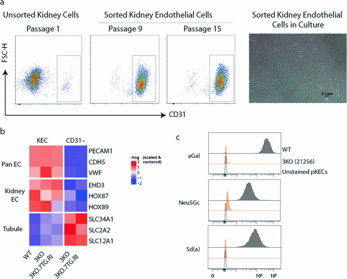
“Three KEC types were identified, including endothelial cells (PECAM1+PLVAP−EHD3−GATA5−), fenestrated endothelial cells (PECAM1+PLVAP+) and glomerular endothelial cells (PECAM1+EHD3+GATA5+) (Fig. 2d).”
§ Design and testing of a humanized porcine donor for xenotransplantation > A humanized porcine donor
plainn = 2- Asserted: passes — The authors put it forward as true.
- Checkable: passes — It can be checked against evidence.
- Load-bearing: unclear — It is unclear whether the paper’s conclusions depend on it.
- Based: passes — The paper gives a reason why it holds.
It may state two separate claims as one.
A result the paper reports from its own data — it cites no other source for it, so there is none to check. The quote shows it is in the paper.
In the paper
“WT KECs showed C3b deposition and 3KO KECs showed less deposition when incubated in the cynomolgus monkey serum.”
§ Design and testing of a humanized porcine donor for xenotransplantation > Porcine molecular incompatibilities
plaincorrelationalSingle case- Asserted: passes — The authors put it forward as true.
- Checkable: passes — It can be checked against evidence.
- Load-bearing: passes — The paper’s conclusions depend on it.
- Based: passes — The paper gives a reason why it holds.
It may state two separate claims as one.
A result the paper reports from its own data — it cites no other source for it, so there is none to check. The quote shows it is in the paper.
In the paper
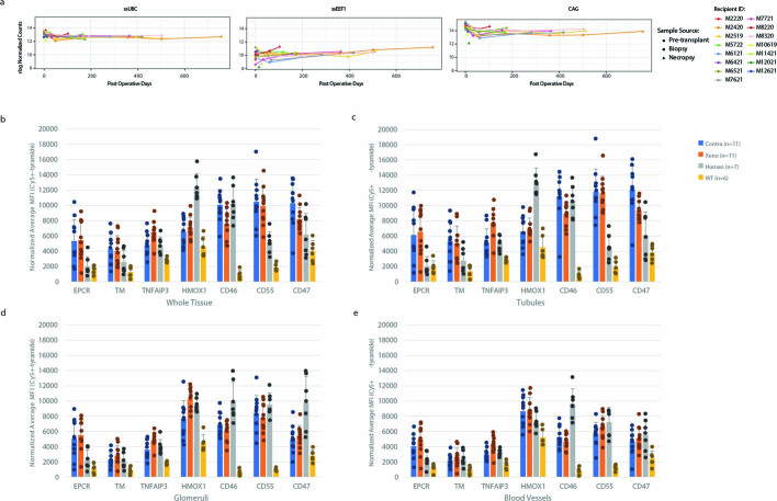
“Among the three units, expression of the CAG and ssUBC units were higher, whereas expression of the ssEEF1A1 unit was at a lower level.”
§ Design and testing of a humanized porcine donor for xenotransplantation > A humanized porcine donor
plainSingle case- Asserted: passes — The authors put it forward as true.
- Checkable: passes — It can be checked against evidence.
- Load-bearing: unclear — It is unclear whether the paper’s conclusions depend on it.
- Based: passes — The paper gives a reason why it holds.
It may state two separate claims as one.
A result the paper reports from its own data — it cites no other source for it, so there is none to check. The quote shows it is in the paper.
In the paper
“When incubated with human serum, human umbilical vein endothelial cells do not show C3b deposition (Fig. 1d), suggesting no antibody binding and/or complete mitigation of complement activation.”
§ Design and testing of a humanized porcine donor for xenotransplantation > Porcine molecular incompatibilities
hedged- Asserted: passes — The authors put it forward as true.
- Checkable: passes — It can be checked against evidence.
- Load-bearing: passes — The paper’s conclusions depend on it.
- Based: passes — The paper gives a reason why it holds.
It may state two separate claims as one.
A result the paper reports from its own data — it cites no other source for it, so there is none to check. The quote shows it is in the paper.
In the paper
“Binding by OWM serum IgG and IgM was similar, although substantial residual antibody binding (especially IgM binding) was detected with the 3KO KECs (Fig. 1b).”
§ Design and testing of a humanized porcine donor for xenotransplantation > Porcine molecular incompatibilities
plain- Asserted: passes — The authors put it forward as true.
- Checkable: passes — It can be checked against evidence.
- Load-bearing: passes — The paper’s conclusions depend on it.
- Based: passes — The paper gives a reason why it holds.
It may state two separate claims as one.
A result the paper reports from its own data — it cites no other source for it, so there is none to check. The quote shows it is in the paper.
In the paper
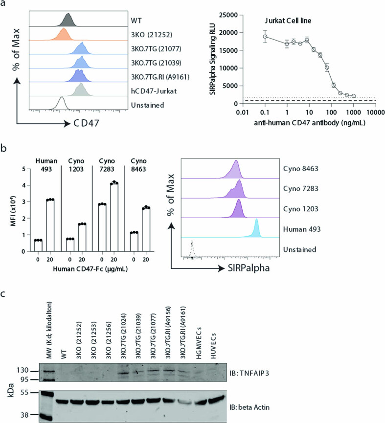
“As expected, the 3KO KECs lacked α-Gal, Neu5Gc and Sd(a) (Extended Data Fig. 3c) and the 3KO.7TG ± RI KECs expressed human transgenes, as analysed by flow cytometry (Extended Data Fig. 4a, CD46, CD55, EPCR and thrombomodulin; Extended Data Fig. 5a, CD47) and by western blot (Extended Data Fig. 5c, TNFAIP3).”
§ Design and testing of a humanized porcine donor for xenotransplantation > Genomic edits confer protection
plain- Asserted: passes — The authors put it forward as true.
- Checkable: passes — It can be checked against evidence.
- Load-bearing: passes — The paper’s conclusions depend on it.
- Based: passes — The paper gives a reason why it holds.
It may state two separate claims as one.
A result the paper reports from its own data — it cites no other source for it, so there is none to check. The quote shows it is in the paper.
In the paper
“Althoughthe 3KO KECs showed significant reduction in C3b deposition compared with the WT KECs, they retained substantial residual C3b deposition (Fig. 1d), suggesting that porcine complement regulators are less effective in mitigating human complement activation.”
§ Design and testing of a humanized porcine donor for xenotransplantation > Porcine molecular incompatibilities
plaindirect- Asserted: passes — The authors put it forward as true.
- Checkable: passes — It can be checked against evidence.
- Load-bearing: passes — The paper’s conclusions depend on it.
- Based: passes — The paper gives a reason why it holds.
It may state two separate claims as one.
The paper states this without a source or data behind it here, so there is nothing to check it against. The quote shows it is in the paper.
In the paper
“In comparison, OWMs express the Neu5Gc antigen.”
§ Design and testing of a humanized porcine donor for xenotransplantation > Porcine molecular incompatibilities
plain- Asserted: passes — The authors put it forward as true.
- Checkable: passes — It can be checked against evidence.
- Load-bearing: fails — The paper’s conclusions may not depend on it.
- Based: fails — The paper may give no reason why it holds.
Probably not a substantial claim. The paper’s conclusions may not depend on it. The paper may give no reason why it holds.
A result the paper reports from its own data — it cites no other source for it, so there is none to check. The quote shows it is in the paper.
In the paper
“Furthermore, when subjected to fluid challenge studies, both groups responded to the challenges similarly, providing further evidence that the 3KO.7TG kidneys functioned normally (Extended Data Fig. 1d).”
§ Design and testing of a humanized porcine donor for xenotransplantation > A humanized porcine donor
plainn = 2- Asserted: passes — The authors put it forward as true.
- Checkable: passes — It can be checked against evidence.
- Load-bearing: passes — The paper’s conclusions depend on it.
- Based: passes — The paper gives a reason why it holds.
It may state two separate claims as one.
A result the paper reports from its own data — it cites no other source for it, so there is none to check. The quote shows it is in the paper.
In the paper
“For RNA-seq, contralateral, biopsy and necropsy samples were analysed and showed that all human transgenes were expressed and, again, those under the CAG and ssUBC promoters were at a higher level, whereas those under the ssEEF1A1 promoter were at a lower level (Extended Data Fig. 2a).”
§ Design and testing of a humanized porcine donor for xenotransplantation > A humanized porcine donor
plaincorrelationaln = 11- Asserted: passes — The authors put it forward as true.
- Checkable: passes — It can be checked against evidence.
- Load-bearing: passes — The paper’s conclusions depend on it.
- Based: passes — The paper gives a reason why it holds.
It may state two separate claims as one.
A result the paper reports from its own data — it cites no other source for it, so there is none to check. The quote shows it is in the paper.
In the paper
“All seven transgenic proteins were detected in the contralateral kidney, in both the glomeruli and the tubular cells, and expression was maintained in the renal graft at necropsy on post-transplantation day 176.”
§ Design and testing of a humanized porcine donor for xenotransplantation > A humanized porcine donor
plainn = 11- Asserted: passes — The authors put it forward as true.
- Checkable: passes — It can be checked against evidence.
- Load-bearing: passes — The paper’s conclusions depend on it.
- Based: passes — The paper gives a reason why it holds.
It may state two separate claims as one.
A result the paper reports from its own data — it cites no other source for it, so there is none to check. The quote shows it is in the paper.
In the paper
“These edits disrupted the three glycan synthesis genes (eight alleles; Extended Data Fig. 1b) (3KO), had a transgenic construct (referred to as Payload 15S (PL15S)) inserted hemizygously into the AAVS1 site (Extended Data Fig. 1a) (7TG), and inactivated the PERV elements (59 copies) (RI) (Extended Data Fig. 1c) carried in the Yucatan female cells Yuc25F.”
§ Design and testing of a humanized porcine donor for xenotransplantation > A humanized porcine donor
plainSingle case- Asserted: passes — The authors put it forward as true.
- Checkable: passes — It can be checked against evidence.
- Load-bearing: passes — The paper’s conclusions depend on it.
- Based: passes — The paper gives a reason why it holds.
It may state two separate claims as one.
The paper states this without a source or data behind it here, so there is nothing to check it against. The quote shows it is in the paper.
In the paper
“Enhanced clotting of human whole blood ex vivo was observed, measured as thrombin–antithrombin (TAT) complex formation, probably due to the inability of the porcine cells to generate aPC (Fig. 1g).”
§ Design and testing of a humanized porcine donor for xenotransplantation > Porcine molecular incompatibilities
hedgedconditional- Asserted: passes — The authors put it forward as true.
- Checkable: passes — It can be checked against evidence.
- Load-bearing: passes — The paper’s conclusions depend on it.
- Based: passes — The paper gives a reason why it holds.
It may state two separate claims as one.
A result the paper reports from its own data — it cites no other source for it, so there is none to check. The quote shows it is in the paper.
In the paper
“Transcripts from the CAG and ssUBC cassettes were readily detected in the three endothelial cell types, whereas ssEEF1A1 cassette transcripts were found in endothelial cells and fenestrated endothelial cells, but not in glomerular endothelial cells.”
§ Design and testing of a humanized porcine donor for xenotransplantation > A humanized porcine donor
plainn = 2- Asserted: passes — The authors put it forward as true.
- Checkable: passes — It can be checked against evidence.
- Load-bearing: passes — The paper’s conclusions depend on it.
- Based: passes — The paper gives a reason why it holds.
It may state two separate claims as one.
A result the paper reports from its own data — it cites no other source for it, so there is none to check. The quote shows it is in the paper.
In the paper
“Nonetheless, both the human TNFAIP3 and HMOX1 proteins were detected in kidneys by IHC (Fig. 2c), and TNFAIP3 was detected by western blot (Extended Data Fig. 5c).”
§ Design and testing of a humanized porcine donor for xenotransplantation > A humanized porcine donor
plainn = 2- Asserted: passes — The authors put it forward as true.
- Checkable: passes — It can be checked against evidence.
- Load-bearing: passes — The paper’s conclusions depend on it.
- Based: passes — The paper gives a reason why it holds.
It may state two separate claims as one.
A result the paper reports from its own data — it cites no other source for it, so there is none to check. The quote shows it is in the paper.
In the paper
“When WT and 3KO porcine KECs were incubated with cynomolgus monkey serum, similar results were obtained, although higher residual C3b levels on 3KO porcine cells were observed, compared with those in human serum (Fig. 1e).”
§ Design and testing of a humanized porcine donor for xenotransplantation > Porcine molecular incompatibilities
plaincorrelational- Asserted: passes — The authors put it forward as true.
- Checkable: passes — It can be checked against evidence.
- Load-bearing: passes — The paper’s conclusions depend on it.
- Based: passes — The paper gives a reason why it holds.
It may state two separate claims as one.
A result the paper reports from its own data — it cites no other source for it, so there is none to check. The quote shows it is in the paper.
In the paper
“In an antibody-binding assay, the porcine wild-type (WT) kidney endothelial cells (KECs) bound a high level of human IgG and IgM and binding was significantly reduced when the three xenoantigens were eliminated (3KO KECs) (Fig. 1b).”
§ Design and testing of a humanized porcine donor for xenotransplantation > Porcine molecular incompatibilities
plaindirect- Asserted: passes — The authors put it forward as true.
- Checkable: passes — It can be checked against evidence.
- Load-bearing: passes — The paper’s conclusions depend on it.
- Based: passes — The paper gives a reason why it holds.
It may state two separate claims as one.
A result the paper reports from its own data — it cites no other source for it, so there is none to check. The quote shows it is in the paper.
In the paper
“The IHC photomicrographs from all completed NHP studies were scanned and quantified (Extended Data Fig. 2b–e), which showed that all transgenic proteins were detected.”
§ Design and testing of a humanized porcine donor for xenotransplantation > A humanized porcine donor
plainn = 11- Asserted: passes — The authors put it forward as true.
- Checkable: passes — It can be checked against evidence.
- Load-bearing: passes — The paper’s conclusions depend on it.
- Based: passes — The paper gives a reason why it holds.
It may state two separate claims as one.
A result the paper reports from its own data — it cites no other source for it, so there is none to check. The quote shows it is in the paper.
In the paper
“PL15S carries seven human genes (Supplementary Table 3), including CD46 and CD55 from the complement cascade, THBD and PROCR from the coagulation pathway, CD47, which is involved in innate immunity, and TNFAIP3 and HMOX1, which dampen ischaemia–reperfusion injury, apoptosis and inflammation.”
§ Design and testing of a humanized porcine donor for xenotransplantation > A humanized porcine donor
plaindirectSingle case- Asserted: passes — The authors put it forward as true.
- Checkable: passes — It can be checked against evidence.
- Load-bearing: passes — The paper’s conclusions depend on it.
- Based: passes — The paper gives a reason why it holds.
It may state two separate claims as one.
A result the paper reports from its own data — it cites no other source for it, so there is none to check. The quote shows it is in the paper.
In the paper
“IHC confirmed that the three glycan antigens are expressed in the WT porcine kidney, but have been eliminated from the 3KO.7TG (donor ID 21077) and the 3KO.7TG.RI (donor ID A9161) kidneys, a pattern similar to that of the human kidney.”
§ Design and testing of a humanized porcine donor for xenotransplantation > Porcine molecular incompatibilities
plaincorrelational- Asserted: passes — The authors put it forward as true.
- Checkable: passes — It can be checked against evidence.
- Load-bearing: passes — The paper’s conclusions depend on it.
- Based: passes — The paper gives a reason why it holds.
It may state two separate claims as one.
A result the paper reports from its own data — it cites no other source for it, so there is none to check. The quote shows it is in the paper.
In the paper
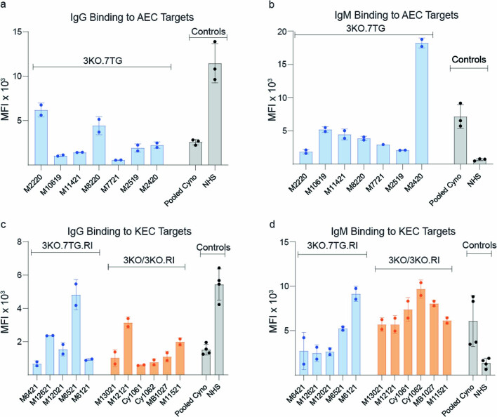
“WT porcine KECs bound human and NHP preformed antibodies, whereas 3KO porcine KECs showed markedly reduced antibody binding.”
§ Design and testing of a humanized porcine donor for xenotransplantation > Porcine molecular incompatibilities
plaindirect- Asserted: passes — The authors put it forward as true.
- Checkable: passes — It can be checked against evidence.
- Load-bearing: passes — The paper’s conclusions depend on it.
- Based: passes — The paper gives a reason why it holds.
It may state two separate claims as one.
A result the paper reports from its own data — it cites no other source for it, so there is none to check. The quote shows it is in the paper.
In the paper
“Porcine KECs (WT or 3KO) did not produce aPC, whereas HUVECs readily produced aPC.”
§ Design and testing of a humanized porcine donor for xenotransplantation > Porcine molecular incompatibilities
plainSingle case- Asserted: passes — The authors put it forward as true.
- Checkable: passes — It can be checked against evidence.
- Load-bearing: passes — The paper’s conclusions depend on it.
- Based: passes — The paper gives a reason why it holds.
It may state two separate claims as one.
A result the paper reports from its own data — it cites no other source for it, so there is none to check. The quote shows it is in the paper.
In the paper
“Here we present data produced from the porcine donor, A9161, carrying the 3KO.7TG.RI genotype, whose kidney was transplanted into NHP recipient M6521 and achieved a graft survival time of 176 days.”
§ Design and testing of a humanized porcine donor for xenotransplantation > A humanized porcine donor
plainSingle case- Asserted: passes — The authors put it forward as true.
- Checkable: passes — It can be checked against evidence.
- Load-bearing: passes — The paper’s conclusions depend on it.
- Based: passes — The paper gives a reason why it holds.
It may state two separate claims as one.
A result the paper reports from its own data — it cites no other source for it, so there is none to check. The quote shows it is in the paper.
In the paper
“In addition, antibody-mediated rejection (AMR)/thrombotic microangiopathy (TMA) (M12121), TMA (CY1061, CY1062 and MB1027) and T cell-mediated rejection (TCMR) (M11521) were also observed.”
§ Design and testing of a humanized porcine donor for xenotransplantation > Engineered porcine kidney supports life
plain- Asserted: passes — The authors put it forward as true.
- Checkable: passes — It can be checked against evidence.
- Load-bearing: passes — The paper’s conclusions depend on it.
- Based: passes — The paper gives a reason why it holds.
It may state two separate claims as one.
A result the paper reports from its own data — it cites no other source for it, so there is none to check. The quote shows it is in the paper.
In the paper
“Other parameters, including serum albumin, serum potassium and blood platelet counts, generally remained within normal range, except when associated with renal failure (serum albumin and potassium) (Supplementary Fig. 5).”
§ Design and testing of a humanized porcine donor for xenotransplantation > Engineered porcine kidney supports life
plaincorrelational- Asserted: passes — The authors put it forward as true.
- Checkable: passes — It can be checked against evidence.
- Load-bearing: unclear — It is unclear whether the paper’s conclusions depend on it.
- Based: passes — The paper gives a reason why it holds.
It may state two separate claims as one.
A result the paper reports from its own data — it cites no other source for it, so there is none to check. The quote shows it is in the paper.
In the paper
“Five out of the six 3KO ± RI kidneys showed evidence of acute tubular injury (ATI), a feature not included in the Banff criteria but representing general kidney injury.”
§ Design and testing of a humanized porcine donor for xenotransplantation > Engineered porcine kidney supports life
plain- Asserted: passes — The authors put it forward as true.
- Checkable: passes — It can be checked against evidence.
- Load-bearing: unclear — It is unclear whether the paper’s conclusions depend on it.
- Based: passes — The paper gives a reason why it holds.
It may state two separate claims as one.
A result the paper reports from its own data — it cites no other source for it, so there is none to check. The quote shows it is in the paper.
In the paper
“Using a SIRPα reporter cell line, we found that 3KO.7TG ± RI KECs expressing transgenic human CD47 (Extended Data Fig. 5a) activated the SIRPα signalling pathway, and pre-incubation with the anti-human CD47 antibody blocked SIRPα signalling in a dose-dependent manner (Fig. 3h).”
§ Design and testing of a humanized porcine donor for xenotransplantation > Genomic edits confer protection
plaindirect- Asserted: passes — The authors put it forward as true.
- Checkable: passes — It can be checked against evidence.
- Load-bearing: passes — The paper’s conclusions depend on it.
- Based: passes — The paper gives a reason why it holds.
It may state two separate claims as one.
A result the paper reports from its own data — it cites no other source for it, so there is none to check. The quote shows it is in the paper.
In the paper
“Although 3KO substantially reduced the level of pre-formed anti-porcine antibody binding, minor xenoantigens remain and contributed to residual antibody binding (Fig. 1b).”
§ Design and testing of a humanized porcine donor for xenotransplantation > Discussion
plaindirect- Asserted: passes — The authors put it forward as true.
- Checkable: passes — It can be checked against evidence.
- Load-bearing: passes — The paper’s conclusions depend on it.
- Based: passes — The paper gives a reason why it holds.
It may state two separate claims as one.
A result the paper reports from its own data — it cites no other source for it, so there is none to check. The quote shows it is in the paper.
In the paper
“When the incubation time was extended to 15 h, the 3KO modification was insufficient to fully protect the cells even from cell death by human serum, whereas 3KO.7TG ± RI KECs were protected against both human and cynomolgus monkey serum cytotoxicity, beyond the protection afforded by 3KO (Extended Data Fig. 4b).”
§ Design and testing of a humanized porcine donor for xenotransplantation > Genomic edits confer protection
plaindirect- Asserted: passes — The authors put it forward as true.
- Checkable: passes — It can be checked against evidence.
- Load-bearing: passes — The paper’s conclusions depend on it.
- Based: passes — The paper gives a reason why it holds.
It may state two separate claims as one.
A result the paper reports from its own data — it cites no other source for it, so there is none to check. The quote shows it is in the paper.
In the paper
“When incubated with human or cynomolgus monkey serum for 45 min, WT cells were lysed, whereas the 3KO modifications alone almost completely abolished complement-dependent cytotoxicity of human serum, but not cynomolgus monkey serum (Fig. 3c and Extended Data Fig. 4b,c).”
§ Design and testing of a humanized porcine donor for xenotransplantation > Genomic edits confer protection
plaindirect- Asserted: passes — The authors put it forward as true.
- Checkable: passes — It can be checked against evidence.
- Load-bearing: passes — The paper’s conclusions depend on it.
- Based: passes — The paper gives a reason why it holds.
It may state two separate claims as one.
A result the paper reports from its own data — it cites no other source for it, so there is none to check. The quote shows it is in the paper.
In the paper
“Unlike WT or 3KO porcine cells, 3KO.7TG ± RI KECs readily produced aPC (Fig. 3f), reduced TAT formation (Fig. 3g) and regulated coagulation of human whole blood as efficiently as human control cells (Extended Data Fig. 4h).”
§ Design and testing of a humanized porcine donor for xenotransplantation > Genomic edits confer protection
plaindirect- Asserted: passes — The authors put it forward as true.
- Checkable: passes — It can be checked against evidence.
- Load-bearing: passes — The paper’s conclusions depend on it.
- Based: passes — The paper gives a reason why it holds.
It may state two separate claims as one.
A result the paper reports from its own data — it cites no other source for it, so there is none to check. The quote shows it is in the paper.
In the paper
“In 3KO.7TG ± RI kidney recipients, those who developed renal failure exhibited TMA with or without AMR at time of euthanasia and only two showed evidence of ATI.”
§ Design and testing of a humanized porcine donor for xenotransplantation > Engineered porcine kidney supports life
plaincorrelational- Asserted: passes — The authors put it forward as true.
- Checkable: passes — It can be checked against evidence.
- Load-bearing: passes — The paper’s conclusions depend on it.
- Based: passes — The paper gives a reason why it holds.
It may state two separate claims as one.
A result the paper reports from its own data — it cites no other source for it, so there is none to check. The quote shows it is in the paper.
In the paper
“In addition, 3KO KECs overexpressing EPCR, thrombomodulin or both were engineered and showed that both EPCR and thrombomodulin contributed to aPC production, with thrombomodulin having a more significant effect in this experimental system (Extended Data Fig. 4g).”
§ Design and testing of a humanized porcine donor for xenotransplantation > Genomic edits confer protection
plaindirect- Asserted: passes — The authors put it forward as true.
- Checkable: passes — It can be checked against evidence.
- Load-bearing: passes — The paper’s conclusions depend on it.
- Based: passes — The paper gives a reason why it holds.
It may state two separate claims as one.
A result the paper reports from its own data — it cites no other source for it, so there is none to check. The quote shows it is in the paper.
In the paper
“Among the long-term survivors, simple appearing, benign cysts in the kidney were observed.”
§ Design and testing of a humanized porcine donor for xenotransplantation > Engineered porcine kidney supports life
plain- Asserted: passes — The authors put it forward as true.
- Checkable: passes — It can be checked against evidence.
- Load-bearing: fails — The paper’s conclusions may not depend on it.
- Based: passes — The paper gives a reason why it holds.
Probably not a substantial claim. The paper’s conclusions may not depend on it.
A result the paper reports from its own data — it cites no other source for it, so there is none to check. The quote shows it is in the paper.
In the paper
“Using blocking antibodies (Extended Data Fig. 4f), we showed that both EPCR and thrombomodulin contributed to aPC production (Fig. 3f), with thrombomodulin having a more prominent effect.”
§ Design and testing of a humanized porcine donor for xenotransplantation > Genomic edits confer protection
boosteddirect- Asserted: passes — The authors put it forward as true.
- Checkable: passes — It can be checked against evidence.
- Load-bearing: passes — The paper’s conclusions depend on it.
- Based: passes — The paper gives a reason why it holds.
It may state two separate claims as one.
A result the paper reports from its own data — it cites no other source for it, so there is none to check. The quote shows it is in the paper.
In the paper
“Unlike allotransplantation, T cell-mediated rejection was not a major pathology in xenotransplantation, with only one graft loss meeting Banff criteria for TCMR.”
§ Design and testing of a humanized porcine donor for xenotransplantation > Engineered porcine kidney supports life
plainSingle case- Asserted: passes — The authors put it forward as true.
- Checkable: passes — It can be checked against evidence.
- Load-bearing: passes — The paper’s conclusions depend on it.
- Based: passes — The paper gives a reason why it holds.
It may state two separate claims as one.
What the paper did — it cites no source for this, so there is none to check. The quote shows it is in the paper.
In the paper
“IHC analyses of 3KO were performed for all contralateral kidney samples included in this study and 3KO phenotypes confirmed for all.”
§ Design and testing of a humanized porcine donor for xenotransplantation > Porcine molecular incompatibilities
plain- Asserted: passes — The authors put it forward as true.
- Checkable: passes — It can be checked against evidence.
- Load-bearing: passes — The paper’s conclusions depend on it.
- Based: passes — The paper gives a reason why it holds.
It may state two separate claims as one.
In the paper
“Similar to humans, OWMs carry preformed antibodies to α-Gal and Sd(a), but unlike humans, they lack preformed antibodies to Neu5Gc, as they possess a functional CMAH gene25 (Fig. 1a).”
§ Design and testing of a humanized porcine donor for xenotransplantation > Porcine molecular incompatibilities
What the sources say
- Not checked — no full text of the sourcecites [25] Morozumi, K. et al. Significance of histochemical expression of hanganutziu–deicher antigens in pig, baboon and human tissues. Transplant. Proc. 31, 942–944 (1999). source
plaindirect- Asserted: passes — The authors put it forward as true.
- Checkable: passes — It can be checked against evidence.
- Load-bearing: passes — The paper’s conclusions depend on it.
- Based: passes — The paper gives a reason why it holds.
It may state two separate claims as one.
What the paper did — it cites no source for this, so there is none to check. The quote shows it is in the paper.
In the paper
“RNA-seq and immunohistochemistry (IHC) were performed for all completed 3KO.7TG ± RI renal transplants, except for donor 21405 whose contralateral kidney biopsy sample was not available (n = 11).”
§ Design and testing of a humanized porcine donor for xenotransplantation > A humanized porcine donor
plainn = 11- Asserted: passes — The authors put it forward as true.
- Checkable: passes — It can be checked against evidence.
- Load-bearing: passes — The paper’s conclusions depend on it.
- Based: passes — The paper gives a reason why it holds.
Probably not a substantial claim.
A conclusion the paper draws from its own results — it cites no other source for it, so there is none to check. The quote shows it is in the paper.
In the paper
“Given the synergistic effect of the two proteins, we reasoned that coagulation may be better regulated when both are provided.”
§ Design and testing of a humanized porcine donor for xenotransplantation > Genomic edits confer protection
hedgedconditional- Asserted: fails — The authors may not put it forward as true.
- Checkable: passes — It can be checked against evidence.
- Load-bearing: passes — The paper’s conclusions depend on it.
- Based: passes — The paper gives a reason why it holds.
Probably not a substantial claim. The authors may not put it forward as true.
A conclusion the paper draws from its own results — it cites no other source for it, so there is none to check. The quote shows it is in the paper.
In the paper
“The variable graft survival time may be inherent to kidney xenotransplantation and/or unique to the OWM model.”
§ Design and testing of a humanized porcine donor for xenotransplantation > Discussion
hedgedconditional- Asserted: passes — The authors put it forward as true.
- Checkable: passes — It can be checked against evidence.
- Load-bearing: fails — The paper’s conclusions may not depend on it.
- Based: passes — The paper gives a reason why it holds.
Probably not a substantial claim. The paper’s conclusions may not depend on it. It may state two separate claims as one.
What the paper did — it cites no source for this, so there is none to check. The quote shows it is in the paper.
In the paper
“In addition, the study was not blind for all involved.”
§ Design and testing of a humanized porcine donor for xenotransplantation > Engineered porcine kidney supports life
plain- Asserted: passes — The authors put it forward as true.
- Checkable: passes — It can be checked against evidence.
- Load-bearing: fails — The paper’s conclusions may not depend on it.
- Based: passes — The paper gives a reason why it holds.
Probably not a substantial claim. The paper’s conclusions may not depend on it.
What the paper did — it cites no source for this, so there is none to check. The quote shows it is in the paper.
In the paper
“The pairing of a porcine donor with an NHP recipient was dictated by availability.”
§ Design and testing of a humanized porcine donor for xenotransplantation > Engineered porcine kidney supports life
plaindirect- Asserted: passes — The authors put it forward as true.
- Checkable: passes — It can be checked against evidence.
- Load-bearing: fails — The paper’s conclusions may not depend on it.
- Based: passes — The paper gives a reason why it holds.
Probably not a substantial claim. The paper’s conclusions may not depend on it.
In the paper
“Finally, activation of innate immune cells in the xenograft induces inflammation and apoptosis of endothelial cells13.”
§ Design and testing of a humanized porcine donor for xenotransplantation > Genomic edits confer protection
What the sources say
- Not checked — no full text of the sourcecites [13] Robson, S. C., Am Esch, J. S. & Bach, F. H. Factors in xenograft rejection. Ann. N. Y. Acad. Sci. 875, 261–276 (1999). source
plaindirect- Asserted: passes — The authors put it forward as true.
- Checkable: passes — It can be checked against evidence.
- Load-bearing: passes — The paper’s conclusions depend on it.
- Based: passes — The paper gives a reason why it holds.
It may state two separate claims as one.
In the paper
“Thrombomodulin binds to thrombin and functions as a cofactor in the thrombin-induced activation of protein C, whereas EPCR binds to protein C and presents it to the thrombomodulin–thrombin activation complex.”
§ Design and testing of a humanized porcine donor for xenotransplantation > Genomic edits confer protection
What the sources say
- Not checked — no full text of the sourcecites [36] Esmon, C. T. The protein C pathway. Chest 124, 26S–32S (2003). source
plaindirect- Asserted: passes — The authors put it forward as true.
- Checkable: passes — It can be checked against evidence.
- Load-bearing: passes — The paper’s conclusions depend on it.
- Based: passes — The paper gives a reason why it holds.
It may state two separate claims as one.
What the paper did — it cites no source for this, so there is none to check. The quote shows it is in the paper.
In the paper
“Therefore, the number of NHP transplants per porcine donor genotype group was empirically determined, based on historical data reported in literature and what could be reasonably achieved.”
§ Design and testing of a humanized porcine donor for xenotransplantation > Engineered porcine kidney supports life
plain- Asserted: passes — The authors put it forward as true.
- Checkable: passes — It can be checked against evidence.
- Load-bearing: passes — The paper’s conclusions depend on it.
- Based: passes — The paper gives a reason why it holds.
Probably not a substantial claim.
A conclusion the paper draws from its own results — it cites no other source for it, so there is none to check. The quote shows it is in the paper.
In the paper
“In allotransplantation, TMA usually reflects an antibody-mediated or drug-related process, whereas in xenotransplantation, substantial species coagulation or complement regulatory protein incompatibility may contribute to TMA, even in the absence of de novo donor-specific antibody (dnDSA).”
§ Design and testing of a humanized porcine donor for xenotransplantation > Engineered porcine kidney supports life
hedgedconditional- Asserted: passes — The authors put it forward as true.
- Checkable: passes — It can be checked against evidence.
- Load-bearing: unclear — It is unclear whether the paper’s conclusions depend on it.
- Based: passes — The paper gives a reason why it holds.
It may state two separate claims as one.
What the paper did — it cites no source for this, so there is none to check. The quote shows it is in the paper.
In the paper
“Given the similar performance of KECs isolated from 3KO.7TG or 3KO.7TG.RI donors in functional assays (Fig. 3 and Extended Data Figs. 3–5), we analysed transplants performed with 3KO kidneys with and without RI as one group and 3KO.7TG and 3KO.7TG.RI kidneys as a second group.”
§ Design and testing of a humanized porcine donor for xenotransplantation > Engineered porcine kidney supports life
plain- Asserted: passes — The authors put it forward as true.
- Checkable: passes — It can be checked against evidence.
- Load-bearing: passes — The paper’s conclusions depend on it.
- Based: passes — The paper gives a reason why it holds.
Probably not a substantial claim.
What the paper did — it cites no source for this, so there is none to check. The quote shows it is in the paper.
In the paper
“With these considerations, pathology samples were assessed with a ‘xenotransplantation-adapted’ criteria (Extended Data Table 1 and Supplementary Table 6).”
§ Design and testing of a humanized porcine donor for xenotransplantation > Engineered porcine kidney supports life
plain- Asserted: passes — The authors put it forward as true.
- Checkable: passes — It can be checked against evidence.
- Load-bearing: fails — The paper’s conclusions may not depend on it.
- Based: passes — The paper gives a reason why it holds.
Probably not a substantial claim. The paper’s conclusions may not depend on it.
In the paper
“Although both thrombomodulin and EPCR contribute to aPC production, their mechanisms of action differ36.”
§ Design and testing of a humanized porcine donor for xenotransplantation > Genomic edits confer protection
What the sources say
- Not checked — no full text of the sourcecites [36] Esmon, C. T. The protein C pathway. Chest 124, 26S–32S (2003). source
plaindirect- Asserted: passes — The authors put it forward as true.
- Checkable: passes — It can be checked against evidence.
- Load-bearing: unclear — It is unclear whether the paper’s conclusions depend on it.
- Based: passes — The paper gives a reason why it holds.
It may state two separate claims as one.
What the paper did — it cites no source for this, so there is none to check. The quote shows it is in the paper.
In the paper
“Monkeys were screened for the presence of anti-porcine IgG and IgM (described above), and animals with low anti-porcine IgG and anti-IgM were selected as recipients.”
§ Design and testing of a humanized porcine donor for xenotransplantation > Methods > NHP recipients
plain- Asserted: passes — The authors put it forward as true.
- Checkable: passes — It can be checked against evidence.
- Load-bearing: passes — The paper’s conclusions depend on it.
- Based: passes — The paper gives a reason why it holds.
Probably not a substantial claim. It may state two separate claims as one.
The paper relies on this without support.
To proceed to clinical studies, a clinically ready porcine donor must be engineered and its xenograft successfully tested in a nonhuman primate model to assess its safety and efficacy.
“To proceed to clinical studies, a clinically ready porcine donor must be engineered and its xenograft successfully tested in a nonhuman primate (NHP) model to assess its safety and efficacy.”
The paper relies on this without support.
The commercial pig breeds used for previous donors have heart and kidney sizes too large for human application.
“First, the donors were often created on a commercial pig breed whose heart and kidney sizes are too large for human application.”
The paper relies on this without support.
The previously created donors were designed for testing in OWMs; they lacked the α-Gal or the α-Gal and Sd(a) glycans but expressed the Neu5Gc glycan to match with Neu5Gc expression in OWMs.
“Second, the donors were designed for testing in OWMs. They lacked the α-Gal (galactose-α-1,3-galactose) or the α-Gal and Sd(a) (Sia-α2.3-[GalNAc-β1.4]Gal-β1.4-GlcNAc) glycans but expressed the Neu5Gc (N-glycolylneuraminic acid) glycan to match with Neu5Gc expression in OWMs.”
The paper relies on this without support.
The immunosuppressive regimen used in the NHP transplants is clinically relevant.
“We show that a humanized porcine renal graft, combined with a clinically relevant immunosuppressive regimen, supported long-term NHP survival for up to 2 years (758 days).”
The paper relies on this without support.
The Banff classification of renal allograft pathology, with modifications, can be satisfactorily applied to xenotransplantation.
“It remains to be determined whether the current Banff scoring system developed for allotransplantation in humans can be satisfactorily applied to xenotransplantation, as rejection mechanisms may differ43.”
The paper relies on this without support.
C4 activation is upstream of CD46 and CD55 intervention.
“C4 activation is upstream of CD46 and CD55 intervention and positive C4d staining may be expected in the 3KO.7TG ± RI kidney samples in the presence of dnDSA.”
The paper relies on this without support.
Humans have a CMAH pseudogene genotype.
“It is worth noting that in a clinical setting, the complication around CMAH knockout unique to the OWMs will not be relevant, as it will be a match with the CMAH pseudogene genotype of the human recipient.”
The paper relies on this without support.
Agents targeting the CD40–CD40L pathway have become standard of care in NHP xenotransplantation studies.
“Of note, agents targeting the CD40–CD40L pathway have become standard of care in NHP xenotransplantation studies.”
Self-check
Percentages, sums and reported differences re-computed from the paper’s own numbers.
Each contradiction between two passages listed here was confirmed by an independent check.
Rated minor: it would not touch a result — a value no result rests on, a p-value that stays on the same side of the significance level, or a conflict that is clerical, resolved elsewhere in the paper, or outside its own findings.
In the paper
§ Design and testing of a humanized porcine donor for xenotransplantation > Genomic edits confer protection
“h, Human CD47 (hCD47) on 3KO.7TG ± RI KECs signalled through SIRPα on human Jurkat cells. WT KECs (n = 1), 3KO KECs (n = 1), 3KO.7TG ± RI (n = 2 for 3KO.7TG and n = 1 for 3KO.7TG.RI).”
§ Design and testing of a humanized porcine donor for xenotransplantation > Genomic edits confer protection
“In a–d,f–i, WT (n = 1), 3KO (n = 3), 3KO.7TG (n = 3, solid blue circles), 3KO.7TG.RI (n = 2, open circles)”
Rated minor: it would not touch a result — a value no result rests on, a p-value that stays on the same side of the significance level, or a conflict that is clerical, resolved elsewhere in the paper, or outside its own findings.
In the paper
§ Design and testing of a humanized porcine donor for xenotransplantation > Engineered porcine kidney supports life
“In 3KO.7TG ± RI kidney recipients, those who developed renal failure exhibited TMA with or without AMR at time of euthanasia and only two showed evidence of ATI.”
§ Design and testing of a humanized porcine donor for xenotransplantation > Engineered porcine kidney supports life
“| 3KO.7TG.RIf | M6421g,i | 6 | Renal failure | T | ATI, TMA | No | | 3KO.7TG.RIf | M12621g,i | 9 | Renal failure | – | AMR, ATI, TMA | No |”
Citations
Cites
Multiplex Genome Engineering Using CRISPR/Cas Systems
Cong, L., Ran, F. A., Cox, D., Lin, S., Barretto, R., Habib, N., Hsu, P. D., Wu, X., Jiang, W., Marraffini, L. A., et al. (2013) · Science · doi:10.1126/science.1231143 · Cited by 15,339
RNA-Guided Human Genome Engineering via Cas9
Mali, P., Yang, L., Esvelt, K. M., Aach, J., Guell, M., DiCarlo, J. E., Norville, J. E., & Church, G. M. (2013) · Science · doi:10.1126/science.1232033 · Cited by 9,210
Self-citationSheep cloned by nuclear transfer from a cultured cell line
Campbell, K., McWhir, J., Ritchie, W. A., & Wilmut, I. (1996) · Nature · doi:10.1038/380064a0 · Cited by 1,868
Failure to Regulate TNF-Induced NF-κB and Cell Death Responses in A20-Deficient Mice
Lee, E. G., Boone, D. L., Chai, S., Libby, S. L., Chien, M., Lodolce, J. P., & Ma, A. (2000) · Science · doi:10.1126/science.289.5488.2350 · Cited by 1,409
Kim, J. H., Lee, S. R., Li, L.-H., Park, H., Park, J.-H., Lee, K. Y., Kim, M., Shin, B. A., & Choi, S. (2011) · PLoS ONE · doi:10.1371/journal.pone.0018556 · Cited by 1,367
Using DECIPHER v2.0 to Analyze Big Biological Sequence Data in R
Erik, S. W. (2016) · The R Journal · doi:10.32614/rj-2016-025 · Cited by 995
Infection of human cells by an endogenous retrovirus of pigs
Patience, C., Takeuchi, Y., & Weiss, R. A. (1997) · Nature Medicine · doi:10.1038/nm0397-282 · Cited by 953
Galili, U., Shohet, S. B., Kobrin, E., Stults, C. L., & Macher, B. A. (1988) · Journal of Biological Chemistry · doi:10.1016/s0021-9258(19)77900-9 · Cited by 817
A 2018 Reference Guide to the Banff Classification of Renal Allograft Pathology
Roufosse, C., Simmonds, N., Clahsen-van Groningen, M., Haas, M., Henriksen, K. J., Horsfield, C., Loupy, A., Mengel, M., Perkowska-Ptasińska, A., Rabant, M., et al. (2018) · Transplantation · doi:10.1097/tp.0000000000002366 · Cited by 751
Genetically Modified Porcine-to-Human Cardiac Xenotransplantation
Griffith, B. P., Goerlich, C. E., Singh, A. K., Rothblatt, M., Lau, C. L., Shah, A., Lorber, M., Grazioli, A., Saharia, K. K., Hong, S. N., et al. (2022) · New England Journal of Medicine · doi:10.1056/nejmoa2201422 · Cited by 732
Cited by
Sorted by citations
Xenotransplantation of a Porcine Kidney for End-Stage Kidney Disease
Kawai, T., Williams, W. W., Elias, N., Fishman, J. A., Crisalli, K., Longchamp, A., Rosales, I. A., Duggan, M., Kimura, S., Morena, L., et al. (2025) · New England Journal of Medicine · doi:10.1056/nejmoa2412747 · Cited by 184
Self-citationEisenson, D. L., Hisadome, Y., Santillan, M. R., Iwase, H., Chen, W., Shimizu, A., Schulick, A. C., Gu, D., Akbar, A. F., Zhou, A. L., et al. (2024) · Nature Communications · doi:10.1038/s41467-024-47679-6 · Cited by 84
First pig kidney transplant in a person: what it means for the future
Mallapaty, S., & Kozlov, M. (2024) · Nature · doi:10.1038/d41586-024-00879-y · Cited by 76
Physiological basis for xenotransplantation from genetically modified pigs to humans
Peterson, L., Yacoub, M. H., Ayares, D., Yamada, K., Eisenson, D. L., Griffith, B. P., Mohiuddin, M. M., Eyestone, W. H., Venter, J. C., Smolenski, R. T., et al. (2024) · Physiological Reviews · doi:10.1152/physrev.00041.2023 · Cited by 64
Engineering immune-evasive allogeneic cellular immunotherapies
Martin, K. E., Hammer, Q., Perica, K., Sadelain, M., & Malmberg, K. (2024) · Nature reviews. Immunology · doi:10.1038/s41577-024-01022-8 · Cited by 64
Metadata
- Authors
- Anand, R. P., Layer, J. V., Heja, D., Hirose, T., Lassiter, G., Firl, D. J., Paragas, V. B., Akkad, A., Chhangawala, S., Colvin, R. B., Ernst, R. J., Esch, N., Getchell, K., Griffin, A. K., Guo, X., Hall, K. C., Hamilton, P., Kalekar, L. A., Kan, Y., Karadagi, A., Li, F., Low, S. C., Matheson, R., Nehring, C., Otsuka, R., Pandelakis, M., Policastro, R. A., Pols, R., Queiroz, L., Rosales, I. A., Serkin, W. T., Stiede, K., Tomosugi, T., Xue, Y., Zentner, G. E., Angeles-Albores, D., Chris Chao, J., Crabtree, J. N., Harken, S., Hinkle, N., Lemos, T., Li, M., Pantano, L., Stevens, D., Subedar, O. D., Tan, X., Yin, S., Anwar, I. J., Aufhauser, D., Capuano, S., Kaufman, D. B., Knechtle, S. J., Kwun, J., Shanmuganayagam, D., Markmann, J. F., Church, G. M., Curtis, M., Kawai, T., Youd, M. E., & Qin, W.
- Affiliations
- Harvard University (US)
- Duke Medical Center (US)
- University of Wisconsin–Madison (US)
- Corresponding author
- Youd, Michele E.
- Qin, Wenning
- ORCID
- Anand, Ranjith P.
- Hirose, Takayuki
- Lassiter, Grace
- Firl, Daniel J.
- Paragas, Violette B.
- Chhangawala, Sagar
- Colvin, Robert B.
- Ernst, Russell J.
- Getchell, Kristen
- Guo, Xiaoyun
- Hall, Katherine C.
- Kalekar, Lokesh A.
- Karadagi, Ahmad
- Li, Feng
- Low, Susan C.
- Matheson, Rudy
- Otsuka, Ryo
- Policastro, Robert A.
- Rosales, Ivy A.
- Serkin, William T.
- Stiede, Kathryn
- Tomosugi, Toshihide
- Zentner, Gabriel E.
- Angeles-Albores, David
- Crabtree, Juliet N.
- Li, Mailin
- Pantano, Lorena
- Tan, Xiaoqing
- Yin, Shiyi
- Anwar, Imran J.
- Aufhauser, David
- Capuano, Saverio
- Kaufman, Dixon B.
- Knechtle, Stuart J.
- Kwun, Jean
- Shanmuganayagam, Dhanansayan
- Markmann, James F.
- Church, George M.
- Kawai, Tatsuo
- Youd, Michele E.
- Qin, Wenning
- Venue
- Nature
- Published
- 2023-10-11
- Volume
- 622
- Issue
- 7982
- Pages
- 393-401
- ISSN
- 0028-0836 (ISSN-L)
- 1476-4687
- PMC
- PMC10567564
- OpenAlex
- W4387541325
- Cited by
- 302
- References
- 50
- Citations by year
- 2026: 92 · 2025: 130 · 2024: 72 · 2023: 8
- FWCI
- 67.86
- Type
- journal article
- Language
- en
- Open access
- hybrid
- doi.org
- Locations
- Nature (publishedVersion, cc-by)
- PubMed Central (publishedVersion, cc-by)
- PubMed (publishedVersion)
- RePEc: Research Papers in Economics (submittedVersion)
- Funders
- National Institutes of Health (P51OD011106)
- National Institute of Allergy and Infectious Diseases (T32AI007529)
- Topics
- Health Sciences › Medicine › Surgery › Xenotransplantation and immune response
- Life Sciences › Biochemistry, Genetics and Molecular Biology › Genetics › Animal Genetics and Reproduction
- Health Sciences › Medicine › Surgery › Organ Transplantation Techniques and Outcomes
- Keywords
- xenotransplantation, genetically modified pigs, porcine endogenous retroviruses, endothelial cells, graft survival, cynomolgus monkey model, non-human primates, immunocompatibility, inflammation modulation
- MeSH terms
- Animals, Graft Rejection, Humans, Macaca fascicularis, Polysaccharides, Swine, Transplantation, Heterologous, Kidney Transplantation, Transgenes, Animals, Genetically Modified, Endothelial Cells
About these checks
Registry, retraction, Cites and statistics results are computed, not judged, with one exception: a printed reference that no index lists is read once by a model before it counts as a citation error. Claims are extracted by a model and kept only when their quote appears verbatim in the text; each cited source is then checked, with a model’s help, for whether it says what the claim relies on. Those verdicts can be wrong — read the claim’s quote and the cited source before relying on one.
A language model drew the claims from the paper on 2026-11-09, under version 3 of the claim definition (model deepseek-v4.1-flash, prompt extract-v3).
A second model labelled each claim on 2026-11-09 (model jev-1.13-20260917, definition revision gold-guideline-v0.2).
Cited sources are checked with a language model’s help (gpt-6-sol).
Statistics and arithmetic are recomputed, not judged.
Confidence bands are shown only for fields calibrated on our annotated test set. None is calibrated yet, so no band is shown.
What counts as a claim ›How we measure confidence ›How we check this ›
Are you an author, or do you see a problem with this page? Request a review
