

The DOI matches its Crossref record; 29 of the 29 references it cites were found in a registry or an index; 2 of the 27 sources its claims cite were checked for support.
Loss of autophagy in dopaminergic neurons causes Lewy pathology and motor dysfunction in aged mice
Open accessAbstract
Inactivation of constitutive autophagy results in the formation of cytoplasmic inclusions in neurons, but the relationship between impaired autophagy and Lewy bodies (LBs) as well as the in vivo process of formation remains unknown. Synuclein, a component of LBs, is the defining characteristic of Parkinson’s disease (PD). Here, we characterize dopamine (DA) neuron–specific autophagy-deficient mice and provide in vivo evidence for LB formation. Synuclein deposition is preceded by p62 and resulted in the formation of inclusions containing synuclein and p62. The number and size of these inclusions were gradually increased in neurites rather than soma with aging. These inclusions may facilitate peripheral failures. As a result, DA neuron loss and motor dysfunction including the hindlimb defect were observed in 120-week-old mice. P62 aggregates derived from an autophagic defect might serve as “seeds” and can potentially be cause of LB formation.
Overview
AI snapshot
Written from the paper’s full text.
TL;DR
Dopamine neuron–specific Atg7 deletion in mice causes age-related p62 inclusions that precede synuclein accumulation, Lewy-like pathology, loss of TH neurons, and motor dysfunction. The findings suggest autophagic p62 aggregates may serve as “seeds” for Lewy body formation.
Key findings
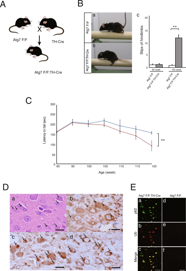
- Atg7flox/flox:TH-Cre mice had 13.54 ± 2.27% residual Atg7 in midbrain versus controls and developed motor coordination impairment around 100 weeks, with apparent deficits around 110 weeks, including frequent hindlimb slips on the runway and reduced rotarod fall latency.
- p62-positive inclusions appeared in substantia nigra dopaminergic neurons by 2 months, increased in number and size with age, and were mainly localized outside the soma along neurites; synuclein accumulation was observed around 9 months and colocalized with p62 inclusions, with 89.30 ± 1.65% of synuclein inclusions located in TH fibers (n = 5, each 2 slices).
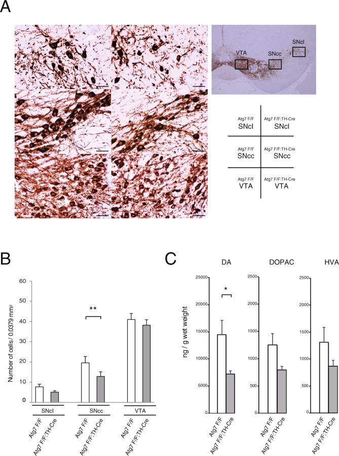
- In 120-week-old mice, Atg7flox/flox:TH-Cre mice had fewer TH neurons than controls, most prominently in the center area of substantia nigra pars compacta (SNcc) (n = 4 cKO; n = 5 control), and HPLC showed reduced striatal DA and metabolites (n = 3 per group; *P = 0.05).
- Ultrastructural analysis showed electron-dense filamentous inclusions in SN somata and neurites containing mitochondria and autophagosome-like structures; the authors speculate p62 aggregates containing mitochondria represent early Lewy body biogenesis.
Methods
- Generated tyrosine hydroxylase (TH) neuron–specific Atg7 conditional knockout mice (Atg7flox/flox:TH-Cre) by crossing Atg7flox/flox mice with TH-Cre mice; controls were Atg7flox/flox mice.
- Assessed locomotor behavior from 90 to 120 weeks using an accelerating rotarod (3 to 35 rpm over 5 min) and a narrow beam runway test; performed immunoblot, immunohistochemistry, immunofluorescence, electron microscopy/immunoelectron microscopy, and HPLC of dorsal striatum.
- Statistical significance was determined by Student’s t-test; behavioral tests used n = 10 per genotype, TH neuron counts used n = 4 cKO and n = 5 control, and HPLC used n = 3 per group.
Limitations
- The paper states that many questions remain about Lewy body formation, that none of the current genetic PD mouse models recapitulates all features of PD, and that only a few develop mild DA neurodegeneration; it also notes that compensatory mechanisms during development may make degenerative phenotypes hard to observe over the mouse lifespan.
- The paper states that the reason the significant decrease in dopamine was not accompanied by concomitant reductions in DOPAC and HVA was not clarified, and it describes the p62 “seed” role as speculation.
- Evident from the text: survival was not observed over the entire lifespan, some synuclein data at early stages are reported as “data not shown,” and terminal analyses used small sample sizes (n = 3–5 per group).
Model-written summary. Check it against the paper before relying on it.
Integrity signals
Registry
Cites
Statistics
Figures
Verification
Claims the paper makes
31 Own result · 20 Not checked yet · 10 Own interpretation · 1 Supported · 1 Partly supported · 1 Own method · 1 No basis given · accuracy not yet measured
A result the paper reports from its own data — it cites no other source for it, so there is none to check. The quote shows it is in the paper.
In the paper
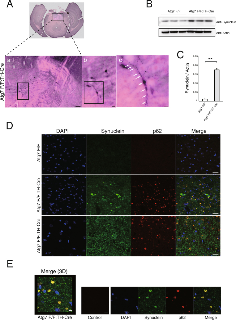
“Interestingly, we observed these inclusions not only within the soma, but also outside the soma (Fig. 3D lower panel).”
§ Loss of autophagy in dopaminergic neurons causes Lewy pathology and motor dysfunction in aged mice > Results > P62-positive inclusions contain endogenous synuclein
plain- Asserted: passes — The authors put it forward as true.
- Checkable: passes — It can be checked against evidence.
- Load-bearing: unclear — It is unclear whether the paper’s conclusions depend on it.
- Based: passes — The paper gives a reason why it holds.
A result the paper reports from its own data — it cites no other source for it, so there is none to check. The quote shows it is in the paper.
In the paper
“To determine the localization of these inclusions, we analyzed many samples by confocal microscopy, and found that most inclusions localized in neurites, as demonstrated in Fig. 2D.”
§ Loss of autophagy in dopaminergic neurons causes Lewy pathology and motor dysfunction in aged mice > Results > P62-positive inclusions contain endogenous synuclein
boosted- Asserted: passes — The authors put it forward as true.
- Checkable: passes — It can be checked against evidence.
- Load-bearing: unclear — It is unclear whether the paper’s conclusions depend on it.
- Based: passes — The paper gives a reason why it holds.
A result the paper reports from its own data — it cites no other source for it, so there is none to check. The quote shows it is in the paper.
In the paper
“In Atg7flox/flox:TH-Cre mice, these neurons contained 7 eosinophilic aggregates, which are characteristic of LBs (Fig. 1Da).”
§ Loss of autophagy in dopaminergic neurons causes Lewy pathology and motor dysfunction in aged mice > Results > Loss of Atg7 leads to age-related development of p62 inclusions in the DA neuron
plain- Asserted: passes — The authors put it forward as true.
- Checkable: passes — It can be checked against evidence.
- Load-bearing: unclear — It is unclear whether the paper’s conclusions depend on it.
- Based: passes — The paper gives a reason why it holds.
A result the paper reports from its own data — it cites no other source for it, so there is none to check. The quote shows it is in the paper.
In the paper
“Furthermore, the level of synuclein increased in the midbrain of Atg7flox/flox:TH-Cre mice (Fig. 3B and C).”
§ Loss of autophagy in dopaminergic neurons causes Lewy pathology and motor dysfunction in aged mice > Results > P62-positive inclusions contain endogenous synuclein
plain- Asserted: passes — The authors put it forward as true.
- Checkable: passes — It can be checked against evidence.
- Load-bearing: passes — The paper’s conclusions depend on it.
- Based: passes — The paper gives a reason why it holds.
A result the paper reports from its own data — it cites no other source for it, so there is none to check. The quote shows it is in the paper.
In the paper
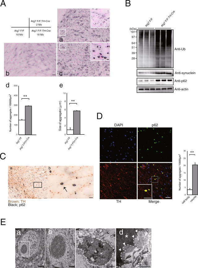
“Specifically, we labeled dopaminergic neurons with TH antibody (Fig. 2D red) and followed individual fibers, revealing that p62-positive aggregates (Fig. 2D green) were present along neurites.”
§ Loss of autophagy in dopaminergic neurons causes Lewy pathology and motor dysfunction in aged mice > Results > Loss of Atg7 leads to age-related development of p62 inclusions in the DA neuron
plain- Asserted: passes — The authors put it forward as true.
- Checkable: passes — It can be checked against evidence.
- Load-bearing: passes — The paper’s conclusions depend on it.
- Based: passes — The paper gives a reason why it holds.
A result the paper reports from its own data — it cites no other source for it, so there is none to check. The quote shows it is in the paper.
In the paper
“Fibrous inclusions localized in the soma (Fig. 2Ea,b) and neurites (Fig. 2Ec,d) in DA neurons.”
§ Loss of autophagy in dopaminergic neurons causes Lewy pathology and motor dysfunction in aged mice > Results > Loss of Atg7 leads to age-related development of p62 inclusions in the DA neuron
plain- Asserted: passes — The authors put it forward as true.
- Checkable: passes — It can be checked against evidence.
- Load-bearing: passes — The paper’s conclusions depend on it.
- Based: passes — The paper gives a reason why it holds.
A result the paper reports from its own data — it cites no other source for it, so there is none to check. The quote shows it is in the paper.
In the paper
“Surprisingly, around 9 months, we pathologically confirmed endogenous synuclein accumulation in somata and neurites (Fig. 3A).”
§ Loss of autophagy in dopaminergic neurons causes Lewy pathology and motor dysfunction in aged mice > Results > P62-positive inclusions contain endogenous synuclein
boosted- Asserted: passes — The authors put it forward as true.
- Checkable: passes — It can be checked against evidence.
- Load-bearing: passes — The paper’s conclusions depend on it.
- Based: passes — The paper gives a reason why it holds.
A result the paper reports from its own data — it cites no other source for it, so there is none to check. The quote shows it is in the paper.
In the paper
“Atg7flox/flox:TH-Cre mice had fewer TH neurons than Atg7flox/flox mice.”
§ Loss of autophagy in dopaminergic neurons causes Lewy pathology and motor dysfunction in aged mice > Results > The number of TH neurons decreases in aged Atg7flox/flox:TH-Cre mice, contributing to locomotor dysfunction
plaincorrelationaln = 4- Asserted: passes — The authors put it forward as true.
- Checkable: passes — It can be checked against evidence.
- Load-bearing: passes — The paper’s conclusions depend on it.
- Based: passes — The paper gives a reason why it holds.
A result the paper reports from its own data — it cites no other source for it, so there is none to check. The quote shows it is in the paper.
In the paper
“In Atg7flox/flox:TH-Cre midbrains, the quantification of Atg7 after normalization by actin (Supple. Figure 1B) showed 13.54 ± 2.27% residual amount, compared to controls.”
§ Loss of autophagy in dopaminergic neurons causes Lewy pathology and motor dysfunction in aged mice > Results > Generation of DA neuron–specific Atg7 conditional knockout mice, and characterization of their locomotor impairments
plain- Asserted: passes — The authors put it forward as true.
- Checkable: passes — It can be checked against evidence.
- Load-bearing: passes — The paper’s conclusions depend on it.
- Based: passes — The paper gives a reason why it holds.
A result the paper reports from its own data — it cites no other source for it, so there is none to check. The quote shows it is in the paper.
In the paper
“On the other hand, Atg7flox/flox:TH-Cre mice exhibited synuclein deposition and inclusions that colocalized with p62 in cell bodies (Fig. 3D middle panel).”
§ Loss of autophagy in dopaminergic neurons causes Lewy pathology and motor dysfunction in aged mice > Results > P62-positive inclusions contain endogenous synuclein
plaincorrelational- Asserted: passes — The authors put it forward as true.
- Checkable: passes — It can be checked against evidence.
- Load-bearing: passes — The paper’s conclusions depend on it.
- Based: passes — The paper gives a reason why it holds.
- Probably not a substantial claim. 16
- It may state two separate claims as one. 19
Every source the paper cites for this claim was checked and supports it.
In the paper
“and Bedford et al. also reported deposits of mitochondria in early LBs (pale bodies) in PD patients26.”
§ Loss of autophagy in dopaminergic neurons causes Lewy pathology and motor dysfunction in aged mice > Discussion
What the sources say
- Supportedcites [26] Bedford, L. et al. Depletion of 26S proteasomes in mouse brain neurons causes neurodegeneration and Lewy-like inclusions resembling human pale bodies. J Neurosci 28, 8189–8198 (2008). source
In “Mitochondrial involvement in inclusion body formation,” the authors report examining substantia nigra tissue from PD patients and finding mitochondria in pale bodies, which they describe as an early form of nigral Lewy body. Figure 5A also shows mitochondria within a human nigral pale body. (LLM, conf=0.94)
plaincorrelational- Asserted: passes — The authors put it forward as true.
- Checkable: passes — It can be checked against evidence.
- Load-bearing: fails — The paper’s conclusions may not depend on it.
- Based: passes — The paper gives a reason why it holds.
Probably not a substantial claim. The paper’s conclusions may not depend on it.
- Supported
The cited sources support only part of this claim — read each source’s verdict below.
In the paper
“During mitochondrial degradation in cultured cells, depolarized mitochondria cluster with p6224. Depolarized mitochondria are ubiquitylated by Parkin, recognized by p62, transported via microtubules to aggregates, and degraded by autophagy.”
§ Loss of autophagy in dopaminergic neurons causes Lewy pathology and motor dysfunction in aged mice > Discussion
What the sources say
- Partly supportedcites [24] Okatsu, K. et al. p62/SQSTM1 cooperates with Parkin for perinuclear clustering of depolarized mitochondria. Genes Cells 15, 887–900 (2010). sourceExperimental — accuracy not yet measured
The Discussion section on juxtanuclear clustering describes Parkin ubiquitylation, p62 recognition, and microtubule transport to aggregates. It says the mitochondria are *sometimes* degraded, while the Results section says degradation occurs *in part* via mitophagy. The claim omits those limits and presents autophagic degradation as the endpoint for the clustered mitochondria generally. (LLM, conf=0.88)
plaindirect- Asserted: passes — The authors put it forward as true.
- Checkable: passes — It can be checked against evidence.
- Load-bearing: passes — The paper’s conclusions depend on it.
- Based: passes — The paper gives a reason why it holds.
Probably not a substantial claim. It may state two separate claims as one.
- Partly supported
A result the paper reports from its own data — it cites no other source for it, so there is none to check. The quote shows it is in the paper.
In the paper
“Although Atg7flox/flox:TH-Cre mice have not yet been observed over the entire lifespan, they began to show impairment in motor coordination tasks around 100 weeks and apparent motor behavioral deficits around 110 weeks.”
§ Loss of autophagy in dopaminergic neurons causes Lewy pathology and motor dysfunction in aged mice > Results > Generation of DA neuron–specific Atg7 conditional knockout mice, and characterization of their locomotor impairments
plain- Asserted: passes — The authors put it forward as true.
- Checkable: passes — It can be checked against evidence.
- Load-bearing: passes — The paper’s conclusions depend on it.
- Based: passes — The paper gives a reason why it holds.
It may state two separate claims as one.
A result the paper reports from its own data — it cites no other source for it, so there is none to check. The quote shows it is in the paper.
In the paper
“No neuronal loss was observed in young mice, which did not exhibit motor dysfunction (data not shown).”
§ Loss of autophagy in dopaminergic neurons causes Lewy pathology and motor dysfunction in aged mice > Results > The number of TH neurons decreases in aged Atg7flox/flox:TH-Cre mice, contributing to locomotor dysfunction
plain- Asserted: passes — The authors put it forward as true.
- Checkable: passes — It can be checked against evidence.
- Load-bearing: passes — The paper’s conclusions depend on it.
- Based: passes — The paper gives a reason why it holds.
It may state two separate claims as one.
A result the paper reports from its own data — it cites no other source for it, so there is none to check. The quote shows it is in the paper.
In the paper
“P62 inclusions were present along the TH fibers, and were mainly located in the branch of dopaminergic neurons (Fig. 2Cb).”
§ Loss of autophagy in dopaminergic neurons causes Lewy pathology and motor dysfunction in aged mice > Results > Loss of Atg7 leads to age-related development of p62 inclusions in the DA neuron
plain- Asserted: passes — The authors put it forward as true.
- Checkable: passes — It can be checked against evidence.
- Load-bearing: unclear — It is unclear whether the paper’s conclusions depend on it.
- Based: passes — The paper gives a reason why it holds.
It may state two separate claims as one.
A result the paper reports from its own data — it cites no other source for it, so there is none to check. The quote shows it is in the paper.
In the paper
“When we observed these inclusions at higher magnification, they have multiple cavities (Fig. 3E), consistent with the results of ultrastructural analysis (Fig. 2Ed).”
§ Loss of autophagy in dopaminergic neurons causes Lewy pathology and motor dysfunction in aged mice > Results > P62-positive inclusions contain endogenous synuclein
plain- Asserted: passes — The authors put it forward as true.
- Checkable: passes — It can be checked against evidence.
- Load-bearing: fails — The paper’s conclusions may not depend on it.
- Based: passes — The paper gives a reason why it holds.
Probably not a substantial claim. The paper’s conclusions may not depend on it.
A result the paper reports from its own data — it cites no other source for it, so there is none to check. The quote shows it is in the paper.
In the paper
“In contrast to Atg7flox/flox mice, in which no aggregates were seen even at the age of 18 months (Fig. 2Ab), p62-positive inclusions were present in 2-month-old Atg7flox/flox:TH-Cre mice (Fig. 2Aa).”
§ Loss of autophagy in dopaminergic neurons causes Lewy pathology and motor dysfunction in aged mice > Results > Loss of Atg7 leads to age-related development of p62 inclusions in the DA neuron
plain- Asserted: passes — The authors put it forward as true.
- Checkable: passes — It can be checked against evidence.
- Load-bearing: passes — The paper’s conclusions depend on it.
- Based: passes — The paper gives a reason why it holds.
It may state two separate claims as one.
A result the paper reports from its own data — it cites no other source for it, so there is none to check. The quote shows it is in the paper.
In the paper
“In addition, these inclusions were observed in TH-positive neurons (Fig. 1Db), including ubiquitin (Fig. 1Dc) and p62 (Fig. 1Dd).”
§ Loss of autophagy in dopaminergic neurons causes Lewy pathology and motor dysfunction in aged mice > Results > Loss of Atg7 leads to age-related development of p62 inclusions in the DA neuron
plain- Asserted: passes — The authors put it forward as true.
- Checkable: passes — It can be checked against evidence.
- Load-bearing: passes — The paper’s conclusions depend on it.
- Based: passes — The paper gives a reason why it holds.
It may state two separate claims as one.
A result the paper reports from its own data — it cites no other source for it, so there is none to check. The quote shows it is in the paper.
In the paper
“In our careful observation of Atg7flox/flox:TH-Cre mice, we found that the level of endogenous synuclein protein increased at around 9 months in cell bodies and neurites (Fig. 3A,B and C), and these accumulated synuclein deposits contained p62 positive aggregates (Fig. 3D).”
§ Loss of autophagy in dopaminergic neurons causes Lewy pathology and motor dysfunction in aged mice > Discussion
plaincorrelational- Asserted: passes — The authors put it forward as true.
- Checkable: passes — It can be checked against evidence.
- Load-bearing: passes — The paper’s conclusions depend on it.
- Based: passes — The paper gives a reason why it holds.
It may state two separate claims as one.
A result the paper reports from its own data — it cites no other source for it, so there is none to check. The quote shows it is in the paper.
In the paper
“In 18-month-old mice, p62 inclusions were larger (Fig. 2Ae and B) and mainly localized outside the soma (Fig. 2Ac).”
§ Loss of autophagy in dopaminergic neurons causes Lewy pathology and motor dysfunction in aged mice > Results > Loss of Atg7 leads to age-related development of p62 inclusions in the DA neuron
plain- Asserted: passes — The authors put it forward as true.
- Checkable: passes — It can be checked against evidence.
- Load-bearing: passes — The paper’s conclusions depend on it.
- Based: passes — The paper gives a reason why it holds.
It may state two separate claims as one.
A result the paper reports from its own data — it cites no other source for it, so there is none to check. The quote shows it is in the paper.
In the paper
“Notably, Western blotting revealed p62 and ubiquitin accumulations in the midbrain of 2 month-old Atg7flox/flox:TH-Cre mice, whereas synuclein did not accumulate in the midbrain, at least at this stage (data not shown).”
§ Loss of autophagy in dopaminergic neurons causes Lewy pathology and motor dysfunction in aged mice > Results > Loss of Atg7 leads to age-related development of p62 inclusions in the DA neuron
hedged- Asserted: passes — The authors put it forward as true.
- Checkable: passes — It can be checked against evidence.
- Load-bearing: passes — The paper’s conclusions depend on it.
- Based: passes — The paper gives a reason why it holds.
It may state two separate claims as one.
A result the paper reports from its own data — it cites no other source for it, so there is none to check. The quote shows it is in the paper.
In the paper
“Atg7flox/flox:TH-Cre mice were viable at birth and indistinguishable in appearance from their littermates, and their survival rate was not markedly diminished.”
§ Loss of autophagy in dopaminergic neurons causes Lewy pathology and motor dysfunction in aged mice > Results > Generation of DA neuron–specific Atg7 conditional knockout mice, and characterization of their locomotor impairments
plain- Asserted: passes — The authors put it forward as true.
- Checkable: passes — It can be checked against evidence.
- Load-bearing: unclear — It is unclear whether the paper’s conclusions depend on it.
- Based: passes — The paper gives a reason why it holds.
It may state two separate claims as one.
None of the sources cited for this claim have been checked yet.
In the paper
“Although the phenotypes of mice harboring brain-specific deletion of Atg5 or Atg7 reveal the critical role of autophagy in the removal of aggregated proteins7,8”
§ Loss of autophagy in dopaminergic neurons causes Lewy pathology and motor dysfunction in aged mice > Introduction
What the sources say
- Not checked yetcites [7] Hara, T. et al. Suppression of basal autophagy in neural cells causes neurodegenerative disease in mice. Nature 441, 885–889 (2006). source
- Not checked yetcites [8] Komatsu, M. et al. Loss of autophagy in the central nervous system causes neurodegeneration in mice. Nature 441, 880–884 (2006). source
boosteddirect- Asserted: passes — The authors put it forward as true.
- Checkable: passes — It can be checked against evidence.
- Load-bearing: passes — The paper’s conclusions depend on it.
- Based: passes — The paper gives a reason why it holds.
Probably not a substantial claim.
In the paper
“Friedman et al.9 demonstrated that DA neuron-specific autophagy deficiency leads to the restrictive presynaptic accumulation of synuclein in the dorsal striatum, suggesting that impaired autophagy plays a role in PD pathogenesis.”
§ Loss of autophagy in dopaminergic neurons causes Lewy pathology and motor dysfunction in aged mice > Introduction
What the sources say
- Not checked yetcites [9] Friedman, L. G. et al. Disrupted autophagy leads to dopaminergic axon and dendrite degeneration and promotes presynaptic accumulation of alpha-synuclein and LRRK2 in the brain. J Neurosci 32, 7585–7593 (2012). source
boosteddirect- Asserted: passes — The authors put it forward as true.
- Checkable: passes — It can be checked against evidence.
- Load-bearing: passes — The paper’s conclusions depend on it.
- Based: passes — The paper gives a reason why it holds.
Probably not a substantial claim.
In the paper
“Parkinson’s disease (PD), the most common neurodegenerative disorder, is characterized by the loss of nigrostriatal dopamine neurons and the formation of intracellular Lewy bodies (LBs), which consist primarily of α-synuclein (hereafter referred to as synuclein) and ubiquitin1.”
§ Loss of autophagy in dopaminergic neurons causes Lewy pathology and motor dysfunction in aged mice > Introduction
What the sources say
- Not checked yetcites [1] Spillantini, M. G. et al. Alpha-synuclein in Lewy bodies. Nature 388, 839–840 (1997). source
plaincorrelational- Asserted: passes — The authors put it forward as true.
- Checkable: passes — It can be checked against evidence.
- Load-bearing: passes — The paper’s conclusions depend on it.
- Based: passes — The paper gives a reason why it holds.
Probably not a substantial claim. It may state two separate claims as one.
In the paper
“Indeed, p62 is present in neuronal inclusions of individuals with Alzheimer’s disease and other neurodegenerative diseases11”
§ Loss of autophagy in dopaminergic neurons causes Lewy pathology and motor dysfunction in aged mice > Introduction
What the sources say
- Not checked yetcites [11] Bartlett, B. J. et al. p62, Ref(2)P and ubiquitinated proteins are conserved markers of neuronal aging, aggregate formation and progressive autophagic defects. Autophagy 7, 572–583 (2011). source
plaincorrelational- Asserted: passes — The authors put it forward as true.
- Checkable: passes — It can be checked against evidence.
- Load-bearing: fails — The paper’s conclusions may not depend on it.
- Based: passes — The paper gives a reason why it holds.
Probably not a substantial claim. The paper’s conclusions may not depend on it.
In the paper
“In this context, the stress-inducible protein p62 (encoded by the gene Sqstm1) is of importance because it is the ubiquitin- and LC3-binding protein that functions as a selective autophagy adaptor/receptor for degradation of ubiquitinated substrates10.”
§ Loss of autophagy in dopaminergic neurons causes Lewy pathology and motor dysfunction in aged mice > Introduction
What the sources say
- Not checked yetcites [10] Komatsu, M. et al. Homeostatic levels of p62 control cytoplasmic inclusion body formation in autophagy-deficient mice. Cell 131, 1149–1163 (2007). source
plaindirect- Asserted: passes — The authors put it forward as true.
- Checkable: passes — It can be checked against evidence.
- Load-bearing: fails — The paper’s conclusions may not depend on it.
- Based: passes — The paper gives a reason why it holds.
Probably not a substantial claim. The paper’s conclusions may not depend on it. It may state two separate claims as one.
None of the sources cited for this claim have been checked yet.
In the paper
“Several lines of evidence indicate a role for autophagy in the regulation of synuclein homeostasis5,6,15, suggesting that dysfunctional autophagic clearance may contribute to the development of synuclein inclusions.”
§ Loss of autophagy in dopaminergic neurons causes Lewy pathology and motor dysfunction in aged mice > Discussion
What the sources say
- Not checked yetcites [5] Spencer, B. et al. Beclin 1 gene transfer activates autophagy and ameliorates the neurodegenerative pathology in alpha-synuclein models of Parkinson’s and Lewy body diseases. J Neurosci 29, 13578–13588 (2009). source
- Not checked yetcites [6] Yu, W. H. et al. Metabolic activity determines efficacy of macroautophagic clearance of pathological oligomeric alpha-synuclein. Am J Pathol 175, 736–747 (2009). source
- Not checked yetcites [15] Ahmed, I. et al. Development and characterization of a new Parkinson’s disease model resulting from impaired autophagy. J Neurosci 32, 16503–16509 (2012). source
hedgedconditional- Asserted: passes — The authors put it forward as true.
- Checkable: passes — It can be checked against evidence.
- Load-bearing: passes — The paper’s conclusions depend on it.
- Based: passes — The paper gives a reason why it holds.
It may state two separate claims as one.
In the paper
“In fact, these autophagosome-like structures were previously reported in Atg7-deficient brains14.”
§ Loss of autophagy in dopaminergic neurons causes Lewy pathology and motor dysfunction in aged mice > Results > Loss of Atg7 leads to age-related development of p62 inclusions in the DA neuron
What the sources say
- Not checked yetcites [14] Komatsu, M. et al. Essential role for autophagy protein Atg7 in the maintenance of axonal homeostasis and the prevention of axonal degeneration. Proc Natl Acad Sci USA 104, 14489–14494 (2007). source
plain- Asserted: passes — The authors put it forward as true.
- Checkable: passes — It can be checked against evidence.
- Load-bearing: fails — The paper’s conclusions may not depend on it.
- Based: passes — The paper gives a reason why it holds.
Probably not a substantial claim. The paper’s conclusions may not depend on it.
A conclusion the paper draws from its own results — it cites no other source for it, so there is none to check. The quote shows it is in the paper.
In the paper
“Thus, loss of autophagy induces synuclein accumulation and formation of LBs, similar to the pathology observed in DA neurons.”
§ Loss of autophagy in dopaminergic neurons causes Lewy pathology and motor dysfunction in aged mice > Results > P62-positive inclusions contain endogenous synuclein
plaindirect- Asserted: passes — The authors put it forward as true.
- Checkable: passes — It can be checked against evidence.
- Load-bearing: passes — The paper’s conclusions depend on it.
- Based: passes — The paper gives a reason why it holds.
It may state two separate claims as one.
In the paper
“Furthermore, LB-like synuclein aggregates resist degradation and impair autophagy20.”
§ Loss of autophagy in dopaminergic neurons causes Lewy pathology and motor dysfunction in aged mice > Discussion
What the sources say
- Not checked yetcites [20] Tanik, S. A., Schultheiss, C. E., Volpicelli-Daley, L. A., Brunden, K. R. & Lee, V. M. Lewy body-like alpha-synuclein aggregates resist degradation and impair macroautophagy. J Biol Chem 288, 15194–15210 (2013). source
plaindirect- Asserted: passes — The authors put it forward as true.
- Checkable: passes — It can be checked against evidence.
- Load-bearing: unclear — It is unclear whether the paper’s conclusions depend on it.
- Based: passes — The paper gives a reason why it holds.
It may state two separate claims as one.
In the paper
“It is well known that homeostasis of p62 is strictly regulated by autophagy in the brain10, and that dysregulation of autophagy causes formation of p62 aggregates.”
§ Loss of autophagy in dopaminergic neurons causes Lewy pathology and motor dysfunction in aged mice > Discussion
What the sources say
- Not checked yetcites [10] Komatsu, M. et al. Homeostatic levels of p62 control cytoplasmic inclusion body formation in autophagy-deficient mice. Cell 131, 1149–1163 (2007). source
boosteddirect- Asserted: passes — The authors put it forward as true.
- Checkable: passes — It can be checked against evidence.
- Load-bearing: passes — The paper’s conclusions depend on it.
- Based: passes — The paper gives a reason why it holds.
Probably not a substantial claim. It may state two separate claims as one.
A conclusion the paper draws from its own results — it cites no other source for it, so there is none to check. The quote shows it is in the paper.
In the paper
“In this study, we carried out detailed observations of Atg7-deficient mice, and observed a loss of autophagy in dopaminergic neurons, resulting in Lewy pathology and motor dysfunctions associated with aging.”
§ Loss of autophagy in dopaminergic neurons causes Lewy pathology and motor dysfunction in aged mice > Discussion
plaindirect- Asserted: passes — The authors put it forward as true.
- Checkable: passes — It can be checked against evidence.
- Load-bearing: passes — The paper’s conclusions depend on it.
- Based: passes — The paper gives a reason why it holds.
It may state two separate claims as one.
What the paper did — it cites no source for this, so there is none to check. The quote shows it is in the paper.
In the paper
“Our synuclein antibody efficiently detects endogenous mouse synuclein, and a virtual slide system could be used to observe neurons extensively.”
§ Loss of autophagy in dopaminergic neurons causes Lewy pathology and motor dysfunction in aged mice > Discussion
plain- Asserted: passes — The authors put it forward as true.
- Checkable: passes — It can be checked against evidence.
- Load-bearing: fails — The paper’s conclusions may not depend on it.
- Based: passes — The paper gives a reason why it holds.
Probably not a substantial claim. The paper’s conclusions may not depend on it.
In the paper
“DA neurons have a unique structural characteristic, long hyper-branched projection axons, that innervate wide areas in the brain22.”
§ Loss of autophagy in dopaminergic neurons causes Lewy pathology and motor dysfunction in aged mice > Discussion
What the sources say
- Not checked yetcites [22] Sulzer, D. & Surmeier, D. J. Neuronal vulnerability, pathogenesis, and Parkinson’s disease. Mov Disord 28, 715–724 (2013). source
plain- Asserted: passes — The authors put it forward as true.
- Checkable: passes — It can be checked against evidence.
- Load-bearing: passes — The paper’s conclusions depend on it.
- Based: passes — The paper gives a reason why it holds.
Probably not a substantial claim. It may state two separate claims as one.
In the paper
“The protein catabolic pathways that regulate synuclein degradation have been discussed by multiple authors4,16,17, and the extent to which autophagy contributes to synuclein degradation depends on the presence of mutations18.”
§ Loss of autophagy in dopaminergic neurons causes Lewy pathology and motor dysfunction in aged mice > Discussion
What the sources say
- Not checked yetcites [18] Cuervo, A. M., Stefanis, L., Fredenburg, R., Lansbury, P. T. & Sulzer, D. Impaired degradation of mutant alpha-synuclein by chaperone-mediated autophagy. Science 305, 1292–1295 (2004). source
plaincorrelational- Asserted: passes — The authors put it forward as true.
- Checkable: passes — It can be checked against evidence.
- Load-bearing: fails — The paper’s conclusions may not depend on it.
- Based: passes — The paper gives a reason why it holds.
Probably not a substantial claim. The paper’s conclusions may not depend on it.
In the paper
“But, as to autophagosome formation, ATG conjugation systems including Atg7 are not absolutely essential13.”
§ Loss of autophagy in dopaminergic neurons causes Lewy pathology and motor dysfunction in aged mice > Results > Loss of Atg7 leads to age-related development of p62 inclusions in the DA neuron
What the sources say
- Not checked yetcites [13] Tsuboyama, K. et al. The ATG conjugation systems are important for degradation of the inner autophagosomal membrane. Science 354, 1036–1041 (2016). source
plain- Asserted: passes — The authors put it forward as true.
- Checkable: passes — It can be checked against evidence.
- Load-bearing: fails — The paper’s conclusions may not depend on it.
- Based: passes — The paper gives a reason why it holds.
Probably not a substantial claim. The paper’s conclusions may not depend on it.
In the paper
“Until now, we did not clarify the reason, but one of the explanation may be presynaptic dysfunction in Atg7-deficient mice9.”
§ Loss of autophagy in dopaminergic neurons causes Lewy pathology and motor dysfunction in aged mice > Discussion
What the sources say
- Not checked yetcites [9] Friedman, L. G. et al. Disrupted autophagy leads to dopaminergic axon and dendrite degeneration and promotes presynaptic accumulation of alpha-synuclein and LRRK2 in the brain. J Neurosci 32, 7585–7593 (2012). source
hedgedconditional- Asserted: passes — The authors put it forward as true.
- Checkable: passes — It can be checked against evidence.
- Load-bearing: fails — The paper’s conclusions may not depend on it.
- Based: passes — The paper gives a reason why it holds.
Probably not a substantial claim. The paper’s conclusions may not depend on it.
In the paper
“Electron microscopic analysis by Gai et al. revealed that many mitochondria are concentrated in the early forms of LBs25”
§ Loss of autophagy in dopaminergic neurons causes Lewy pathology and motor dysfunction in aged mice > Discussion
What the sources say
- Not checked yetcites [25] Gai, W. P. et al. In situ and in vitro study of colocalization and segregation of alpha-synuclein, ubiquitin, and lipids in Lewy bodies. Exp Neurol 166, 324–333 (2000). source
plaincorrelational- Asserted: passes — The authors put it forward as true.
- Checkable: passes — It can be checked against evidence.
- Load-bearing: fails — The paper’s conclusions may not depend on it.
- Based: passes — The paper gives a reason why it holds.
Probably not a substantial claim. The paper’s conclusions may not depend on it.
A conclusion the paper draws from its own results — it cites no other source for it, so there is none to check. The quote shows it is in the paper.
In the paper
“Our Atg7-deficient mice demonstrate typical Lewy pathology, including endogenous synuclein and neuronal loss, which resembles PD.”
§ Loss of autophagy in dopaminergic neurons causes Lewy pathology and motor dysfunction in aged mice > Discussion
boostedcorrelational- Asserted: passes — The authors put it forward as true.
- Checkable: passes — It can be checked against evidence.
- Load-bearing: passes — The paper’s conclusions depend on it.
- Based: passes — The paper gives a reason why it holds.
It may state two separate claims as one.
The paper relies on this without support.
The relationship between impaired autophagy and Lewy body formation and the in vivo process of formation remains unknown.
“Inactivation of constitutive autophagy results in the formation of cytoplasmic inclusions in neurons, but the relationship between impaired autophagy and Lewy bodies (LBs) as well as the in vivo process of formation remains unknown.”
The paper relies on this without support.
In DA neurons, no detailed reports to date have examined endogenous synuclein accumulation in dopaminergic cell bodies and neurites associated with Lewy pathology.
“In DA neurons, the primary site of endogenous pathology, no detailed reports to date have examined the endogenous synuclein accumulation in dopaminergic cell bodies and neurites, which is associated with Lewy pathology.”
The paper relies on this without support.
The mechanisms underlying age-related progression and intracellular localization of p62 inclusions in dopaminergic neurons remain elusive.
“However, the mechanisms underlying age-related progression and intracellular localization of these inclusions in dopaminergic neurons remain elusive.”
The paper relies on this without support.
It remained unclear how synuclein accumulation is associated with PD pathology.
“Previous in vivo analysis of DA neurons suggested that synuclein regulation is linked to autophagy9,15, but it remained unclear how synuclein accumulation is associated with PD pathology.”
The paper relies on this without support.
Most genetically modified mouse PD models do not exhibit neuronal loss.
“To date, many genetically modified mice have been developed as PD models, but most of them do not exhibit neuronal loss.”
The paper relies on this without support.
None of the current genetic PD models in mouse recapitulates all features of PD.
“None of the current genetic PD models in mouse recapitulates all features of PD.”
The paper relies on this without support.
Only a few genetic PD models develop mild DA neurodegeneration.
“Additionally, only a few of these models develop mild DA neurodegeneration.”
Claims other papers make about this one
Each is checked against this paper’s own text: does it say what the citing paper says it does?
In the citing paper
“In support of the importance of the autophagic process in DA neurons, autophagy impairment has been strongly linked to the degeneration of SNpc DA neurons in PD (Hunn et al., 2019; Isidoro et al., 2009; Sato et al., 2018)”
From Nilotinib restores memory function by preventing dopaminergic neuron degeneration in a mouse model of Alzheimer’s Disease (2021) · cites this paper as Sato et al., 2018
Self-check
Percentages, sums and reported differences re-computed from the paper’s own numbers.
Each contradiction between two passages listed here was confirmed by an independent check.
Rated major: it would touch a result — a value a result rests on, reported and recomputed p-values on opposite sides of the significance level, two passages that conflict with no sign which is right, or a cited work retracted before this paper was published.
In the paper
§ Loss of autophagy in dopaminergic neurons causes Lewy pathology and motor dysfunction in aged mice > Results > The number of TH neurons decreases in aged Atg7flox/flox:TH-Cre mice, contributing to locomotor dysfunction
“High-performance liquid chromatography (HPLC) revealed a reduction in striatal DA levels and metabolites in Atg7flox/flox:TH-Cre versus control mice (Fig. 4C).”
§ Loss of autophagy in dopaminergic neurons causes Lewy pathology and motor dysfunction in aged mice > Discussion
“Intriguingly, the significant decrease in dopamine is not accompanied by concomitant reductions in the dopamine catabolites DOPAC and HVA.”
Citations
Cites
Spillantini, M. G., Schmidt, M. L., Lee, V. M.-Y., Trojanowski, J. Q., Jakes, R., & Goedert, M. (1997) · Nature · doi:10.1038/42166 · Cited by 8,382
Dauer, W., & Przedborski, S. (2003) · Neuron · doi:10.1016/s0896-6273(03)00568-3 · Cited by 5,469
Suppression of basal autophagy in neural cells causes neurodegenerative disease in mice
Hara, T., Nakamura, K., Matsui, M., Yamamoto, A., Nakahara, Y., Suzuki-Migishima, R., Yokoyama, M., Mishima, K., Saito, I., Okano, H., et al. (2006) · Nature · doi:10.1038/nature04724 · Cited by 3,882
Levine, B., & Klionsky, D. J. (2004) · Developmental Cell · doi:10.1016/s1534-5807(04)00099-1 · Cited by 3,741
Loss of autophagy in the central nervous system causes neurodegeneration in mice
Komatsu, M., Waguri, S., Chiba, T., Murata, S., Iwata, J.-I., Tanida, I., Ueno, T., Koike, M., Uchiyama, Y., Kominami, E., et al. (2006) · Nature · doi:10.1038/nature04723 · Cited by 3,500
Self-citationHomeostatic levels of p62 control cytoplasmic inclusion body formation in autophagy-deficient mice.
Komatsu, M., Waguri, S., Koike, M., Sou, Y.-S., Ueno, T., Hara, T., Mizushima, N., Iwata, J.-I., Ezaki, J., Murata, S., et al. (2007) · Cell · doi:10.1016/j.cell.2007.10.035 · Cited by 2,124
Self-citationImpaired Degradation of Mutant α-Synuclein by Chaperone-Mediated Autophagy
Cuervo, A. M., Stefanis, L., Fredenburg, R., Lansbury, P. T., & Sulzer, D. (2004) · Science · doi:10.1126/science.1101738 · Cited by 2,024
α-Synuclein Is Degraded by Both Autophagy and the Proteasome
Webb, J. L., Ravikumar, B., Atkins, J., Skepper, J. N., & Rubinsztein, D. C. (2003) · Journal of Biological Chemistry · doi:10.1074/jbc.m300227200 · Cited by 1,429
α-Synuclein impairs macroautophagy: implications for Parkinson’s disease
Winslow, A. R., Chen, C.-W., Corrochano, S., Acevedo-Arozena, A., Gordon, D. E., Peden, A. A., Lichtenberg, M., Menzies, F. M., Ravikumar, B., Imarisio, S., et al. (2010) · Journal of Cell Biology · doi:10.1083/jcb.201003122 · Cited by 825
Komatsu, M., Wang, Q. J., Holstein, G. R., Friedrich, V. L., Iwata, J.-I., Kominami, E., Chait, B. T., Tanaka, K., & Yue, Z. (2007) · Proceedings of the National Academy of Sciences · doi:10.1073/pnas.0701311104 · Cited by 614
Self-citation
Cited by
Sorted by citations
Parkinson's disease: Mechanisms, translational models and management strategies
Raza, C., Anjum, R., & Shakeel, N. U. A. (2019) · Life Sciences · doi:10.1016/j.lfs.2019.03.057 · Cited by 628
“Aberrant protein homeostasis and PD Protein homeostasis is under intrinsic surveillance mechanisms (such as ubiquitin-protease, chaperone mediated process and autophagy system) for rapid detection of altered protein and its processing or elimination [51,52].”
BackgroundAutophagy in Parkinson's Disease
Hou, X., Watzlawik, J. O., Fiesel, F. C., & Springer, W. (2020) · Journal of Molecular Biology · doi:10.1016/j.jmb.2020.01.037 · Cited by 361
Stavoe, A. K. H., & Holzbaur, E. L. F. (2019) · Annual Review of Cell and Developmental Biology · doi:10.1146/annurev-cellbio-100818-125242 · Cited by 328
Autophagy and the hallmarks of aging
Kaushik, S., Tasset, I., Arias, E., Pampliega, O., Wong, E., Martinez-Vicente, M., & Cuervo, A. M. (2021) · Ageing Research Reviews · doi:10.1016/j.arr.2021.101468 · Cited by 285
Autophagy in Neurodegenerative Diseases: A Hunter for Aggregates
Park, H., Kang, J., & Lee, S. (2020) · International Journal of Molecular Sciences · doi:10.3390/ijms21093369 · Cited by 217
“Knockout of ATG7 was shown to induce an age-dependent increase in the formation of p62-containing α-synuclein inclusion bodies in dopaminergic neurons and motor function deficits in aged mice [73].”
BackgroundSQSTM1/p62: a potential target for neurodegenerative disease
Ma, S., Attarwala, I. Y., & Xie, X.-Q. (2019) · ACS Chemical Neuroscience · doi:10.1021/acschemneuro.8b00516 · Cited by 185
Magnesium Is a Key Player in Neuronal Maturation and Neuropathology
Yamanaka, R., Shindo, Y., & Oka, K. (2019) · International Journal of Molecular Sciences · doi:10.3390/ijms20143439 · Cited by 184
“Thus, the impairment of basal autophagy causes abnormal accumulation and protein aggregation [211,212], and consequently, pathological features of PD in dopaminergic neurons [213].”
BackgroundZhang, K., Zhu, S., Li, J., Jiang, T., Feng, L., Pei, J., Wang, G., Ouyang, L., & Liu, B. (2021) · Acta Pharmaceutica Sinica B · doi:10.1016/j.apsb.2021.02.016 · Cited by 159
Macroautophagy in CNS health and disease
Griffey, C. J., & Yamamoto, A. (2022) · Nature reviews. Neuroscience · doi:10.1038/s41583-022-00588-3 · Cited by 147
Wulansari, N., Darsono, W., Woo, H.-J., Chang, M., Kim, J., Bae, E.-J., Sun, W., Lee, J.-H., Cho, I.-J., Shin, H., et al. (2021) · Science Advances · doi:10.1126/sciadv.abb1540 · Cited by 127
“Impairment of autophagic clearance of -syn is one of the main causes of -syn accumulation in PD (46, 47).”
Autophagy in health and disease: From molecular mechanisms to therapeutic target
Lu, G. X., Wang, Y., Shi, Y. P., Zhang, Z., Huang, C., He, W., Wang, C., & Shen, H. (2022) · MedComm · doi:10.1002/mco2.150 · Cited by 135
Deneubourg, C., Ramm, M., Smith, L. J., Baron, O., Singh, K., Byrne, S., Duchen, M. R., Gautel, M., Eskelinen, E., Fanto, M., et al. (2021) · Autophagy · doi:10.1080/15548627.2021.1943177 · Cited by 115
“In addition to these basic considerations and in a more clinical context, autophagy has been associated with adultonset neurodegenerative disorders in several ways (for review [148],), nonspecifically through its complex interactions with the potentially toxic protein aggregates implicated in these conditions, and, sp…”
BackgroundLa Barbera, L., Vedele, F., Nobili, A., Krashia, P., Spoleti, E., Latagliata, E. C., Cutuli, D., Cauzzi, E., Marino, R., Viscomi, M. T., et al. (2021) · Progress in Neurobiology · doi:10.1016/j.pneurobio.2021.102031 · Cited by 98
Alpha-Synuclein defects autophagy by impairing SNAP29-mediated autophagosome-lysosome fusion
Tang, Q., Gao, P., Arzberger, T., Höllerhage, M., Herms, J., Höglinger, G., & Koeglsperger, T. (2021) · Cell Death & Disease · doi:10.1038/s41419-021-04138-0 · Cited by 103
Loss of Parkin contributes to mitochondrial turnover and dopaminergic neuronal loss in aged mice
Noda, S., Sato, S., Fukuda, T., Tada, N., Uchiyama, Y., Tanaka, K., & Hattori, N. (2019) · Neurobiology of Disease · doi:10.1016/j.nbd.2019.104717 · Cited by 94
Self-citationPromiscuous Roles of Autophagy and Proteasome in Neurodegenerative Proteinopathies
Limanaqi, F., Biagioni, F., Gambardella, S., Familiari, P., Frati, A., & Fornai, F. (2020) · International Journal of Molecular Sciences · doi:10.3390/ijms21083028 · Cited by 93
“These studies indicate that both UPS and autophagy are seminal for the survival of DA neurons, since inhibition of either pathway leads to parkinsonism featuring a massive loss of midbrain DA neurons and α-syn accumulation within ubiquitin-positive Lewy body-like inclusions in surviving neurons [119,121,122,140].”
BackgroundAutophagy as a Cellular Stress Response Mechanism in the Nervous System
Peker, N., & Gözüaçık, D. (2020) · Journal of Molecular Biology · doi:10.1016/j.jmb.2020.01.017 · Cited by 87
Autophagy in Synucleinopathy: The Overwhelmed and Defective Machinery
Arotçarena, M.-L., Teil, M., & Dehay, B. (2019) · Cells · doi:10.3390/cells8060565 · Cited by 91
“Recently, Sato and collaborators generated a Tyrosine-Hydroxylase cell-specific Atg7 conditional knock-out (KO) mouse model that presented α-syn accumulation and LB-like inclusions (positive for α-syn, p62, and ubiquitin) after autophagy impairment [62].”
BackgroundPhytochemicals Bridging Autophagy Induction and Alpha-Synuclein Degradation in Parkinsonism
Limanaqi, F., Biagioni, F., Busceti, C. L., Ryskalin, L., Polzella, M., Frati, A., & Fornai, F. (2019) · International Journal of Molecular Sciences · doi:10.3390/ijms20133274 · Cited by 87
“In fact, the loss of autophagy in these models produces DA cell loss along with neuronal inclusions featuring protein aggregates such as α-syn, Parkin, PINK1, LRRK2, ubiquitin, and p62 [136,184].”
BackgroundROS networks: designs, aging, Parkinson’s disease and precision therapies
Kolodkin, A. N., Sharma, R. P., Colangelo, A. M., Ignatenko, A., Martorana, F., Jennen, D. G. J., Briedé, J. J., Brady, N. R., Barberis, M., Mondeel, T. D. G. A., et al. (2020) · npj Systems Biology and Applications · doi:10.1038/s41540-020-00150-w · Cited by 89
The beneficial role of autophagy in multiple sclerosis: Yes or No?
Al-Kuraishy, H. M., Jabir, M. S., Al-Gareeb, A. I., Saad, H. M., Batiha, G. E., & Klionsky, D. J. (2023) · Autophagy · doi:10.1080/15548627.2023.2259281 · Cited by 89
“In addition, neuronal autophagy is highly dysregulated in proteinopa-thies proposing an association between autophagy dysfunction and the development of neurodegeneration [91].”
BackgroundGreen Tea Catechins Attenuate Neurodegenerative Diseases and Cognitive Deficits
Afzal, O., Dalhat, M. H., Altamimi, A. S. A., Rasool, R., Alzarea, S. I., Almalki, W. H., Murtaza, B. N., Iftikhar, S., Nadeem, S. I., Nadeem, M. S., et al. (2022) · Molecules · doi:10.3390/molecules27217604 · Cited by 89
“Autophagy is linked to aging, which protects neural cells by removing protein aggregates and degrading obsolete cell structures [83,84].”
BackgroundLim, J., Bang, Y., & Choi, H. J. (2018) · Archives of Pharmacal Research · doi:10.1007/s12272-018-1063-x · Cited by 82
Limanaqi, F., Biagioni, F., Mastroiacovo, F., Polzella, M., Lazzeri, G., & Fornai, F. (2020) · Antioxidants · doi:10.3390/antiox9101022 · Cited by 80
Targeting α-Synuclein for PD Therapeutics: A Pursuit on All Fronts
Teil, M., Arotçarena, M.-L., Faggiani, E., Laferrière, F., Bézard, E., & Dehay, B. (2020) · Biomolecules · doi:10.3390/biom10030391 · Cited by 80
Loss of nuclear REST/NRSF in aged-dopaminergic neurons in Parkinson’s disease patients
Kawamura, M., Sato, S., Matsumoto, G., Fukuda, T., Shiba‐Fukushima, K., Noda, S., Takanashi, M., Mori, N., & Hattori, N. (2019) · Neuroscience Letters · doi:10.1016/j.neulet.2019.01.042 · Cited by 74
“We recently reported that Atg7:TH-Cre mice demonstrate neuronal loss and typical Lewy body disease pathology, including p62, ubiquitin, and synuclein, a pattern which resembles PD [11].”
Self-citationBayati, A., Ayoubi, R., Aguila, A., Zorca, C. E., Deyab, G., Han, C., Recinto, S. J., Nguyen, E., Rocha, C., Maussion, G., et al. (2024) · Nature Neuroscience · doi:10.1038/s41593-024-01775-4 · Cited by 67
“Consistent with protein degradation defects in LB formation, lysosomal proteins, including GBA and TMEM175, are implicated in PD pathophysiology 19,20 .”
Obergasteiger, J., Frapporti, G., Lamonaca, G., Pizzi, S., Picard, A., Lavdas, A. Α., Pischedda, F., Piccoli, G., Hilfiker, S., Lobbestael, E., et al. (2020) · Cell Death Discovery · doi:10.1038/s41420-020-0279-y · Cited by 56
Proteostasis of α-Synuclein and Its Role in the Pathogenesis of Parkinson’s Disease
Han, D., Zheng, W., Wang, X., & Chen, Z. (2020) · Frontiers in Cellular Neuroscience · doi:10.3389/fncel.2020.00045 · Cited by 55
The PARK10 gene USP24 is a negative regulator of autophagy and ULK1 protein stability
Thayer, J. A., Awad, O., Hegdekar, N., Sarkar, C., Tesfay, H., Burt, C. I., Zeng, X., Feldman, R. A., & Lipinski, M. M. (2019) · Autophagy · doi:10.1080/15548627.2019.1598754 · Cited by 52
“In animal models inhibition of autophagy in dopaminergic neurons causes protein aggregation and progressive neuronal loss, leading to decrease in dopamine levels and age-dependent motor deficits [7,8].”
BackgroundLimanaqi, F., Biagioni, F., Gambardella, S., Ryskalin, L., & Fornai, F. (2018) · Frontiers in Molecular Neuroscience · doi:10.3389/fnmol.2018.00299 · Cited by 50
Regulation of α-synuclein homeostasis and inflammasome activation by microglial autophagy
Choi, I., Heaton, G. R., Lee, Y.-K., & Yue, Z. (2022) · Science Advances · doi:10.1126/sciadv.abn1298 · Cited by 54
“To address this, several research groups have examined the effects of neuron-specific deletion of Atg7 in -synuclein in vivo (6, 68, 69).”
BackgroundPradas, E., & Martínez‐Vicente, M. (2023) · Cells · doi:10.3390/cells12010191 · Cited by 52
Zhou, T., Lin, D., Chen, Y., Peng, S., Jing, X., Lei, M., Tao, E., & Liang, Y. (2019) · Epigenomics · doi:10.2217/epi-2019-0222 · Cited by 47
Prolyl oligopeptidase inhibition activates autophagy via protein phosphatase 2A
Svarcbahs, R., Jäntti, M. H., Kilpeläinen, T. P., Julku, U. H., Urvas, L., Kivioja, S., Norrbacka, S., & Myöhänen, T. T. (2019) · Pharmacological Research · doi:10.1016/j.phrs.2019.104558 · Cited by 50
p62: Friend or Foe? Evidences for OncoJanus and NeuroJanus Roles
Emanuele, S., Lauricella, M., D’Anneo, A., Carlisi, D., Blasio, A. D., Liberto, D. D., & Giuliano, M. (2020) · International Journal of Molecular Sciences · doi:10.3390/ijms21145029 · Cited by 48
“For instance, loss of p62-dependent autophagy in neurons was associated with Lewy pathology and motor dysfunction in aged mice [111].”
Therapeutic Effects of Catechins in Less Common Neurological and Neurodegenerative Disorders
Sebastiani, G., Almeida-Toledano, L., Serra-Delgado, M., Navarro‐Tapia, E., Sailer, S., Valverde, O., García‐Algar, Ó., & Andreu-Férnández, V. (2021) · Nutrients · doi:10.3390/nu13072232 · Cited by 48
“Aging is associated with dysfunction of the autophagy activity, which protects neuronal cells by recycling obsolete cellular constituents and removing protein aggregates, contributors to neurodegenerative disorders, such as PD [40,41].”
BackgroundNorwitz, N. G., Soto-Mota, A., Norwitz, S. G., & Clarke, K. (2019) · Frontiers in Aging Neuroscience · doi:10.3389/fnagi.2019.00184 · Cited by 42
Living with the enemy: from protein-misfolding pathologies we know, to those we want to know
Emwas, A.-H. M., Alghrably, M., Dhahri, M., Sharfalddin, A. A., Alsiary, R., Jaremko, M., Faa, G., Campagna, M., Congiu, T., Piras, M., et al. (2021) · Ageing Research Reviews · doi:10.1016/j.arr.2021.101391 · Cited by 43
“In an aged mouse model, Sato et al. showed that ATG7-knockout leads to dopamine neuron loss and motor dysfunction as a result of the increasing formation of inclusions consisting of α-syn and p62, the autophagy adaptor protein, with age (Sato et al., 2018).”
Spermidine as an epigenetic regulator of autophagy in neurodegenerative disorders
Satarker, S., Wilson, J., Kolathur, K. K., Mudgal, J., Lewis, S. A., Arora, D. S., & Nampoothiri, M. (2024) · European Journal of Pharmacology · doi:10.1016/j.ejphar.2024.176823 · Cited by 43
Homeostatic p62 levels and inclusion body formation in CHCHD2 knockout mice
Sato, S., Noda, S., Torii, S., Amo, T., Ikeda, A., Funayama, M., Yamaguchi, J., Fukuda, T., Kondo, H., Tada, N., et al. (2021) · Human Molecular Genetics · doi:10.1093/hmg/ddab057 · Cited by 38
Self-citationPadilla-Godínez, F. J., Vázquez-García, E. R., Trujillo-Villagrán, M. I., Soto-Rojas, L. O., Palomero‐Rivero, M., Hernández-González, O., Pérez-Eugenio, F., Collazo-Navarrete, O., Arias-Carrión, Ó., & Guerra‐Crespo, M. (2025) · Frontiers in Neuroscience · doi:10.3389/fnins.2025.1570553 · Cited by 42
“Additionally, abnormal proteins can directly or indirectly disrupt UPS processes, further impairing associated pathways and ultimately leading to irreversible neuronal protein imbalance and degeneration (Cookson, 2009; Sato et al., 2018).”
Role of melatonin in TLR4-mediated inflammatory pathway in the MTPT-induced mouse model
Yıldırım, S., Özkan, A., Aytaç, G., Ağar, A., & Tanrιöver, G. (2021) · NeuroToxicology · doi:10.1016/j.neuro.2021.11.011 · Cited by 36
Limanaqi, F., Busceti, C. L., Celli, R., Biagioni, F., & Fornai, F. (2021) · Progress in Neurobiology · doi:10.1016/j.pneurobio.2021.102112 · Cited by 40
“In fact, genetically inactivating autophagy in murine models reproduces both psychiatric alterations and key features of neurodegeneration; rescuing autophagy improves behavior and provides neuroprotection instead (Feng et al., 2020; Hara et al., 2006; Hu et al., 2017; Hui and Tanaka, 2019; Komatsu et al., 2006, 2007;…”
BackgroundInfluentialAutophagy and lysosomal pathways in nervous system disorders
Bingol, B. (2018) · Molecular and Cellular Neuroscience · doi:10.1016/j.mcn.2018.04.009 · Cited by 32
Lamonaca, G., & Volta, M. (2020) · Cells · doi:10.3390/cells9051115 · Cited by 30
“Indeed, impaired autophagic clearance leads to deposition of aSyn in presynaptic terminals of mice lacking Atg7, supporting the hypothesis that synapses are a major target of aSyn pathology [29,30].”
BackgroundPitcairn, C., Murata, N., Zalon, A. J., Stojkovska, I., & Mazzulli, J. R. (2023) · Journal of Neuroscience · doi:10.1523/jneurosci.0610-22.2023 · Cited by 29
Liu, M., Liu, S., Lin, Z.-H., Chen, X., Jiao, Q., Du, X., & Jiang, H. F. (2025) · Biomolecules · doi:10.3390/biom15010149 · Cited by 32
Cell Clearing Systems Bridging Neuro-Immunity and Synaptic Plasticity
Limanaqi, F., Biagioni, F., Busceti, C. L., Ryskalin, L., Soldani, P., Frati, A., & Fornai, F. (2019) · International Journal of Molecular Sciences · doi:10.3390/ijms20092197 · Cited by 28
Macroautophagy and Mitophagy in Neurodegenerative Disorders: Focus on Therapeutic Interventions
Magalhães, J. D., Fão, L., Vilaça, R., Cardoso, S. M., & Rego, A. C. (2021) · Biomedicines · doi:10.3390/biomedicines9111625 · Cited by 29
“For example, specific ablation of ATG7, essential for autophagosome elongation in dopaminergic neurons from aged mice, induces characteristic PD features, such as motor frailty, loss of TH-positive neurons, and aSyn deposition [208].”
BackgroundAutophagy and Redox Homeostasis in Parkinson’s: A Crucial Balancing Act
Jiménez-Moreno, N., & Lane, J. D. (2020) · Oxidative Medicine and Cellular Longevity · doi:10.1155/2020/8865611 · Cited by 26
“In the absence of autophagy in dopaminergic neurons (in Atg7 knockout mice), inclusions containing αsyn and p62/SQSTM1 are observed predominantly in neurites, and these increase with age, preempting neurodegeneration and motor dysfunction [96].”
BackgroundATG8-dependent LMX1B-autophagy crosstalk shapes human midbrain dopaminergic neuronal resilience
Jiménez-Moreno, N., Kollareddy, M., Stathakos, P., Moss, J. J., Antón, Z., Shoemark, D. K., Sessions, R. B., Witzgall, R., Caldwell, M. A., & Lane, J. D. (2023) · The Journal of Cell Biology · doi:10.1083/jcb.201910133 · Cited by 26
Real-state of autophagy signaling pathway in neurodegenerative disease; focus on multiple sclerosis
Hassanpour, M., Hajihassani, F., Hiradfar, A., Aghamohammadzadeh, N., Rahbarghazi, R., Safaie, N., Nouri, M., & Panahi, Y. (2020) · Journal of Inflammation · doi:10.1186/s12950-020-0237-8 · Cited by 25
Zuo, L., Dai, C., Yi, L., & Dong, Z. (2021) · Cell Death Discovery · doi:10.1038/s41420-021-00643-5 · Cited by 24
Dementia associated with disorders of the basal ganglia
Vitanova, K. S., Stringer, K. M., Benítez, D. P., Brenton, J., & Cummings, D. M. (2019) · Journal of Neuroscience Research · doi:10.1002/jnr.24508 · Cited by 24
Zhang, Z.-R., Guo, P.-W., Huang, S.-N., Jia, Z.-C., Chen, T.-N., Liu, X., Feng, H., & Chen, Y.-J. (2022) · Oxidative Medicine and Cellular Longevity · doi:10.1155/2022/4177317 · Cited by 24
“Much of the evidence to support this derives from studies adjusting autophagy in neurons and observing neurodegeneration, especially in AD and PD [48 – 50].”
Impaired mitochondrial accumulation and Lewy pathology in neuron-specific FBXO7-deficient mice
Noda, S., Sato, S., Fukuda, T., Ueno, S., Tada, N., & Hattori, N. (2022) · Molecular Brain · doi:10.1186/s13041-022-00936-5 · Cited by 20
Self-citationMSA: From basic mechanisms to experimental therapeutics
Heras‐Garvin, A., & Stefanova, N. (2020) · Parkinsonism & Related Disorders · doi:10.1016/j.parkreldis.2020.01.010 · Cited by 21
Autophagy status as a gateway for stress-induced catecholamine interplay in neurodegeneration
Fornai, F., & Puglisi‐Allegra, S. (2021) · Neuroscience & Biobehavioral Reviews · doi:10.1016/j.neubiorev.2021.01.015 · Cited by 20
LRRK2 along the Golgi and lysosome connection: a jamming situation
Piccoli, G., & Volta, M. (2021) · Biochemical Society Transactions · doi:10.1042/bst20201146 · Cited by 17
Sukseree, S., László, L., Грубер, Ф., Bergmann, S., Narzt, M., Nagelreiter, I., Höftberger, R., Molnár, K., Rauter, G., Birngruber, T., et al. (2018) · Molecular Neurobiology · doi:10.1007/s12035-018-0996-x · Cited by 17
Oizumi, H., Yamasaki, K., Suzuki, H., Ohshiro, S., Saito, Y., Murayama, S., Sugimura, Y., Hasegawa, T., Fukunaga, K., & Takeda, A. (2022) · Annals of Clinical and Translational Neurology · doi:10.1002/acn3.51610 · Cited by 14
“To detect cytoplasmic autophagy-associated proteins, rabbit anti-p62/SQSTM1 polyclonal antibodies (1:100, ProteinTech, Rosemont, IL) were used.(20) The sections were double-immunostained overnight with combinations of primary antibodies: (1) Mouse anti-p-aSyn (1:1000 dilution) and rabbit anti-DBH (1:100 dilution), (2)…”
BackgroundMethodSebti, S., Zou, Z., & Shiloh, M. U. (2022) · Autophagy · doi:10.1080/15548627.2022.2111852 · Cited by 14
Pinelli, R., Biagioni, F., Limanaqi, F., Bertelli, M., Scaffidi, E., Polzella, M., Busceti, C. L., & Fornai, F. (2020) · International Journal of Molecular Sciences · doi:10.3390/ijms21155563 · Cited by 16
Dopaminergic Neuron-Specific Autophagy-Deficient Mice
Sato, S., & Hattori, N. (2018) · Methods in molecular biology · doi:10.1007/7651_2018_156 · Cited by 14
Self-citationYao, L., Yang, Y., Yang, X., & Rezaei, M. J. (2024) · Molecular Neurobiology · doi:10.1007/s12035-024-04151-2 · Cited by 15
“The presence of anti-synuclein staining in the SN is associated with motor dysfunction [45].”
Association of ATG5 gene polymorphism with Parkinson’s disease in a Han Chinese population
Han, J., Feng, G., Wu, J., Zhang, Y., Long, Z., & Yao, X. (2021) · Acta Neurologica Belgica · doi:10.1007/s13760-021-01814-y · Cited by 13
Clearance Pathways for α‐Synuclein in Parkinson's Disease
Ho, M. S.-C. (2025) · Journal of Neurochemistry · doi:10.1111/jnc.70124 · Cited by 16
“Conditional deletion of Atg7 in DA neurons leads to the accumulation of ubiquitinated inclusions and age-dependent neurodegeneration (Hunn et al. 2019; Sato et al. 2018; Friedman et al. 2012).”
MicroRNAs regulating autophagy: opportunities in treating neurodegenerative diseases
Mohseni, M., Behzad, G., Farhadi, A., Behroozi, J., Mohseni, H., & Valipour, B. (2024) · Frontiers in Neuroscience · doi:10.3389/fnins.2024.1397106 · Cited by 12
Jo, Y. R., Kim, H. R., Jang, S. Y., Go, H., Song, M., Park, D. K., Oh, Y., Jo, J.-Y., Shin, Y. K., Lee, S. J., et al. (2020) · The FASEB Journal · doi:10.1096/fj.202001366rr · Cited by 10
Wise, J. P., Price, C. G., Amaro, J. A., & Cannon, J. R. (2018) · Frontiers in Neuroscience · doi:10.3389/fnins.2018.00289 · Cited by 11
“In their report, these autophagy-deficient mice exhibited behavior and pathology in LC and SNpc akin to other PD models (Sato et al., 2018).”
BackgroundResultIdentifying Disease Signatures in the Spinocerebellar Ataxia Type 1 Mouse Cortex
Luttik, K. P., Olmos, V., Owens, A., Khan, A., Yun, J., Driessen, T. M., & Lim, J. (2022) · Cells · doi:10.3390/cells11172632 · Cited by 9
“The functional impact of this colocalization is of interest, especially given that the regulation and role of p62 has been quite well studied in other neurodegenerative disease, including Alzheimer’s disease (AD) [49], Parkinson’s disease (PD) [50,51],”
BackgroundAre Proteinopathy and Oxidative Stress Two Sides of the Same Coin?
Mehta, N. J., Marwah, P. K., & Njus, D. (2019) · Cells · doi:10.3390/cells8010059 · Cited by 10
“For example, p62 has been reported in α-synuclein deposits in neuron-specific autophagy-deficient mice [24].”
BackgroundAutophagic signatures in peripheral blood mononuclear cells from Parkinson's disease patients
Lee, M. S., Kim, J. W., Park, D. G., Heo, H., Kim, J., Yoon, J. H., & Chang, J. (2024) · Molecules and Cells · doi:10.1016/j.mocell.2024.100173 · Cited by 8
The regulation of microglia by aging and autophagy in multiple sclerosis
Wang, X., Sun, Y., Yu, H., Xue, C., Pei, X., Chen, Y., & Guan, Y. (2025) · Pharmacological Research · doi:10.1016/j.phrs.2025.107786 · Cited by 11
Moudio, S., Rodin, F., Albargothy, N. J., Karlsson, U., Reyes, J. F., & Hallbeck, M. (2022) · Frontiers in Neurology · doi:10.3389/fneur.2022.826102 · Cited by 11
“Moreover, we also detected direct co-localization between α-synuclein inclusions and p62 proteins indicating impaired autophagic turnover, further contributing to an increase in cellular toxicity and cell death as previously described (41, 49, 50, 53).”
Zhang, J., Fu, Z., Wen, F., Lyu, P., Huang, S., Cai, X., Zhang, Z., Zhang, Y., Fan, C. C., Man, W., et al. (2025) · Journal of Neurophysiology · doi:10.1152/jn.00497.2024 · Cited by 9
Pathogenic insights to Parkin-linked model mice
Sato, S., Noda, S., & Hattori, N. (2020) · Neuroscience Research · doi:10.1016/j.neures.2020.03.014 · Cited by 8
Self-citationNoda, S., Sato, S., Yamakado, H., Takahashi, R., & Hattori, N. (2025) · Biochemical and Biophysical Research Communications · doi:10.1016/j.bbrc.2025.151514 · Cited by 7
Self-citationMa, C., Wei, X., Wang, F., Zhang, T., Jiang, Y., Meng, Z., & Zhang, Z. (2023) · Neuroscience Letters · doi:10.1016/j.neulet.2023.137223 · Cited by 8
Ferrucci, M., Lenzi, P., Lazzeri, G., et al. (2024) · Journal of Neural Transmission · doi:10.1007/s00702-024-02741-x · Cited by 7
Lenzi, P., Lazzeri, G., Ferrucci, M., Scotto, M., Frati, A., Puglisi‐Allegra, S., Busceti, C. L., & Fornai, F. (2024) · International Journal of Molecular Sciences · doi:10.3390/ijms25073929 · Cited by 7
“Apart from lipid membranes, other proteins than alpha-syn are now claimed as potential seeds of LBs, which confirms that the catalytic role of alpha-syn as a building block to start neuronal inclusions is debatable [11,15,16].”
BackgroundResultRepairing the Brain: Gene Therapy
Björklund, T. (2018) · Journal of Parkinson s Disease · doi:10.3233/jpd-181485 · Cited by 8
“Macroautophagy is impaired with aging and with its decrease comes protein aggregation and neurodegeneration [70, 71].”
BackgroundHo, D. H., Nam, D., Jeong, S., Seo, M. K., Park, S. W., Seol, W., & Son, I. (2021) · Neurobiology of Disease · doi:10.1016/j.nbd.2021.105349 · Cited by 8
Mantas, I., Contestabile, A., Skara, V., Loiseau, C., Santos, I., Cramb, K. M. L., Filograna, R., Wade‐Martins, R., Magill, P. J., & Meletis, K. (2024) · bioRxiv (Cold Spring Harbor Laboratory) · doi:10.1101/2024.12.22.629963 · Cited by 12
Gait analysis in swine, sheep, and goats after neurologic injury: a literature review
Sveum, J. W., Mishra, R. R., Marti, T. L., et al. (2023) · Neural Regeneration Research · doi:10.4103/1673-5374.367839 · Cited by 6
“The rotarod is a rotating cylinder which the animal must run on to avoid falling (Farr et al., 2006; Navarro et al., 2012; Imani et al., 2016; Sato et al., 2018).”
BackgroundEmerging Promise of Phytochemicals in Ameliorating Neurological Disorders
Jayaraman, M., Dutta, P., B., S. K. B., Arora, K., Sivakumar, D., & Raghavendran, H. R. B. (2022) · CNS & Neurological Disorders - Drug Targets · doi:10.2174/1871527321666220701153926 · Cited by 6
In vitro modulation of mTOR and mGlur5 influence α-synuclein accumulation
Xing, V., Biggar, K. K., Ferguson, S. S. G., & Hayley, S. P. (2024) · Molecular Brain · doi:10.1186/s13041-023-01074-2 · Cited by 6
“Parkinson’s disease (PD) is associated with the accumulation of pathological proteinaceous inclusions, referred to as Lewy bodies [4, 9, 34, 36, 38].”
BackgroundRelating aging and autophagy – a new perspective towards the welfare of human health
Chowdhury, S. G., Ray, R., & Karmakar, P. (2023) · EXCLI journal · doi:10.17179/excli2023-6300 · Cited by 8
Abubakar, M., Alom, S., Ali, F., Ali, S. R., Adhikary, K., Sarika, S., Gupta, P. K., Puppala, E. R., Murti, K., Nand, S., et al. (2026) · Current Neuropharmacology · doi:10.2174/011570159x446831260303200516 · Cited by 7
“Several internal monitoring systems are in place to ensure protein homeostasis, including the chaperone-mediated process, the ubiquitin-proteasome system, and the autophagy system, which enable the rapid identification and processing or removal of altered proteins [165, 166].”
Lenzi, P., Lazzeri, G., Ferrucci, M., Busceti, C. L., Puglisi‐Allegra, S., & Fornai, F. (2024) · Journal of Neural Transmission · doi:10.1007/s00702-024-02795-x · Cited by 5
Iron‐inhibited autophagy via transcription factor ZFP27 in Parkinson's disease
Wang, Y., Wen, Q., Chen, R., Gan, Z., Huang, X., Wang, P., Cao, X., Zhao, N., Yang, Z., & Yan, J. (2023) · Journal of Cellular and Molecular Medicine · doi:10.1111/jcmm.17946 · Cited by 4
“PD continues to be a world challenge for ageing population due to its 48,49 However, activated autophagy may be detrimental when numerous undergraded autophagic vacuoles accumulate in neurodegenerative diseases.”
BackgroundAutophagosome marker, LC 3, is released extracellularly via several distinct pathways
Saito, K., Arakawa, M., Maeda, K., et al. (2025) · FEBS Open Bio · doi:10.1002/2211-5463.70150 · Cited by 4
“Disruption of this molecular mechanism of intracellular homeostasis has been linked to the development of neurodegenerative diseases, cancer, bacterial infections, and inflammatory conditions [17 – 21].”
Morphology, clearing efficacy, and mTOR dependency of the organelle autophagoproteasome
Limanaqi, F., Biagioni, F., Salvetti, A., Puglisi‐Allegra, S., Lenzi, P., & Fornai, F. (2021) · European Journal of Histochemistry · doi:10.4081/ejh.2021.3220 · Cited by 4
Sequestosome-1 (SQSTM1/p62) as a target in dopamine catabolite-mediated cellular dyshomeostasis
Masato, A., Andolfo, A., Favetta, G., Bellini, E. N., Cogo, S., Valle, L. D., Boassa, D., Greggio, E., Plotegher, N., & Bubacco, L. (2024) · Cell Death and Disease · doi:10.1038/s41419-024-06763-x · Cited by 6
“In the context of PD, loss-of-function of p62 contributes to dysregulated proteasome-and autophagy-mediated protein degradation, leading to aberrant accumulation of misfolded proteins and dysfunctional organelles in dopaminergic neurons [15, 20, 21].”
BackgroundInhibition of DYRK1A can alleviate MPTP/MPP+-induced neuronal apoptosis and mitochondrial damage
Cai, Q., Xia, H., Li, P., Yao, Y., Luo, Q., & Yang, X. (2025) · Brain Research Bulletin · doi:10.1016/j.brainresbull.2025.111516 · Cited by 2
Mukhtar, S., Kataria, S., Cimaglia, G., & Beccano-Kelly, D. A. (2025) · Current Research in Physiology · doi:10.1016/j.crphys.2025.100166 · Cited by 2
“Furthermore, enhanced deposition of α -synuclein in LBs and in presynaptic terminals has been observed when there are deficits in autophagy and in Atg7 (cid:0) / (cid:0) mice, respectively (Friedman et al., 2012; Sato et al., 2018).”
Shimizu, T., Isik, S., Kamath, N., & Yue, Z. (2026) · Translational Neurodegeneration · doi:10.1186/s40035-026-00555-3 · Cited by 3
“…of intracellular accumulation of α-synuclein and formation of Lewy body-like inclusions in autophagy-deficient neurons support a potential role of autophagy in digesting large α-synuclein aggregates [31, 36, 100], the direct evidence of autophagy clearance of fibril through autophagy is lacking.”
Absence of motor impairments or pathological changes in TMEM230 knockout rats
Zhang, W., Peng, H., Yang, D., Song, G., He, J., Zhou, Y., Huang, C., & Huang, B. (2024) · Neuroscience Letters · doi:10.1016/j.neulet.2024.137921 · Cited by 2
“The loss of p62 in dopaminergic neurons causes Lewy pathology and motor dysfunction in aged mice, likely due to changes in several biological processes [29].”
BackgroundLIR-dependent LMX1A/LMX1B autophagy crosstalk shapes human midbrain dopaminergic neuronal resiliencePreprint
Jiménez-Moreno, N., Stathakos, P., Antón, Z., Shoemark, D. K., Sessions, R. B., Witzgall, R., Caldwell, M. A., & Lane, J. D. (2019) · bioRxiv (Cold Spring Harbor Laboratory) · doi:10.1101/636712 · Cited by 2
Metadata
- Authors
- Sato, S., Uchihara, T., Fukuda, T., Noda, S., Kondo, H., Saiki, S., Komatsu, M., Uchiyama, Y., Tanaka, K., & Hattori, N.
- Affiliations
- Juntendo University (JP)
- Tokyo Metropolitan Institute of Medical Science (JP)
- Jikei University School of Medicine (JP)
- Niigata University (JP)
- Corresponding author
- Sato, Shigeto
- Hattori, Nobutaka
- ORCID
- Venue
- Scientific Reports
- Published
- 2018-02-12
- Volume
- 8
- Issue
- 1
- Pages
- 2813-2813
- ISSN
- 2045-2322 (ISSN-L)
- PMC
- PMC5809579
- OpenAlex
- W2800535259
- Semantic Scholar
- 515cfb0f749654c17b9ffeb4dc295ee16d8ba92c
- Cited by
- 125
- References
- 29
- Citations by year
- 2026: 6 · 2025: 13 · 2024: 14 · 2023: 10 · 2022: 13 · 2021: 20 · 2020: 23 · 2019: 19 · 2018: 7
- FWCI
- 11.72
- Type
- journal article
- Language
- en
- Open access
- gold
- doi.org
- Locations
- Scientific Reports (publishedVersion, cc-by)
- Europe PMC (PubMed Central) (publishedVersion, cc-by)
- PubMed Central (publishedVersion, cc-by)
- PubMed (publishedVersion)
- DOAJ (DOAJ: Directory of Open Access Journals) (submittedVersion)
- Funders
- Japan Society for the Promotion of Science (25111006, 17H03555, 25111007, 17K07797, 15H04843, 18H04043, 26000014, 16K14572)
- Topics
- Health Sciences › Medicine › Clinical Neurology › Parkinson's Disease Mechanisms and Treatments
- Life Sciences › Biochemistry, Genetics and Molecular Biology › Cell Biology › Autophagy in Disease and Therapy
- Life Sciences › Biochemistry, Genetics and Molecular Biology › Molecular Biology › Nuclear Receptors and Signaling
- Keywords
- autophagy, dopaminergic neurons, Lewy bodies, Parkinson's disease, synuclein, p62, neuronal inclusions, aging, motor impairment, autophagy-deficient mice, protein aggregation, neurodegeneration
- MeSH terms
- Animals, Autophagy, Brain, Inclusion Bodies, Disease Models, Animal, Parkinson Disease, Transcription Factors, Neurites, Lewy Bodies, Motor Skills Disorders, Mice, Transcription Factor TFIIH, Synucleins, Dopaminergic Neurons
About these checks
Registry, retraction, Cites and statistics results are computed, not judged, with one exception: a printed reference that no index lists is read once by a model before it counts as a citation error. Claims are extracted by a model and kept only when their quote appears verbatim in the text; each cited source is then checked, with a model’s help, for whether it says what the claim relies on. Those verdicts can be wrong — read the claim’s quote and the cited source before relying on one.
A language model drew the claims from the paper on 2026-10-10, under version 3 of the claim definition (model deepseek-v4.1-flash, prompt extract-v3).
A second model labelled each claim on 2026-10-10 (model jev-1.13-20260917, definition revision gold-guideline-v0.2).
Cited sources are checked with a language model’s help (gpt-6-sol).
Statistics and arithmetic are recomputed, not judged.
Confidence bands are shown only for fields calibrated on our annotated test set. None is calibrated yet, so no band is shown.
What counts as a claim ›How we measure confidence ›How we check this ›
Are you an author, or do you see a problem with this page? Request a review
