

The DOI matches its Crossref record; 69 of the 71 references it cites were found in a registry or an index; 54 of the 79 sources its claims cite were checked for support.
The roles of APOBEC-mediated RNA editing in SARS-CoV-2 mutations, replication and fitness
Open accessPublished version of a preprint
Abstract
During COVID-19 pandemic, mutations of SARS-CoV-2 produce new strains that can be more infectious or evade vaccines. Viral RNA mutations can arise from misincorporation by RNA-polymerases and modification by host factors. Analysis of SARS-CoV-2 sequence from patients showed a strong bias toward C-to-U mutation, suggesting a potential mutational role by host APOBEC cytosine deaminases that possess broad anti-viral activity. We report the first experimental evidence demonstrating that APOBEC3A, APOBEC1, and APOBEC3G can edit on specific sites of SARS-CoV-2 RNA to produce C-to-U mutations. However, SARS-CoV-2 replication and viral progeny production in Caco-2 cells are not inhibited by the expression of these APOBECs. Instead, expression of wild-type APOBEC3 greatly promotes viral replication/propagation, suggesting that SARS-CoV-2 utilizes the APOBEC-mediated mutations for fitness and evolution. Unlike the random mutations, this study suggests the predictability of all possible viral genome mutations by these APOBECs based on the UC/AC motifs and the viral genomic RNA structure.
Overview
AI snapshot
Written from the paper’s full text.
TL;DR
This study provides experimental evidence that APOBEC3A, APOBEC1 plus A1CF, and APOBEC3G can directly edit specific SARS-CoV-2 RNA sites to cause C-to-U mutations. Rather than inhibiting SARS-CoV-2, wild-type APOBEC3A expression promoted viral RNA replication and progeny production in Caco-2 cells, suggesting the virus can use APOBEC-mediated mutations for fitness and evolution.
Key findings
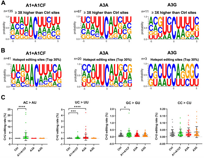
- Among 307 total C sites in seven selected viral RNA segments, significant C-to-U editing sites numbered 135 for A1 + A1CF, 67 for A3A, and 11 for A3G; editing efficiencies reached as high as 22.2% for A1 + A1CF, 4.5% for A3A, and 0.18% for A3G.
- A3A preferred a UC motif and A1 + A1CF preferred an AC motif; among C-to-U variations analyzed, AC-to-AU accounted for 38.23% and UC-to-UU for 31.83%, versus 15.44% for GC-to-GU and 14.50% for CC-to-CU.
- In Caco-2 cells, A1 + A1CF or A3G expression did not significantly change viral RNA levels up to 96 h post-infection, but A3A expression significantly increased viral RNA at 72 and 96 h and produced approximately 10–100-fold higher viral progeny at 72 h.
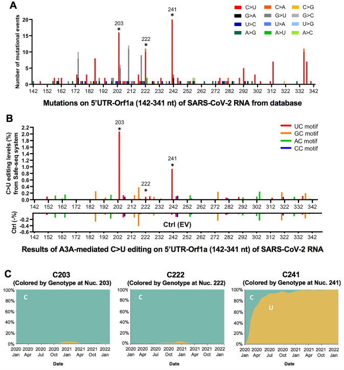
- Database analysis found C-to-U was the predominant mutation in the 5′UTR-Orf1a region, accounting for ~55.8% of single nucleotide variants; the A3A-edited UC241 mutation became a signature of dominant strains, with 90% of circulating variants carrying it by July 2020 and almost all by March 2021.
Methods
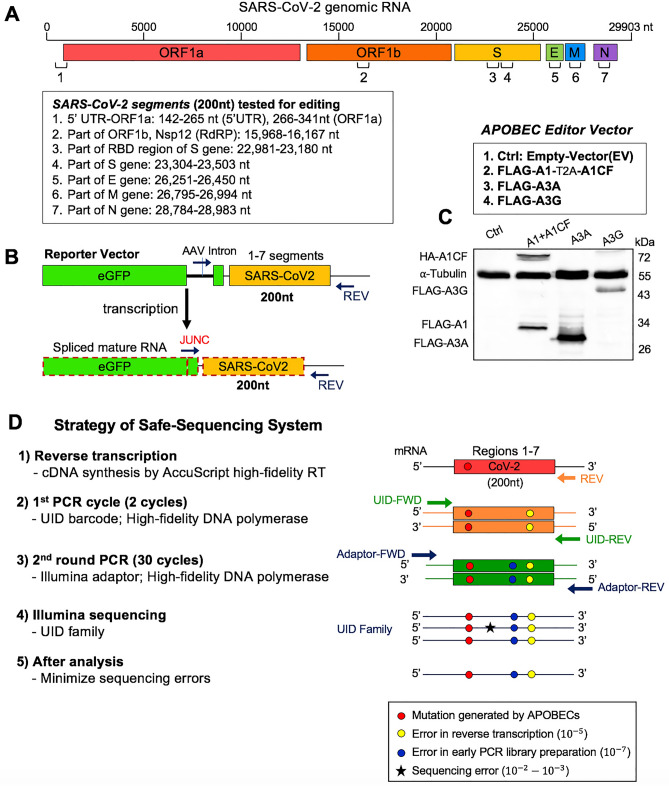
- Cell-based RNA editing assay: seven 200 nt SARS-CoV-2 RNA segments were inserted into reporter vectors, co-transfected with APOBEC editor vectors (A1 + A1CF, A3A, or A3G) in HEK293T cells, and analyzed by Safe-Sequencing-System (SSS) deep sequencing with UID barcodes to distinguish true editing from PCR/sequencing errors.
- Infection experiments: Caco-2 stable cell lines expressing A1 + A1CF, A3A, or A3G under a tetracycline-controlled promoter were infected with SARS-CoV-2 (MOI = 0.05); viral RNA replication was measured by qPCR for Nsp12, S, and N regions, and progeny yield by plaque assay on Vero E6 cells.
- Sequence analysis: publicly available SARS-CoV-2 genome data from Nextstrain/GISAID (Dec. 2019 to Jan. 22, 2022) were analyzed for mutational events and C-to-U prevalence.
Limitations
- The paper states that SSS sequencing is several times more expensive than regular NGS, each read covers ~150–200 nt, and it was too expensive to cover the entire ~30,000 nt SARS-CoV-2 genome, so only seven viral RNA segments were selected.
- The paper states that A3G edited only a small number of sites (n = 11) and did not show a clear motif preference, so more SARS-CoV-2 regions and/or folded RNA structures are needed to verify A3G-mediated editing; UC/AC motifs alone are insufficient for efficient editing, and the exact RNA structural features remain to be defined.
- The paper notes that a minor deaminase-independent enhancement by inactive A3A-E72A was observed and warrants further validation, and that host RNA or DNA editing by WT A3A could also contribute to increased viral replication/progeny; it also states that multiple viral passages in cell culture or animal models are needed to better assess mutation effects on fitness.
Model-written summary. Check it against the paper before relying on it.
Integrity signals
Registry
Cites
Statistics
Figures
Verification
Claims the paper makes
55 Own result · 28 Own interpretation · 14 Partly supported · 11 Not checked yet · 6 Own method · 5 Supported · 1 Couldn’t confirm · 1 No full text of the sources · accuracy not yet measured
Every source the paper cites for this claim was checked and supports it.
In the paper
“These pro-viral effect of A3A contrast to the known anti-viral effect of APOBECs19–22.”
§ The roles of APOBEC-mediated RNA editing in SARS-CoV-2 mutations, replication and fitness > Discussion
What the sources say
- Supportedcites [19] Olson, M. E., Harris, R. S. & Harki, D. A. APOBEC enzymes as targets for virus and cancer therapy. Cell Chem. Biol. 25, 36–49. https://doi.org/10.1016/j.chembiol.2017.10.007 (2018). source
The “HIV-Restrictive APOBECs” section describes A3 enzymes as antiviral defenses and states that they restrict HIV-1 and other viruses. The citation supports the known antiviral effect used as the contrast; the citing authors’ pro-viral A3A finding is not attributed to this source. (LLM, conf=0.93)
- Supportedcites [20] Salter, J. D., Bennett, R. P. & Smith, H. C. The APOBEC protein family: United by structure, Divergent in Function. Trends Biochem. Sci. 41, 578–594. https://doi.org/10.1016/j.tibs.2016.05.001 (2016). source
The review’s “RNA viruses” section describes APOBEC3 proteins’ anti-HIV activity, and its “DNA viruses” section calls A3A a potent inhibitor of AAV-2. These passages support the known antiviral effect invoked as the contrast; the citing authors’ observation of a pro-viral effect is not being attributed to this review. Page numbers are not provided in our copy. (LLM, conf=0.92)
- Supportedcites [21] Harris, R. S. & Dudley, J. P. APOBECs and virus restriction. Virology 479–480, 131–145. https://doi.org/10.1016/j.virol.2015.03.012 (2015). source
The Conclusions describe APOBEC family proteins as virus restriction factors that protect against exogenous viral infections. The HIV restriction section also attributes HIV-1 restriction to several APOBEC3 proteins. The citation supports the known antiviral effect being contrasted, not the citing paper’s observation of a pro-viral A3A effect. (LLM, conf=0.92)
- Supportedcites [22] Chen, X. S. Insights into the structures and multimeric status of APOBEC proteins involved in viral restriction and other cellular functions. Viruses https://doi.org/10.3390/v13030497 (2021). source
The citation supports the background point that APOBEC proteins have antiviral effects. Section 5 describes the APOBEC3 subfamily as restricting RNA and DNA viral infections, and Section 8 reports strong HIV-restricting activity for several APOBEC3 proteins. The pro-viral effect of A3A is the citing authors’ contrasting finding, not a finding attributed to this source. (LLM, conf=0.92)
boosteddirect- Asserted: passes — The authors put it forward as true.
- Checkable: passes — It can be checked against evidence.
- Load-bearing: passes — The paper’s conclusions depend on it.
- Based: passes — The paper gives a reason why it holds.
- Supported
The cited sources support only part of this claim — read each source’s verdict below.
In the paper
“Prior bioinformatic analysis of the SARS-CoV-2 sequences suggested that some of the C-to-U mutations may be caused by APOBEC proteins instead of the random mutations caused by viral RNA polymerase during replication or ROS11,12,14,27,51,52.”
§ The roles of APOBEC-mediated RNA editing in SARS-CoV-2 mutations, replication and fitness > Discussion
What the sources say
- Partly supportedcites [11] Di Giorgio, S., Martignano, F., Torcia, M. G., Mattiuz, G. & Conticello, S. G. Evidence for host-dependent RNA editing in the transcriptome of SARS-CoV-2. Sci. Adv. 6, eabb5813. https://doi.org/10.1126/sciadv.abb5813 (2020). sourceExperimental — accuracy not yet measured
The Results (page unavailable) suggest that some SARS-CoV-2 C-to-U changes could result from APOBEC-mediated editing, and say that polymerase errors account for only a marginal fraction of the observed variants. The paper does not address ROS as an alternative cause, so it does not support that part of the comparison. (LLM, conf=0.84)
- Partly supportedcites [12] Simmonds, P. Rampant C–>U hypermutation in the genomes of SARS-CoV-2 and other coronaviruses: Causes and consequences for their short- and long-term evolutionary trajectories. mSphere. https://doi.org/10.1128/mSphere.00408-20 (2020). sourceExperimental — accuracy not yet measured
The Discussion (‘The mechanism of SARS-CoV-2 hypermutation’; page unavailable) says the C→U pattern provides circumstantial grounds for suspecting APOBEC proteins and argues that its asymmetry cannot be explained by errors of the viral RNA-dependent RNA polymerase. The paper does not address ROS as an alternative cause, so that part of the attributed comparison is unsupported. (LLM, conf=0.86)
- Partly supportedcites [14] Matyasek, R. & Kovarik, A. Mutation patterns of human SARS-CoV-2 and Bat RaTG13 coronavirus genomes are strongly biased towards C>U transitions, indicating rapid evolution in their hosts. Genes (Basel). https://doi.org/10.3390/genes11070761 (2020). sourceExperimental — accuracy not yet measured
Section 4.1 argues that polymerase errors are unlikely to explain the strong C-to-U bias and suggests enzymatic cytidine deamination as a possible contributor, mentioning the APOBEC enzyme Apo3G. But it also proposes spontaneous deamination and does not assess ROS as an alternative, so the full comparison in the claim is not supported. (LLM, conf=0.84)
- Supportedcites [27] Klimczak, L. J., Randall, T. A., Saini, N., Li, J. L. & Gordenin, D. A. Similarity between mutation spectra in hypermutated genomes of rubella virus and in SARS-CoV-2 genomes accumulated during the COVID-19 pandemic. PLoS One 15, e0237689. https://doi.org/10.1371/journal.pone.0237689 (2020). source
The Discussion reports that SARS-CoV-2 C-to-U substitutions show sequence motifs characteristic of APOBEC cytidine deaminases, supporting APOBEC activity as a possible source of some mutations. It also acknowledges other possible sources of C-to-U mutagenesis, including oxidative mutagenesis; the citing sentence’s qualified “some” and “may” are consistent with that uncertainty. The Introduction identifies viral polymerase errors as another source of mutations. (LLM, conf=0.88)
- Partly supportedcites [51] Sadykov, M., Mourier, T., Guan, Q. & Pain, A. Short sequence motif dynamics in the SARS-CoV-2 genome suggest a role for cytosine deamination in CpG reduction. J. Mol. Cell Biol. https://doi.org/10.1093/jmcb/mjab011 (2021). sourceExperimental — accuracy not yet measured
In the paper’s main text (page not specified), the authors report a bioinformatic analysis of SARS-CoV-2 genomes and suggest APOBEC-mediated editing as a potential driver of C-to-U substitutions. The copy does not compare that explanation with random mutations from viral RNA polymerase or ROS, so the stated contrast is not supported by this source. (LLM, conf=0.85)
- Supportedcites [52] Wang, R., Hozumi, Y., Zheng, Y. H., Yin, C. & Wei, G. W. Host immune response driving SARS-CoV-2 evolution. Viruses https://doi.org/10.3390/v12101095 (2020). source
The Introduction (page not provided) says APOBEC deaminates cytosine to uracil and identifies APOBEC editing as a likely source of SARS-CoV-2 C-to-T mutations. The Results (page not provided) find that C-to-T mutations occur far more often than expected under a random-mutation model, supporting the claim that some arise through APOBEC editing rather than random processes. (LLM, conf=0.90)
hedgedconditional- Asserted: passes — The authors put it forward as true.
- Checkable: passes — It can be checked against evidence.
- Load-bearing: passes — The paper’s conclusions depend on it.
- Based: passes — The paper gives a reason why it holds.
- Partly supported
The cited sources support only part of this claim — read each source’s verdict below.
In the paper
“In fact, the UC241 is within the viral packaging signal sequence that has a stem-loop secondary structure which is in close proximity to the stem-loop structures involved in replication and leader transcription regulatory sequence (TRS-L)39 (Supplementary Fig. S14).”
§ The roles of APOBEC-mediated RNA editing in SARS-CoV-2 mutations, replication and fitness > Discussion
What the sources say
- Partly supportedcites [39] Miao, Z., Tidu, A., Eriani, G. & Martin, F. Secondary structure of the SARS-CoV-2 5’-UTR. RNA Biol. 18, 447–456. https://doi.org/10.1080/15476286.2020.1814556 (2021). sourceExperimental — accuracy not yet measured
In Results and discussion (5′-UTR inline probing; page not provided), the paper describes SL5 as a stem-loop region whose loops are most likely involved in RNA packaging. It also identifies nearby SL2 with replication and SL3 with TRS-L. The copy does not identify UC241 as lying within a packaging signal, and it presents SL5’s packaging role as likely rather than established. (LLM, conf=0.85)
plaincorrelational- Asserted: passes — The authors put it forward as true.
- Checkable: passes — It can be checked against evidence.
- Load-bearing: passes — The paper’s conclusions depend on it.
- Based: passes — The paper gives a reason why it holds.
- Partly supported
The cited sources support only part of this claim — read each source’s verdict below.
In the paper
“Interestingly, analysis of SARS-CoV-2 variants from the database also showed higher AC motif mutations (38.3%) than UC motif mutations (31.2%) (Supplementary Fig. S4), as also reported in the previous bioinformatic database analysis3,11,27.”
§ The roles of APOBEC-mediated RNA editing in SARS-CoV-2 mutations, replication and fitness > Discussion
What the sources say
- Partly supportedcites [3] Kockler, Z. W. & Gordenin, D. A. From RNA world to SARS-CoV-2: The edited story of RNA viral evolution. Cells https://doi.org/10.3390/cells10061557 (2021). sourceExperimental — accuracy not yet measured
Section 5.3 describes an analysis of more than 30,000 SARS-CoV-2 genome sequences and reports enrichment of a UC-containing mutation signature. It does not report that AC-motif mutations exceeded UC-motif mutations, or give the cited 38.3% and 31.2% figures. No page number is available in our copy. (LLM, conf=0.83)
- Partly supportedcites [11] Di Giorgio, S., Martignano, F., Torcia, M. G., Mattiuz, G. & Conticello, S. G. Evidence for host-dependent RNA editing in the transcriptome of SARS-CoV-2. Sci. Adv. 6, eabb5813. https://doi.org/10.1126/sciadv.abb5813 (2020). sourceExperimental — accuracy not yet measured
The Results (page unavailable) report that C-to-U changes preferentially occur after either adenosine or uridine, supporting the relevance of AC and UC motifs. Our copy does not give the claimed 38.3% and 31.2% frequencies or state that AC mutations are more frequent than UC mutations. (LLM, conf=0.78)
- Partly supportedcites [27] Klimczak, L. J., Randall, T. A., Saini, N., Li, J. L. & Gordenin, D. A. Similarity between mutation spectra in hypermutated genomes of rubella virus and in SARS-CoV-2 genomes accumulated during the COVID-19 pandemic. PLoS One 15, e0237689. https://doi.org/10.1371/journal.pone.0237689 (2020). sourceExperimental — accuracy not yet measured
Table 1 in the Results identifies both uCn-to-uUn (UC) and aCn-to-aUn (AC) as enriched SARS-CoV-2 mutation motifs. Neither that table nor the Discussion reports that AC mutations were more frequent than UC mutations, or gives the cited 38.3% and 31.2% figures; the citing sentence attributes those figures to its own Supplementary Fig. S4. (LLM, conf=0.85)
plaincorrelational- Asserted: passes — The authors put it forward as true.
- Checkable: passes — It can be checked against evidence.
- Load-bearing: passes — The paper’s conclusions depend on it.
- Based: passes — The paper gives a reason why it holds.
- Partly supported
Every source the paper cites for this claim was checked and supports it.
In the paper
“Interestingly, both A1 cofactors A1CF and RBM47 were shown to physically interact with SARS-CoV-2 RNA in an interactome study56, offering indirect evidence that these RNA-binding A1-cofactors could recruit A1 to target SARS-CoV-2 RNA for editing in the infected tissue.”
§ The roles of APOBEC-mediated RNA editing in SARS-CoV-2 mutations, replication and fitness > Discussion
What the sources say
- Supportedcites [56] Schmidt, N. et al. The SARS-CoV-2 RNA-protein interactome in infected human cells. Nat. Microbiol. 6, 339–353. https://doi.org/10.1038/s41564-020-00846-z (2021). source
The Discussion lists RBM47 and A1CF as RNA-editing cofactors among the SARS-CoV-2 RNA binders identified in the study. The citing sentence’s suggestion that they could recruit A1 for editing is its own inference, not a finding attributed to this source. (LLM, conf=0.93)
plaincorrelational- Asserted: passes — The authors put it forward as true.
- Checkable: passes — It can be checked against evidence.
- Load-bearing: passes — The paper’s conclusions depend on it.
- Based: passes — The paper gives a reason why it holds.
- Supported
A result the paper reports from its own data — it cites no other source for it, so there is none to check. The quote shows it is in the paper.
In the paper
“Even the very recent omicron variants, which began to spread from Nov. 2021 rapidly, continue to show noticeable C-to-U editing pattern (Supplementary Fig. S7B).”
§ The roles of APOBEC-mediated RNA editing in SARS-CoV-2 mutations, replication and fitness > Results > The potential effect of APOBEC-mediated RNA editing on SARS-CoV-2 variants
plaincorrelational- Asserted: passes — The authors put it forward as true.
- Checkable: passes — It can be checked against evidence.
- Load-bearing: passes — The paper’s conclusions depend on it.
- Based: passes — The paper gives a reason why it holds.
A result the paper reports from its own data — it cites no other source for it, so there is none to check. The quote shows it is in the paper.
In the paper
“Instead, expression of wild-type APOBEC3 greatly promotes viral replication/propagation, suggesting that SARS-CoV-2 utilizes the APOBEC-mediated mutations for fitness and evolution.”
§ The roles of APOBEC-mediated RNA editing in SARS-CoV-2 mutations, replication and fitness > Abstract
plaindirect- Asserted: passes — The authors put it forward as true.
- Checkable: passes — It can be checked against evidence.
- Load-bearing: passes — The paper’s conclusions depend on it.
- Based: passes — The paper gives a reason why it holds.
A result the paper reports from its own data — it cites no other source for it, so there is none to check. The quote shows it is in the paper.
In the paper
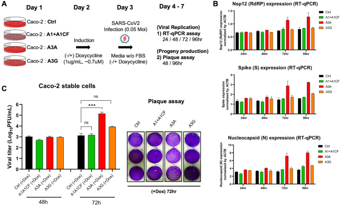
“the virus titer from the cell line expressing A3A consistently showed an approximately 10–100 fold higher than the control and A1 + A1CF and A3G expressing cell lines at 72 h (Fig. 5C).”
§ The roles of APOBEC-mediated RNA editing in SARS-CoV-2 mutations, replication and fitness > Results > SARS-CoV-2 replication and progeny yield in cells overexpressing APOBECs
plaincorrelational- Asserted: passes — The authors put it forward as true.
- Checkable: passes — It can be checked against evidence.
- Load-bearing: passes — The paper’s conclusions depend on it.
- Based: passes — The paper gives a reason why it holds.
A result the paper reports from its own data — it cites no other source for it, so there is none to check. The quote shows it is in the paper.
In the paper
“Consistent with the increased viral RNA replication, A3A expression in the stable Caco-2 cell line also correlates with significantly higher viral progeny yield after 3 days of infection.”
§ The roles of APOBEC-mediated RNA editing in SARS-CoV-2 mutations, replication and fitness > Results > SARS-CoV-2 replication and progeny yield in cells overexpressing APOBECs
plaincorrelational- Asserted: passes — The authors put it forward as true.
- Checkable: passes — It can be checked against evidence.
- Load-bearing: passes — The paper’s conclusions depend on it.
- Based: passes — The paper gives a reason why it holds.
A result the paper reports from its own data — it cites no other source for it, so there is none to check. The quote shows it is in the paper.
In the paper
“Notably, the AC to AU mutation at C23525 resulted an H655Y mutation on the spike protein (Supplementary Fig. S7B), and the H655Y mutation was shown to alter cell entry pathways, i.e. the mutation is responsible for the preferential usage of endosomal pathway over cell surface entry pathways41.”
§ The roles of APOBEC-mediated RNA editing in SARS-CoV-2 mutations, replication and fitness > Results > The potential effect of APOBEC-mediated RNA editing on SARS-CoV-2 variants
plaindirect- Asserted: passes — The authors put it forward as true.
- Checkable: passes — It can be checked against evidence.
- Load-bearing: passes — The paper’s conclusions depend on it.
- Based: passes — The paper gives a reason why it holds.
A result the paper reports from its own data — it cites no other source for it, so there is none to check. The quote shows it is in the paper.
In the paper
“However, the preferred motif by A3G is not a clear cut from our data, possibly due to the low activity on the limited number of edited sites among the seven tested viral segments.”
§ The roles of APOBEC-mediated RNA editing in SARS-CoV-2 mutations, replication and fitness > Results > Sequence motifs near the APOBEC-edited C on SARS-CoV-2 RNA
hedged- Asserted: passes — The authors put it forward as true.
- Checkable: passes — It can be checked against evidence.
- Load-bearing: unclear — It is unclear whether the paper’s conclusions depend on it.
- Based: passes — The paper gives a reason why it holds.
A result the paper reports from its own data — it cites no other source for it, so there is none to check. The quote shows it is in the paper.
In the paper
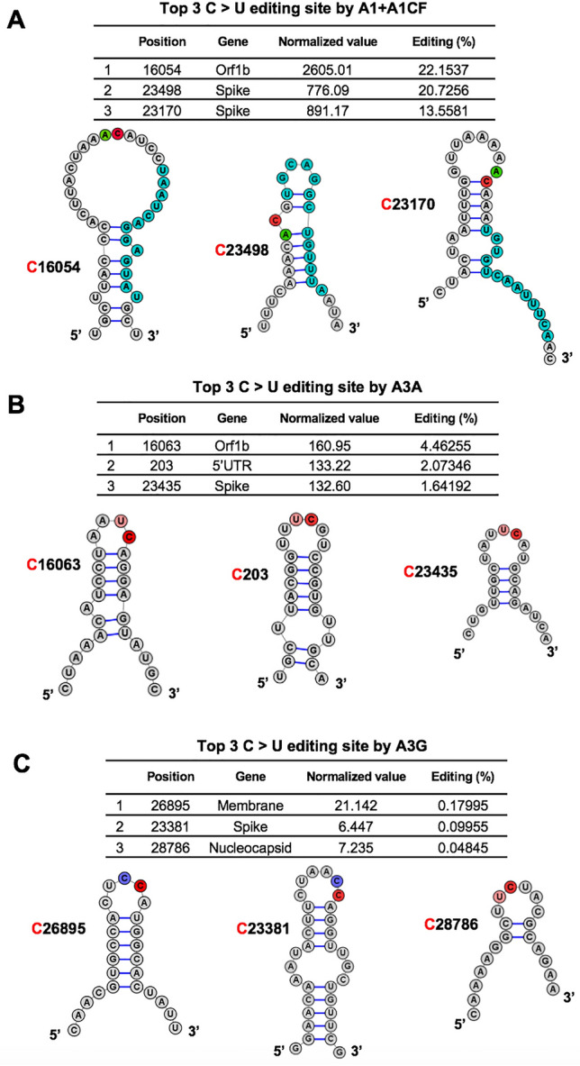
“Interestingly, our top 3 highest A3A-mediated editing sites on SARS-CoV-2 RNA reported here all have the UC motifs in the loop of a predicted stem-loop secondary structure (Fig. 3B).”
§ The roles of APOBEC-mediated RNA editing in SARS-CoV-2 mutations, replication and fitness > Results > Features of the efficiently APOBEC-edited RNA sites on SARS-CoV-2
plaincorrelational- Asserted: passes — The authors put it forward as true.
- Checkable: passes — It can be checked against evidence.
- Load-bearing: unclear — It is unclear whether the paper’s conclusions depend on it.
- Based: passes — The paper gives a reason why it holds.
A result the paper reports from its own data — it cites no other source for it, so there is none to check. The quote shows it is in the paper.
In the paper
“However, SARS-CoV-2 replication and viral progeny production in Caco-2 cells are not inhibited by the expression of these APOBECs.”
§ The roles of APOBEC-mediated RNA editing in SARS-CoV-2 mutations, replication and fitness > Abstract
plain- Asserted: passes — The authors put it forward as true.
- Checkable: passes — It can be checked against evidence.
- Load-bearing: passes — The paper’s conclusions depend on it.
- Based: passes — The paper gives a reason why it holds.
A result the paper reports from its own data — it cites no other source for it, so there is none to check. The quote shows it is in the paper.
In the paper
“While no difference in viral titer was observed at 48 h from all cell lines with or without APOBEC expression, the virus titer from the cell line expressing A3A consistently showed an approximately 10–100 fold higher than the control and A1 + A1CF and A3G expressing cell lines at 72 h (Fig. 5C).”
§ The roles of APOBEC-mediated RNA editing in SARS-CoV-2 mutations, replication and fitness > Results > SARS-CoV-2 replication and progeny yield in cells overexpressing APOBECs
plain- Asserted: passes — The authors put it forward as true.
- Checkable: passes — It can be checked against evidence.
- Load-bearing: unclear — It is unclear whether the paper’s conclusions depend on it.
- Based: passes — The paper gives a reason why it holds.
A result the paper reports from its own data — it cites no other source for it, so there is none to check. The quote shows it is in the paper.
In the paper
“We report the first experimental evidence demonstrating that APOBEC3A, APOBEC1, and APOBEC3G can edit on specific sites of SARS-CoV-2 RNA to produce C-to-U mutations.”
§ The roles of APOBEC-mediated RNA editing in SARS-CoV-2 mutations, replication and fitness > Abstract
boosteddirect- Asserted: passes — The authors put it forward as true.
- Checkable: passes — It can be checked against evidence.
- Load-bearing: passes — The paper’s conclusions depend on it.
- Based: passes — The paper gives a reason why it holds.
A result the paper reports from its own data — it cites no other source for it, so there is none to check. The quote shows it is in the paper.
In the paper
“The sequence context near the unedited C sites by the corresponding APOBECs also shows no particular trend except for a relatively small ratios change of the − 1 position (e.g., less A by A1 + A1CF and less U by A3A, respectively) (Supplementary Fig. S3B–D).”
§ The roles of APOBEC-mediated RNA editing in SARS-CoV-2 mutations, replication and fitness > Results > Sequence motifs near the APOBEC-edited C on SARS-CoV-2 RNA
plain- Asserted: passes — The authors put it forward as true.
- Checkable: passes — It can be checked against evidence.
- Load-bearing: unclear — It is unclear whether the paper’s conclusions depend on it.
- Based: passes — The paper gives a reason why it holds.
A result the paper reports from its own data — it cites no other source for it, so there is none to check. The quote shows it is in the paper.
In the paper
“Because Caco-2 cell lines have no detectible endogenous expression of A1, A3A, and A3G protein (Supplementary Fig. S8), we first constructed Caco-2 stable cell lines expressing one of the three APOBEC genes.”
§ The roles of APOBEC-mediated RNA editing in SARS-CoV-2 mutations, replication and fitness > Results > SARS-CoV-2 replication and progeny yield in cells overexpressing APOBECs
plain- Asserted: passes — The authors put it forward as true.
- Checkable: passes — It can be checked against evidence.
- Load-bearing: passes — The paper’s conclusions depend on it.
- Based: passes — The paper gives a reason why it holds.
A result the paper reports from its own data — it cites no other source for it, so there is none to check. The quote shows it is in the paper.
In the paper
“Again, the editing efficiency at C16063 (4.5%) is about threefold higher than the third efficiently edited site at C23453 (1.6%) (Fig. 3B).”
§ The roles of APOBEC-mediated RNA editing in SARS-CoV-2 mutations, replication and fitness > Results > Features of the efficiently APOBEC-edited RNA sites on SARS-CoV-2
plain- Asserted: passes — The authors put it forward as true.
- Checkable: passes — It can be checked against evidence.
- Load-bearing: unclear — It is unclear whether the paper’s conclusions depend on it.
- Based: passes — The paper gives a reason why it holds.
A result the paper reports from its own data — it cites no other source for it, so there is none to check. The quote shows it is in the paper.
In the paper
“Although each of the three APOBEC proteins showed a strong preference for specific dinucleotide sequence motifs (i.e. AC, UC, or CC sequence motifs) for editing on the viral RNA, the relative editing efficiency of these motif sites vary greatly, such as between 0.0041 and 22.15% for A1 + A1CF, and between 0.0040 and 4.46% for A3A (Supplementary Dataset File 2).”
§ The roles of APOBEC-mediated RNA editing in SARS-CoV-2 mutations, replication and fitness > Results > Features of the efficiently APOBEC-edited RNA sites on SARS-CoV-2
boostedcorrelational- Asserted: passes — The authors put it forward as true.
- Checkable: passes — It can be checked against evidence.
- Load-bearing: unclear — It is unclear whether the paper’s conclusions depend on it.
- Based: passes — The paper gives a reason why it holds.
A result the paper reports from its own data — it cites no other source for it, so there is none to check. The quote shows it is in the paper.
In the paper
“As a control, the sequence context near all C sites (edited + unedited sites) in the selected viral region does not show a specific feature (Supplementary Fig. S3A).”
§ The roles of APOBEC-mediated RNA editing in SARS-CoV-2 mutations, replication and fitness > Results > Sequence motifs near the APOBEC-edited C on SARS-CoV-2 RNA
plain- Asserted: passes — The authors put it forward as true.
- Checkable: passes — It can be checked against evidence.
- Load-bearing: unclear — It is unclear whether the paper’s conclusions depend on it.
- Based: passes — The paper gives a reason why it holds.
A result the paper reports from its own data — it cites no other source for it, so there is none to check. The quote shows it is in the paper.
In the paper
“The analysis revealed that the C-to-U is the predominant mutation for the entire genome, accounting for ~ 55.8% among all single nucleotide variants (SNVs) within the SARS-CoV-2 5′UTR-Orf1a region (142–341 nt, the segment tested in our reporter 1 vector) (Fig. 4A and Supplementary Fig. S6A).”
§ The roles of APOBEC-mediated RNA editing in SARS-CoV-2 mutations, replication and fitness > Results > The potential effect of APOBEC-mediated RNA editing on SARS-CoV-2 variants
plain- Asserted: passes — The authors put it forward as true.
- Checkable: passes — It can be checked against evidence.
- Load-bearing: passes — The paper’s conclusions depend on it.
- Based: passes — The paper gives a reason why it holds.
A result the paper reports from its own data — it cites no other source for it, so there is none to check. The quote shows it is in the paper.
In the paper
“Unexpectedly, the abundance of viral RNAs has increased significantly in the cell lines expressing A3A at 72 h and 96 h post SARS-CoV-2 infection.”
§ The roles of APOBEC-mediated RNA editing in SARS-CoV-2 mutations, replication and fitness > Results > SARS-CoV-2 replication and progeny yield in cells overexpressing APOBECs
plaindirect- Asserted: passes — The authors put it forward as true.
- Checkable: passes — It can be checked against evidence.
- Load-bearing: passes — The paper’s conclusions depend on it.
- Based: passes — The paper gives a reason why it holds.
A result the paper reports from its own data — it cites no other source for it, so there is none to check. The quote shows it is in the paper.
In the paper
“Analysis of the top 3 sites edited by A3G also showed the CC (or UC) motifs in the single-stranded loop region of a predicted stem-loop secondary structure (Fig. 3C).”
§ The roles of APOBEC-mediated RNA editing in SARS-CoV-2 mutations, replication and fitness > Results > Features of the efficiently APOBEC-edited RNA sites on SARS-CoV-2
plaincorrelational- Asserted: passes — The authors put it forward as true.
- Checkable: passes — It can be checked against evidence.
- Load-bearing: unclear — It is unclear whether the paper’s conclusions depend on it.
- Based: passes — The paper gives a reason why it holds.
A result the paper reports from its own data — it cites no other source for it, so there is none to check. The quote shows it is in the paper.
In the paper
“In all representative clades of SARS-CoV-2 emerged over the last two years since the initial outbreak, the C-to-U mutations have been much more pronounced than other types of single nucleotide variations (Supplementary Fig. S7A).”
§ The roles of APOBEC-mediated RNA editing in SARS-CoV-2 mutations, replication and fitness > Results > The potential effect of APOBEC-mediated RNA editing on SARS-CoV-2 variants
plaincorrelational- Asserted: passes — The authors put it forward as true.
- Checkable: passes — It can be checked against evidence.
- Load-bearing: passes — The paper’s conclusions depend on it.
- Based: passes — The paper gives a reason why it holds.
A result the paper reports from its own data — it cites no other source for it, so there is none to check. The quote shows it is in the paper.
In the paper
“The replication assay results showed that, compared with the control Caco-2 cell line, no significant change of viral RNA level was detected by qPCR with the cell lines expressing A1 + A1CF or A3G even up to 96 h post-infection (Fig. 5B).”
§ The roles of APOBEC-mediated RNA editing in SARS-CoV-2 mutations, replication and fitness > Results > SARS-CoV-2 replication and progeny yield in cells overexpressing APOBECs
plain- Asserted: passes — The authors put it forward as true.
- Checkable: passes — It can be checked against evidence.
- Load-bearing: passes — The paper’s conclusions depend on it.
- Based: passes — The paper gives a reason why it holds.
A result the paper reports from its own data — it cites no other source for it, so there is none to check. The quote shows it is in the paper.
In the paper
“However, the UC and AC motifs alone are not sufficient for efficient editing by A3A or A1 + A1CF.”
§ The roles of APOBEC-mediated RNA editing in SARS-CoV-2 mutations, replication and fitness > Discussion
plain- Asserted: passes — The authors put it forward as true.
- Checkable: passes — It can be checked against evidence.
- Load-bearing: passes — The paper’s conclusions depend on it.
- Based: passes — The paper gives a reason why it holds.
A result the paper reports from its own data — it cites no other source for it, so there is none to check. The quote shows it is in the paper.
In the paper
“Many of the UC and AC motifs in the SARS-CoV-2 RNA showed only the background level of C-to-U mutation (Supplementary Dataset File 2), which suggest that additional RNA structural features around the UC/AC motifs should play an important role in dictating whether a UC or AC site can be efficiently edited (Fig. 3).”
§ The roles of APOBEC-mediated RNA editing in SARS-CoV-2 mutations, replication and fitness > Discussion
plain- Asserted: passes — The authors put it forward as true.
- Checkable: passes — It can be checked against evidence.
- Load-bearing: passes — The paper’s conclusions depend on it.
- Based: passes — The paper gives a reason why it holds.
A result the paper reports from its own data — it cites no other source for it, so there is none to check. The quote shows it is in the paper.
In the paper
“Our results demonstrated that the three tested APOBECs show different editing rate at specific viral RNA sites, reaching as high as 22.2% for A1 + A1CF, 4.5% for A3A, and 0.18% for A3G, respectively, on the selected viral RNA transcript populations (Fig. 2).”
§ The roles of APOBEC-mediated RNA editing in SARS-CoV-2 mutations, replication and fitness > Discussion
boostedn = 11- Asserted: passes — The authors put it forward as true.
- Checkable: passes — It can be checked against evidence.
- Load-bearing: unclear — It is unclear whether the paper’s conclusions depend on it.
- Based: passes — The paper gives a reason why it holds.
A result the paper reports from its own data — it cites no other source for it, so there is none to check. The quote shows it is in the paper.
In the paper
“Surprisingly, expression of WT A3A in the tested cells showed significant increases of the viral RNA replication and the viral progeny production after 72 h postinfection (Fig. 5B,C, Supplementary Fig. S10), even though some increase of replication and progeny production was also observed in the inactive-A3A mutant expressing cell line (Supplementary Fig. S10).”
§ The roles of APOBEC-mediated RNA editing in SARS-CoV-2 mutations, replication and fitness > Discussion
plaindirect- Asserted: passes — The authors put it forward as true.
- Checkable: passes — It can be checked against evidence.
- Load-bearing: passes — The paper’s conclusions depend on it.
- Based: passes — The paper gives a reason why it holds.
A result the paper reports from its own data — it cites no other source for it, so there is none to check. The quote shows it is in the paper.
In the paper
“Our results demonstrated that the three tested APOBECs show different editing rate at specific viral RNA sites, reaching as high as 22.2% for A1 + A1CF, 4.5% for A3A, and 0.18% for A3G, respectively, on the selected viral RNA transcript populations (Fig. 2).”
§ The roles of APOBEC-mediated RNA editing in SARS-CoV-2 mutations, replication and fitness > Discussion
boosted- Asserted: passes — The authors put it forward as true.
- Checkable: passes — It can be checked against evidence.
- Load-bearing: passes — The paper’s conclusions depend on it.
- Based: passes — The paper gives a reason why it holds.
A result the paper reports from its own data — it cites no other source for it, so there is none to check. The quote shows it is in the paper.
In the paper
“Contrary to the editing at C241, our sequencing results showed that the nearby C203 and C222, where C203U and C222U mutations displayed a neutral effect on viral replication, showed a slight but statistically non-significant increase in C to U mutations 72 h post viral infection in WT A3A overexpression (Supplementary Fig. S11B).”
§ The roles of APOBEC-mediated RNA editing in SARS-CoV-2 mutations, replication and fitness > Results > SARS-CoV-2 replication and progeny yield in cells overexpressing APOBECs
plain- Asserted: passes — The authors put it forward as true.
- Checkable: passes — It can be checked against evidence.
- Load-bearing: unclear — It is unclear whether the paper’s conclusions depend on it.
- Based: passes — The paper gives a reason why it holds.
A result the paper reports from its own data — it cites no other source for it, so there is none to check. The quote shows it is in the paper.
In the paper
“Our results demonstrated that the three tested APOBECs show different editing rate at specific viral RNA sites, reaching as high as 22.2% for A1 + A1CF, 4.5% for A3A, and 0.18% for A3G, respectively, on the selected viral RNA transcript populations (Fig. 2).”
§ The roles of APOBEC-mediated RNA editing in SARS-CoV-2 mutations, replication and fitness > Discussion
boosted- Asserted: passes — The authors put it forward as true.
- Checkable: passes — It can be checked against evidence.
- Load-bearing: passes — The paper’s conclusions depend on it.
- Based: passes — The paper gives a reason why it holds.
A result the paper reports from its own data — it cites no other source for it, so there is none to check. The quote shows it is in the paper.
In the paper
“Because the AC-to-AU mutations was not detected in A1 alone (Supplementary Fig. S5), it indicates that the AC-to-AU editing also requires a cofactor A1CF.”
§ The roles of APOBEC-mediated RNA editing in SARS-CoV-2 mutations, replication and fitness > Discussion
plain- Asserted: passes — The authors put it forward as true.
- Checkable: passes — It can be checked against evidence.
- Load-bearing: passes — The paper’s conclusions depend on it.
- Based: passes — The paper gives a reason why it holds.
A result the paper reports from its own data — it cites no other source for it, so there is none to check. The quote shows it is in the paper.
In the paper
“A3G edits a small number (n = 11) of SARS-CoV-2 RNA in our study (Fig. 2).”
§ The roles of APOBEC-mediated RNA editing in SARS-CoV-2 mutations, replication and fitness > Discussion
plainn = 11- Asserted: passes — The authors put it forward as true.
- Checkable: passes — It can be checked against evidence.
- Load-bearing: unclear — It is unclear whether the paper’s conclusions depend on it.
- Based: passes — The paper gives a reason why it holds.
A result the paper reports from its own data — it cites no other source for it, so there is none to check. The quote shows it is in the paper.
In the paper
“This significant C241U mutation correlates with the time points (72 and 96 h) with detectible increases in viral RNA replication and viral progeny production.”
§ The roles of APOBEC-mediated RNA editing in SARS-CoV-2 mutations, replication and fitness > Results > SARS-CoV-2 replication and progeny yield in cells overexpressing APOBECs
plaincorrelational- Asserted: passes — The authors put it forward as true.
- Checkable: passes — It can be checked against evidence.
- Load-bearing: passes — The paper’s conclusions depend on it.
- Based: passes — The paper gives a reason why it holds.
A result the paper reports from its own data — it cites no other source for it, so there is none to check. The quote shows it is in the paper.
In the paper
“Among many of the detected A3A edited sites on SARS-CoV-2 RNA in our study are three C-to-U mutations located in the 5’UTR region, UC203, UC222, UC241 (Fig. 4A,B).”
§ The roles of APOBEC-mediated RNA editing in SARS-CoV-2 mutations, replication and fitness > Discussion
plain- Asserted: passes — The authors put it forward as true.
- Checkable: passes — It can be checked against evidence.
- Load-bearing: passes — The paper’s conclusions depend on it.
- Based: passes — The paper gives a reason why it holds.
A result the paper reports from its own data — it cites no other source for it, so there is none to check. The quote shows it is in the paper.
In the paper
“The SARS-CoV-2 from patients detected all of these three mutations at different stages since early 2020, but only major circulating viral strains since early 2020 acquired the mutation at UC241 (Fig. 4C, Supplementary Fig. S6C), suggesting that C-to-U mutation at UC241 is selected for better viral fitness.”
§ The roles of APOBEC-mediated RNA editing in SARS-CoV-2 mutations, replication and fitness > Discussion
plain- Asserted: passes — The authors put it forward as true.
- Checkable: passes — It can be checked against evidence.
- Load-bearing: passes — The paper’s conclusions depend on it.
- Based: passes — The paper gives a reason why it holds.
In the paper
“This editing efficiency at a specific site is a few orders of magnitude higher compared with the estimated ~ 10–6–10–7 random incorporation error rate for Coronaviruses replication53.”
§ The roles of APOBEC-mediated RNA editing in SARS-CoV-2 mutations, replication and fitness > Discussion
What the sources say
- Not checked — no full text of the sourcecites [53] Smith, E. C., Sexton, N. R. & Denison, M. R. Thinking outside the triangle: Replication fidelity of the largest RNA viruses. Annu. Rev. Virol. 1, 111–132. https://doi.org/10.1146/annurev-virology-031413-085507 (2014). source
plain- Asserted: passes — The authors put it forward as true.
- Checkable: passes — It can be checked against evidence.
- Load-bearing: passes — The paper’s conclusions depend on it.
- Based: passes — The paper gives a reason why it holds.
A result the paper reports from its own data — it cites no other source for it, so there is none to check. The quote shows it is in the paper.
In the paper
“In our experimental system, the editing efficiency of A1 + A1CF on the AC motif is much higher than A3A on the UC motif (Fig. 2C,D).”
§ The roles of APOBEC-mediated RNA editing in SARS-CoV-2 mutations, replication and fitness > Discussion
plain- Asserted: passes — The authors put it forward as true.
- Checkable: passes — It can be checked against evidence.
- Load-bearing: passes — The paper’s conclusions depend on it.
- Based: passes — The paper gives a reason why it holds.
A result the paper reports from its own data — it cites no other source for it, so there is none to check. The quote shows it is in the paper.
In the paper
“Surprisingly, expression of WT A3A in the tested cells showed significant increases of the viral RNA replication and the viral progeny production after 72 h postinfection (Fig. 5B,C, Supplementary Fig. S10), even though some increase of replication and progeny production was also observed in the inactive-A3A mutant expressing cell line (Supplementary Fig. S10).”
§ The roles of APOBEC-mediated RNA editing in SARS-CoV-2 mutations, replication and fitness > Discussion
plaindirect- Asserted: passes — The authors put it forward as true.
- Checkable: passes — It can be checked against evidence.
- Load-bearing: unclear — It is unclear whether the paper’s conclusions depend on it.
- Based: passes — The paper gives a reason why it holds.
A result the paper reports from its own data — it cites no other source for it, so there is none to check. The quote shows it is in the paper.
In the paper
“The data here demonstrate that A1 plus its cofactor A1CF can efficiently edit specific AC motif sties of the viral RNA (Figs. 2, 3).”
§ The roles of APOBEC-mediated RNA editing in SARS-CoV-2 mutations, replication and fitness > Discussion
boosteddirect- Asserted: passes — The authors put it forward as true.
- Checkable: passes — It can be checked against evidence.
- Load-bearing: passes — The paper’s conclusions depend on it.
- Based: passes — The paper gives a reason why it holds.
A result the paper reports from its own data — it cites no other source for it, so there is none to check. The quote shows it is in the paper.
In the paper
“In summary, we report here the experimental evidence demonstrating that certain sites of SARS-CoV-2 genomic RNA can be directly edited with high efficiency by A3A and A1 + A1CF to cause C-to-U mutation.”
§ The roles of APOBEC-mediated RNA editing in SARS-CoV-2 mutations, replication and fitness > Discussion
boosteddirect- Asserted: passes — The authors put it forward as true.
- Checkable: passes — It can be checked against evidence.
- Load-bearing: passes — The paper’s conclusions depend on it.
- Based: passes — The paper gives a reason why it holds.
A result the paper reports from its own data — it cites no other source for it, so there is none to check. The quote shows it is in the paper.
In the paper
“The SARS-CoV-2 from patients detected all of these three mutations at different stages since early 2020, but only major circulating viral strains since early 2020 acquired the mutation at UC241 (Fig. 4C, Supplementary Fig. S6C), suggesting that C-to-U mutation at UC241 is selected for better viral fitness.”
§ The roles of APOBEC-mediated RNA editing in SARS-CoV-2 mutations, replication and fitness > Discussion
plaincorrelational- Asserted: passes — The authors put it forward as true.
- Checkable: passes — It can be checked against evidence.
- Load-bearing: passes — The paper’s conclusions depend on it.
- Based: passes — The paper gives a reason why it holds.
A result the paper reports from its own data — it cites no other source for it, so there is none to check. The quote shows it is in the paper.
In the paper
“The results demonstrated that the C241 started to show clearly detectible (and statistically significant) C-to-U mutation 72 and 96 h post infection in Caco-2 cells with WT A3A (Supplementary Fig. S11A,B).”
§ The roles of APOBEC-mediated RNA editing in SARS-CoV-2 mutations, replication and fitness > Results > SARS-CoV-2 replication and progeny yield in cells overexpressing APOBECs
boostedcorrelational- Asserted: passes — The authors put it forward as true.
- Checkable: passes — It can be checked against evidence.
- Load-bearing: passes — The paper’s conclusions depend on it.
- Based: passes — The paper gives a reason why it holds.
A result the paper reports from its own data — it cites no other source for it, so there is none to check. The quote shows it is in the paper.
In the paper
“Even though some APOBECs, including A3A, are regarded as host antiviral factors, we show here that RNA editing of SARS-CoV-2 by A3A can promote viral replication/propagation.”
§ The roles of APOBEC-mediated RNA editing in SARS-CoV-2 mutations, replication and fitness > Discussion
plainconditional- Asserted: passes — The authors put it forward as true.
- Checkable: passes — It can be checked against evidence.
- Load-bearing: passes — The paper’s conclusions depend on it.
- Based: passes — The paper gives a reason why it holds.
A result the paper reports from its own data — it cites no other source for it, so there is none to check. The quote shows it is in the paper.
In the paper
“Our results demonstrated that the three tested APOBECs show different editing rate at specific viral RNA sites, reaching as high as 22.2% for A1 + A1CF, 4.5% for A3A, and 0.18% for A3G, respectively, on the selected viral RNA transcript populations (Fig. 2).”
§ The roles of APOBEC-mediated RNA editing in SARS-CoV-2 mutations, replication and fitness > Discussion
boostedcorrelational- Asserted: passes — The authors put it forward as true.
- Checkable: passes — It can be checked against evidence.
- Load-bearing: passes — The paper’s conclusions depend on it.
- Based: passes — The paper gives a reason why it holds.
None of the sources cited for this claim have been checked yet.
In the paper
“and the H655Y mutation was shown to alter cell entry pathways, i.e. the mutation is responsible for the preferential usage of endosomal pathway over cell surface entry pathways41.”
§ The roles of APOBEC-mediated RNA editing in SARS-CoV-2 mutations, replication and fitness > Results > The potential effect of APOBEC-mediated RNA editing on SARS-CoV-2 variants
What the sources say
- Not checked yetcites [41] Yamamoto, M. et al. SARS-CoV-2 Omicron spike H655Y mutation is responsible for enhancement of the endosomal entry pathway and reduction of cell surface entry pathways. BioRxiv https://doi.org/10.1101/2022.03.21.485084 (2022). source
Checked before — its earlier verdict is held back until a new check runs.
plaindirect- Asserted: passes — The authors put it forward as true.
- Checkable: passes — It can be checked against evidence.
- Load-bearing: passes — The paper’s conclusions depend on it.
- Based: passes — The paper gives a reason why it holds.
- Not checked yet
A conclusion the paper draws from its own results — it cites no other source for it, so there is none to check. The quote shows it is in the paper.
In the paper
“suggesting that SARS-CoV-2 utilizes the APOBEC-mediated mutations for fitness and evolution.”
§ The roles of APOBEC-mediated RNA editing in SARS-CoV-2 mutations, replication and fitness > Abstract
hedgeddirect- Asserted: passes — The authors put it forward as true.
- Checkable: passes — It can be checked against evidence.
- Load-bearing: passes — The paper’s conclusions depend on it.
- Based: passes — The paper gives a reason why it holds.
A conclusion the paper draws from its own results — it cites no other source for it, so there is none to check. The quote shows it is in the paper.
In the paper
“These results suggest that, despite the general viral restriction function of APOBECs, presence of A3A expression appears to endow an advantage for the viral RNA replication.”
§ The roles of APOBEC-mediated RNA editing in SARS-CoV-2 mutations, replication and fitness > Results > SARS-CoV-2 replication and progeny yield in cells overexpressing APOBECs
hedgedconditional- Asserted: passes — The authors put it forward as true.
- Checkable: passes — It can be checked against evidence.
- Load-bearing: passes — The paper’s conclusions depend on it.
- Based: passes — The paper gives a reason why it holds.
None of the sources cited for this claim have been checked yet.
In the paper
“While most APOBECs use single-stranded DNA (ssDNA) as substrate for C-deamination (reviewed in Ref.19), three APOBECs, APOBEC1 (A1)23,24, APOBEC3A (A3A)25, and APOBEC3G (A3G)26, are also shown to deaminate certain cellular single-stranded RNA (ssRNA) targets to cause C-to-U editing.”
- Also stated in: § APOBEC-mediated editing test of SARS-CoV-2 RNA · plain
“Because A1 + A1CF (APOBEC1 Complementation Factor), A3A, and A3G are the three APOBEC proteins shown to possess RNA editing activities24–26, we tested each of these three APOBEC proteins for their ability to edit SARS-CoV-2 RNA.”
§ The roles of APOBEC-mediated RNA editing in SARS-CoV-2 mutations, replication and fitness > Introduction
What the sources say
- Not checked — no full text of the sourcecites [23] Driscoll, D. M., Wynne, J. K., Wallis, S. C. & Scott, J. An in vitro system for the editing of apolipoprotein B mRNA. Cell 58, 519–525. https://doi.org/10.1016/0092-8674(89)90432-7 (1989). source
- Not checked — no full text of the sourcecites [24] Teng, B., Burant, C. F. & Davidson, N. O. Molecular cloning of an apolipoprotein B messenger RNA editing protein. Science 260, 1816–1819. https://doi.org/10.1126/science.8511591 (1993). source
- Not checked yetcites [25] Sharma, S. et al. APOBEC3A cytidine deaminase induces RNA editing in monocytes and macrophages. Nat. Commun. 6, 6881. https://doi.org/10.1038/ncomms7881 (2015). source
Checked before — its earlier verdict is held back until a new check runs.
- Not checked yetcites [26] Sharma, S., Patnaik, S. K., Taggart, R. T. & Baysal, B. E. The double-domain cytidine deaminase APOBEC3G is a cellular site-specific RNA editing enzyme. Sci. Rep. 6, 39100. https://doi.org/10.1038/srep39100 (2016). source
Checked before — its earlier verdict is held back until a new check runs.
plaindirect- Asserted: passes — The authors put it forward as true.
- Checkable: passes — It can be checked against evidence.
- Load-bearing: passes — The paper’s conclusions depend on it.
- Based: passes — The paper gives a reason why it holds.
- Also stated in: § APOBEC-mediated editing test of SARS-CoV-2 RNA · plain
A conclusion the paper draws from its own results — it cites no other source for it, so there is none to check. The quote shows it is in the paper.
In the paper
“this study suggests the predictability of all possible viral genome mutations by these APOBECs based on the UC/AC motifs and the viral genomic RNA structure.”
§ The roles of APOBEC-mediated RNA editing in SARS-CoV-2 mutations, replication and fitness > Abstract
hedgeddirect- Asserted: passes — The authors put it forward as true.
- Checkable: passes — It can be checked against evidence.
- Load-bearing: passes — The paper’s conclusions depend on it.
- Based: passes — The paper gives a reason why it holds.
What the paper did — it cites no source for this, so there is none to check. The quote shows it is in the paper.
In the paper
“The probability that three out of three reads will all have the same sequencing error at a site is then 10–7 (= (0.013)/(32)).”
§ The roles of APOBEC-mediated RNA editing in SARS-CoV-2 mutations, replication and fitness > Methods and materials > Analysis of safe-sequencing-system
plain- Asserted: passes — The authors put it forward as true.
- Checkable: passes — It can be checked against evidence.
- Load-bearing: passes — The paper’s conclusions depend on it.
- Based: passes — The paper gives a reason why it holds.
A conclusion the paper draws from its own results — it cites no other source for it, so there is none to check. The quote shows it is in the paper.
In the paper
“Because the AC-to-AU mutations was not detected in A1 alone (Supplementary Fig. S5), it indicates that the AC-to-AU editing also requires a cofactor A1CF.”
§ The roles of APOBEC-mediated RNA editing in SARS-CoV-2 mutations, replication and fitness > Discussion
hedgeddirect- Asserted: passes — The authors put it forward as true.
- Checkable: passes — It can be checked against evidence.
- Load-bearing: passes — The paper’s conclusions depend on it.
- Based: passes — The paper gives a reason why it holds.
A conclusion the paper draws from its own results — it cites no other source for it, so there is none to check. The quote shows it is in the paper.
In the paper
“While most APOBEC-mediated C-to-U mutations detected in our assay system may be either detrimental or neutral to the virus and will be lost in the viral infection cycle, thus missing in the databank consensus viral sequence, some mutations beneficial to the virus's fitness will be selected for in the new viral strains.”
§ The roles of APOBEC-mediated RNA editing in SARS-CoV-2 mutations, replication and fitness > Discussion
hedgedconditional- Asserted: passes — The authors put it forward as true.
- Checkable: passes — It can be checked against evidence.
- Load-bearing: passes — The paper’s conclusions depend on it.
- Based: passes — The paper gives a reason why it holds.
A conclusion the paper draws from its own results — it cites no other source for it, so there is none to check. The quote shows it is in the paper.
In the paper
“Unlike the random mutations caused by RNA replication or ROS, the presence of the finite number of the UC/AC motifs on the SARS-CoV-2 genomic RNA and the potentially correct prediction of the viral RNA structures suggest that it is possible to predict all the possible target C sites in both the coding and non-coding regions to be edited by these APOBECs.”
§ The roles of APOBEC-mediated RNA editing in SARS-CoV-2 mutations, replication and fitness > Discussion
hedged- Asserted: passes — The authors put it forward as true.
- Checkable: passes — It can be checked against evidence.
- Load-bearing: passes — The paper’s conclusions depend on it.
- Based: passes — The paper gives a reason why it holds.
A conclusion the paper draws from its own results — it cites no other source for it, so there is none to check. The quote shows it is in the paper.
In the paper
“Many of the UC and AC motifs in the SARS-CoV-2 RNA showed only the background level of C-to-U mutation (Supplementary Dataset File 2), which suggest that additional RNA structural features around the UC/AC motifs should play an important role in dictating whether a UC or AC site can be efficiently edited (Fig. 3).”
§ The roles of APOBEC-mediated RNA editing in SARS-CoV-2 mutations, replication and fitness > Discussion
hedgedconditional- Asserted: passes — The authors put it forward as true.
- Checkable: passes — It can be checked against evidence.
- Load-bearing: passes — The paper’s conclusions depend on it.
- Based: passes — The paper gives a reason why it holds.
A conclusion the paper draws from its own results — it cites no other source for it, so there is none to check. The quote shows it is in the paper.
In the paper
“While most APOBEC-mediated C-to-U mutations detected in our assay system may be either detrimental or neutral to the virus and will be lost in the viral infection cycle, thus missing in the databank consensus viral sequence, some mutations beneficial to the virus's fitness will be selected for in the new viral strains.”
§ The roles of APOBEC-mediated RNA editing in SARS-CoV-2 mutations, replication and fitness > Discussion
plaindirect- Asserted: passes — The authors put it forward as true.
- Checkable: passes — It can be checked against evidence.
- Load-bearing: passes — The paper’s conclusions depend on it.
- Based: passes — The paper gives a reason why it holds.
A conclusion the paper draws from its own results — it cites no other source for it, so there is none to check. The quote shows it is in the paper.
In the paper
“The SARS-CoV-2 from patients detected all of these three mutations at different stages since early 2020, but only major circulating viral strains since early 2020 acquired the mutation at UC241 (Fig. 4C, Supplementary Fig. S6C), suggesting that C-to-U mutation at UC241 is selected for better viral fitness.”
§ The roles of APOBEC-mediated RNA editing in SARS-CoV-2 mutations, replication and fitness > Discussion
hedgeddirect- Asserted: passes — The authors put it forward as true.
- Checkable: passes — It can be checked against evidence.
- Load-bearing: passes — The paper’s conclusions depend on it.
- Based: passes — The paper gives a reason why it holds.
A conclusion the paper draws from its own results — it cites no other source for it, so there is none to check. The quote shows it is in the paper.
In the paper
“Interestingly, both A1 cofactors A1CF and RBM47 were shown to physically interact with SARS-CoV-2 RNA in an interactome study56, offering indirect evidence that these RNA-binding A1-cofactors could recruit A1 to target SARS-CoV-2 RNA for editing in the infected tissue.”
§ The roles of APOBEC-mediated RNA editing in SARS-CoV-2 mutations, replication and fitness > Discussion
hedgedconditional- Asserted: passes — The authors put it forward as true.
- Checkable: passes — It can be checked against evidence.
- Load-bearing: passes — The paper’s conclusions depend on it.
- Based: passes — The paper gives a reason why it holds.
A conclusion the paper draws from its own results — it cites no other source for it, so there is none to check. The quote shows it is in the paper.
In the paper
“Such expression profiles make it possible for A3A and A1 + A1CF to edit the viral RNA genome in the real world.”
§ The roles of APOBEC-mediated RNA editing in SARS-CoV-2 mutations, replication and fitness > Results > SARS-CoV-2 replication and progeny yield in cells overexpressing APOBECs
hedgedconditional- Asserted: passes — The authors put it forward as true.
- Checkable: passes — It can be checked against evidence.
- Load-bearing: passes — The paper’s conclusions depend on it.
- Based: passes — The paper gives a reason why it holds.
A conclusion the paper draws from its own results — it cites no other source for it, so there is none to check. The quote shows it is in the paper.
In the paper
“These results indicate that many of the AC-to-AU mutations in the SARS-CoV-2 genome from patients can be driven by A1 + A1CF mediated RNA-editing in the small intestine and liver with SARS-CoV-2 infection.”
§ The roles of APOBEC-mediated RNA editing in SARS-CoV-2 mutations, replication and fitness > Discussion
hedgeddirect- Asserted: passes — The authors put it forward as true.
- Checkable: passes — It can be checked against evidence.
- Load-bearing: passes — The paper’s conclusions depend on it.
- Based: passes — The paper gives a reason why it holds.
- Rests on an assumption: A1 + A1CF is expressed in the small intestine and liver during SARS-CoV-2 infection, enabling AC-to-AU editing in patients.
A conclusion the paper draws from its own results — it cites no other source for it, so there is none to check. The quote shows it is in the paper.
In the paper
“which could explain why this mutation becomes prevalent in the widely circulating SARS-CoV-2 strains after January 2020 (Fig. 4A–C).”
§ The roles of APOBEC-mediated RNA editing in SARS-CoV-2 mutations, replication and fitness > Results > SARS-CoV-2 replication and progeny yield in cells overexpressing APOBECs
hedgedconditional- Asserted: passes — The authors put it forward as true.
- Checkable: passes — It can be checked against evidence.
- Load-bearing: passes — The paper’s conclusions depend on it.
- Based: passes — The paper gives a reason why it holds.
A conclusion the paper draws from its own results — it cites no other source for it, so there is none to check. The quote shows it is in the paper.
In the paper
“Taken together, our results suggest that the A3A deaminase activity plays a major role for promoting the viral replication and viral progeny production.”
§ The roles of APOBEC-mediated RNA editing in SARS-CoV-2 mutations, replication and fitness > Results > SARS-CoV-2 replication and progeny yield in cells overexpressing APOBECs
hedgeddirect- Asserted: passes — The authors put it forward as true.
- Checkable: passes — It can be checked against evidence.
- Load-bearing: passes — The paper’s conclusions depend on it.
- Based: passes — The paper gives a reason why it holds.
A conclusion the paper draws from its own results — it cites no other source for it, so there is none to check. The quote shows it is in the paper.
In the paper
“Here we described the first experimental evidence demonstrating APOBEC enzymes, A1 + A1CF and A3A can target specific SARS-CoV-2 viral sequences for RNA editing, and the resulting mutations likely contribute to the viral replication and fitness.”
§ The roles of APOBEC-mediated RNA editing in SARS-CoV-2 mutations, replication and fitness > Discussion
hedgedconditional- Asserted: passes — The authors put it forward as true.
- Checkable: passes — It can be checked against evidence.
- Load-bearing: passes — The paper’s conclusions depend on it.
- Based: passes — The paper gives a reason why it holds.
- Rests on an assumption: The HEK293T cell-based editing assay reflects APOBEC-mediated editing in natural SARS-CoV-2 infection.
None of the sources cited for this claim have been checked yet.
In the paper
“We used lentiviral transfection to construct stable Caco-2 cell lines expressing A3A, A3G, and A1 + A1CF to study the effect of APOBEC on SARS-CoV-2 replication because Caco-2 expresses the virus receptor ACE242.”
§ The roles of APOBEC-mediated RNA editing in SARS-CoV-2 mutations, replication and fitness > Methods and materials > Caco-2 stable cell line expressing APOBEC proteins
What the sources say
- Not checked yetcites [42] Chu, H. et al. Comparative tropism, replication kinetics, and cell damage profiling of SARS-CoV-2 and SARS-CoV with implications for clinical manifestations, transmissibility, and laboratory studies of COVID-19: An observational study. Lancet Microbe 1, e14–e23. https://doi.org/10.1016/S2666-5247(20)30004-5 (2020). source
Checked before — its earlier verdict is held back until a new check runs.
plain- Asserted: passes — The authors put it forward as true.
- Checkable: passes — It can be checked against evidence.
- Load-bearing: passes — The paper’s conclusions depend on it.
- Based: passes — The paper gives a reason why it holds.
- Not checked yet
A conclusion the paper draws from its own results — it cites no other source for it, so there is none to check. The quote shows it is in the paper.
In the paper
“Therefore, this C241 to U mutation should not be related to a change of a viral protein function, such as cell surface receptor binding or polyprotein processing.”
§ The roles of APOBEC-mediated RNA editing in SARS-CoV-2 mutations, replication and fitness > Discussion
plain- Asserted: passes — The authors put it forward as true.
- Checkable: passes — It can be checked against evidence.
- Load-bearing: passes — The paper’s conclusions depend on it.
- Based: passes — The paper gives a reason why it holds.
A conclusion the paper draws from its own results — it cites no other source for it, so there is none to check. The quote shows it is in the paper.
In the paper
“The data indicate that the deaminase activity of A3A to mutate the viral genome plays a critical role in the pro-viral effect even though a minor deamination-independent enhancement effect could not be ruled out.”
§ The roles of APOBEC-mediated RNA editing in SARS-CoV-2 mutations, replication and fitness > Discussion
hedgeddirect- Asserted: passes — The authors put it forward as true.
- Checkable: passes — It can be checked against evidence.
- Load-bearing: passes — The paper’s conclusions depend on it.
- Based: passes — The paper gives a reason why it holds.
- Rests on an assumption: The inactive-A3A mutant used lacks deaminase activity.
A conclusion the paper draws from its own results — it cites no other source for it, so there is none to check. The quote shows it is in the paper.
In the paper
“These results suggest that SARS-CoV-2 can take advantage of APOBEC-mediated mutation for their fitness and evolution.”
§ The roles of APOBEC-mediated RNA editing in SARS-CoV-2 mutations, replication and fitness > Discussion
hedgeddirect- Asserted: passes — The authors put it forward as true.
- Checkable: passes — It can be checked against evidence.
- Load-bearing: passes — The paper’s conclusions depend on it.
- Based: passes — The paper gives a reason why it holds.
None of the sources cited for this claim have been checked yet.
In the paper
“Given the closely overlapping RNA target and editing efficiency by another A1-cofactor RBM4728,32,35,50, it’s likely that RBM47 may also enable A1 to perform AC-to-AU editing of SARS-CoV-2 RNA.”
§ The roles of APOBEC-mediated RNA editing in SARS-CoV-2 mutations, replication and fitness > Discussion
What the sources say
- Not checked yetcites [28] Wolfe, A. D., Arnold, D. B. & Chen, X. S. Comparison of RNA editing activity of APOBEC1-A1CF and APOBEC1-RBM47 complexes reconstituted in HEK293T cells. J. Mol. Biol. 431, 1506–1517. https://doi.org/10.1016/j.jmb.2019.02.025 (2019). source
Checked before — its earlier verdict is held back until a new check runs.
- Not checked yetcites [32] Fossat, N. et al. C to U RNA editing mediated by APOBEC1 requires RNA-binding protein RBM47. EMBO Rep. 15, 903–910. https://doi.org/10.15252/embr.201438450 (2014). source
Checked before — its earlier verdict is held back until a new check runs.
- Not checked yetcites [35] Wolfe, A. D., Li, S., Goedderz, C. & Chen, X. S. The structure of APOBEC1 and insights into its RNA and DNA substrate selectivity. NAR Cancer 2, zcaa027. https://doi.org/10.1093/narcan/zcaa027 (2020). source
Checked before — its earlier verdict is held back until a new check runs.
- Not checked yetcites [50] Blanc, V. et al. Apobec1 complementation factor (A1CF) and RBM47 interact in tissue-specific regulation of C to U RNA editing in mouse intestine and liver. RNA 25, 70–81. https://doi.org/10.1261/rna.068395.118 (2019). source
Checked before — its earlier verdict is held back until a new check runs.
hedgedconditional- Asserted: passes — The authors put it forward as true.
- Checkable: passes — It can be checked against evidence.
- Load-bearing: unclear — It is unclear whether the paper’s conclusions depend on it.
- Based: passes — The paper gives a reason why it holds.
- Not checked yet
A conclusion the paper draws from its own results — it cites no other source for it, so there is none to check. The quote shows it is in the paper.
In the paper
“This observation suggests that the C241U mutation may be a beneficial mutation for viral replication and progeny production even in cell culture infection assay.”
§ The roles of APOBEC-mediated RNA editing in SARS-CoV-2 mutations, replication and fitness > Results > SARS-CoV-2 replication and progeny yield in cells overexpressing APOBECs
hedgedconditional- Asserted: passes — The authors put it forward as true.
- Checkable: passes — It can be checked against evidence.
- Load-bearing: passes — The paper’s conclusions depend on it.
- Based: passes — The paper gives a reason why it holds.
- Probably not a substantial claim. 20
- It may state two separate claims as one. 35
- The quote only partly states this claim. 1
The cited sources support only part of this claim — read each source’s verdict below.
In the paper
“The seven APOBEC3 subfamilies (including A3A to A3H) are reported to restrict DNA and RNA viruses (reviewed in Refs.21,22).”
§ The roles of APOBEC-mediated RNA editing in SARS-CoV-2 mutations, replication and fitness > Introduction
What the sources say
- Partly supportedcites [21] Harris, R. S. & Dudley, J. P. APOBECs and virus restriction. Virology 479–480, 131–145. https://doi.org/10.1016/j.virol.2015.03.012 (2015). sourceExperimental — accuracy not yet measured
The ‘Positive selection and copy number variation’ section (page unavailable) identifies seven human APOBEC3 genes. The HIV-restriction section reports restriction by A3D, A3F, A3G and A3H, while ‘DNA viruses and the APOBEC family’ describes restriction by other APOBEC3 members. The review therefore supports restriction of RNA retroviruses and DNA viruses by APOBEC3 proteins, but not the implication that all seven have been shown to restrict both types. (LLM, conf=0.88)
- Supportedcites [22] Chen, X. S. Insights into the structures and multimeric status of APOBEC proteins involved in viral restriction and other cellular functions. Viruses https://doi.org/10.3390/v13030497 (2021). source
Section 5 (APOBEC3A) identifies the seven APOBEC3 members, from A3A through A3H, and states that the subfamily shows activity restricting RNA and DNA viral infections. The claim describes the subfamily collectively, as the passage does. (LLM, conf=0.94)
hedgeddirect- Asserted: passes — The authors put it forward as true.
- Checkable: passes — It can be checked against evidence.
- Load-bearing: fails — The paper’s conclusions may not depend on it.
- Based: passes — The paper gives a reason why it holds.
Probably not a substantial claim. The paper’s conclusions may not depend on it.
- Partly supported
The cited sources support only part of this claim — read each source’s verdict below.
In the paper
“The APOBEC proteins are a family of cytosine deaminases that can deaminate cytosine to uracil (C-to-U) in single-stranded nucleic acids and function in a variety of biological processes, including innate and adaptive immune responses to viral pathogens (Refs.19–22 and references therein).”
§ The roles of APOBEC-mediated RNA editing in SARS-CoV-2 mutations, replication and fitness > Introduction
What the sources say
- Partly supportedcites [19] Olson, M. E., Harris, R. S. & Harki, D. A. APOBEC enzymes as targets for virus and cancer therapy. Cell Chem. Biol. 25, 36–49. https://doi.org/10.1016/j.chembiol.2017.10.007 (2018). sourceExperimental — accuracy not yet measured
The Introduction identifies APOBEC-family enzymes as cytosine-to-uracil editors, and “HIV-Restrictive APOBECs” describes APOBEC3 enzymes deaminating viral single-stranded DNA as an innate defense. “Considerations for APOBEC Inhibitor Design” says the family member AID is essential for antibody diversification, supporting an adaptive-immune role, but the paper does not specifically connect that role to responses to viral pathogens. (LLM, conf=0.84)
- Supportedcites [20] Salter, J. D., Bennett, R. P. & Smith, H. C. The APOBEC protein family: United by structure, Divergent in Function. Trends Biochem. Sci. 41, 578–594. https://doi.org/10.1016/j.tibs.2016.05.001 (2016). source
The common-core section describes the APOBEC family’s cytidine deaminase domain and conversion of (deoxy)cytidine to (deoxy)uridine; the RNA-editing section identifies single-stranded DNA and RNA substrates. The diversity section links AID to antibody diversification and A3G to restriction of HIV, supporting the adaptive- and innate-immune aspects of the paraphrase. Page numbers are unavailable in our copy. (LLM, conf=0.90)
- Supportedcites [21] Harris, R. S. & Dudley, J. P. APOBECs and virus restriction. Virology 479–480, 131–145. https://doi.org/10.1016/j.virol.2015.03.012 (2015). source
The DNA deamination section states that APOBEC enzymes convert cytosine to uracil, largely on single-stranded DNA. The Abstract identifies APOBECs as part of innate immunity and describes AID’s role in antibody diversification; the Conclusions also connect the family to defense against viral infections. The paper additionally notes APOBEC1’s RNA-editing function. (LLM, conf=0.90)
- Supportedcites [22] Chen, X. S. Insights into the structures and multimeric status of APOBEC proteins involved in viral restriction and other cellular functions. Viruses https://doi.org/10.3390/v13030497 (2021). source
The Introduction states that nine of the eleven APOBEC members can deaminate cytosine to uracil on DNA and/or RNA. It also identifies AID with adaptive immunity against infectious pathogens and APOBEC3 proteins with innate immunity against viral pathogens. The claim describes a capability of the family, not of every member. (LLM, conf=0.92)
plaindirect- Asserted: passes — The authors put it forward as true.
- Checkable: passes — It can be checked against evidence.
- Load-bearing: passes — The paper’s conclusions depend on it.
- Based: passes — The paper gives a reason why it holds.
Probably not a substantial claim. It may state two separate claims as one.
- Partly supported
The cited sources support only part of this claim — read each source’s verdict below.
In the paper
“The RNA editing by A1 + A1CF was previously reported to require a so-called mooring sequence that has a general stem-loop structure around the target C and contains relatively high U/G/A content downstream of the target C33,34.”
§ The roles of APOBEC-mediated RNA editing in SARS-CoV-2 mutations, replication and fitness > Results > Features of the efficiently APOBEC-edited RNA sites on SARS-CoV-2
What the sources say
- Partly supportedcites [33] Sowden, M., Hamm, J. K. & Smith, H. C. Overexpression of APOBEC-1 results in mooring sequence-dependent promiscuous RNA editing. J. Biol. Chem. 271, 3011–3017. https://doi.org/10.1074/jbc.271.6.3011 (1996). sourceExperimental — accuracy not yet measured
The opening section says an 11-nucleotide mooring sequence, UGAUCAGUAUA, is necessary and sufficient for site-specific editing and places it downstream of the target cytidine; that sequence is predominantly U, G, and A. The paper does not describe a required stem-loop structure around the target C or identify the auxiliary protein as A1CF. No page numbers are available in our copy. (LLM, conf=0.87)
- Partly supportedcites [34] Maris, C., Masse, J., Chester, A., Navaratnam, N. & Allain, F. H. NMR structure of the apoB mRNA stem-loop and its interaction with the C to U editing APOBEC1 complementary factor. RNA 11, 173–186. https://doi.org/10.1261/rna.7190705 (2005). sourceExperimental — accuracy not yet measured
The Introduction (page unavailable) identifies an indispensable mooring sequence downstream of the edited cytidine in apoB mRNA, and the Abstract and structural discussion place it in a stem-loop. The structural discussion identifies U, G, and A residues in that sequence, but the paper does not establish high U/G/A content as a requirement for editing or extend its finding beyond apoB mRNA. (LLM, conf=0.84)
plaindirect- Asserted: passes — The authors put it forward as true.
- Checkable: passes — It can be checked against evidence.
- Load-bearing: fails — The paper’s conclusions may not depend on it.
- Based: passes — The paper gives a reason why it holds.
Probably not a substantial claim. The paper’s conclusions may not depend on it.
- Partly supported
The cited sources support only part of this claim — read each source’s verdict below.
In the paper
“Recent analysis of SARS-CoV-2 and rubella vaccine virus genome data derived from patients show predominant mutational patterns with specific signatures rather than random genetic variations, suggesting that the host-factor induced mutations play an important role in shaping the viral genomic RNA mutational outcome and evolution11–14.”
§ The roles of APOBEC-mediated RNA editing in SARS-CoV-2 mutations, replication and fitness > Introduction
What the sources say
- Supportedcites [11] Di Giorgio, S., Martignano, F., Torcia, M. G., Mattiuz, G. & Conticello, S. G. Evidence for host-dependent RNA editing in the transcriptome of SARS-CoV-2. Sci. Adv. 6, eabb5813. https://doi.org/10.1126/sciadv.abb5813 (2020). source
Split into 2 atoms — 2 confirmed, 0 partially supported, 0 disputed, 0 misattributed, 0 unverifiable. Expand the claim to see per-atom verdicts.
- Partly supportedcites [12] Simmonds, P. Rampant C–>U hypermutation in the genomes of SARS-CoV-2 and other coronaviruses: Causes and consequences for their short- and long-term evolutionary trajectories. mSphere. https://doi.org/10.1128/mSphere.00408-20 (2020). sourceExperimental — accuracy not yet measured
Split into 2 atoms — 1 confirmed, 1 partially supported, 0 disputed, 0 misattributed, 0 unverifiable. Expand the claim to see per-atom verdicts.
- Supportedcites [13] Perelygina, L. et al. Infectious vaccine-derived rubella viruses emerge, persist, and evolve in cutaneous granulomas of children with primary immunodeficiencies. PLoS Pathog. 15, e1008080. https://doi.org/10.1371/journal.ppat.1008080 (2019). source
Split into 2 atoms — 2 confirmed, 0 partially supported, 0 disputed, 0 misattributed, 0 unverifiable. Expand the claim to see per-atom verdicts.
- Partly supportedcites [14] Matyasek, R. & Kovarik, A. Mutation patterns of human SARS-CoV-2 and Bat RaTG13 coronavirus genomes are strongly biased towards C>U transitions, indicating rapid evolution in their hosts. Genes (Basel). https://doi.org/10.3390/genes11070761 (2020). sourceExperimental — accuracy not yet measured
Split into 2 atoms — 1 confirmed, 1 partially supported, 0 disputed, 0 misattributed, 0 unverifiable. Expand the claim to see per-atom verdicts.
hedgedconditional- Asserted: passes — The authors put it forward as true.
- Checkable: passes — It can be checked against evidence.
- Load-bearing: passes — The paper’s conclusions depend on it.
- Based: passes — The paper gives a reason why it holds.
It may state two separate claims as one.
- Supported
The cited sources support only part of this claim — read each source’s verdict below.
In the paper
“Analysis of the sequence contexts around the significant target C sites (with ± 5 nucleotides from target C) showed that A3A prefers for an UCa/u trinucleotide motif and A1 + A1CF prefers ACu/a motif (Fig. 2A), which is consistent with the reported motif preference for RNA editing by A3A and A125,31.”
§ The roles of APOBEC-mediated RNA editing in SARS-CoV-2 mutations, replication and fitness > Results > Sequence motifs near the APOBEC-edited C on SARS-CoV-2 RNA
What the sources say
- Partly supportedcites [25] Sharma, S. et al. APOBEC3A cytidine deaminase induces RNA editing in monocytes and macrophages. Nat. Commun. 6, 6881. https://doi.org/10.1038/ncomms7881 (2015). sourceExperimental — accuracy not yet measured
The Results section on sequence and structural contexts reports a sequence preference for C>U editing sites, and the Discussion links CAUC and its one-nucleotide variants to APOBEC3A RNA editing. It does not report the specific UCa/u motif attributed to A3A. The A1 + A1CF motif is attached to a separate citation in the citing sentence and is not judged here. (LLM, conf=0.86)
- Partly supportedcites [31] Rosenberg, B. R., Hamilton, C. E., Mwangi, M. M., Dewell, S. & Papavasiliou, F. N. Transcriptome-wide sequencing reveals numerous APOBEC1 mRNA-editing targets in transcript 3’ UTRs. Nat. Struct. Mol. Biol. 18, 230–236. https://doi.org/10.1038/nsmb.1975 (2011). sourceExperimental — accuracy not yet measured
The Results section reports that APOBEC1-edited cytidines are usually flanked by A or U at both the −1 and +1 positions, and the paper says APOBEC1 associates with ACF. It does not establish the narrower ACu/a preference for A1 + A1CF or report the claimed UCa/u preference for A3A. Page numbers are unavailable in our copy. (LLM, conf=0.86)
plaincorrelational- Asserted: passes — The authors put it forward as true.
- Checkable: passes — It can be checked against evidence.
- Load-bearing: unclear — It is unclear whether the paper’s conclusions depend on it.
- Based: passes — The paper gives a reason why it holds.
It may state two separate claims as one.
- Partly supported
Every source the paper cites for this claim was checked and supports it.
In the paper
“To minimize the sequencing errors when evaluating the C-to-U RNA editing, we employed the SSS system, a targeted next generation deep-sequencing system, with slightly adapted protocols (Fig. 1D, see Methods in SI)29,30.”
§ The roles of APOBEC-mediated RNA editing in SARS-CoV-2 mutations, replication and fitness > Results > APOBEC-mediated editing test of SARS-CoV-2 RNA
What the sources say
- Supportedcites [29] Kinde, I., Wu, J., Papadopoulos, N., Kinzler, K. W. & Vogelstein, B. Detection and quantification of rare mutations with massively parallel sequencing. Proc. Natl. Acad. Sci. U. S. A. 108, 9530–9535. https://doi.org/10.1073/pnas.1105422108 (2011). source
The paper describes Safe-SeqS as using unique identifiers and redundant sequencing to improve the accuracy of massively parallel sequencing. The Discussion identifies both PCR-generated artifacts and residual sequencing errors as obstacles to detecting rare variants, supporting the claim that the system minimizes these errors. The paper does not claim to eliminate them. (LLM, conf=0.90)
- Supportedcites [30] Eboreime, J., Choi, S. K., Yoon, S. R., Arnheim, N. & Calabrese, P. Estimating exceptionally rare germline and somatic mutation frequencies via next generation sequencing. PLoS One 11, e0158340. https://doi.org/10.1371/journal.pone.0158340 (2016). source
The Results (Fig. 2; page not provided) explain that analyzing UID families can eliminate an error introduced during later PCR amplification or sequencing. The Results also explain how reads sharing a UID help distinguish likely errors from mutations in the original DNA. The paper notes that errors arising in the first two PCR cycles can remain, but the claim says SSS minimizes errors, not that it eliminates all of them. (LLM, conf=0.90)
plaindirect- Asserted: passes — The authors put it forward as true.
- Checkable: passes — It can be checked against evidence.
- Load-bearing: passes — The paper’s conclusions depend on it.
- Based: passes — The paper gives a reason why it holds.
It may state two separate claims as one. The quote only partly states this claim.
- Supported
The cited sources support only part of this claim — read each source’s verdict below.
In the paper
“Interestingly, the database analysis of SARS-CoV-2 genomic variations showed an overwhelmingly high C-to-U mutation rate, account for about 40% of all single nucleotide variations, which was interpreted as a result of RNA editing by host APOBECs rather than random mutations11,12,14,27.”
§ The roles of APOBEC-mediated RNA editing in SARS-CoV-2 mutations, replication and fitness > Introduction
What the sources say
- Partly supportedcites [11] Di Giorgio, S., Martignano, F., Torcia, M. G., Mattiuz, G. & Conticello, S. G. Evidence for host-dependent RNA editing in the transcriptome of SARS-CoV-2. Sci. Adv. 6, eabb5813. https://doi.org/10.1126/sciadv.abb5813 (2020). sourceExperimental — accuracy not yet measured
Split into 2 atoms — 1 confirmed, 1 partially supported, 0 disputed, 0 misattributed, 0 unverifiable. Expand the claim to see per-atom verdicts.
- Supportedcites [12] Simmonds, P. Rampant C–>U hypermutation in the genomes of SARS-CoV-2 and other coronaviruses: Causes and consequences for their short- and long-term evolutionary trajectories. mSphere. https://doi.org/10.1128/mSphere.00408-20 (2020). source
Split into 2 atoms — 2 confirmed, 0 partially supported, 0 disputed, 0 misattributed, 0 unverifiable. Expand the claim to see per-atom verdicts.
- Partly supportedcites [14] Matyasek, R. & Kovarik, A. Mutation patterns of human SARS-CoV-2 and Bat RaTG13 coronavirus genomes are strongly biased towards C>U transitions, indicating rapid evolution in their hosts. Genes (Basel). https://doi.org/10.3390/genes11070761 (2020). sourceExperimental — accuracy not yet measured
Split into 2 atoms — 0 confirmed, 2 partially supported, 0 disputed, 0 misattributed, 0 unverifiable. Expand the claim to see per-atom verdicts.
- Partly supportedcites [27] Klimczak, L. J., Randall, T. A., Saini, N., Li, J. L. & Gordenin, D. A. Similarity between mutation spectra in hypermutated genomes of rubella virus and in SARS-CoV-2 genomes accumulated during the COVID-19 pandemic. PLoS One 15, e0237689. https://doi.org/10.1371/journal.pone.0237689 (2020). sourceExperimental — accuracy not yet measured
Split into 2 atoms — 0 confirmed, 2 partially supported, 0 disputed, 0 misattributed, 0 unverifiable. Expand the claim to see per-atom verdicts.
plaindirect- Asserted: passes — The authors put it forward as true.
- Checkable: passes — It can be checked against evidence.
- Load-bearing: passes — The paper’s conclusions depend on it.
- Based: passes — The paper gives a reason why it holds.
Probably not a substantial claim. It may state two separate claims as one.
- Partly supported
The cited sources that could be checked couldn’t confirm or dispute this claim.
In the paper
“A3A is expressed in lung epithelial cells, and, importantly, the A3A expression level is significantly stimulated by SARS-CoV-2 infection in patients47–49 (Supplementary Fig. S13A).”
§ The roles of APOBEC-mediated RNA editing in SARS-CoV-2 mutations, replication and fitness > Results > SARS-CoV-2 replication and progeny yield in cells overexpressing APOBECs
What the sources say
- Not checked — no full text of the sourcecites [47] Heng, T. S., Painter, M. W. & Immunological Genome Project, C. The Immunological Genome Project: Networks of gene expression in immune cells. Nat. Immunol. 9, 1091–1094. https://doi.org/10.1038/ni1008-1091 (2008). source
- Could not verifycites [48] Blanco-Melo, D. et al. Imbalanced host response to SARS-CoV-2 drives development of COVID-19. Cell 181, 1036–10451039. https://doi.org/10.1016/j.cell.2020.04.026 (2020). sourceExperimental — accuracy not yet measured
The Results section on COVID-19 patients reports induced interferon-stimulated genes in lung tissue, but does not identify A3A among them. Searches of our copy for A3A, APOBEC3A, and its expanded name found no passage establishing its expression in lung epithelial cells or significant induction in patients. (LLM, conf=0.73)
- Could not verifycites [49] Xiong, Y. et al. Transcriptomic characteristics of bronchoalveolar lavage fluid and peripheral blood mononuclear cells in COVID-19 patients. Emerg. Microbes Infect. 9, 761–770. https://doi.org/10.1080/22221751.2020.1747363 (2020). sourceExperimental — accuracy not yet measured
The Results section reports gene-expression changes in patients’ bronchoalveolar lavage fluid and blood cells, but does not identify A3A as one of the changed genes or establish its expression in lung epithelial cells. Searches for A3A, APOBEC3A, and lung epithelial cells found no passage addressing the specific claim. (LLM, conf=0.72)
plaindirect- Asserted: passes — The authors put it forward as true.
- Checkable: passes — It can be checked against evidence.
- Load-bearing: passes — The paper’s conclusions depend on it.
- Based: passes — The paper gives a reason why it holds.
It may state two separate claims as one.
Every source the paper cites for this claim was checked and supports it.
In the paper
“Since APOBEC proteins play an important role in immune responses against DNA and RNA viral pathogens (Refs.19–22 and references therein), we also investigated the possible effects of the three APOBECs on SARS-CoV-2 replication and progeny production in our experimental system (Fig. 5A).”
§ The roles of APOBEC-mediated RNA editing in SARS-CoV-2 mutations, replication and fitness > Discussion
What the sources say
- Supportedcites [19] Olson, M. E., Harris, R. S. & Harki, D. A. APOBEC enzymes as targets for virus and cancer therapy. Cell Chem. Biol. 25, 36–49. https://doi.org/10.1016/j.chembiol.2017.10.007 (2018). source
The “HIV-Restrictive APOBECs” section (page not provided) calls APOBEC3 enzymes vital innate immune defenses against DNA-based viruses and reports their restriction of HIV-1 and human T-cell leukemia virus type 1, both retroviruses. This supports the attributed role against DNA and RNA viral pathogens; it does not establish an effect on SARS-CoV-2, which the citing sentence presents as a question for its own experiment. (LLM, conf=0.90)
- Supportedcites [20] Salter, J. D., Bennett, R. P. & Smith, H. C. The APOBEC protein family: United by structure, Divergent in Function. Trends Biochem. Sci. 41, 578–594. https://doi.org/10.1016/j.tibs.2016.05.001 (2016). source
The paper’s “RNA viruses” section describes APOBEC3 proteins’ anti-HIV activity and proposed defense against SIV. Its “DNA viruses” section describes inhibition of a parvovirus and possible protection against hepatitis B virus. Together, these passages support the attributed role in defense against both types of viral pathogen; they do not establish an effect on SARS-CoV-2, which the citing sentence presents as a question for its own experiment. (LLM, conf=0.93)
- Supportedcites [21] Harris, R. S. & Dudley, J. P. APOBECs and virus restriction. Virology 479–480, 131–145. https://doi.org/10.1016/j.virol.2015.03.012 (2015). source
The Abstract (page unavailable) describes APOBECs as part of the innate immune system that restricts viruses. The sections on endogenous virus and transposon restriction and on DNA viruses (pages unavailable) describe restriction of retroviruses and DNA viruses, respectively. Retroviruses are RNA viruses, though the review emphasizes their DNA replication intermediates; the cited claim does not assert a mechanism or a SARS-CoV-2 finding. (LLM, conf=0.88)
- Supportedcites [22] Chen, X. S. Insights into the structures and multimeric status of APOBEC proteins involved in viral restriction and other cellular functions. Viruses https://doi.org/10.3390/v13030497 (2021). source
The Introduction describes APOBEC3 proteins as a crucial part of innate immunity against viral pathogens. Section 5 explicitly states that the APOBEC3 subfamily restricts RNA and DNA viral infections. (LLM, conf=0.93)
plaindirect- Asserted: passes — The authors put it forward as true.
- Checkable: passes — It can be checked against evidence.
- Load-bearing: passes — The paper’s conclusions depend on it.
- Based: passes — The paper gives a reason why it holds.
Probably not a substantial claim.
- Supported
The cited sources support only part of this claim — read each source’s verdict below.
In the paper
“Prior reports show that some cellular RNA targets with stem-loop structures are favored by A3A and A1 editing25,28,34,54.”
§ The roles of APOBEC-mediated RNA editing in SARS-CoV-2 mutations, replication and fitness > Discussion
What the sources say
- Supportedcites [25] Sharma, S. et al. APOBEC3A cytidine deaminase induces RNA editing in monocytes and macrophages. Nat. Commun. 6, 6881. https://doi.org/10.1038/ncomms7881 (2015). source
The Results section, “Sequence and structural contexts of C>U RNA-editing sites” (page not supplied), reports that the edited cytidine lay in a stem-loop for 72% of sites in MEPs and 67% in macrophages, and suggests a target-structure preference. This supports the A3A portion of the multi-citation sentence; this paper is not being relied on for the A1 portion. (LLM, conf=0.89)
- Supportedcites [28] Wolfe, A. D., Arnold, D. B. & Chen, X. S. Comparison of RNA editing activity of APOBEC1-A1CF and APOBEC1-RBM47 complexes reconstituted in HEK293T cells. J. Mol. Biol. 431, 1506–1517. https://doi.org/10.1016/j.jmb.2019.02.025 (2019). source
This paper supports the A1 portion of the multi-citation sentence. In Results (“Editing is observed for additional RNA targets”; page unavailable), it describes the edited APOB target as a stem-loop and says some other observed editing sites may have similar local structures. The reporter-design Results section also says local secondary structure may play a large role in APOBEC1 substrate recognition. (LLM, conf=0.87)
- Supportedcites [34] Maris, C., Masse, J., Chester, A., Navaratnam, N. & Allain, F. H. NMR structure of the apoB mRNA stem-loop and its interaction with the C to U editing APOBEC1 complementary factor. RNA 11, 173–186. https://doi.org/10.1261/rna.7190705 (2005). source
The paper identifies the cellular apoB mRNA stem-loop as an APOBEC1 (A1) editing substrate. In “Implication for C to U editing of the two apoB mRNA stem-loop solution structures” (page unavailable), it links the loop’s structure and flexibility to recognition and editing specificity. This supports the A1 example for which this paper is cited in the multi-citation sentence. (LLM, conf=0.88)
- Partly supportedcites [54] Buisson, R. et al. Passenger hotspot mutations in cancer driven by APOBEC3A and mesoscale genomic features. Science https://doi.org/10.1126/science.aaw2872 (2019). sourceExperimental — accuracy not yet measured
In “APOBEC3A prefers substrate sites in DNA stem-loops” (page not provided), the paper reports that A3A preferentially targets DNA stem-loops. That supports the structural-preference aspect, but the cited evidence concerns DNA, not cellular RNA editing, and it does not establish a preference for A1. (LLM, conf=0.84)
plaincorrelational- Asserted: passes — The authors put it forward as true.
- Checkable: passes — It can be checked against evidence.
- Load-bearing: fails — The paper’s conclusions may not depend on it.
- Based: passes — The paper gives a reason why it holds.
Probably not a substantial claim. The paper’s conclusions may not depend on it.
- Supported
The cited sources support only part of this claim — read each source’s verdict below.
In the paper
“A3A showed a preferred dinucleotide UC motif, consistent with previous reports about the preferred motif for A3A-mediated editing of cellular RNA targets25,54.”
§ The roles of APOBEC-mediated RNA editing in SARS-CoV-2 mutations, replication and fitness > Discussion
What the sources say
- Supportedcites [25] Sharma, S. et al. APOBEC3A cytidine deaminase induces RNA editing in monocytes and macrophages. Nat. Commun. 6, 6881. https://doi.org/10.1038/ncomms7881 (2015). source
The Results section, “Sequence and structural contexts of C>U RNA-editing sites” (page unavailable), reports that editing sites most commonly occurred in motifs including CAUC and several variants with U immediately before the edited C. The Discussion likewise describes CAUC and its variants as preferred contexts for APOBEC3A-mediated RNA editing. This supports UC as a preferred dinucleotide, though not an exclusive one. (LLM, conf=0.88)
- Partly supportedcites [54] Buisson, R. et al. Passenger hotspot mutations in cancer driven by APOBEC3A and mesoscale genomic features. Science https://doi.org/10.1126/science.aaw2872 (2019). sourceExperimental — accuracy not yet measured
In “APOBEC3A prefers substrate sites in DNA stem-loops” (page unavailable), the paper links A3A activity to TpC dinucleotides in DNA. “Sequence contexts regulating APOBEC3A and mutation accumulation in tumors” further identifies preferred DNA sequence contexts. It does not establish a UC preference for editing cellular RNA targets; the supported finding concerns the analogous DNA motif. (LLM, conf=0.86)
plaincorrelational- Asserted: passes — The authors put it forward as true.
- Checkable: passes — It can be checked against evidence.
- Load-bearing: fails — The paper’s conclusions may not depend on it.
- Based: passes — The paper gives a reason why it holds.
Probably not a substantial claim. The paper’s conclusions may not depend on it.
- Supported
The cited sources support only part of this claim — read each source’s verdict below.
In the paper
“A high mutation rate of C in AC motif was also noted in SARS-CoV-2 and Rubella Virus by prior bioinformatic analysis3,11,27.”
§ The roles of APOBEC-mediated RNA editing in SARS-CoV-2 mutations, replication and fitness > Discussion
What the sources say
- Partly supportedcites [3] Kockler, Z. W. & Gordenin, D. A. From RNA world to SARS-CoV-2: The edited story of RNA viral evolution. Cells https://doi.org/10.3390/cells10061557 (2021). sourceExperimental — accuracy not yet measured
Section 5.3 reports bioinformatic comparisons of SARS-CoV-2 and hypermutated vaccine-derived rubella virus and identifies C-to-U mutation signatures in both. It reports enrichment of the uCn motif, particularly uCa, rather than a high mutation rate of C in an AC motif; that specific motif is not supported. (LLM, conf=0.86)
- Partly supportedcites [11] Di Giorgio, S., Martignano, F., Torcia, M. G., Mattiuz, G. & Conticello, S. G. Evidence for host-dependent RNA editing in the transcriptome of SARS-CoV-2. Sci. Adv. 6, eabb5813. https://doi.org/10.1126/sciadv.abb5813 (2020). sourceExperimental — accuracy not yet measured
The Results (page unavailable) report that SARS-CoV-2 C-to-U changes preferentially occur after A or U, a sequence context that includes AC. They also report that C-to-U changes predominate in rubella virus, but do not establish an AC-motif preference for rubella. The paper describes editing patterns rather than quantifying a high mutation rate at AC sites in both viruses. (LLM, conf=0.87)
- Partly supportedcites [27] Klimczak, L. J., Randall, T. A., Saini, N., Li, J. L. & Gordenin, D. A. Similarity between mutation spectra in hypermutated genomes of rubella virus and in SARS-CoV-2 genomes accumulated during the COVID-19 pandemic. PLoS One 15, e0237689. https://doi.org/10.1371/journal.pone.0237689 (2020). sourceExperimental — accuracy not yet measured
Table 1 in the Results reports an enriched aCn-to-aUn motif—C-to-U mutation in an AC context—for SARS-CoV-2. For rubella, it lists uCn-to-uUn instead, so this source does not support the claim that the AC motif was enriched in both viruses. (LLM, conf=0.90)
plaincorrelational- Asserted: passes — The authors put it forward as true.
- Checkable: passes — It can be checked against evidence.
- Load-bearing: fails — The paper’s conclusions may not depend on it.
- Based: passes — The paper gives a reason why it holds.
Probably not a substantial claim. The paper’s conclusions may not depend on it.
- Partly supported
The cited sources support only part of this claim — read each source’s verdict below.
In the paper
“A1 and its two known cofactors, A1CF and RBM47, are not expressed in the lungs but are expressed in the small intestine or liver50 that can also be infected by SARS-CoV-2 (Supplementary Fig. S13B).”
§ The roles of APOBEC-mediated RNA editing in SARS-CoV-2 mutations, replication and fitness > Results > SARS-CoV-2 replication and progeny yield in cells overexpressing APOBECs
What the sources say
- Partly supportedcites [50] Blanc, V. et al. Apobec1 complementation factor (A1CF) and RBM47 interact in tissue-specific regulation of C to U RNA editing in mouse intestine and liver. RNA 25, 70–81. https://doi.org/10.1261/rna.068395.118 (2019). sourceExperimental — accuracy not yet measured
The Abstract and Discussion describe APOBEC1, A1CF, and RBM47 in adult mouse intestine and liver, supporting their presence in those tissues. Our copy does not establish that all three are absent from lungs (searched for lung expression). The SARS-CoV-2 statement is attached to a supplementary-figure citation, not this source. (LLM, conf=0.86)
plain- Asserted: passes — The authors put it forward as true.
- Checkable: passes — It can be checked against evidence.
- Load-bearing: passes — The paper’s conclusions depend on it.
- Based: passes — The paper gives a reason why it holds.
It may state two separate claims as one.
- Partly supported
The cited sources support only part of this claim — read each source’s verdict below.
In the paper
“Many human cell types expressing ACE2 in multiple organs can be infected by SARS-CoV-2, including (but not limited to) the lungs, heart, small intestine, and liver45,46.”
§ The roles of APOBEC-mediated RNA editing in SARS-CoV-2 mutations, replication and fitness > Results > SARS-CoV-2 replication and progeny yield in cells overexpressing APOBECs
What the sources say
- Partly supportedcites [45] Zheng, Y. Y., Ma, Y. T., Zhang, J. Y. & Xie, X. COVID-19 and the cardiovascular system. Nat. Rev. Cardiol. 17, 259–260. https://doi.org/10.1038/s41569-020-0360-5 (2020). sourceExperimental — accuracy not yet measured
The “SARS-CoV-2 and ACE2” section says the virus enters cells through ACE2, which is highly expressed in the heart and lungs, and that it mainly invades alveolar epithelial cells. The copy does not support the claim’s specific inclusion of the small intestine and liver as sites of infection. (LLM, conf=0.87)
- Partly supportedcites [46] Trypsteen, W., Van Cleemput, J., Snippenberg, W. V., Gerlo, S. & Vandekerckhove, L. On the whereabouts of SARS-CoV-2 in the human body: A systematic review. PLoS Pathog. 16, e1009037. https://doi.org/10.1371/journal.ppat.1009037 (2020). sourceExperimental — accuracy not yet measured
The Results and discussion sections report SARS-CoV-2 infection in respiratory cells and infectable gut enterocytes; the Cardiovascular system section describes evidence suggestive of heart infection. But the Digestive system section says that, despite ACE2 expression in liver cells, there were no reports of SARS-CoV-2 presence in the liver at the time. The claim therefore overstates the evidence for liver infection. (LLM, conf=0.90)
plain- Asserted: passes — The authors put it forward as true.
- Checkable: passes — It can be checked against evidence.
- Load-bearing: fails — The paper’s conclusions may not depend on it.
- Based: passes — The paper gives a reason why it holds.
Probably not a substantial claim. The paper’s conclusions may not depend on it.
- Partly supported
Every source the paper cites for this claim was checked and supports it.
In the paper
“A1 was not previously thought as a candidate which could edit the SARS-CoV-2 genome because of its target specificity and lack of expression in lungs, the primary target tissues of SARS-CoV-2 infection55.”
§ The roles of APOBEC-mediated RNA editing in SARS-CoV-2 mutations, replication and fitness > Discussion
What the sources say
- Supportedcites [55] Ratcliff, J. & Simmonds, P. Potential APOBEC-mediated RNA editing of the genomes of SARS-CoV-2 and other coronaviruses and its impact on their longer term evolution. Virology 556, 62–72. https://doi.org/10.1016/j.virol.2020.12.018 (2021). source
In “A perspective on future functional investigations,” the paper calls A1 an unlikely candidate for SARS-CoV-2 editing in humans because of its target specificity and restricted hepatic expression, given that SARS-CoV-2 is principally a respiratory pathogen. This supports the citing sentence’s paraphrase; the supplied passage has no page number. (LLM, conf=0.91)
plain- Asserted: passes — The authors put it forward as true.
- Checkable: passes — It can be checked against evidence.
- Load-bearing: passes — The paper’s conclusions depend on it.
- Based: passes — The paper gives a reason why it holds.
Probably not a substantial claim.
- Supported
A result the paper reports from its own data — it cites no other source for it, so there is none to check. The quote shows it is in the paper.
In the paper
“Interestingly, among all the C-to-U variations of the SARS-CoV-2 sequences in our analysis, AC-to-AU (preferred by A1) and UC-to-UU (preferred by A3A) account for 38.23% and 31.83%, respectively, significantly higher than GC-to-GU and CC-to-CU that account for 15.44% and 14.50%, respectively (Supplementary Fig. S4), indicating the significance of APOBEC editing on the viral mutation.”
§ The roles of APOBEC-mediated RNA editing in SARS-CoV-2 mutations, replication and fitness > Results > Sequence motifs near the APOBEC-edited C on SARS-CoV-2 RNA
plaincorrelational- Asserted: passes — The authors put it forward as true.
- Checkable: passes — It can be checked against evidence.
- Load-bearing: passes — The paper’s conclusions depend on it.
- Based: passes — The paper gives a reason why it holds.
It may state two separate claims as one.
A result the paper reports from its own data — it cites no other source for it, so there is none to check. The quote shows it is in the paper.
In the paper
“as these 3 sites all feature UC motifs and showed significant C-to-U editing by A3A in our assay results (Fig. 4B, Supplementary Fig S6B, and Supplementary Dataset File 2).”
§ The roles of APOBEC-mediated RNA editing in SARS-CoV-2 mutations, replication and fitness > Results > The potential effect of APOBEC-mediated RNA editing on SARS-CoV-2 variants
plaincorrelational- Asserted: passes — The authors put it forward as true.
- Checkable: passes — It can be checked against evidence.
- Load-bearing: passes — The paper’s conclusions depend on it.
- Based: passes — The paper gives a reason why it holds.
It may state two separate claims as one.
A result the paper reports from its own data — it cites no other source for it, so there is none to check. The quote shows it is in the paper.
In the paper
“However, A3G did not show a clear motif preference here, possibly due to generally inefficient editing by A3G on a small number of edited sites (n = 11) in the limitedly selected SARS-CoV-2 regions in this study (Fig. 2A).”
§ The roles of APOBEC-mediated RNA editing in SARS-CoV-2 mutations, replication and fitness > Results > Sequence motifs near the APOBEC-edited C on SARS-CoV-2 RNA
hedgedconditionaln = 11- Asserted: passes — The authors put it forward as true.
- Checkable: passes — It can be checked against evidence.
- Load-bearing: unclear — It is unclear whether the paper’s conclusions depend on it.
- Based: passes — The paper gives a reason why it holds.
It may state two separate claims as one.
A result the paper reports from its own data — it cites no other source for it, so there is none to check. The quote shows it is in the paper.
In the paper
“Out of 307 total C in the selected viral RNA segments, the number of significant target sites with A1 + A1CF is 135, A3A is 67, and A3G is 11 (Fig. 2A and Supplementary Dataset File 2).”
§ The roles of APOBEC-mediated RNA editing in SARS-CoV-2 mutations, replication and fitness > Results > Sequence motifs near the APOBEC-edited C on SARS-CoV-2 RNA
plain- Asserted: passes — The authors put it forward as true.
- Checkable: passes — It can be checked against evidence.
- Load-bearing: passes — The paper’s conclusions depend on it.
- Based: passes — The paper gives a reason why it holds.
It may state two separate claims as one.
A result the paper reports from its own data — it cites no other source for it, so there is none to check. The quote shows it is in the paper.
In the paper
“It distinctly shows that A3A strongly prefers the UC motif, whereas A1 + A1CF has a strong bias toward the AC motif.”
§ The roles of APOBEC-mediated RNA editing in SARS-CoV-2 mutations, replication and fitness > Results > Sequence motifs near the APOBEC-edited C on SARS-CoV-2 RNA
boostedcorrelational- Asserted: passes — The authors put it forward as true.
- Checkable: passes — It can be checked against evidence.
- Load-bearing: passes — The paper’s conclusions depend on it.
- Based: passes — The paper gives a reason why it holds.
It may state two separate claims as one.
A result the paper reports from its own data — it cites no other source for it, so there is none to check. The quote shows it is in the paper.
In the paper
“The C-to-U editing by all three APOBECs is detected, with A1 + A1CF and A3A showing much higher editing than A3G (Fig S2).”
§ The roles of APOBEC-mediated RNA editing in SARS-CoV-2 mutations, replication and fitness > Results > Sequence motifs near the APOBEC-edited C on SARS-CoV-2 RNA
plaincorrelational- Asserted: passes — The authors put it forward as true.
- Checkable: passes — It can be checked against evidence.
- Load-bearing: passes — The paper’s conclusions depend on it.
- Based: passes — The paper gives a reason why it holds.
It may state two separate claims as one.
A result the paper reports from its own data — it cites no other source for it, so there is none to check. The quote shows it is in the paper.
In the paper
“We analyzed the RNA features around the top 3 AC sites with the highest editing efficiency by A1 + A1CF (Fig. 3A). The result showed that they could form a relatively stable stem-loop structure, with relatively high U/G/A contents downstream of the target C (Fig. 3A).”
§ The roles of APOBEC-mediated RNA editing in SARS-CoV-2 mutations, replication and fitness > Results > Features of the efficiently APOBEC-edited RNA sites on SARS-CoV-2
hedgedcorrelational- Asserted: passes — The authors put it forward as true.
- Checkable: passes — It can be checked against evidence.
- Load-bearing: unclear — It is unclear whether the paper’s conclusions depend on it.
- Based: passes — The paper gives a reason why it holds.
It may state two separate claims as one.
A result the paper reports from its own data — it cites no other source for it, so there is none to check. The quote shows it is in the paper.
In the paper
“No DNA C-to-U mutation was detected, but specific RNA C-to-U changes on the mRNA transcript were present in a frequency consistent with our SSS results obtained in the presence of A1 + A1CF (e.g., C16049, C16054, and C16092, etc.) or A3A (C16063) (Supplementary Fig. S5).”
§ The roles of APOBEC-mediated RNA editing in SARS-CoV-2 mutations, replication and fitness > Results > Features of the efficiently APOBEC-edited RNA sites on SARS-CoV-2
plaincorrelational- Asserted: passes — The authors put it forward as true.
- Checkable: passes — It can be checked against evidence.
- Load-bearing: passes — The paper’s conclusions depend on it.
- Based: passes — The paper gives a reason why it holds.
It may state two separate claims as one.
A result the paper reports from its own data — it cites no other source for it, so there is none to check. The quote shows it is in the paper.
In the paper
“Interestingly, A3A-E72A also showed slight enhancement of viral RNA replication (Supplementary Fig. S10A) and viral progeny yield (Supplementary Fig. S10B).”
§ The roles of APOBEC-mediated RNA editing in SARS-CoV-2 mutations, replication and fitness > Results > SARS-CoV-2 replication and progeny yield in cells overexpressing APOBECs
plaindirect- Asserted: passes — The authors put it forward as true.
- Checkable: passes — It can be checked against evidence.
- Load-bearing: unclear — It is unclear whether the paper’s conclusions depend on it.
- Based: passes — The paper gives a reason why it holds.
It may state two separate claims as one.
A result the paper reports from its own data — it cites no other source for it, so there is none to check. The quote shows it is in the paper.
In the paper
“the relative editing efficiency of these motif sites vary greatly, such as between 0.0041 and 22.15% for A1 + A1CF, and between 0.0040 and 4.46% for A3A (Supplementary Dataset File 2).”
§ The roles of APOBEC-mediated RNA editing in SARS-CoV-2 mutations, replication and fitness > Results > Features of the efficiently APOBEC-edited RNA sites on SARS-CoV-2
plain- Asserted: passes — The authors put it forward as true.
- Checkable: passes — It can be checked against evidence.
- Load-bearing: unclear — It is unclear whether the paper’s conclusions depend on it.
- Based: passes — The paper gives a reason why it holds.
It may state two separate claims as one.
A result the paper reports from its own data — it cites no other source for it, so there is none to check. The quote shows it is in the paper.
In the paper
“While the editing of SARS-CoV-2 RNA by A3A and A1 + A1CF has been demonstrated in our cell culture system, the analysis of the expression profiles reveals that A3A and A1 + A1CF, but not A3G, are expressed in the human organs and cell types infected by SARS-CoV-2 (Supplementary Fig. S13A,B).”
§ The roles of APOBEC-mediated RNA editing in SARS-CoV-2 mutations, replication and fitness > Results > SARS-CoV-2 replication and progeny yield in cells overexpressing APOBECs
plain- Asserted: passes — The authors put it forward as true.
- Checkable: passes — It can be checked against evidence.
- Load-bearing: passes — The paper’s conclusions depend on it.
- Based: passes — The paper gives a reason why it holds.
It may state two separate claims as one.
A result the paper reports from its own data — it cites no other source for it, so there is none to check. The quote shows it is in the paper.
In the paper
“Here we described the first experimental evidence demonstrating APOBEC enzymes, A1 + A1CF and A3A can target specific SARS-CoV-2 viral sequences for RNA editing, and the resulting mutations likely contribute to the viral replication and fitness.”
§ The roles of APOBEC-mediated RNA editing in SARS-CoV-2 mutations, replication and fitness > Discussion
boosteddirect- Asserted: passes — The authors put it forward as true.
- Checkable: passes — It can be checked against evidence.
- Load-bearing: passes — The paper’s conclusions depend on it.
- Based: passes — The paper gives a reason why it holds.
It may state two separate claims as one.
- Rests on an assumption: The HEK293T cell-based editing assay reflects APOBEC-mediated editing in natural SARS-CoV-2 infection.
A result the paper reports from its own data — it cites no other source for it, so there is none to check. The quote shows it is in the paper.
In the paper
“In summary, while APOBEC-mediated C to U mutations were detected in all five sites on the viral RNA from infected Caco-2 cells using Sanger sequencing, significant C to U mutation was detected only for C241U at 72 and 96 h post infection.”
§ The roles of APOBEC-mediated RNA editing in SARS-CoV-2 mutations, replication and fitness > Results > SARS-CoV-2 replication and progeny yield in cells overexpressing APOBECs
plain- Asserted: passes — The authors put it forward as true.
- Checkable: passes — It can be checked against evidence.
- Load-bearing: passes — The paper’s conclusions depend on it.
- Based: passes — The paper gives a reason why it holds.
It may state two separate claims as one.
A result the paper reports from its own data — it cites no other source for it, so there is none to check. The quote shows it is in the paper.
In the paper
“This is consistent, among other things, with the observation that the UC241 to UU241 mutation, a site highly edited by A3A in our study, is within the viral packaging signal and near the viral replication regulation area at the 5′UTR (Supplementary Fig. S14), which could explain why this mutation becomes prevalent in the widely circulating SARS-CoV-2 strains after January 2020 (Fig. 4A–C).”
§ The roles of APOBEC-mediated RNA editing in SARS-CoV-2 mutations, replication and fitness > Results > SARS-CoV-2 replication and progeny yield in cells overexpressing APOBECs
plain- Asserted: passes — The authors put it forward as true.
- Checkable: passes — It can be checked against evidence.
- Load-bearing: passes — The paper’s conclusions depend on it.
- Based: passes — The paper gives a reason why it holds.
It may state two separate claims as one.
A result the paper reports from its own data — it cites no other source for it, so there is none to check. The quote shows it is in the paper.
In the paper
“The results showed a slight but statistically non-significant increase in editing at C16054 and C23535 96 h post viral infection (Supplementary Fig. S12A,B).”
§ The roles of APOBEC-mediated RNA editing in SARS-CoV-2 mutations, replication and fitness > Results > SARS-CoV-2 replication and progeny yield in cells overexpressing APOBECs
plain- Asserted: passes — The authors put it forward as true.
- Checkable: passes — It can be checked against evidence.
- Load-bearing: fails — The paper’s conclusions may not depend on it.
- Based: passes — The paper gives a reason why it holds.
Probably not a substantial claim. The paper’s conclusions may not depend on it.
None of the sources cited for this claim have been checked yet.
In the paper
“The dominant new viral strain is the Omicron BA.5 which is the most antibody immune evasive against currently available vaccines9.”
§ The roles of APOBEC-mediated RNA editing in SARS-CoV-2 mutations, replication and fitness > Introduction
What the sources say
- Not checked yetcites [9] Wang, Q. et al. Antibody evasion by SARS-CoV-2 Omicron subvariants BA.2.12.1, BA.4, & BA.5. Nature https://doi.org/10.1038/s41586-022-05053-w (2022). source
Checked before — its earlier verdict is held back until a new check runs.
plain- Asserted: passes — The authors put it forward as true.
- Checkable: passes — It can be checked against evidence.
- Load-bearing: fails — The paper’s conclusions may not depend on it.
- Based: passes — The paper gives a reason why it holds.
Probably not a substantial claim. The paper’s conclusions may not depend on it. It may state two separate claims as one.
- Not checked yet
A conclusion the paper draws from its own results — it cites no other source for it, so there is none to check. The quote shows it is in the paper.
In the paper
“Surprisingly, the C-to-U mutation at UC241 occurred in early January 2020 and has rapidly become a signature of the dominant strains (including Delta and Omicron) that spread worldwide (Fig. 4C and Supplementary Fig S6C), strongly suggesting that this C-to-U mutation at UC241 may contribute to the better fitness for SARS-CoV-2.”
§ The roles of APOBEC-mediated RNA editing in SARS-CoV-2 mutations, replication and fitness > Results > The potential effect of APOBEC-mediated RNA editing on SARS-CoV-2 variants
hedgedconditional- Asserted: passes — The authors put it forward as true.
- Checkable: passes — It can be checked against evidence.
- Load-bearing: passes — The paper’s conclusions depend on it.
- Based: passes — The paper gives a reason why it holds.
It may state two separate claims as one.
None of the sources cited for this claim have been checked yet.
In the paper
“However, sequencing data of SARS-CoV-2 from patients revealed persistent accumulation of new mutations over the last two years of the Covid19 pandemic, leading to the emergence of new viral strains that can be more transmissible or virulent5,6 or evade vaccines7–9.”
§ The roles of APOBEC-mediated RNA editing in SARS-CoV-2 mutations, replication and fitness > Introduction
What the sources say
- Not checked yetcites [5] Korber, B. et al. Tracking changes in SARS-CoV-2 spike: Evidence that D614G increases infectivity of the COVID-19 virus. Cell 182, 812–827819. https://doi.org/10.1016/j.cell.2020.06.043 (2020). source
- Not checked yetcites [6] Tasakis, R. N. et al. SARS-CoV-2 variant evolution in the United States: High accumulation of viral mutations over time likely through serial Founder Events and mutational bursts. PLoS One 16, e0255169. https://doi.org/10.1371/journal.pone.0255169 (2021). source
Checked before — its earlier verdict is held back until a new check runs.
- Not checked yetcites [7] Garcia-Beltran, W. F. et al. Multiple SARS-CoV-2 variants escape neutralization by vaccine-induced humoral immunity. Cell 184, 2523. https://doi.org/10.1016/j.cell.2021.04.006 (2021). source
Checked before — its earlier verdict is held back until a new check runs.
- Not checked yetcites [8] Harvey, W. T. et al. SARS-CoV-2 variants, spike mutations and immune escape. Nat. Rev. Microbiol. 19, 409–424. https://doi.org/10.1038/s41579-021-00573-0 (2021). source
- Not checked yetcites [9] Wang, Q. et al. Antibody evasion by SARS-CoV-2 Omicron subvariants BA.2.12.1, BA.4, & BA.5. Nature https://doi.org/10.1038/s41586-022-05053-w (2022). source
Checked before — its earlier verdict is held back until a new check runs.
plaindirect- Asserted: passes — The authors put it forward as true.
- Checkable: passes — It can be checked against evidence.
- Load-bearing: passes — The paper’s conclusions depend on it.
- Based: passes — The paper gives a reason why it holds.
It may state two separate claims as one.
None of the sources cited for this claim have been checked yet.
In the paper
“Unlike most RNA viruses that exhibit high mutation rates (Ref.3 and references therein), SARS-CoV-2 and other coronaviruses have moderate genetic variability because they have built-in proofreading mechanism in their RNA-dependent RNA-polymerase (RdRP) to correct the errors during viral genome replication and transcription4.”
§ The roles of APOBEC-mediated RNA editing in SARS-CoV-2 mutations, replication and fitness > Introduction
What the sources say
- Not checked yetcites [4] Robson, F. et al. Coronavirus RNA proofreading: Molecular basis and therapeutic targeting. Mol. Cell 80, 1136–1138. https://doi.org/10.1016/j.molcel.2020.11.048 (2020). source
Checked before — its earlier verdict is held back until a new check runs.
plaindirect- Asserted: passes — The authors put it forward as true.
- Checkable: passes — It can be checked against evidence.
- Load-bearing: fails — The paper’s conclusions may not depend on it.
- Based: passes — The paper gives a reason why it holds.
Probably not a substantial claim. The paper’s conclusions may not depend on it.
- Not checked yet
A conclusion the paper draws from its own results — it cites no other source for it, so there is none to check. The quote shows it is in the paper.
In the paper
“These results suggest that A3A generated these mutations on the viral RNA genome, and the mutations can be maintained, likely because these three mutations generated by A3A editing are not determinantal to the virus.”
§ The roles of APOBEC-mediated RNA editing in SARS-CoV-2 mutations, replication and fitness > Results > The potential effect of APOBEC-mediated RNA editing on SARS-CoV-2 variants
hedgeddirect- Asserted: passes — The authors put it forward as true.
- Checkable: passes — It can be checked against evidence.
- Load-bearing: passes — The paper’s conclusions depend on it.
- Based: passes — The paper gives a reason why it holds.
It may state two separate claims as one.
None of the sources cited for this claim have been checked yet.
In the paper
“These results suggest that the observed AC-to-AU mutations are most likely generated by A1 plus cofactor A1CF (or other A1 cofactors, such as RBM4732); the UC-to-UU mutations by A3A in the sequence variation detected on the SARS-CoV-2 RNA segments (Fig. 2C).”
§ The roles of APOBEC-mediated RNA editing in SARS-CoV-2 mutations, replication and fitness > Results > Sequence motifs near the APOBEC-edited C on SARS-CoV-2 RNA
What the sources say
- Not checked yetcites [32] Fossat, N. et al. C to U RNA editing mediated by APOBEC1 requires RNA-binding protein RBM47. EMBO Rep. 15, 903–910. https://doi.org/10.15252/embr.201438450 (2014). source
Checked before — its earlier verdict is held back until a new check runs.
hedgedconditional- Asserted: passes — The authors put it forward as true.
- Checkable: passes — It can be checked against evidence.
- Load-bearing: passes — The paper’s conclusions depend on it.
- Based: passes — The paper gives a reason why it holds.
It may state two separate claims as one.
- Not checked yet
A conclusion the paper draws from its own results — it cites no other source for it, so there is none to check. The quote shows it is in the paper.
In the paper
“Two of these three mutations, UC203 and UC222, were detected in some of the SARS-CoV-2 sequences in late 2020 but are not persistently present in the main circulating strains, suggesting these two mutations may be neutral for the viral fitness.”
§ The roles of APOBEC-mediated RNA editing in SARS-CoV-2 mutations, replication and fitness > Results > The potential effect of APOBEC-mediated RNA editing on SARS-CoV-2 variants
hedgedconditional- Asserted: passes — The authors put it forward as true.
- Checkable: passes — It can be checked against evidence.
- Load-bearing: passes — The paper’s conclusions depend on it.
- Based: passes — The paper gives a reason why it holds.
It may state two separate claims as one.
A conclusion the paper draws from its own results — it cites no other source for it, so there is none to check. The quote shows it is in the paper.
In the paper
“Among these top 3 editing sites, editing at C16054 is significantly higher than at C23170 (Fig. 3A), suggesting that, in addition to the possible involvement of long range RNA interactions, the editing efficiency also depends on the detailed local stem-loop structure and the position of the target C.”
§ The roles of APOBEC-mediated RNA editing in SARS-CoV-2 mutations, replication and fitness > Results > Features of the efficiently APOBEC-edited RNA sites on SARS-CoV-2
hedgedconditional- Asserted: passes — The authors put it forward as true.
- Checkable: passes — It can be checked against evidence.
- Load-bearing: passes — The paper’s conclusions depend on it.
- Based: passes — The paper gives a reason why it holds.
It may state two separate claims as one.
A conclusion the paper draws from its own results — it cites no other source for it, so there is none to check. The quote shows it is in the paper.
In the paper
“Furthermore, many of these AC, UC, and CC motif sites on the viral RNA have no detectable editing by A1 + A1CF, A3A, and A3G, respectively, suggesting that other RNA features beyond the dinucleotide sequence motifs, such as the secondary and tertiary structures, must play a role in the editing efficiency of a particular motif site.”
§ The roles of APOBEC-mediated RNA editing in SARS-CoV-2 mutations, replication and fitness > Results > Features of the efficiently APOBEC-edited RNA sites on SARS-CoV-2
hedgedconditional- Asserted: passes — The authors put it forward as true.
- Checkable: passes — It can be checked against evidence.
- Load-bearing: passes — The paper’s conclusions depend on it.
- Based: passes — The paper gives a reason why it holds.
It may state two separate claims as one.
A conclusion the paper draws from its own results — it cites no other source for it, so there is none to check. The quote shows it is in the paper.
In the paper
“These data indicate that the C-to-U changes on the RNA are not caused by DNA mutation, but are the result of direct RNA editing mediated by A1 + A1CF and A3A in our cell-based assay system.”
§ The roles of APOBEC-mediated RNA editing in SARS-CoV-2 mutations, replication and fitness > Results > Features of the efficiently APOBEC-edited RNA sites on SARS-CoV-2
hedgeddirect- Asserted: passes — The authors put it forward as true.
- Checkable: passes — It can be checked against evidence.
- Load-bearing: passes — The paper’s conclusions depend on it.
- Based: passes — The paper gives a reason why it holds.
It may state two separate claims as one.
None of the sources cited for this claim have been checked yet.
In the paper
“we used the human colon epithelial cell line Caco-2 that expresses ACE2 receptor and thus is a model cell line for SARS-CoV-2 infection and replication studies42.”
§ The roles of APOBEC-mediated RNA editing in SARS-CoV-2 mutations, replication and fitness > Results > SARS-CoV-2 replication and progeny yield in cells overexpressing APOBECs
What the sources say
- Not checked yetcites [42] Chu, H. et al. Comparative tropism, replication kinetics, and cell damage profiling of SARS-CoV-2 and SARS-CoV with implications for clinical manifestations, transmissibility, and laboratory studies of COVID-19: An observational study. Lancet Microbe 1, e14–e23. https://doi.org/10.1016/S2666-5247(20)30004-5 (2020). source
Checked before — its earlier verdict is held back until a new check runs.
plaindirect- Asserted: passes — The authors put it forward as true.
- Checkable: passes — It can be checked against evidence.
- Load-bearing: passes — The paper’s conclusions depend on it.
- Based: passes — The paper gives a reason why it holds.
It may state two separate claims as one.
- Not checked yet
What the paper did — it cites no source for this, so there is none to check. The quote shows it is in the paper.
In the paper
“A randomized guide RNA was used to generate a Caco-2 cell line as a negative control.”
§ The roles of APOBEC-mediated RNA editing in SARS-CoV-2 mutations, replication and fitness > Methods and materials > Caco-2 stable cell line expressing APOBEC proteins
plaindirect- Asserted: passes — The authors put it forward as true.
- Checkable: passes — It can be checked against evidence.
- Load-bearing: fails — The paper’s conclusions may not depend on it.
- Based: passes — The paper gives a reason why it holds.
Probably not a substantial claim. The paper’s conclusions may not depend on it.
A conclusion the paper draws from its own results — it cites no other source for it, so there is none to check. The quote shows it is in the paper.
In the paper
“Thus, this UC241 to UU241 mutation may impact the viral RNA packaging and virion production, and may even impact RNA replication, sub-genomic production, or/and the translation efficiency of the downstream proteins to endow the virus with better fitness.”
§ The roles of APOBEC-mediated RNA editing in SARS-CoV-2 mutations, replication and fitness > Discussion
hedgedconditional- Asserted: passes — The authors put it forward as true.
- Checkable: passes — It can be checked against evidence.
- Load-bearing: passes — The paper’s conclusions depend on it.
- Based: passes — The paper gives a reason why it holds.
It may state two separate claims as one.
What the paper did — it cites no source for this, so there is none to check. The quote shows it is in the paper.
In the paper
“Western blot analysis were performed from three independent transfections using FLAG-tagged APOBECs and HA-tagged A1CF.”
§ The roles of APOBEC-mediated RNA editing in SARS-CoV-2 mutations, replication and fitness > Methods and materials > Western blot and antibodies
plain- Asserted: passes — The authors put it forward as true.
- Checkable: passes — It can be checked against evidence.
- Load-bearing: fails — The paper’s conclusions may not depend on it.
- Based: passes — The paper gives a reason why it holds.
Probably not a substantial claim. The paper’s conclusions may not depend on it.
None of the sources cited for this claim have been checked yet.
In the paper
“However, the 5′UTR region of SARS-CoV-2 has important function in regulating protein expression and viral RNA replication and virion packaging39,40,57.”
§ The roles of APOBEC-mediated RNA editing in SARS-CoV-2 mutations, replication and fitness > Discussion
What the sources say
- Not checked yetcites [39] Miao, Z., Tidu, A., Eriani, G. & Martin, F. Secondary structure of the SARS-CoV-2 5’-UTR. RNA Biol. 18, 447–456. https://doi.org/10.1080/15476286.2020.1814556 (2021). source
Checked before — its earlier verdict is held back until a new check runs.
- Not checked yetcites [40] Nomburg, J., Meyerson, M. & DeCaprio, J. A. Pervasive generation of non-canonical subgenomic RNAs by SARS-CoV-2. Genome Med. 12, 108. https://doi.org/10.1186/s13073-020-00802-w (2020). source
Checked before — its earlier verdict is held back until a new check runs.
- Not checked yetcites [57] Guan, B. J., Su, Y. P., Wu, H. Y. & Brian, D. A. Genetic evidence of a long-range RNA–RNA interaction between the genomic 5’ untranslated region and the nonstructural protein 1 coding region in murine and bovine coronaviruses. J. Virol. 86, 4631–4643. https://doi.org/10.1128/JVI.06265-11 (2012). source
Checked before — its earlier verdict is held back until a new check runs.
plaindirect- Asserted: passes — The authors put it forward as true.
- Checkable: passes — It can be checked against evidence.
- Load-bearing: passes — The paper’s conclusions depend on it.
- Based: passes — The paper gives a reason why it holds.
It may state two separate claims as one.
- Not checked yet
What the paper did — it cites no source for this, so there is none to check. The quote shows it is in the paper.
In the paper
“Expression of these APOBEC proteins was verified by Western blot.”
§ The roles of APOBEC-mediated RNA editing in SARS-CoV-2 mutations, replication and fitness > Methods and materials > Caco-2 stable cell line expressing APOBEC proteins
plain- Asserted: passes — The authors put it forward as true.
- Checkable: passes — It can be checked against evidence.
- Load-bearing: passes — The paper’s conclusions depend on it.
- Based: passes — The paper gives a reason why it holds.
Probably not a substantial claim.
What the paper did — it cites no source for this, so there is none to check. The quote shows it is in the paper.
In the paper
“The qPCR of the internal actin RNA abundance level is used as a control by using actin-specific primers.”
§ The roles of APOBEC-mediated RNA editing in SARS-CoV-2 mutations, replication and fitness > Methods and materials > SARS-CoV-2 virus replication and progeny production
plain- Asserted: passes — The authors put it forward as true.
- Checkable: passes — It can be checked against evidence.
- Load-bearing: fails — The paper’s conclusions may not depend on it.
- Based: passes — The paper gives a reason why it holds.
Probably not a substantial claim. The paper’s conclusions may not depend on it.
A conclusion the paper draws from its own results — it cites no other source for it, so there is none to check. The quote shows it is in the paper.
In the paper
“Critical factors dictating the RNA-editing efficiency by these two APOBECs include a dinucleotide motif UC for A3A or AC for A1 as well as certain RNA structural features around the target C.”
§ The roles of APOBEC-mediated RNA editing in SARS-CoV-2 mutations, replication and fitness > Discussion
plaindirect- Asserted: passes — The authors put it forward as true.
- Checkable: passes — It can be checked against evidence.
- Load-bearing: passes — The paper’s conclusions depend on it.
- Based: passes — The paper gives a reason why it holds.
It may state two separate claims as one.
What the paper did — it cites no source for this, so there is none to check. The quote shows it is in the paper.
In the paper
“The SSS approach also avoid the high-error rate associated with the conventional NGS sequencing used for obtaining the currently deposited SARS-CoV-2 viral sequences.”
§ The roles of APOBEC-mediated RNA editing in SARS-CoV-2 mutations, replication and fitness > Discussion
plaindirect- Asserted: passes — The authors put it forward as true.
- Checkable: passes — It can be checked against evidence.
- Load-bearing: fails — The paper’s conclusions may not depend on it.
- Based: fails — The paper may give no reason why it holds.
Probably not a substantial claim. The paper’s conclusions may not depend on it. The paper may give no reason why it holds.
A conclusion the paper draws from its own results — it cites no other source for it, so there is none to check. The quote shows it is in the paper.
In the paper
“If so, SARS-CoV-2 can then turn the tables on the APOBEC mutational defense system for its evolution, including (but not limited to) the improvement of viral RNA replication, protein expression, evasion of host immune responses, and receptor binding and cell entry.”
§ The roles of APOBEC-mediated RNA editing in SARS-CoV-2 mutations, replication and fitness > Discussion
hedgedconditional- Asserted: passes — The authors put it forward as true.
- Checkable: passes — It can be checked against evidence.
- Load-bearing: passes — The paper’s conclusions depend on it.
- Based: fails — The paper may give no reason why it holds.
Probably not a substantial claim. The paper may give no reason why it holds. It may state two separate claims as one.
None of the sources cited for this claim have been checked yet.
In the paper
“Mutations outside protein-coding ORFs of SARS-CoV-2 were previously designated as non-functional changes3.”
§ The roles of APOBEC-mediated RNA editing in SARS-CoV-2 mutations, replication and fitness > Discussion
What the sources say
- Not checked yetcites [3] Kockler, Z. W. & Gordenin, D. A. From RNA world to SARS-CoV-2: The edited story of RNA viral evolution. Cells https://doi.org/10.3390/cells10061557 (2021). source
Checked before — its earlier verdict is held back until a new check runs.
plain- Asserted: passes — The authors put it forward as true.
- Checkable: passes — It can be checked against evidence.
- Load-bearing: passes — The paper’s conclusions depend on it.
- Based: passes — The paper gives a reason why it holds.
Probably not a substantial claim.
- Not checked yet
The paper relies on this without support.
There is no report on direct experimental evidence to demonstrate if APOBECs can edit the SARS-CoV-2 genome.
“However, there is no report on the direct experimental evidence to demonstrate if APOBECs can edit the SARS-CoV-2 genome and, if so, what is the extent of editing on the viral genome by different APOBECs, and the potential effect of the APOBEC-editing on the virus.”
The paper relies on this without support.
A1 + A1CF is expressed in the small intestine and liver during SARS-CoV-2 infection, enabling AC-to-AU editing in patients.
“These results indicate that many of the AC-to-AU mutations in the SARS-CoV-2 genome from patients can be driven by A1 + A1CF mediated RNA-editing in the small intestine and liver with SARS-CoV-2 infection.”
The paper relies on this without support.
The inactive-A3A mutant used lacks deaminase activity.
“even though some increase of replication and progeny production was also observed in the inactive-A3A mutant expressing cell line (Supplementary Fig. S10).”
The paper relies on this without support.
The HEK293T cell-based editing assay reflects APOBEC-mediated editing in natural SARS-CoV-2 infection.
“Here we described the first experimental evidence demonstrating APOBEC enzymes, A1 + A1CF and A3A can target specific SARS-CoV-2 viral sequences for RNA editing, and the resulting mutations likely contribute to the viral replication and fitness.”
Self-check
Percentages, sums and reported differences re-computed from the paper’s own numbers.
Each contradiction between two passages listed here was confirmed by an independent check.
Rated minor: it would not touch a result — a value no result rests on, a p-value that stays on the same side of the significance level, or a conflict that is clerical, resolved elsewhere in the paper, or outside its own findings.
In the paper
§ The roles of APOBEC-mediated RNA editing in SARS-CoV-2 mutations, replication and fitness > Results > APOBEC-mediated editing test of SARS-CoV-2 RNA
“An AAV intron is inserted in the middle of eGFP”
§ The roles of APOBEC-mediated RNA editing in SARS-CoV-2 mutations, replication and fitness > Results > APOBEC-mediated editing test of SARS-CoV-2 RNA
“an RNA containing an AAV intron between the eGFP and the viral RNA segment”
Rated minor: it would not touch a result — a value no result rests on, a p-value that stays on the same side of the significance level, or a conflict that is clerical, resolved elsewhere in the paper, or outside its own findings.
In the paper
§ The roles of APOBEC-mediated RNA editing in SARS-CoV-2 mutations, replication and fitness > Results > Sequence motifs near the APOBEC-edited C on SARS-CoV-2 RNA
“UC-to-UU (preferred by A3A) account for 38.23% and 31.83%, respectively”
§ The roles of APOBEC-mediated RNA editing in SARS-CoV-2 mutations, replication and fitness > Discussion
“higher AC motif mutations (38.3%) than UC motif mutations (31.2%) (Supplementary Fig. S4)”
Citations
Cites
A novel coronavirus from patients with pneumonia in China, 2019
Zhu, N., Zhang, D., Wang, W., Li, X., Yang, B., Song, J., Zhao, X., Huang, B., Shi, W., Lu, R., et al. (2020) · New England Journal of Medicine · doi:10.1056/nejmoa2001017 · Cited by 28,244
WebLogo: A Sequence Logo Generator: Figure 1Preprint
Crooks, G. E., Hon, G. C., Chandonia, J., & Brenner, S. E. (2004) · Genome Research · doi:10.1101/gr.849004 · Cited by 12,480
Characteristics of SARS-CoV-2 and COVID-19
Hu, B., Guo, H., Zhou, P., & Shi, Z.-L. (2021) · Nature Reviews Microbiology · doi:10.1038/s41579-020-00459-7 · Cited by 5,291
Korber, B., Fischer, W. M., Gnanakaran, S., Yoon, H., Theiler, J., Abfalterer, W., Hengartner, N., Giorgi, E. E., Bhattacharya, T., Foley, B., et al. (2020) · Cell · doi:10.1016/j.cell.2020.06.043 · Cited by 4,281
Imbalanced Host Response to SARS-CoV-2 Drives Development of COVID-19
Blanco-Melo, D., Nilsson-Payant, B. E., Liu, W.-C., Uhl, S., Hoagland, D., Møller, R., Jordan, T. X., Oishi, K., Panis, M., Sachs, D., et al. (2020) · Cell · doi:10.1016/j.cell.2020.04.026 · Cited by 4,222
SARS-CoV-2 variants, spike mutations and immune escape
Harvey, W. T., Carabelli, A. M., Jackson, B., Gupta, R. K., Thomson, E. C., Harrison, E. M., Ludden, C., Reeve, R., Rambaut, A., COVID-19 Genomics UK (COG-UK) Consortium, et al. (2021) · Nature Reviews Microbiology · doi:10.1038/s41579-021-00573-0 · Cited by 3,528 · cited 1×
“mutations over time, leading to the emergence of new viral strains that can be more transmissible or virulent (5, 6) or evade vaccines (7, 8), highlighting the importance of understanding the driving force of viral mutation and evolution of SARS-CoV-2 genome.”
Nextstrain: real-time tracking of pathogen evolution
Hadfield, J., Megill, C., Bell, S. M., Huddleston, J., Potter, B., Callender, C., Sagulenko, P., Bedford, T., & Neher, R. A. (2018) · Bioinformatics · doi:10.1093/bioinformatics/bty407 · Cited by 3,154
COVID-19 and the cardiovascular system
Zheng, Y.-Y., Ma, Y.-T., Zhang, J.-Y., & Xie, X. (2020) · Nature Reviews Cardiology · doi:10.1038/s41569-020-0360-5 · Cited by 2,970
RNAstructure: software for RNA secondary structure prediction and analysis
Reuter, J. S., & Mathews, D. H. (2010) · BMC Bioinformatics · doi:10.1186/1471-2105-11-129 · Cited by 2,034
The Immunological Genome Project: networks of gene expression in immune cells
The Immunological Genome Project Consortium, Heng, T. S. P., Painter, M. W., Elpek, K., Lukacs-Kornek, V., Mauermann, N., Turley, S. J., Koller, D., Kim, F. S., Wagers, A. J., et al. (2008) · Nature Immunology · doi:10.1038/ni1008-1091 · Cited by 1,864
Cited by
Sorted by citations
Markov, P. V., Ghafari, M., Beer, M., Lythgoe, K., Simmonds, P., Stilianakis, N. I., & Katzourakis, A. (2023) · Nature Reviews Microbiology · doi:10.1038/s41579-023-00878-2 · Cited by 1,179
Functions and consequences of AID/APOBEC-mediated DNA and RNA deamination
Pecori, R., Giorgio, S. D., Lorenzo, J. P., & Papavasiliou, F. N. (2022) · Nature Reviews Genetics · doi:10.1038/s41576-022-00459-8 · Cited by 262
Omicron: What Makes the Latest SARS-CoV-2 Variant of Concern So Concerning?
Jung, C., Kmieć, D., Koepke, L., Zech, F., Jacob, T., Sparrer, K. M. J., & Kirchhoff, F. (2022) · Journal of Virology · doi:10.1128/jvi.02077-21 · Cited by 207
SARS-CoV-2 and innate immunity: the good, the bad, and the “goldilocks”
Sievers, B. L., Cheng, M. T. K., Csiba, K., Meng, B., & Gupta, R. K. (2023) · Cellular and Molecular Immunology · doi:10.1038/s41423-023-01104-y · Cited by 91
Accelerated evolution of SARS-CoV-2 in free-ranging white-tailed deer
McBride, D. S., Garushyants, S. K., Franks, J., Magee, A. F., Overend, S. H., Huey, D. D., Williams, A. M., Faith, S. A., Kandeil, A., Trifkovic, S., et al. (2023) · Nature Communications · doi:10.1038/s41467-023-40706-y · Cited by 82
Metadata
- Authors
- Kim, K., Calabrese, P., Wang, S., Qin, C., Rao, Y., Feng, P., & Chen, X. S.
- Affiliations
- University of Southern California (US)
- Corresponding author
- Chen, Xiaojiang S.
- Venue
- Scientific Reports
- Published
- 2022-09-13
- Volume
- 12
- Issue
- 1
- Pages
- 14972-14972
- ISSN
- 2045-2322 (ISSN-L)
- PMC
- PMC9470679
- OpenAlex
- W4295346375
- Cited by
- 115
- References
- 71
- Citations by year
- 2026: 13 · 2025: 24 · 2024: 41 · 2023: 27 · 2022: 10
- FWCI
- 7.65
- Type
- journal article
- Language
- en
- Open access
- gold
- doi.org
- Locations
- Scientific Reports (publishedVersion, cc-by)
- bioRxiv (Cold Spring Harbor Laboratory) (acceptedVersion, cc-by-nc-nd)
- Research Square (acceptedVersion, cc-by)
- PubMed Central (publishedVersion, cc-by)
- PubMed (publishedVersion)
- PubMed Central (submittedVersion, cc-by-nc-nd)
- PubMed Central (submittedVersion, cc-by)
- DOAJ (DOAJ: Directory of Open Access Journals) (submittedVersion, cc-by-sa)
- Funders
- National Institutes of Health (AI150524)
- Topics
- Life Sciences › Biochemistry, Genetics and Molecular Biology › Molecular Biology › RNA regulation and disease
- Health Sciences › Medicine › Infectious Diseases › SARS-CoV-2 and COVID-19 Research
- Life Sciences › Biochemistry, Genetics and Molecular Biology › Genetics › Evolution and Genetic Dynamics
- Keywords
- SARS-CoV-2, APOBEC3A, APOBEC1, APOBEC3G, viral RNA replication, viral fitness, viral evolution, host-virus interaction, Caco-2 cells, viral genome structure
About these checks
Registry, retraction, Cites and statistics results are computed, not judged, with one exception: a printed reference that no index lists is read once by a model before it counts as a citation error. Claims are extracted by a model and kept only when their quote appears verbatim in the text; each cited source is then checked, with a model’s help, for whether it says what the claim relies on. Those verdicts can be wrong — read the claim’s quote and the cited source before relying on one.
A language model drew the claims from the paper on 2026-11-10, under version 3 of the claim definition (model deepseek-v4.1-flash, prompt extract-v3).
A second model labelled each claim on 2026-11-10 (model jev-1.13-20260917, definition revision gold-guideline-v0.2).
Cited sources are checked with a language model’s help (gpt-6-sol).
Statistics and arithmetic are recomputed, not judged.
Confidence bands are shown only for fields calibrated on our annotated test set. None is calibrated yet, so no band is shown.
What counts as a claim ›How we measure confidence ›How we check this ›
Are you an author, or do you see a problem with this page? Request a review
