

The DOI matches its Crossref record; 23 of the 23 references it cites were found in a registry or an index.
The role of the blood–brain barrier during neurological disease and infection
Abstract
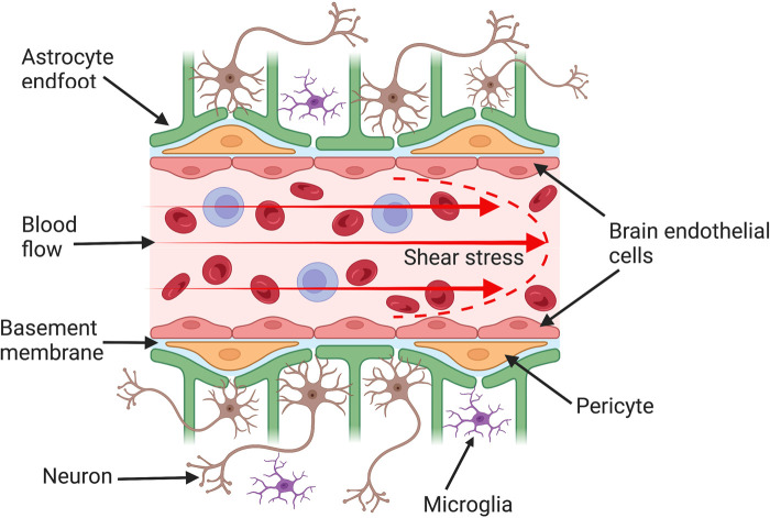
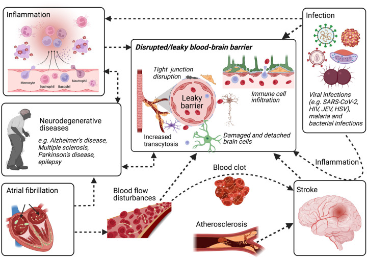
A healthy brain is protected by the blood–brain barrier (BBB), which is formed by the endothelial cells that line brain capillaries. The BBB plays an extremely important role in supporting normal neuronal function by maintaining the homeostasis of the brain microenvironment and restricting pathogen and toxin entry to the brain. Dysfunction of this highly complex and regulated structure can be life threatening. BBB dysfunction is implicated in many neurological diseases such as stroke, Alzheimer's disease, multiple sclerosis, and brain infections. Among other mechanisms, inflammation and/or flow disturbances are major causes of BBB dysfunction in neurological infections and diseases. In particular, in ischaemic stroke, both inflammation and flow disturbances contribute to BBB disruption, leading to devastating consequences. While a transient or minor disruption to the barrier function could be tolerated, chronic or a total breach of the barrier can result in irreversible brain damage. It is worth noting that timing and extent of BBB disruption play an important role in the process of any repair of brain damage and treatment strategies. This review evaluates and summarises some of the latest research on the role of the BBB during neurological disease and infection with a focus on the effects of inflammation and flow disturbances on the BBB. The BBB's crucial role in protecting the brain is also the bottleneck in central nervous system drug development. Therefore, innovative strategies to carry therapeutics across the BBB and novel models to screen drugs, and to study the complex, overlapping mechanisms of BBB disruption are urgently needed.
Overview
The claims and statistics of this paper haven’t been checked yet.
Run the check — free, no account needed. It reads the open-access text and checks the sources it cites.
Integrity signals
Registry
Cites
Statistics
Figures
Verification
Claims the paper makes
The claims and statistics of this paper haven’t been checked yet.
Run the check — free, no account needed. It reads the open-access text and checks the sources it cites.
Claims other papers make about this one
Each is checked against this paper’s own text: does it say what the citing paper says it does?
In the citing paper
“Owing to their intimate association with cerebral vasculature, astrocytes are among the first cells to encounter circulating factors that traverse a compromised BBB (Patabendige and Janigro 2023).”
From LDL Cholesterol Modulates Astrocyte Metabolism, Lipid Handling, and Morphology: Evidence From In Vitro and In Vivo Models (2026) · cites this paper as Patabendige and Janigro 2023
Self-check
Percentages, sums and reported differences re-computed from the paper’s own numbers.
The self-check needs the paper’s full text.
Citations
Cites
2 retracted
RETRACTED ARTICLE: Pericyte loss influences Alzheimer-like neurodegeneration in mice
Sagare, A. P., Bell, R. D., Zhao, Z., Ma, Q., Winkler, E. A., Ramanathan, A., & Zlokovic, B. V. (2013) · Nature Communications · doi:10.1038/ncomms3932 · Cited by 589
RetractedMontagne, A., Nikolakopoulou, A. M., Zhao, Z., Sagare, A. P., Si, G., Lazic, D., Barnes, S. R., Daianu, M., Ramanathan, A., Go, A., et al. (2018) · Nature Medicine · doi:10.1038/nm.4482 · Cited by 344
RetractedAstrocyte-endothelial interactions at the blood-brain barrier
Abbott, N. J., Rönnbäck, L., & Hansson, E. (2006) · Nature Reviews Neuroscience · doi:10.1038/nrn1824 · Cited by 5,595
Pericytes regulate the blood–brain barrier
Armulik, A., Genové, G., Mäe, M., Nisancioglu, M. H., Wallgard, E., Niaudet, C., He, L., Norlin, J., Lindblom, P., Strittmatter, K., et al. (2010) · Nature · doi:10.1038/nature09522 · Cited by 2,843
Pericytes are required for blood–brain barrier integrity during embryogenesis
Daneman, R., Zhou, L., Kebede, A. A., & Barres, B. A. (2010) · Nature · doi:10.1038/nature09513 · Cited by 2,128
Blood-brain barrier breakdown in the aging human hippocampus
Montagne, A., Barnes, S. R., Sweeney, M. D., Halliday, M. R., Sagare, A. P., Zhao, Z., Toga, A. W., Jacobs, R. E., Liu, C. Y., Amezcua, L., et al. (2015) · Neuron · doi:10.1016/j.neuron.2014.12.032 · Cited by 2,102
Meinhardt, J., Radke, J., Dittmayer, C., Franz, J., Thomas, C., Mothes, R., Laue, M., Schneider, J., Brünink, S., Greuel, S., et al. (2021) · Nature Neuroscience · doi:10.1038/s41593-020-00758-5 · Cited by 1,458
Bell, R. D., Winkler, E. A., Sagare, A. P., Singh, I., LaRue, B., Deane, R., & Zlokovic, B. V. (2010) · Neuron · doi:10.1016/j.neuron.2010.09.043 · Cited by 1,448
APOE4 leads to blood–brain barrier dysfunction predicting cognitive decline
Montagne, A., Nation, D. A., Sagare, A. P., Barisano, G., Sweeney, M. D., Chakhoyan, A., Pachicano, M., Joe, E., Nelson, A. R., D’Orazio, L. M., et al. (2020) · Nature · doi:10.1038/s41586-020-2247-3 · Cited by 1,281
Pericytes of the neurovascular unit: key functions and signaling pathways
Sweeney, M. D., Ayyadurai, S., & Zlokovic, B. V. (2016) · Nature Neuroscience · doi:10.1038/nn.4288 · Cited by 1,125
Research works printed but not linked: 169
- 2010, Structure and function of the blood–brain barrier, Neurobiol. Dis., 37, 13, 10.1016/j.nbd.2009.07.030 · DOI · Paper page
- 2022, A human-derived neurovascular unit in vitro model to study the effects of cellular cross-talk and soluble factors on barrier integrity, Front. Cell. Neurosci., 16, 1065193, 10.3389/fncel.2022.1065193 · DOI
- 2021, Astrocytes are necessary for blood–brain barrier maintenance in the adult mouse brain, Glia, 69, 436, 10.1002/glia.23908 · DOI
- 2007, Human astrocytes/astrocyte-conditioned medium and shear stress enhance the barrier properties of human brain microvascular endothelial cells, Brain Res., 1147, 39, 10.1016/j.brainres.2007.02.029 · DOI
- 2003, Synergistic effects of neurons and astrocytes on the differentiation of brain capillary endothelial cells in culture, J. Cell. Mol. Med., 7, 165, 10.1111/j.1582-4934.2003.tb00215.x · DOI
- 2018, A linked organ-on-chip model of the human neurovascular unit reveals the metabolic coupling of endothelial and neuronal cells, Nat. Biotechnol., 36, 865, 10.1038/nbt.4226 · DOI
- 2019, Brain perivascular macrophages: recent advances and implications in health and diseases, CNS Neurosci. Ther., 25, 1318, 10.1111/cns.13263 · DOI · Paper page
- 2017, The movers and shapers in immune privilege of the CNS, Nat. Immunol., 18, 123, 10.1038/ni.3666 · DOI · Paper page
- 1999, Glial cell line-derived neurotrophic factor induces barrier function of endothelial cells forming the blood–brain barrier, Biochem. Biophys. Res. Commun., 261, 108, 10.1006/bbrc.1999.0992 · DOI
- 2003, SSeCKS regulates angiogenesis and tight junction formation in blood–brain barrier, Nat. Med., 9, 900, 10.1038/nm889 · DOI
- 2005, Permeability studies on in vitro blood–brain barrier models: physiology, pathology, and pharmacology, Cell. Mol. Neurobiol., 25, 59, 10.1007/s10571-004-1377-8 · DOI
- 2012, The value of in vitro models of the blood–brain barrier and their uses, Altern. Lab. Anim., 40, 335, 10.1177/026119291204000606 · DOI
- 1999, The cell biology of the blood–brain barrier, Annu. Rev. Neurosci., 22, 11, 10.1146/annurev.neuro.22.1.11 · DOI
- 2013, A detailed method for preparation of a functional and flexible blood–brain barrier model using porcine brain endothelial cells, Brain Res., 1521, 16, 10.1016/j.brainres.2013.04.006 · DOI
- 1997, Induction of various blood–brain barrier properties in non-neural endothelial cells by close apposition to co-cultured astrocytes, Glia, 19, 13, 10.1002/(SICI)1098-1136(199701)19:1<13::AID-GLIA2>3.0.CO;2-B · DOI
- 2001, Mechanisms of glucose transport at the blood–brain barrier: an in vitro study, Brain Res., 904, 20, 10.1016/S0006-8993(01)02418-0 · DOI
- 2012, In vitro evidence for the brain glutamate efflux hypothesis: brain endothelial cells cocultured with astrocytes display a polarized brain-to-blood transport of glutamate, Glia, 60, 882, 10.1002/glia.22321 · DOI
- 2016, Astrocyte-mediated regulation of multidrug resistance p-glycoprotein in fetal and neonatal brain endothelial cells: age-dependent effects, Physiol. Rep., 4, e12853, 10.14814/phy2.12853 · DOI
- 2002, Compartmentalized coculture of rat brain endothelial cells and astrocytes: a syngenic model to study the blood–brain barrier, J. Neurosci. Methods, 121, 21, 10.1016/S0165-0270(02)00225-X · DOI
- 2011, Pericytes: developmental, physiological, and pathological perspectives, problems, and promises, Dev. Cell, 21, 193, 10.1016/j.devcel.2011.07.001 · DOI · Paper page
- 2015, Regional blood flow in the normal and ischemic brain is controlled by arteriolar smooth muscle cell contractility and not by capillary pericytes, Neuron, 87, 95, 10.1016/j.neuron.2015.06.001 · DOI
- 2017, Pericyte degeneration leads to neurovascular uncoupling and limits oxygen supply to brain, Nat. Neurosci., 20, 406, 10.1038/nn.4489 · DOI · Paper page
- 2018, Unique and shared inflammatory profiles of human brain endothelia and pericytes, J. Neuroinflammation, 15, 138, 10.1186/s12974-018-1167-8 · DOI
- 2015, Involvement of platelet-derived growth factor receptor β in fibrosis through extracellular matrix protein production after ischemic stroke, Exp. Neurol., 264, 127, 10.1016/j.expneurol.2014.12.007 · DOI
- 2019, Pericyte loss leads to circulatory failure and pleiotrophin depletion causing neuron loss, Nat. Neurosci., 22, 1089, 10.1038/s41593-019-0434-z · DOI
- 2015, Brain vascular pericytes following ischemia have multipotential stem cell activity to differentiate into neural and vascular lineage cells, Stem Cells, 33, 1962, 10.1002/stem.1977 · DOI
- 2016, Brain pericytes serve as microglia-generating multipotent vascular stem cells following ischemic stroke, J. Neuroinflammation, 13, 57, 10.1186/s12974-016-0523-9 · DOI
- 2007, An intimate interplay between precocious, migrating pericytes and endothelial cells governs human fetal brain angiogenesis, Angiogenesis, 10, 35, 10.1007/s10456-006-9061-x · DOI · Paper page
- 2001, Lack of pericytes leads to endothelial hyperplasia and abnormal vascular morphogenesis, J. Cell Biol., 153, 543, 10.1083/jcb.153.3.543 · DOI
- 1999, Role of PDGF-B and PDGFR-beta in recruitment of vascular smooth muscle cells and pericytes during embryonic blood vessel formation in the mouse, Development, 126, 3047, 10.1242/dev.126.14.3047 · DOI
- 2018, Blood–brain barrier dysfunction in ischemic stroke: targeting tight junctions and transporters for vascular protection, Am. J. Physiol. Cell Physiol., 315, C343, 10.1152/ajpcell.00095.2018 · DOI
- 2019, Profiling the mouse brain endothelial transcriptome in health and disease models reveals a core blood–brain barrier dysfunction module, Nat. Neurosci., 22, 1892, 10.1038/s41593-019-0497-x · DOI
- 2018, Blood–brain barrier breakdown in Alzheimer disease and other neurodegenerative disorders, Nat. Rev. Neurol., 14, 133, 10.1038/nrneurol.2017.188 · DOI · Paper page
- 2016, Immune cell trafficking across the barriers of the central nervous system in multiple sclerosis and stroke, Biochim. Biophys. Acta, 1862, 461, 10.1016/j.bbadis.2015.10.018 · DOI · Paper page
- 2018, Vascular pathology in multiple sclerosis: reframing pathogenesis around the blood–brain barrier, J. Neurol. Neurosurg. Psychiatry, 89, 42, 10.1136/jnnp-2017-316011 · DOI
- 2016, Blood–brain barrier, bulk flow, and interstitial clearance in epilepsy, J. Neurosci. Methods, 260, 118, 10.1016/j.jneumeth.2015.06.011 · DOI
- 2019, Breakdown of blood brain barrier as a mechanism of post-traumatic epilepsy, Neurobiol. Dis., 123, 20, 10.1016/j.nbd.2018.06.022 · DOI
- 2006, The blood–brain barrier and epilepsy, Epilepsia, 47, 1761, 10.1111/j.1528-1167.2006.00817.x · DOI
- 2018, Brain microvascular endothelial-astrocyte cell responses following Japanese encephalitis virus infection in an in vitro human blood–brain barrier model, Mol. Cell Neurosci., 89, 60, 10.1016/j.mcn.2018.04.002 · DOI
- 2020, Astrocyte- and neuron-derived CXCL1 drives neutrophil transmigration and blood–brain barrier permeability in viral encephalitis, Cell Rep., 32, 108150, 10.1016/j.celrep.2020.108150 · DOI
- 2016, The interleukin-1 balance during encephalitis is associated with clinical severity, blood–brain barrier permeability, neuroimaging changes, and disease outcome, J. Infect. Dis., 213, 1651, 10.1093/infdis/jiv771 · DOI
- 2021, Interactions of SARS-CoV-2 with the blood–brain barrier, Int. J. Mol. Sci., 22, 2681, 10.3390/ijms22052681 · DOI
- 2020, Cytokine release syndrome-associated encephalopathy in patients with COVID-19, Eur. J. Neurol., 28, 248, 10.1111/ene.14491 · DOI
- 2020, The SARS-CoV-2 spike protein alters barrier function in 2D static and 3D microfluidic in-vitro models of the human blood–brain barrier, Neurobiol. Dis., 146, 105131, 10.1016/j.nbd.2020.105131 · DOI
- 2006, The blood–brain barrier in neuroAIDS, Curr. HIV Res., 4, 259, 10.2174/157016206777709447 · DOI
- 2022, Cerebral microvascular endothelial glycocalyx damage, its implications on the blood–brain barrier and a possible contributor to cognitive impairment, Brain Res., 1780, 147804, 10.1016/j.brainres.2022.147804 · DOI
- 2012, The crossroads of neuroinflammation in infectious diseases: endothelial cells and astrocytes, Trends Parasitol., 28, 311, 10.1016/j.pt.2012.05.008 · DOI
- 2020, Parasite histones are toxic to brain endothelium and link blood barrier breakdown and thrombosis in cerebral malaria, Blood Adv., 4, 2851, 10.1182/bloodadvances.2019001258 · DOI
- 2009, Validation of serum markers for blood–brain barrier disruption in traumatic brain injury, J. Neurotrauma, 26, 1497, 10.1089/neu.2008.0738 · DOI
- 2021, Blood–brain barrier disruption in atrial fibrillation: a potential contributor to the increased risk of dementia and worsening of stroke outcomes?, Open Biol., 11, 200396, 10.1098/rsob.200396 · DOI
- 2016, Junctional proteins of the blood–brain barrier: new insights into function and dysfunction, Tissue Barriers, 4, e1154641, 10.1080/21688370.2016.1154641 · DOI
- 2019, Transcytosis at the blood–brain barrier, Curr. Opin. Neurobiol., 57, 32, 10.1016/j.conb.2018.12.014 · DOI
- 2019, Intracellular transport and regulation of transcytosis across the blood–brain barrier, Cell. Mol. Life Sci., 76, 1081, 10.1007/s00018-018-2982-x · DOI
- 2020, In vitro modeling of blood–brain barrier and interface functions in neuroimmune communication, Fluids Barriers CNS, 17, 26, 10.1186/s12987-020-00187-3 · DOI
- 2019, Global brain inflammation in stroke, Lancet. Neurol., 18, 1058, 10.1016/S1474-4422(19)30078-X · DOI
- 2017, The role of neurogenic inflammation in blood–brain barrier disruption and development of cerebral oedema following acute central nervous system (CNS) injury, Int. J. Mol. Sci., 18, 1788, 10.3390/ijms18081788 · DOI
- 2007, Therapeutic targeting of leukocyte trafficking across the blood–brain barrier, Inflamm. Allergy Drug Targets, 6, 210, 10.2174/187152807783334328 · DOI
- 2006, Regulation of immune cell entry into the central nervous system, Results Probl. Cell Differ., 43, 259, 10.1007/400_020 · DOI
- 2000, Interleukin-1beta -induced changes in blood–brain barrier permeability, apparent diffusion coefficient, and cerebral blood volume in the rat brain: a magnetic resonance study, J. Neurosci., 20, 8153, 10.1523/JNEUROSCI.20-21-08153.2000 · DOI
- 2009, Antagonism of peripheral inflammation reduces the severity of status epilepticus, Neurobiol. Dis., 33, 171, 10.1016/j.nbd.2008.10.002 · DOI
- 2008, Natalizumab: targeting alpha4-integrins in multiple sclerosis, Neurodegener. Dis., 5, 16, 10.1159/000109933 · DOI
- 2016, ABC transporters at the blood–brain barrier, Expert. Opin. Drug Metab. Toxicol., 12, 499, 10.1517/17425255.2016.1168804 · DOI
- 2014, The role of blood–brain barrier transporters in pathophysiology and pharmacotherapy of stroke, Curr. Pharm. Des., 20, 1510, 10.2174/13816128113199990465 · DOI
- 2015, Establishment and dysfunction of the blood–brain barrier, Cell, 163, 1064, 10.1016/j.cell.2015.10.067 · DOI · Paper page
- 2012, Surgical decompression for cerebral oedema in acute ischaemic stroke, Cochrane Database Syst. Rev., 1, Cd003435, 10.1002/14651858.CD003435.pub2 · DOI
- 2006, Factors influencing hemorrhagic transformation in ischemic stroke: a clinicopathological comparison, Eur. J. Neurol., 13, 1251, 10.1111/j.1468-1331.2006.01489.x · DOI
- 2016, Haemorrhagic transformation in ischaemic stroke is more frequent than clinically suspected - a neuropathological study, J. Neurol. Sci., 368, 4, 10.1016/j.jns.2016.06.055 · DOI
- 2011, Neurological complications of acute ischaemic stroke, Lancet Neurol., 10, 357, 10.1016/S1474-4422(10)70313-6 · DOI
- 2019, Neuroinflammatory mechanisms of blood–brain barrier damage in ischemic stroke, Am. J. Physiol. Cell Physiol., 316, C135, 10.1152/ajpcell.00136.2018 · DOI · Paper page
- 2020, Mechanisms in blood–brain barrier opening and metabolism-challenged cerebrovascular ischemia with emphasis on ischemic stroke, Metab Brain Dis., 35, 851, 10.1007/s11011-020-00573-8 · DOI
- 2016, Implications of MMP9 for blood brain barrier disruption and hemorrhagic transformation following ischemic stroke, Front. Cell. Neurosci., 10, 56, 10.3389/fncel.2016.00056 · DOI
- 2018, Autophagy- and MMP-2/9-mediated reduction and redistribution of ZO-1 contribute to hyperglycemia-increased blood–brain barrier permeability during early reperfusion in stroke, Neuroscience, 377, 126, 10.1016/j.neuroscience.2018.02.035 · DOI
- 2001, Dynamic platelet accumulation at the site of the occluded middle cerebral artery and in downstream microvessels is associated with loss of microvascular integrity after embolic middle cerebral artery occlusion, Brain Res., 912, 181, 10.1016/S0006-8993(01)02735-4 · DOI
- 2016, Matrix metalloproteinases in the brain and blood–brain barrier: versatile breakers and makers, J. Cereb. Blood Flow Metab., 36, 1481, 10.1177/0271678X16655551 · DOI · Paper page
- 2018, Immune responses in stroke: how the immune system contributes to damage and healing after stroke and how this knowledge could be translated to better cures?, Immunology, 154, 363, 10.1111/imm.12918 · DOI
- 2012, Inflammatory cell trafficking across the blood–brain barrier: chemokine regulation and in vitro models, Immunol. Rev., 248, 228, 10.1111/j.1600-065X.2012.01127.x · DOI
- 2017, Blood–brain barrier leakage increases with small vessel disease in acute ischemic stroke, Neurology, 89, 2143, 10.1212/WNL.0000000000004677 · DOI
- 2013, Ultra-sensitive molecular MRI of vascular cell adhesion molecule-1 reveals a dynamic inflammatory penumbra after strokes, Stroke, 44, 1988, 10.1161/STROKEAHA.111.000544 · DOI
- 2016, Molecular magnetic resonance imaging discloses endothelial activation after transient ischaemic attack, Brain, 140, 146, 10.1093/brain/aww260 · DOI
- 2015, Viral infection of the central nervous system and neuroinflammation precede blood–brain barrier disruption during Japanese encephalitis virus infection, J. Virol., 89, 5602, 10.1128/JVI.00143-15 · DOI
- 2011, Breaking down the barrier: the effects of HIV-1 on the blood–brain barrier, Brain Res., 1399, 96, 10.1016/j.brainres.2011.05.015 · DOI · Paper page
- 1999, Microglial and astrocyte chemokines regulate monocyte migration through the blood–brain barrier in human immunodeficiency virus-1 encephalitis, Am. J. Pathol., 155, 1599, 10.1016/S0002-9440(10)65476-4 · DOI
- 2022, Role of aging in blood–brain barrier dysfunction and susceptibility to SARS-CoV-2 infection: impacts on neurological symptoms of COVID-19, Fluids Barriers CNS, 19, 63, 10.1186/s12987-022-00357-5 · DOI
- 2004, Proinflammatory cytokines and chemokines in humans with Japanese encephalitis, J. Infect. Dis., 190, 1618, 10.1086/423328 · DOI
- 2006, A preliminary neuropathological study of Japanese encephalitis in humans and a mouse model, Trans. R. Soc. Trop. Med. Hyg., 100, 1135, 10.1016/j.trstmh.2006.02.008 · DOI
- 2014, Infection of pericytes in vitro by Japanese encephalitis virus disrupts the integrity of the endothelial barrier, J. Virol., 88, 1150, 10.1128/JVI.02738-13 · DOI
- 2020, Neuropathology of patients with COVID-19 in Germany: a post-mortem case series, Lancet Neurol., 19, 919, 10.1016/S1474-4422(20)30308-2 · DOI · Paper page
- 2020, Central nervous system involvement by severe acute respiratory syndrome coronavirus-2 (SARS-CoV-2), J. Med. Virol., 92, 699, 10.1002/jmv.25915 · DOI · Paper page
- 2022, Neurological manifestations of COVID-19 in adults and children, Brain, awac332, 10.1093/brain/awac332 · DOI
- 2022, Neurovascular injury with complement activation and inflammation in COVID-19, Brain, 145, 2555, 10.1093/brain/awac151 · DOI
- 2022, Markers of blood–brain barrier disruption increase early and persistently in COVID-19 patients with neurological manifestations, Front. Immunol., 13, 1070379, 10.3389/fimmu.2022.1070379 · DOI
- 2020, SARS-CoV-2 cell entry depends on ACE2 and TMPRSS2 and is blocked by a clinically proven protease inhibitor, Cell, 181, 271, 10.1016/j.cell.2020.02.052 · DOI · Paper page
- 2020, The spatial and cell-type distribution of SARS-CoV-2 receptor ACE2 in the human and mouse brains, Front. Neurol., 11, 573095, 10.3389/fneur.2020.573095 · DOI
- 2021, The SARS-CoV-2 main protease Mpro causes microvascular brain pathology by cleaving NEMO in brain endothelial cells, Nat. Neurosci., 24, 1522, 10.1038/s41593-021-00926-1 · DOI
- 2021, Neuroinvasion of SARS-CoV-2 in human and mouse brain, J. Exp. Med., 218, 10.1084/jem.20202135 · DOI · Paper page
- 2022, The blood–brain barrier is dysregulated in COVID-19 and serves as a CNS entry route for SARS-CoV-2, Stem Cell Rep., 17, 307, 10.1016/j.stemcr.2021.12.011 · DOI
- 2022, SARS-CoV-2 productively infects human brain microvascular endothelial cells, J. Neuroinflammation, 19, 149, 10.1186/s12974-022-02514-x · DOI
- 2021, Spike proteins of SARS-CoV-2 induce pathological changes in molecular delivery and metabolic function in the brain endothelial cells, Viruses, 13, 10.3390/v13102021 · DOI
- 2021, SARS-CoV-2 crosses the blood–brain barrier accompanied with basement membrane disruption without tight junctions alteration, Signal Transduct. Target Ther., 6, 337, 10.1038/s41392-021-00719-9 · DOI
- 2021, SARS-CoV-2 poorly replicates in cells of the human blood–brain barrier without associated deleterious effects, Front. Immunol., 12, 697329, 10.3389/fimmu.2021.697329 · DOI
- 2021, Astrocyte activation in neurovascular damage and repair following ischaemic stroke, Int. J. Mol. Sci., 22, 4280, 10.3390/ijms22084280 · DOI
- 2022, Shared inflammatory pathology of stroke and COVID-19, Int. J. Mol. Sci., 23, 5150, 10.3390/ijms23095150 · DOI
- 2022, Selective brain entry of lipid nanoparticles in haemorrhagic stroke is linked to biphasic blood–brain barrier disruption, Theranostics, 12, 4477, 10.7150/thno.72167 · DOI
- 2017, Evolution of blood-brain-barrier permeability after acute ischemic stroke, PLoS One, 12, e0171558, 10.1371/journal.pone.0171558 · DOI
- 2020, Late changes in blood–brain barrier permeability in a rat tMCAO model of stroke detected by gadolinium-enhanced MRI, Neurol. Res., 42, 844, 10.1080/01616412.2020.1786637 · DOI
- 2011, Specific role of tight junction proteins claudin-5, occludin, and ZO-1 of the blood–brain barrier in a focal cerebral ischemic insult, J. Mol. Neurosci., 44, 130, 10.1007/s12031-011-9496-4 · DOI · Paper page
- 2022, Glycocalyx is critical for blood–brain barrier integrity by suppressing caveolin1-dependent endothelial transcytosis following ischemic stroke, Brain Pathol., 32, e13006, 10.1111/bpa.13006 · DOI
- 2018, Blast exposure elicits blood–brain barrier disruption and repair mediated by tight junction integrity and nitric oxide dependent processes, Sci. Rep., 8, 11344, 10.1038/s41598-018-29341-6 · DOI
- 2021, Longitudinal characterization of blood–brain barrier permeability after experimental traumatic brain injury by in vivo 2-photon microscopy, J. Neurotrauma, 38, 399, 10.1089/neu.2020.7271 · DOI
- 2021, A validated quantitative method for the assessment of neuroprotective barrier impairment in neurodegenerative disease models, J. Neurochem., 158, 807, 10.1111/jnc.15119 · DOI
- 2011, The role of the blood–brain barrier during Venezuelan equine encephalitis virus infection, J. Virol., 85, 10682, 10.1128/JVI.05032-11 · DOI
- 1985, The biphasic opening of the blood–brain barrier to proteins following temporary middle cerebral artery occlusion, Acta Neuropathol., 68, 122, 10.1007/BF00688633 · DOI
- 1999, Biphasic opening of the blood–brain barrier following transient focal ischemia: effects of hypothermia, Can. J. Neurol. Sci., 26, 298, 10.1017/S0317167100000421 · DOI
- 2005, Hyperbaric oxygen reduces blood–brain barrier damage and edema after transient focal cerebral ischemia, Stroke, 36, 1679, 10.1161/01.STR.0000173408.94728.79 · DOI
- 2009, Post-ischemic blood–brain barrier leakage in rats: one-week follow-up by MRI, Brain Res., 1280, 158, 10.1016/j.brainres.2009.05.025 · DOI
- 2008, The blood–brain barrier is continuously open for several weeks following transient focal cerebral ischemia, Neuroscience, 153, 175, 10.1016/j.neuroscience.2008.02.012 · DOI
- 2008, Dynamic changes in vascular permeability, cerebral blood volume, vascular density, and size after transient focal cerebral ischemia in rats: evaluation with contrast-enhanced magnetic resonance imaging, J. Cereb. Blood Flow Metab., 28, 1491, 10.1038/jcbfm.2008.42 · DOI
- 2013, Early inhibition of MMP activity in ischemic rat brain promotes expression of tight junction proteins and angiogenesis during recovery, J. Cereb. Blood Flow Metab., 33, 1104, 10.1038/jcbfm.2013.56 · DOI · Paper page
- 2012, Matrix metalloproteinase-2-mediated occludin degradation and caveolin-1-mediated claudin-5 redistribution contribute to blood–brain barrier damage in early ischemic stroke stage, J. Neurosci., 32, 3044, 10.1523/JNEUROSCI.6409-11.2012 · DOI · Paper page
- 2001, Effects of matrix metalloproteinase-9 gene knock-out on the proteolysis of blood–brain barrier and white matter components after cerebral ischemia, J. Neurosci., 21, 7724, 10.1523/JNEUROSCI.21-19-07724.2001 · DOI · Paper page
- 2014, Stepwise recruitment of transcellular and paracellular pathways underlies blood–brain barrier breakdown in stroke, Neuron, 82, 603, 10.1016/j.neuron.2014.03.003 · DOI · Paper page
- 2013, Blood–brain barrier breakdown after embolic stroke in rats occurs without ultrastructural evidence for disrupting tight junctions, PLoS One, 8, e56419, 10.1371/journal.pone.0056419 · DOI
- 2016, Ultrastructural analysis of blood–brain barrier breakdown in the peri-infarct zone in young adult and aged mice, J. Cereb. Blood Flow Metab., 36, 413, 10.1177/0271678X15608396 · DOI
- 2021, Caveolae-mediated endothelial transcytosis across the blood–Brain barrier in acute ischemic stroke, J. Clin. Med., 10, 3795, 10.3390/jcm10173795 · DOI
- 2017, The blood–brain barrier after stroke: structural studies and the role of transcytotic vesicles, J. Cereb. Blood Flow Metab., 37, 456, 10.1177/0271678X16629976 · DOI
- 2019, Blood–brain barrier breakdown is an early biomarker of human cognitive dysfunction, Nat. Med., 25, 270, 10.1038/s41591-018-0297-y · DOI · Paper page
- 2016, Blood–brain barrier leakage in patients with early Alzheimer disease, Radiology, 281, 527, 10.1148/radiol.2016152244 · DOI · Paper page
- 1998, Shear stress and the endothelium, Kidney Int. Suppl., 67, S100, 10.1046/j.1523-1755.1998.06720.x · DOI
- 2020, Fluid shear stress sensing by the endothelial layer, Front. Physiol., 11, 861, 10.3389/fphys.2020.00861 · DOI
- 2020, In vitro models of the blood–brain barrier: tools in translational medicine, Front. Med. Technol., 2, 623950, 10.3389/fmedt.2020.623950 · DOI
- 1995, Chronic in vitro flow promotes ultrastructural differentiation of endothelial cells, Endothelium, 3, 21, 10.3109/10623329509024655 · DOI
- 2016, Endothelial cell alignment as a result of anisotropic strain and flow induced shear stress combinations, Sci. Rep., 6, 29510, 10.1038/srep29510 · DOI
- 2017, Flow induced adherens junction remodeling driven by cytoskeletal forces, Exp. Cell Res., 359, 327, 10.1016/j.yexcr.2017.08.009 · DOI
- 2022, Wall shear stress alteration: a local risk factor of atherosclerosis, Curr. Atheroscler. Rep., 24, 143, 10.1007/s11883-022-00993-0 · DOI
- 2011, The role of shear stress in blood–brain barrier endothelial physiology, BMC Neurosci., 12, 40, 10.1186/1471-2202-12-40 · DOI
- 2002, Mechanisms of endothelial survival under shear stress, Endothelium, 9, 89, 10.1080/10623320212004 · DOI
- 2006, Side by side comparison between dynamic versus static models of blood–brain barrier in vitro: a permeability study, Brain Res., 1109, 1, 10.1016/j.brainres.2006.06.027 · DOI
- Desai, S.Y., McAllister, M.A., Goodrich, K., Mayberg, M.M. and Janigro, D. (2001) Gene Expression Changes and Progression to a BBB Phenotype in a Dynamic Model of the BBB. In Blood–Brain Barrier (Kobiler, D., Lustig, S. and Shapira, S., eds), Springer, Boston, MA10.1007/978-1-4615-0579-2_6 · DOI
- 2003, Effects of pulsatile shear stress on nitric oxide production and endothelial cell nitric oxide synthase expression by ovine fetoplacental artery endothelial cells, Biol. Reprod., 69, 1053, 10.1095/biolreprod.102.013474 · DOI
- 1995, Shear stress induces ATP-independent transient nitric oxide release from vascular endothelial cells, measured directly with a porphyrinic microsensor, Circ. Res., 77, 284, 10.1161/01.RES.77.2.284 · DOI
- 2016, Shear-induced nitric oxide production by endothelial cells, Biophys. J., 111, 208, 10.1016/j.bpj.2016.05.034 · DOI
- 2022, Barriers to stroke prevention in atrial fibrillation: insights from the global anticoagulation roundtable, Int. J. Cardiol. Heart Vasc., 42, 101096, 10.1016/j.ijcha.2022.101096 · DOI
- 2022, Screening for atrial fibrillation to prevent stroke: a meta-analysis, Eur. Heart J. Open, 2, oeac044, 10.1093/ehjopen/oeac044 · DOI
- 2021, Global epidemiology of atrial fibrillation: an increasing epidemic and public health challenge, Int. J. Stroke, 16, 217, 10.1177/1747493019897870 · DOI
- 1998, Impact of atrial fibrillation on mortality, stroke, and medical costs, Arch. Intern. Med., 158, 229, 10.1001/archinte.158.3.229 · DOI
- GBD 2019 Stroke Collaborators, 2021, Global, regional, and national burden of stroke and its risk factors, 1990–2019: a systematic analysis for the Global Burden of Disease Study 2019, Lancet Neurol., 20, 795, 10.1016/S1474-4422(21)00252-0 · DOI
- 2021, 2020 ESC guidelines for the diagnosis and management of atrial fibrillation developed in collaboration with the European Association for Cardio-Thoracic Surgery (EACTS): the task force for the diagnosis and management of atrial fibrillation of the European Society of Cardiology (ESC) developed with the special contribution of the European Heart Rhythm Association (EHRA) of the ESC, Eur. Heart J., 42, 373, 10.1093/eurheartj/ehaa612 · DOI
- 2009, Mechanisms of thrombogenesis in atrial fibrillation: Virchow's triad revisited, Lancet, 373, 155, 10.1016/S0140-6736(09)60040-4 · DOI
- 2019, Circulating levels of biomarkers of cerebral injury in patients with atrial fibrillation, Am. J. Cardiol., 124, 1697, 10.1016/j.amjcard.2019.08.027 · DOI
- 2015, Endothelial (dys)function in lone atrial fibrillation, Curr. Pharm. Des., 21, 622, 10.2174/1381612820666140825143028 · DOI
- 2009, Endothelial dysfunction and pathophysiological correlates in atrial fibrillation, Heart, 95, 102, 10.1136/hrt.2007.135277 · DOI
- 2002, Downregulation of endocardial nitric oxide synthase expression and nitric oxide production in atrial fibrillation: potential mechanisms for atrial thrombosis and stroke, Circulation, 106, 2854, 10.1161/01.CIR.0000039327.11661.16 · DOI
- 2013, Atrial fibrillation-induced endothelial dysfunction improves after restoration of sinus rhythm, Int. J. Cardiol., 168, 1280, 10.1016/j.ijcard.2012.12.006 · DOI
- 2013, Atrial fibrillation pacing decreases intravascular shear stress in a New Zealand white rabbit model: implications in endothelial function, Biomech. Model. Mechanobiol., 12, 735, 10.1007/s10237-012-0437-0 · DOI
- 2019, The biophysical role of hemodynamics in the pathogenesis of cerebral aneurysm formation and rupture, Neurosurg. Focus, 47, E11, 10.3171/2019.4.FOCUS19232 · DOI
- 2019, Shear stress and aneurysms: a review, Neurosurg. Focus, 47, E2, 10.3171/2019.4.FOCUS19225 · DOI
- 2017, The neurovascular unit coming of age: a journey through neurovascular coupling in health and disease, Neuron, 96, 17, 10.1016/j.neuron.2017.07.030 · DOI · Paper page
- 2013, The pathobiology of vascular dementia, Neuron, 80, 844, 10.1016/j.neuron.2013.10.008 · DOI · Paper page
- 2006, Loss of flow induces leukocyte-mediated MMP/TIMP imbalance in dynamic in vitro blood–brain barrier model: role of pro-inflammatory cytokines, Am. J. Physiol. Cell Physiol., 291, C740, 10.1152/ajpcell.00516.2005 · DOI
- 2006, Loss of shear stress induces leukocyte-mediated cytokine release and blood–brain barrier failure in dynamic in vitro blood–brain barrier model, J. Cell Physiol., 206, 68, 10.1002/jcp.20429 · DOI
- 2003, Effects of transient loss of shear stress on blood–brain barrier endothelium: role of nitric oxide and IL-6, Brain Res., 977, 239, 10.1016/S0006-8993(03)02689-1 · DOI
- 2020, The global burden of neurological disorders: translating evidence into policy, Lancet Neurol., 19, 255, 10.1016/S1474-4422(19)30411-9 · DOI · Paper page
- 2017, NIH workshop report on the trans-agency blood-brain interface workshop 2016: exploring key challenges and opportunities associated with the blood, brain and their interface, Fluids Barriers CNS, 14, 12, 10.1186/s12987-017-0061-6 · DOI
- 2021, Post-capillary venules are the key locus for transcytosis-mediated brain delivery of therapeutic nanoparticles, Nat. Commun., 12, 4121, 10.1038/s41467-021-24323-1 · DOI · Paper page
- 2022, Variants of the adeno-associated virus serotype 9 with enhanced penetration of the blood–brain barrier in rodents and primates, Nat. Biomed. Eng., 6, 1257, 10.1038/s41551-022-00938-7 · DOI · Paper page
- 2022, Targeting organic cation transporters at the blood–brain barrier to treat ischemic stroke in rats, Exp. Neurol., 357, 114181, 10.1016/j.expneurol.2022.114181 · DOI
- 2023, Neuronal Serpina3n is an endogenous protector against blood brain barrier damage following cerebral ischemic stroke, J. Cereb. Blood Flow Metab, 43, 241, 10.1177/0271678X221113897 · DOI
- 2023, Editing a gateway for cell therapy across the blood–brain barrier, Brain, 146, 823, 10.1093/brain/awac393 · DOI
- 2022, Applications of focused ultrasound-mediated blood–brain barrier opening, Adv. Drug Deliv. Rev., 191, 114583, 10.1016/j.addr.2022.114583 · DOI · Paper page
Cited by
Sorted by citations
Gao, L., Wang, J., & Bi, Y. (2025) · International Journal of Nanomedicine · doi:10.2147/ijn.s549893 · Cited by 52
Kumar, S., Yadav, A., Verma, R. K., Kumar, A., Gupta, P. K., & Shukla, R. (2025) · Molecular Pharmaceutics · doi:10.1021/acs.molpharmaceut.5c00386 · Cited by 14
Baumart, G. J., Rodrigues, M. S., Sholl, J. N., de Sousa, A. C., Weber, A. F., Costa‐Beber, L. C., Farias, H. R., Costa, M. V., Peres, A. M., de Camargo, P. R., et al. (2026) · Journal of Neurochemistry · doi:10.1111/jnc.70403 · Cited by 2
Metadata
- Authors
- Patabendige, A., & Janigro, D.
- Affiliations
- 1Brain Barriers Research Group, Department of Biology, Edge Hill University, Ormskirk, U.K.
- 4Flocel Inc., Cleveland, OH, U.S.A.
- ORCID
- Patabendige, Adjanie
- Venue
- Biochemical Society Transactions
- Publisher
- Portland Press Ltd.
- Published
- 2023-03-17
- Volume
- 51
- Issue
- 2
- Pages
- 613-626
- ISSN
- 0300-5127
- 1470-8752
- PMC
- PMC10212550
- Cited by
- 112
- References
- 23
- Language
- en
About these checks
Registry, retraction, Cites and statistics results are computed, not judged, with one exception: a printed reference that no index lists is read once by a model before it counts as a citation error. Claims are extracted by a model and kept only when their quote appears verbatim in the text; each cited source is then checked, with a model’s help, for whether it says what the claim relies on. Those verdicts can be wrong — read the claim’s quote and the cited source before relying on one.
Statistics and arithmetic are recomputed, not judged.
Confidence bands are shown only for fields calibrated on our annotated test set. None is calibrated yet, so no band is shown.
What counts as a claim ›How we measure confidence ›How we check this ›
Are you an author, or do you see a problem with this page? Request a review
