

The DOI matches its Crossref record; 56 of the 56 references it cites were found in a registry or an index; none of the 52 sources its claims cite is checked for support yet.
Comprehensive pregnancy monitoring with a network of wireless, soft, and flexible sensors in high- and low-resource health settings
Open accessAbstract
Significance: Monitoring vital signs for laboring women and their fetuses is foundational to the delivery of obstetrical care; however, monitoring platforms for pregnancy have undergone little innovation over the last several decades with many low-income settings lacking basic access. We report a new time-synchronized, flexible, and wireless sensor system applicable across the entire continuum of antepartum and postpartum care that provides continuous, comprehensive, and noninvasive monitoring (heart rate, respiratory rate, and pulse oxygenation) compatible with a wide range of mobile devices. This system offers advanced features such as continuous blood pressure, uterine electrohysterography, and automated body position classification. We further demonstrate the performance of this new system among pregnant individuals ( n = 576) in both high-resource settings and low-resource care settings.
Overview
AI snapshot
Written from the paper’s full text.
TL;DR
The paper reports a time-synchronized, wireless, soft, flexible sensor network for comprehensive maternal–fetal pregnancy monitoring across antepartum and postpartum care. It was tested in 576 pregnant individuals in high-resource Chicago and low-resource Lusaka, with comparisons against gold-standard monitors for vital signs, fetal heart rate, uterine contraction, and continuous blood pressure surrogates.
Key findings
- The system uses chest, limb, and abdominal sensors: chest and limb sensors operate for 24 h, the abdominal sensor for 8 h; the three sensors are time-synchronized with 1-ms timing accuracy and an SD of 3.6 ms over 24 consecutive hours.
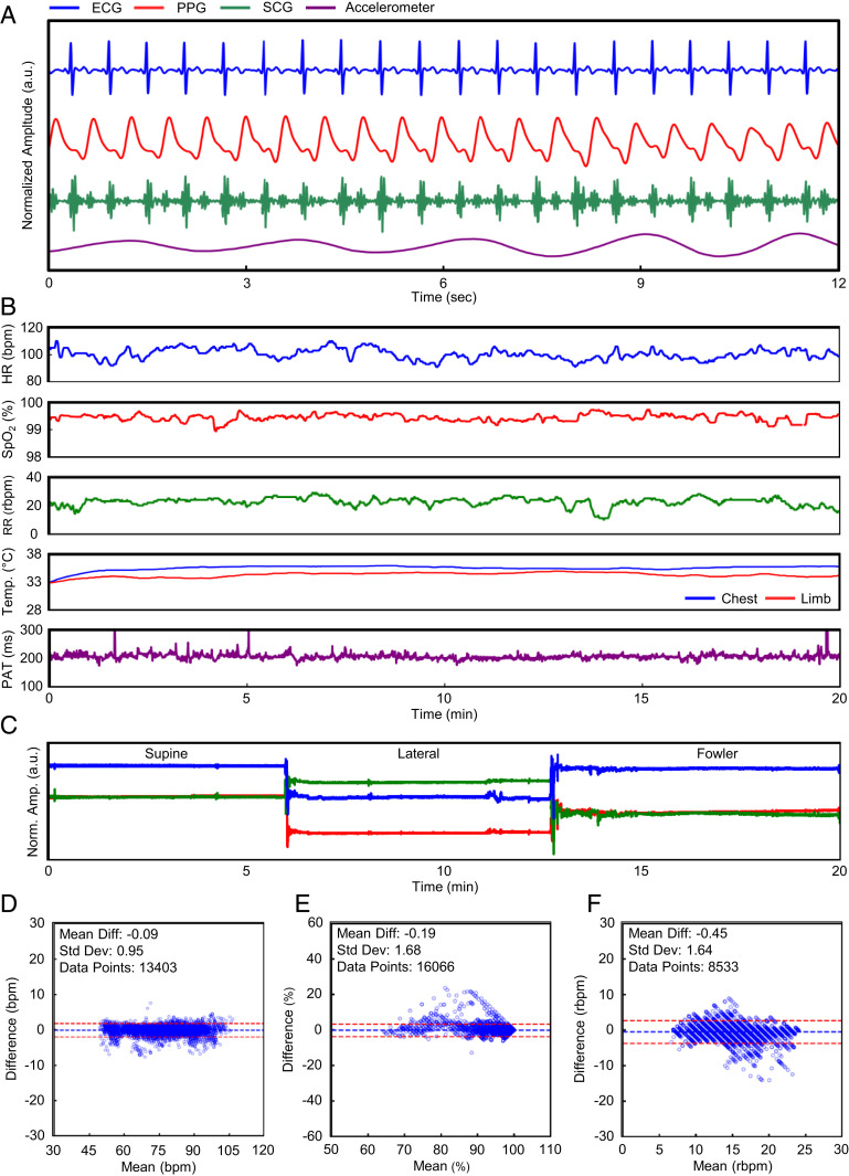
- In 13 healthy nonpregnant adults, compared with a Philips Intellivue MP50, mean differences were −0.09 bpm for HR (SD 0.95 bpm), −0.19% for SpO2 (SD 1.68%), and −0.45 breaths per minute for RR (SD 1.64 breaths per minute), within FDA guidelines for new cardiopulmonary monitoring platforms.
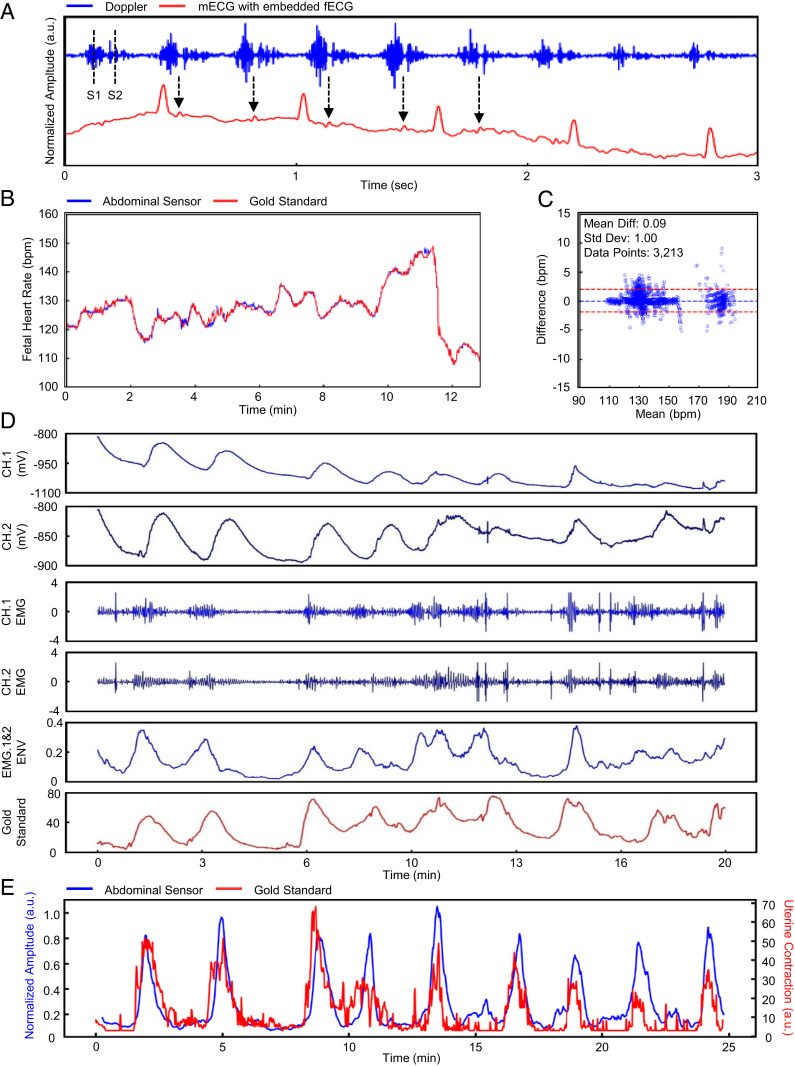
- Fetal heart rate from the abdominal sensor compared with gold-standard Doppler in n = 10 laboring women across 3,213 single data points had a mean difference of 0.1 bpm (SD 1 bpm).
- In the total cohort of 576 pregnant people, 91 (16%) were recruited in Chicago using all three sensors and 485 (84%) in Lusaka using maternal sensors only; more than 85% of surveyed laboring people (41 of 48 respondents) and all 10 surveyed obstetrical providers reported a positive or very positive experience.
Methods
- Design and sample: field trials/validation among pregnant women at 25–41 wk gestation (n = 576), including 91 at Prentice Women’s Hospital in Chicago (59 nonstress testing, 32 active labor) and 485 in Lusaka, Zambia, as part of the LABOR study; additional validation included n = 13 healthy nonpregnant adults, n = 10 laboring women for FHR, and n = 3 healthy subjects for cold-pressor BP testing.
- Sensor construction: four-layer flexible printed circuit boards for chest and limb sensors, two-layer for the abdominal sensor, packaged in silicone; chest sensor measures ECG, SCG, chest-wall movement for RR, and skin temperature; limb sensor measures PPG and skin temperature; abdominal sensor measures Doppler ultrasound, EHG, and ECG.
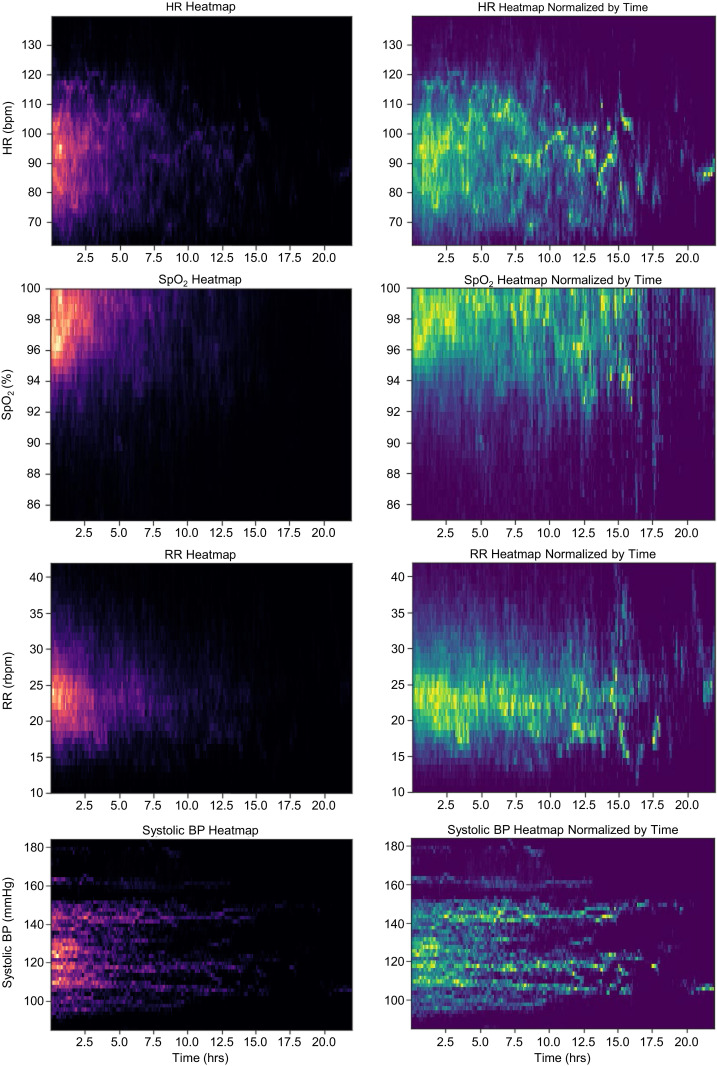
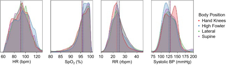
- Analysis: real-time algorithms on mobile devices derived HR, SpO2, RR, temperature, FHR, uterine contraction, PAT-based BP, and body position; Bland–Altman comparisons were made against gold-standard systems; body position was classified using a Gaussian mixture model with n = 8 clusters merged into four postures; time-series heat maps were generated for HR, RR, SpO2, and derived SBP in the low-resource cohort (n = 485).
Limitations
- The paper states that PAT-based BP measurements still require initial calibration with a gold-standard BP device, and this calibration step leads to further errors; further work is needed to validate PAT-based measurements for elevated BP following established guidelines with at least n = 85 subjects before wider clinical use.
- The paper states that further development efforts are targeting enhancement of fetal ECG capabilities of the system.
- Evident from the text: the low-resource cohort of 485 pregnant people in Lusaka used maternal sensors only (chest and limb), so fetal monitoring with the abdominal sensor was not validated in that setting; the abdominal sensor also has a shorter operation time (8 h) than the chest and limb sensors (24 h).
Model-written summary. Check it against the paper before relying on it.
Integrity signals
Registry
Cites
Statistics
Figures
Verification
Claims the paper makes
32 Not checked yet · 30 Own result · 16 No basis given · 6 Own interpretation · 2 Own method
A result the paper reports from its own data — it cites no other source for it, so there is none to check. The quote shows it is in the paper.
In the paper
“We demonstrate the sensor’s capability of detecting uterine contractions during labor.”
§ Comprehensive pregnancy monitoring with a network of wireless, soft, and flexible sensors in high- and low-resource health settings > Results > FHR and Uterine Contraction via a Soft, Flexible, and Wireless Abdominal Sensor.
boostedstates a cause- Asserted: passes — The authors put it forward as true.
- Checkable: passes — It can be checked against evidence.
- Load-bearing: passes — The paper’s conclusions depend on it.
- Based: passes — The paper gives a reason why it holds.
A result the paper reports from its own data — it cites no other source for it, so there is none to check. The quote shows it is in the paper.
In the paper
“In Fig. 2 D–F we demonstrate the agreement in HR, SpO2, and RR calculations between the experimental system and a commercially available gold-standard monitoring system (Intellivue MP50; Philips) operating concomitantly in a cohort of 13 healthy nonpregnant adults.”
§ Comprehensive pregnancy monitoring with a network of wireless, soft, and flexible sensors in high- and low-resource health settings > Results > Real-Time Measurement of Clinical Data for Maternal Health.
boostedn = 13- Asserted: passes — The authors put it forward as true.
- Checkable: passes — It can be checked against evidence.
- Load-bearing: passes — The paper’s conclusions depend on it.
- Based: passes — The paper gives a reason why it holds.
A result the paper reports from its own data — it cites no other source for it, so there is none to check. The quote shows it is in the paper.
In the paper
“Although intended for measuring maternal ECG, maternal HR, and uterine contraction, the abdominal sensor is also capable of capturing incidental fetal ECG, which coincides with fetal Doppler signal.”
§ Comprehensive pregnancy monitoring with a network of wireless, soft, and flexible sensors in high- and low-resource health settings > Results > FHR and Uterine Contraction via a Soft, Flexible, and Wireless Abdominal Sensor.
plainstates an association- Asserted: passes — The authors put it forward as true.
- Checkable: passes — It can be checked against evidence.
- Load-bearing: unclear — It is unclear whether the paper’s conclusions depend on it.
- Based: passes — The paper gives a reason why it holds.
A result the paper reports from its own data — it cites no other source for it, so there is none to check. The quote shows it is in the paper.
In the paper
“An additional 485 (84%) pregnant people were recruited in Lusaka, Zambia using the maternal sensors only (chest and limb).”
§ Comprehensive pregnancy monitoring with a network of wireless, soft, and flexible sensors in high- and low-resource health settings > Results > Clinical Validation and Deployments in Both High-Resource and Low-Resource Settings.
plain- Asserted: passes — The authors put it forward as true.
- Checkable: passes — It can be checked against evidence.
- Load-bearing: passes — The paper’s conclusions depend on it.
- Based: passes — The paper gives a reason why it holds.
A result the paper reports from its own data — it cites no other source for it, so there is none to check. The quote shows it is in the paper.
In the paper
“We further demonstrate the performance of this new system among pregnant individuals (n = 576) in both high-resource settings and low-resource care settings.”
§ Comprehensive pregnancy monitoring with a network of wireless, soft, and flexible sensors in high- and low-resource health settings > Abstract
boostedn = 576- Asserted: passes — The authors put it forward as true.
- Checkable: passes — It can be checked against evidence.
- Load-bearing: passes — The paper’s conclusions depend on it.
- Based: passes — The paper gives a reason why it holds.
A result the paper reports from its own data — it cites no other source for it, so there is none to check. The quote shows it is in the paper.
In the paper
“We present performance, feasibility, and usability of the sensors, both at the individual and population level, in the total cohort of 576 pregnant people.”
§ Comprehensive pregnancy monitoring with a network of wireless, soft, and flexible sensors in high- and low-resource health settings > Results > Clinical Validation and Deployments in Both High-Resource and Low-Resource Settings.
plainn = 576- Asserted: passes — The authors put it forward as true.
- Checkable: passes — It can be checked against evidence.
- Load-bearing: passes — The paper’s conclusions depend on it.
- Based: passes — The paper gives a reason why it holds.
A result the paper reports from its own data — it cites no other source for it, so there is none to check. The quote shows it is in the paper.
In the paper
“More than 85% of laboring people surveyed (41 out of n = 48 total respondents) indicated a positive or very positive experience with the wireless sensors.”
§ Comprehensive pregnancy monitoring with a network of wireless, soft, and flexible sensors in high- and low-resource health settings > Results > Clinical Validation and Deployments in Both High-Resource and Low-Resource Settings.
plainn = 48- Asserted: passes — The authors put it forward as true.
- Checkable: passes — It can be checked against evidence.
- Load-bearing: unclear — It is unclear whether the paper’s conclusions depend on it.
- Based: passes — The paper gives a reason why it holds.
The paper states this without a source or data behind it here, so there is nothing to check it against. The quote shows it is in the paper.
In the paper
“We note that the participants recruited in the low-resource setting were all actively in labor with imminent delivery.”
§ Comprehensive pregnancy monitoring with a network of wireless, soft, and flexible sensors in high- and low-resource health settings > Results > Clinical Validation and Deployments in Both High-Resource and Low-Resource Settings.
plain- Asserted: passes — The authors put it forward as true.
- Checkable: passes — It can be checked against evidence.
- Load-bearing: unclear — It is unclear whether the paper’s conclusions depend on it.
- Based: passes — The paper gives a reason why it holds.
A result the paper reports from its own data — it cites no other source for it, so there is none to check. The quote shows it is in the paper.
In the paper
“This includes 91 (16%) participants recruited at Prentice Women’s Hospital (Chicago, IL) using all three sensor systems (chest, limb, and abdomen).”
§ Comprehensive pregnancy monitoring with a network of wireless, soft, and flexible sensors in high- and low-resource health settings > Results > Clinical Validation and Deployments in Both High-Resource and Low-Resource Settings.
plain- Asserted: passes — The authors put it forward as true.
- Checkable: passes — It can be checked against evidence.
- Load-bearing: unclear — It is unclear whether the paper’s conclusions depend on it.
- Based: passes — The paper gives a reason why it holds.
A result the paper reports from its own data — it cites no other source for it, so there is none to check. The quote shows it is in the paper.
In the paper
“The sensor tracks expected FHR variation over time.”
§ Comprehensive pregnancy monitoring with a network of wireless, soft, and flexible sensors in high- and low-resource health settings > Results > FHR and Uterine Contraction via a Soft, Flexible, and Wireless Abdominal Sensor.
plainn = 10- Asserted: passes — The authors put it forward as true.
- Checkable: passes — It can be checked against evidence.
- Load-bearing: passes — The paper’s conclusions depend on it.
- Based: passes — The paper gives a reason why it holds.
A result the paper reports from its own data — it cites no other source for it, so there is none to check. The quote shows it is in the paper.
In the paper
“This monitoring system is able to automatically categorize pregnancy body positions with comprehensive vital signs monitoring.”
§ Comprehensive pregnancy monitoring with a network of wireless, soft, and flexible sensors in high- and low-resource health settings > Results > Real-Time Tracking of Maternal Body Position during Labor.
plainstates a cause- Asserted: passes — The authors put it forward as true.
- Checkable: passes — It can be checked against evidence.
- Load-bearing: passes — The paper’s conclusions depend on it.
- Based: passes — The paper gives a reason why it holds.
A result the paper reports from its own data — it cites no other source for it, so there is none to check. The quote shows it is in the paper.
In the paper
“As the majority of the monitoring period during labor did not extend beyond 5 h”
§ Comprehensive pregnancy monitoring with a network of wireless, soft, and flexible sensors in high- and low-resource health settings > Results > Time-Series Data of Vital Signs during Labor.
plainn = 485- Asserted: passes — The authors put it forward as true.
- Checkable: passes — It can be checked against evidence.
- Load-bearing: unclear — It is unclear whether the paper’s conclusions depend on it.
- Based: passes — The paper gives a reason why it holds.
A result the paper reports from its own data — it cites no other source for it, so there is none to check. The quote shows it is in the paper.
In the paper
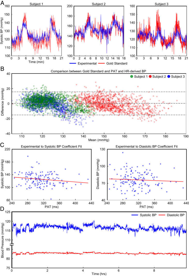
“In Fig. 4 C and D, we demonstrate the ability of our methods to capture measurements over 10 h of labor with our sensors after a single point calibration with a sphygmomanometer.”
§ Comprehensive pregnancy monitoring with a network of wireless, soft, and flexible sensors in high- and low-resource health settings > Results > Continuous Surrogates of Systolic BP Derived from PAT and Pulse Transit Time.
boostedstates a cause- Asserted: passes — The authors put it forward as true.
- Checkable: passes — It can be checked against evidence.
- Load-bearing: passes — The paper’s conclusions depend on it.
- Based: passes — The paper gives a reason why it holds.
A result the paper reports from its own data — it cites no other source for it, so there is none to check. The quote shows it is in the paper.
In the paper
“Herein, we demonstrate the derivation of a continuous surrogate of systolic BP (SBP) via PAT and HR without the need for clinical staff to initiate, collect, and then record any measurements after an initial calibration.”
§ Comprehensive pregnancy monitoring with a network of wireless, soft, and flexible sensors in high- and low-resource health settings > Results > Continuous Surrogates of Systolic BP Derived from PAT and Pulse Transit Time.
boostedstates a cause- Asserted: passes — The authors put it forward as true.
- Checkable: passes — It can be checked against evidence.
- Load-bearing: passes — The paper’s conclusions depend on it.
- Based: passes — The paper gives a reason why it holds.
A result the paper reports from its own data — it cites no other source for it, so there is none to check. The quote shows it is in the paper.
In the paper
“We present differences in average maternal vital signs in SI Appendix, Table S2 based on positional changes.”
- Also stated in: § Real-Time Tracking of Maternal Body Position during Labor. · boosted
“Small, but clear, differences are seen in maternal vital signs based on body position for HR, SpO2, RR, and SBP derived from PAT.”
§ Comprehensive pregnancy monitoring with a network of wireless, soft, and flexible sensors in high- and low-resource health settings > Results > Real-Time Tracking of Maternal Body Position during Labor.
boostedstates an association- Asserted: passes — The authors put it forward as true.
- Checkable: passes — It can be checked against evidence.
- Load-bearing: unclear — It is unclear whether the paper’s conclusions depend on it.
- Based: passes — The paper gives a reason why it holds.
- Also stated in: § Real-Time Tracking of Maternal Body Position during Labor. · boosted
A result the paper reports from its own data — it cites no other source for it, so there is none to check. The quote shows it is in the paper.
In the paper
“We automatically categorize these four common laboring positions based on the three-axis IMU outputs.”
§ Comprehensive pregnancy monitoring with a network of wireless, soft, and flexible sensors in high- and low-resource health settings > Results > Real-Time Tracking of Maternal Body Position during Labor.
plainstates a cause- Asserted: passes — The authors put it forward as true.
- Checkable: passes — It can be checked against evidence.
- Load-bearing: passes — The paper’s conclusions depend on it.
- Based: passes — The paper gives a reason why it holds.
A result the paper reports from its own data — it cites no other source for it, so there is none to check. The quote shows it is in the paper.
In the paper
“We derive an inverse relationship between PAT and BP measurements across the entire cohort of pregnant women in the low-resource setting.”
§ Comprehensive pregnancy monitoring with a network of wireless, soft, and flexible sensors in high- and low-resource health settings > Results > Continuous Surrogates of Systolic BP Derived from PAT and Pulse Transit Time.
plainstates an association- Asserted: passes — The authors put it forward as true.
- Checkable: passes — It can be checked against evidence.
- Load-bearing: passes — The paper’s conclusions depend on it.
- Based: passes — The paper gives a reason why it holds.
A result the paper reports from its own data — it cites no other source for it, so there is none to check. The quote shows it is in the paper.
In the paper
“In our cohort of n = 485 laboring participants in low-resource setting (Lusaka, Zambia) we demonstrate heat maps illustrating median vital signs for HR, SpO2, RR, and PAT converted to BP in Fig. 5.”
§ Comprehensive pregnancy monitoring with a network of wireless, soft, and flexible sensors in high- and low-resource health settings > Results > Time-Series Data of Vital Signs during Labor.
boostedn = 485- Asserted: passes — The authors put it forward as true.
- Checkable: passes — It can be checked against evidence.
- Load-bearing: passes — The paper’s conclusions depend on it.
- Based: passes — The paper gives a reason why it holds.
A result the paper reports from its own data — it cites no other source for it, so there is none to check. The quote shows it is in the paper.
In the paper
“Among surveyed providers (n = 10), all indicated a positive or very positive experience with the wireless sensors.”
§ Comprehensive pregnancy monitoring with a network of wireless, soft, and flexible sensors in high- and low-resource health settings > Results > Clinical Validation and Deployments in Both High-Resource and Low-Resource Settings.
plainn = 10- Asserted: passes — The authors put it forward as true.
- Checkable: passes — It can be checked against evidence.
- Load-bearing: unclear — It is unclear whether the paper’s conclusions depend on it.
- Based: passes — The paper gives a reason why it holds.
The paper states this without a source or data behind it here, so there is nothing to check it against. The quote shows it is in the paper.
In the paper
“Furthermore, the sensors are able to be used seamlessly across the entire continuum of maternal, fetal, and neonatal care.”
§ Comprehensive pregnancy monitoring with a network of wireless, soft, and flexible sensors in high- and low-resource health settings > Discussion
plain- Asserted: passes — The authors put it forward as true.
- Checkable: passes — It can be checked against evidence.
- Load-bearing: passes — The paper’s conclusions depend on it.
- Based: passes — The paper gives a reason why it holds.
The paper states this without a source or data behind it here, so there is nothing to check it against. The quote shows it is in the paper.
In the paper
“Finally, the system leverages a wide range of widely available mobile devices (Android or iOS) to display, store, and transmit data.”
§ Comprehensive pregnancy monitoring with a network of wireless, soft, and flexible sensors in high- and low-resource health settings > Discussion
plainstates a cause- Asserted: passes — The authors put it forward as true.
- Checkable: passes — It can be checked against evidence.
- Load-bearing: passes — The paper’s conclusions depend on it.
- Based: passes — The paper gives a reason why it holds.
A result the paper reports from its own data — it cites no other source for it, so there is none to check. The quote shows it is in the paper.
In the paper
“In this scenario, we demonstrated the responsiveness of our system to controlled changes of BP with a cold-pressor test”
§ Comprehensive pregnancy monitoring with a network of wireless, soft, and flexible sensors in high- and low-resource health settings > Materials and Methods > Continuous BP Derivation.
boostedstates an associationn = 3- Asserted: passes — The authors put it forward as true.
- Checkable: passes — It can be checked against evidence.
- Load-bearing: unclear — It is unclear whether the paper’s conclusions depend on it.
- Based: passes — The paper gives a reason why it holds.
None of the sources cited for this claim have been checked yet.
In the paper
“The outputs of the chest and limb sensors are then capable of calculating pulse arrival time (PAT), which serves as a surrogate measurement for BP (24, 25).”
§ Comprehensive pregnancy monitoring with a network of wireless, soft, and flexible sensors in high- and low-resource health settings > Results > Wireless Maternal–Fetal Sensor System Design.
What the sources say
- Not checked yetcites [24] M. Gao, N. B. Olivier, R. Mukkamala, Comparison of noninvasive pulse transit time estimates as markers of blood pressure using invasive pulse transit time measurements as a reference. Physiol. Rep. 4, e12768 (2016). 27233300 PMC4886159 source
- Not checked yetcites [25] Y. Ma., Relation between blood pressure and pulse wave velocity for human arteries. Proc Natl. Acad. Sci. USA 115, 11144– 11149 (2018). 30322935 PMC6217416 source
plainstates an association- Asserted: passes — The authors put it forward as true.
- Checkable: passes — It can be checked against evidence.
- Load-bearing: passes — The paper’s conclusions depend on it.
- Based: passes — The paper gives a reason why it holds.
In the paper
“Standard monitoring systems have changed minimally over the past 30 y (10).”
§ Comprehensive pregnancy monitoring with a network of wireless, soft, and flexible sensors in high- and low-resource health settings > Abstract
What the sources say
- Not checked yetcites [10] S. Xu, A. Jayaraman, J. A. Rogers, Skin sensors are the future of health care. Nature 571, 319– 321 (2019). 31316200 source
plain- Asserted: passes — The authors put it forward as true.
- Checkable: passes — It can be checked against evidence.
- Load-bearing: passes — The paper’s conclusions depend on it.
- Based: passes — The paper gives a reason why it holds.
None of the sources cited for this claim have been checked yet.
In the paper
“The failure to recognize and act on deteriorating vital signs is identified as a key contributor to preventable maternal deaths (12, 13).”
§ Comprehensive pregnancy monitoring with a network of wireless, soft, and flexible sensors in high- and low-resource health settings > Abstract
What the sources say
- Not checked yetcites [12] M. Knight, Ed.., Saving Lives, Improving Mothers’ Care—Lessons Learned to Inform Maternity Care from the UK and Ireland Confidential Enquiries into Maternal Deaths and Morbidity 2016-18, (National Perinatal Epidemiology Unit, University of Oxford, Oxford, UK, 2020), pp. 1– 96.
- Not checked yetcites [13] L. Bowyer, The Confidential Enquiry into Maternal and Child Health (CEMACH). Saving mothers’ lives: Reviewing maternal deaths to make motherhood safer 2003–2005. The seventh report of the confidential enquiries into maternal deaths in the UK. Obstet. Med. 1, 54 (2008). source
plainstates a cause- Asserted: passes — The authors put it forward as true.
- Checkable: passes — It can be checked against evidence.
- Load-bearing: passes — The paper’s conclusions depend on it.
- Based: passes — The paper gives a reason why it holds.
The paper states this without a source or data behind it here, so there is nothing to check it against. The quote shows it is in the paper.
In the paper
“Cumulatively, the high base costs, expensive consumables, and operational burden of existing monitoring systems often preclude deployment in LMIC settings where maternal morbidity is the highest.”
§ Comprehensive pregnancy monitoring with a network of wireless, soft, and flexible sensors in high- and low-resource health settings > Abstract
hedgedstates a cause- Asserted: passes — The authors put it forward as true.
- Checkable: passes — It can be checked against evidence.
- Load-bearing: passes — The paper’s conclusions depend on it.
- Based: passes — The paper gives a reason why it holds.
A conclusion the paper draws from its own results — it cites no other source for it, so there is none to check. The quote shows it is in the paper.
In the paper
“The chest, limb, and abdominal sensors all integrate commercially available components to not only take advantage of state-of-the-art technology but also to facilitate manufacturing for mass production.”
§ Comprehensive pregnancy monitoring with a network of wireless, soft, and flexible sensors in high- and low-resource health settings > Results > Wireless Maternal–Fetal Sensor System Design.
plainstates a cause- Asserted: passes — The authors put it forward as true.
- Checkable: passes — It can be checked against evidence.
- Load-bearing: unclear — It is unclear whether the paper’s conclusions depend on it.
- Based: passes — The paper gives a reason why it holds.
None of the sources cited for this claim have been checked yet.
In the paper
“The differences are within Food and Drug Administration guidelines for new cardiopulmonary monitoring platforms (29, 30).”
§ Comprehensive pregnancy monitoring with a network of wireless, soft, and flexible sensors in high- and low-resource health settings > Results > Real-Time Measurement of Clinical Data for Maternal Health.
What the sources say
- Not checked yetcites [29] FDA, “Pulse oximeters—Premarket Notification Submissions: Guidance for industry and Food and Drug Administration staff” (US Food and Drug Administration, 2013).
- Not checked yetcites [30] FDA, “Cardiac monitor guidance (including cardiotachometer and rate alarm)—Guidance for industry” (US Food and Drug Administration, 1998).
plainn = 13- Asserted: passes — The authors put it forward as true.
- Checkable: passes — It can be checked against evidence.
- Load-bearing: passes — The paper’s conclusions depend on it.
- Based: passes — The paper gives a reason why it holds.
In the paper
“These systems require a large central monitor with rigid pucks (7 to 9 cm in diameter) strapped to the maternal abdomen that are wired to a local receiver and monitor (33).”
§ Comprehensive pregnancy monitoring with a network of wireless, soft, and flexible sensors in high- and low-resource health settings > Discussion
What the sources say
- Not checked yetcites [33] R. E. Bailey, Intrapartum fetal monitoring. Am. Fam. Physician 80, 1388– 1396 (2009). 20000301
plain- Asserted: passes — The authors put it forward as true.
- Checkable: passes — It can be checked against evidence.
- Load-bearing: passes — The paper’s conclusions depend on it.
- Based: passes — The paper gives a reason why it holds.
In the paper
“Prior work, pending publication in n = 23 subjects with in-dwelling arterial lines show an average error of 8.8 mmHg for SBP and 6.5 mmHg for diastolic BP”
§ Comprehensive pregnancy monitoring with a network of wireless, soft, and flexible sensors in high- and low-resource health settings > Results > Continuous Surrogates of Systolic BP Derived from PAT and Pulse Transit Time.
What the sources say
- Not checked yetcites [31] C. Liu., Wireless, skin-interfaced devices for pediatric critical care: Application to continuous, noninvasive blood pressure monitoring. Adv. Healthc. Mater. in press. 33938638
plainn = 23- Asserted: passes — The authors put it forward as true.
- Checkable: passes — It can be checked against evidence.
- Load-bearing: passes — The paper’s conclusions depend on it.
- Based: passes — The paper gives a reason why it holds.
None of the sources cited for this claim have been checked yet.
In the paper
“In low-resource settings, lower-complexity tools such as partographs, Pinard stethoscopes, and intermittent handheld Doppler US have been used to track fetal well-being and labor progress (14, 34, 35).”
§ Comprehensive pregnancy monitoring with a network of wireless, soft, and flexible sensors in high- and low-resource health settings > Discussion
What the sources say
- Not checked yetcites [14] T. Lavender, A. Hart, R. M. Smyth, Effect of partogram use on outcomes for women in spontaneous labour at term. Cochrane Database Syst. Rev. 8, CD005461 (2012). PMC4161496 22895950
- Not checked yetcites [34] P. F. Mdoe., Intermittent fetal heart rate monitoring using a fetoscope or hand held Doppler in rural Tanzania: A randomized controlled trial. BMC Pregnancy Childbirth 18, 134 (2018). 29728142 PMC5935915 source
- Not checked yetcites [35] N. Housseine., Strategies for intrapartum foetal surveillance in low- and middle-income countries: A systematic review. PLoS One 13, e0206295 (2018). 30365564 PMC6203373 source
plain- Asserted: passes — The authors put it forward as true.
- Checkable: passes — It can be checked against evidence.
- Load-bearing: unclear — It is unclear whether the paper’s conclusions depend on it.
- Based: passes — The paper gives a reason why it holds.
In the paper
“meeting the tolerable error of 10 mmHg from consensus guidelines (32) for BP measurement systems.”
§ Comprehensive pregnancy monitoring with a network of wireless, soft, and flexible sensors in high- and low-resource health settings > Results > Continuous Surrogates of Systolic BP Derived from PAT and Pulse Transit Time.
What the sources say
- Not checked yetcites [32] G. S. Stergiou., A universal standard for the validation of blood pressure measuring devices: Association for the Advancement of Medical Instrumentation/European Society of Hypertension/International Organization for Standardization (AAMI/ESH/ISO) collaboration statement. Hypertension 71, 368– 374 (2018). 29386350 source
plainn = 3- Asserted: passes — The authors put it forward as true.
- Checkable: passes — It can be checked against evidence.
- Load-bearing: passes — The paper’s conclusions depend on it.
- Based: passes — The paper gives a reason why it holds.
In the paper
“which also meets consensus guidelines for tolerable error (31).”
§ Comprehensive pregnancy monitoring with a network of wireless, soft, and flexible sensors in high- and low-resource health settings > Results > Continuous Surrogates of Systolic BP Derived from PAT and Pulse Transit Time.
What the sources say
- Not checked yetcites [31] C. Liu., Wireless, skin-interfaced devices for pediatric critical care: Application to continuous, noninvasive blood pressure monitoring. Adv. Healthc. Mater. in press. 33938638
plainn = 23- Asserted: passes — The authors put it forward as true.
- Checkable: passes — It can be checked against evidence.
- Load-bearing: unclear — It is unclear whether the paper’s conclusions depend on it.
- Based: passes — The paper gives a reason why it holds.
A conclusion the paper draws from its own results — it cites no other source for it, so there is none to check. The quote shows it is in the paper.
In the paper
“Ultimately, with the technologies presently in use, evaluation of the full spectrum of both maternal and fetal health requires a complex and expensive configuration of wired sensors and large, stationary base units.”
§ Comprehensive pregnancy monitoring with a network of wireless, soft, and flexible sensors in high- and low-resource health settings > Discussion
plainstates a cause- Asserted: passes — The authors put it forward as true.
- Checkable: passes — It can be checked against evidence.
- Load-bearing: passes — The paper’s conclusions depend on it.
- Based: passes — The paper gives a reason why it holds.
- Rests on an assumption: Of all wireless pregnancy monitoring devices currently approved for clinical use, none incorporates maternal vital signs.
- Rests on an assumption: Assessing maternal vital signs such as HR, RR, SpO2, and BP requires additional systems wired to the body and configured to another monitoring system that is often not interoperable.
None of the sources cited for this claim have been checked yet.
In the paper
“While there are newer platforms that assess fetal ECG (17, 36), these still require bulky systems adhered to the body or wired to base units.”
§ Comprehensive pregnancy monitoring with a network of wireless, soft, and flexible sensors in high- and low-resource health settings > Discussion
What the sources say
- Not checked yetcites [17] A. Wacker-Gussmann., Fetal cardiac time intervals in healthy pregnancies–An observational study by fetal ECG (Monica Healthcare System). J. Perinat. Med. 46, 587– 592 (2018). 28453441 source
- Not checked yetcites [36] R. Sameni, G. D. Clifford, A review of fetal ECG signal processing; issues and promising directions. Open Pacing Electrophysiol. Ther. J. 3, 4– 20 (2010). 21614148 PMC3100207
plain- Asserted: passes — The authors put it forward as true.
- Checkable: passes — It can be checked against evidence.
- Load-bearing: passes — The paper’s conclusions depend on it.
- Based: passes — The paper gives a reason why it holds.
None of the sources cited for this claim have been checked yet.
In the paper
“However, there is concern that these tools lack clear evidence of clinical benefit in low-resource settings (14, 35).”
§ Comprehensive pregnancy monitoring with a network of wireless, soft, and flexible sensors in high- and low-resource health settings > Discussion
What the sources say
- Not checked yetcites [14] T. Lavender, A. Hart, R. M. Smyth, Effect of partogram use on outcomes for women in spontaneous labour at term. Cochrane Database Syst. Rev. 8, CD005461 (2012). PMC4161496 22895950
- Not checked yetcites [35] N. Housseine., Strategies for intrapartum foetal surveillance in low- and middle-income countries: A systematic review. PLoS One 13, e0206295 (2018). 30365564 PMC6203373 source
plain- Asserted: passes — The authors put it forward as true.
- Checkable: passes — It can be checked against evidence.
- Load-bearing: passes — The paper’s conclusions depend on it.
- Based: passes — The paper gives a reason why it holds.
In the paper
“The use of medical devices in low-income settings often is not sustainable even when donated.”
§ Comprehensive pregnancy monitoring with a network of wireless, soft, and flexible sensors in high- and low-resource health settings > Discussion
What the sources say
- Not checked yetcites [50] B. Compton., “Access to medical devices in low-income countries: Addressing sustainability challenges in medical device donations” (NAM Perspectives, National Academy of Medicine, 2018). source
plain- Asserted: passes — The authors put it forward as true.
- Checkable: passes — It can be checked against evidence.
- Load-bearing: passes — The paper’s conclusions depend on it.
- Based: passes — The paper gives a reason why it holds.
In the paper
“Cost remains a critical barrier particularly in low-resource settings for acquisition and use of perinatal monitoring technologies (49).”
§ Comprehensive pregnancy monitoring with a network of wireless, soft, and flexible sensors in high- and low-resource health settings > Discussion
What the sources say
- Not checked yetcites [49] B. Martinez., Agile development of a smartphone app for perinatal monitoring in a resource-constrained setting. J. Health Inform. Dev. Ctries. 11 (2017). PMC5604479 28936111
plainstates a cause- Asserted: passes — The authors put it forward as true.
- Checkable: passes — It can be checked against evidence.
- Load-bearing: passes — The paper’s conclusions depend on it.
- Based: passes — The paper gives a reason why it holds.
A conclusion the paper draws from its own results — it cites no other source for it, so there is none to check. The quote shows it is in the paper.
In the paper
“The reusability of the sensors dramatically lowers the daily cost of patient monitoring.”
§ Comprehensive pregnancy monitoring with a network of wireless, soft, and flexible sensors in high- and low-resource health settings > Discussion
boostedstates a cause- Asserted: passes — The authors put it forward as true.
- Checkable: passes — It can be checked against evidence.
- Load-bearing: passes — The paper’s conclusions depend on it.
- Based: passes — The paper gives a reason why it holds.
- It may state two separate claims as one. 38
- Probably not a substantial claim. 17
- The quote only partly states this claim. 1
A result the paper reports from its own data — it cites no other source for it, so there is none to check. The quote shows it is in the paper.
In the paper
“The mean difference is −0.09 beats per minute (bpm) for HR (SD 0.95 bpm), −0.19% for SpO2 (SD 1.68%), and −0.45 breaths per minute for RR (SD 1.64 breaths per minute).”
§ Comprehensive pregnancy monitoring with a network of wireless, soft, and flexible sensors in high- and low-resource health settings > Results > Real-Time Measurement of Clinical Data for Maternal Health.
plainn = 13- Asserted: passes — The authors put it forward as true.
- Checkable: passes — It can be checked against evidence.
- Load-bearing: passes — The paper’s conclusions depend on it.
- Based: passes — The paper gives a reason why it holds.
It may state two separate claims as one.
The paper states this without a source or data behind it here, so there is nothing to check it against. The quote shows it is in the paper.
In the paper
“The chest and limb sensors have an operation time of 24 h and the abdominal sensor has an operation time of 8 h.”
§ Comprehensive pregnancy monitoring with a network of wireless, soft, and flexible sensors in high- and low-resource health settings > Results > Wireless Maternal–Fetal Sensor System Design.
plain- Asserted: passes — The authors put it forward as true.
- Checkable: passes — It can be checked against evidence.
- Load-bearing: fails — The paper’s conclusions may not depend on it.
- Based: passes — The paper gives a reason why it holds.
Probably not a substantial claim. The paper’s conclusions may not depend on it. It may state two separate claims as one.
A result the paper reports from its own data — it cites no other source for it, so there is none to check. The quote shows it is in the paper.
In the paper
“In a subset of people the abdominal sensor output was analyzed to isolate fetal ECG; the R-to-R peak interval for maternal ECG averaged 635 ms, and the fetal ECG R-to-R peak interval averaged 344 ms.”
§ Comprehensive pregnancy monitoring with a network of wireless, soft, and flexible sensors in high- and low-resource health settings > Results > FHR and Uterine Contraction via a Soft, Flexible, and Wireless Abdominal Sensor.
plain- Asserted: passes — The authors put it forward as true.
- Checkable: passes — It can be checked against evidence.
- Load-bearing: unclear — It is unclear whether the paper’s conclusions depend on it.
- Based: passes — The paper gives a reason why it holds.
It may state two separate claims as one.
A result the paper reports from its own data — it cites no other source for it, so there is none to check. The quote shows it is in the paper.
In the paper
“The final cohort recruited in the pilot study included a wide range of maternal ages (18 to 43 y), races and ethnicities, and gestational ages (25 to 41 wk) (SI Appendix, Table S1).”
§ Comprehensive pregnancy monitoring with a network of wireless, soft, and flexible sensors in high- and low-resource health settings > Results > Clinical Validation and Deployments in Both High-Resource and Low-Resource Settings.
plain- Asserted: passes — The authors put it forward as true.
- Checkable: passes — It can be checked against evidence.
- Load-bearing: unclear — It is unclear whether the paper’s conclusions depend on it.
- Based: passes — The paper gives a reason why it holds.
It may state two separate claims as one.
The paper states this without a source or data behind it here, so there is nothing to check it against. The quote shows it is in the paper.
In the paper
“We report a new time-synchronized, flexible, and wireless sensor system applicable across the entire continuum of antepartum and postpartum care that provides continuous, comprehensive, and noninvasive monitoring (heart rate, respiratory rate, and pulse oxygenation) compatible with a wide range of mobile devices.”
§ Comprehensive pregnancy monitoring with a network of wireless, soft, and flexible sensors in high- and low-resource health settings > Abstract
plainn = 576- Asserted: passes — The authors put it forward as true.
- Checkable: passes — It can be checked against evidence.
- Load-bearing: passes — The paper’s conclusions depend on it.
- Based: passes — The paper gives a reason why it holds.
It may state two separate claims as one.
A result the paper reports from its own data — it cites no other source for it, so there is none to check. The quote shows it is in the paper.
In the paper
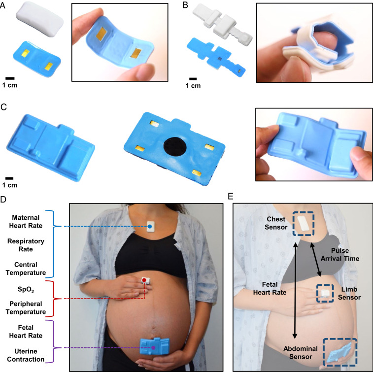
“The abdominal sensor (Fig. 1C) offers an onboard wireless Doppler ultrasound (US), EHG, and ECG, each sampled at 504 and 500 Hz, respectively.”
§ Comprehensive pregnancy monitoring with a network of wireless, soft, and flexible sensors in high- and low-resource health settings > Results > Wireless Maternal–Fetal Sensor System Design.
plain- Asserted: passes — The authors put it forward as true.
- Checkable: passes — It can be checked against evidence.
- Load-bearing: passes — The paper’s conclusions depend on it.
- Based: passes — The paper gives a reason why it holds.
It may state two separate claims as one.
A result the paper reports from its own data — it cites no other source for it, so there is none to check. The quote shows it is in the paper.
In the paper
“The chest sensor (Fig. 1A) measures ECG, seismocardiogram (SCG), chest wall movement for respiratory rate (RR), and skin temperature, each sampled at 512, 416, 52, and 0.2 Hz, respectively.”
§ Comprehensive pregnancy monitoring with a network of wireless, soft, and flexible sensors in high- and low-resource health settings > Results > Wireless Maternal–Fetal Sensor System Design.
plain- Asserted: passes — The authors put it forward as true.
- Checkable: passes — It can be checked against evidence.
- Load-bearing: fails — The paper’s conclusions may not depend on it.
- Based: passes — The paper gives a reason why it holds.
Probably not a substantial claim. The paper’s conclusions may not depend on it. It may state two separate claims as one.
A result the paper reports from its own data — it cites no other source for it, so there is none to check. The quote shows it is in the paper.
In the paper
“A Bland–Altman plot comparing data from the abdominal sensor to the gold standard is presented in Fig. 3C for a subset of n = 10 laboring women who contributed 3,213 single data points.”
- Also stated in: § FHR and Uterine Contraction via a Soft, Flexible, and Wireless Abdominal Sensor. · boosted
“The mean difference is 0.1 bpm with an SD of 1 bpm, showing strong agreement.”
§ Comprehensive pregnancy monitoring with a network of wireless, soft, and flexible sensors in high- and low-resource health settings > Results > FHR and Uterine Contraction via a Soft, Flexible, and Wireless Abdominal Sensor.
boostedn = 10- Asserted: passes — The authors put it forward as true.
- Checkable: passes — It can be checked against evidence.
- Load-bearing: passes — The paper’s conclusions depend on it.
- Based: passes — The paper gives a reason why it holds.
It may state two separate claims as one. The quote only partly states this claim.
- Also stated in: § FHR and Uterine Contraction via a Soft, Flexible, and Wireless Abdominal Sensor. · boosted
The paper states this without a source or data behind it here, so there is nothing to check it against. The quote shows it is in the paper.
In the paper
“This system offers advanced features such as continuous blood pressure, uterine electrohysterography, and automated body position classification.”
§ Comprehensive pregnancy monitoring with a network of wireless, soft, and flexible sensors in high- and low-resource health settings > Abstract
plain- Asserted: passes — The authors put it forward as true.
- Checkable: passes — It can be checked against evidence.
- Load-bearing: passes — The paper’s conclusions depend on it.
- Based: passes — The paper gives a reason why it holds.
It may state two separate claims as one.
The paper states this without a source or data behind it here, so there is nothing to check it against. The quote shows it is in the paper.
In the paper
“The sensor is able to obtain high signal quality even in the presence of motion and is water-resistant such that it can be worn in the shower.”
§ Comprehensive pregnancy monitoring with a network of wireless, soft, and flexible sensors in high- and low-resource health settings > Results > Wireless Maternal–Fetal Sensor System Design.
plain- Asserted: passes — The authors put it forward as true.
- Checkable: passes — It can be checked against evidence.
- Load-bearing: fails — The paper’s conclusions may not depend on it.
- Based: fails — The paper may give no reason why it holds.
Probably not a substantial claim. The paper’s conclusions may not depend on it. The paper may give no reason why it holds. It may state two separate claims as one.
The paper states this without a source or data behind it here, so there is nothing to check it against. The quote shows it is in the paper.
In the paper
“The chest and abdominal sensors also communicate, which enables maternal ECG elimination from the abdominal sensor’s outputs, allowing for fetal ECG isolation.”
§ Comprehensive pregnancy monitoring with a network of wireless, soft, and flexible sensors in high- and low-resource health settings > Results > Wireless Maternal–Fetal Sensor System Design.
plainstates a cause- Asserted: passes — The authors put it forward as true.
- Checkable: passes — It can be checked against evidence.
- Load-bearing: passes — The paper’s conclusions depend on it.
- Based: passes — The paper gives a reason why it holds.
It may state two separate claims as one.
A result the paper reports from its own data — it cites no other source for it, so there is none to check. The quote shows it is in the paper.
In the paper
“The limb sensor (Fig. 1B) measures photoplethysmogram (PPG) and skin temperature, sampled at 256 and 0.2 Hz, respectively.”
§ Comprehensive pregnancy monitoring with a network of wireless, soft, and flexible sensors in high- and low-resource health settings > Results > Wireless Maternal–Fetal Sensor System Design.
plain- Asserted: passes — The authors put it forward as true.
- Checkable: passes — It can be checked against evidence.
- Load-bearing: fails — The paper’s conclusions may not depend on it.
- Based: passes — The paper gives a reason why it holds.
Probably not a substantial claim. The paper’s conclusions may not depend on it. It may state two separate claims as one.
A result the paper reports from its own data — it cites no other source for it, so there is none to check. The quote shows it is in the paper.
In the paper
“While our data show cold-pressor-induced hypertensive episodes”
§ Comprehensive pregnancy monitoring with a network of wireless, soft, and flexible sensors in high- and low-resource health settings > Results > Continuous Surrogates of Systolic BP Derived from PAT and Pulse Transit Time.
plainstates a causen = 3- Asserted: passes — The authors put it forward as true.
- Checkable: passes — It can be checked against evidence.
- Load-bearing: fails — The paper’s conclusions may not depend on it.
- Based: passes — The paper gives a reason why it holds.
Probably not a substantial claim. The paper’s conclusions may not depend on it.
A result the paper reports from its own data — it cites no other source for it, so there is none to check. The quote shows it is in the paper.
In the paper
“In Fig. 4A we demonstrate a high degree of agreement with traditional sphygmomanometers, capturing BP at discrete time points with average errors less than 10 mmHg.”
§ Comprehensive pregnancy monitoring with a network of wireless, soft, and flexible sensors in high- and low-resource health settings > Results > Continuous Surrogates of Systolic BP Derived from PAT and Pulse Transit Time.
boostedstates an association- Asserted: passes — The authors put it forward as true.
- Checkable: passes — It can be checked against evidence.
- Load-bearing: passes — The paper’s conclusions depend on it.
- Based: passes — The paper gives a reason why it holds.
It may state two separate claims as one.
A result the paper reports from its own data — it cites no other source for it, so there is none to check. The quote shows it is in the paper.
In the paper
“A Bland–Altman plot in Fig. 4B demonstrates the accuracy of our method, which has a mean difference from the sphygmomanometer of 0.4 mmHg (SD 7.8 mmHg).”
§ Comprehensive pregnancy monitoring with a network of wireless, soft, and flexible sensors in high- and low-resource health settings > Results > Continuous Surrogates of Systolic BP Derived from PAT and Pulse Transit Time.
boosted- Asserted: passes — The authors put it forward as true.
- Checkable: passes — It can be checked against evidence.
- Load-bearing: passes — The paper’s conclusions depend on it.
- Based: passes — The paper gives a reason why it holds.
It may state two separate claims as one.
A result the paper reports from its own data — it cites no other source for it, so there is none to check. The quote shows it is in the paper.
In the paper
“Our demonstration here in n = 3 healthy normal subjects in highly controlled settings with variable BP achieved via the cold-pressor method shows an average mean and root mean square error of 0.6 and 5.31 mmHg for SBP and 0.2 and 7.07 mmHg for diastolic BP”
§ Comprehensive pregnancy monitoring with a network of wireless, soft, and flexible sensors in high- and low-resource health settings > Results > Continuous Surrogates of Systolic BP Derived from PAT and Pulse Transit Time.
boostedn = 3- Asserted: passes — The authors put it forward as true.
- Checkable: passes — It can be checked against evidence.
- Load-bearing: passes — The paper’s conclusions depend on it.
- Based: passes — The paper gives a reason why it holds.
It may state two separate claims as one.
The paper states this without a source or data behind it here, so there is nothing to check it against. The quote shows it is in the paper.
In the paper
“We present a wireless monitoring platform that provides comprehensive assessment of both the mother and fetus with compatibility across a wide range of mobile devices.”
§ Comprehensive pregnancy monitoring with a network of wireless, soft, and flexible sensors in high- and low-resource health settings > Discussion
plain- Asserted: passes — The authors put it forward as true.
- Checkable: passes — It can be checked against evidence.
- Load-bearing: passes — The paper’s conclusions depend on it.
- Based: passes — The paper gives a reason why it holds.
It may state two separate claims as one.
The paper states this without a source or data behind it here, so there is nothing to check it against. The quote shows it is in the paper.
In the paper
“This system is able to provide continuous body positioning categorization and continuous BP assessments and has been deployed in a significant number of pregnant women.”
§ Comprehensive pregnancy monitoring with a network of wireless, soft, and flexible sensors in high- and low-resource health settings > Discussion
plain- Asserted: passes — The authors put it forward as true.
- Checkable: passes — It can be checked against evidence.
- Load-bearing: passes — The paper’s conclusions depend on it.
- Based: unclear — It is unclear why the paper says it holds.
It may state two separate claims as one.
The paper states this without a source or data behind it here, so there is nothing to check it against. The quote shows it is in the paper.
In the paper
“The system we present here offers comprehensive and advanced measurement modalities with evidence of feasibility, performance, and user-preference acceptability among laboring women in both high-resource and low-resource settings for both in-hospital and remote monitoring applications.”
§ Comprehensive pregnancy monitoring with a network of wireless, soft, and flexible sensors in high- and low-resource health settings > Discussion
plain- Asserted: passes — The authors put it forward as true.
- Checkable: passes — It can be checked against evidence.
- Load-bearing: passes — The paper’s conclusions depend on it.
- Based: passes — The paper gives a reason why it holds.
It may state two separate claims as one.
- Rests on an assumption: In both high- and low-resource settings the burden placed on clinical staff in order to obtain physiologic information on a woman and her fetus is significant.
The paper states this without a source or data behind it here, so there is nothing to check it against. The quote shows it is in the paper.
In the paper
“The system provided here can be configured so as to alert providers only when thresholds of vital signs that are indicative of concern are crossed.”
§ Comprehensive pregnancy monitoring with a network of wireless, soft, and flexible sensors in high- and low-resource health settings > Discussion
plain- Asserted: passes — The authors put it forward as true.
- Checkable: passes — It can be checked against evidence.
- Load-bearing: passes — The paper’s conclusions depend on it.
- Based: passes — The paper gives a reason why it holds.
Probably not a substantial claim.
The paper states this without a source or data behind it here, so there is nothing to check it against. The quote shows it is in the paper.
In the paper
“The system we present here provides objective and real-time data outputs on the impact of maternal body position on both maternal and fetal health with the ability to assess how position may be associated with clinical outcomes and the potential to include automated alerts to patients and providers for body position changes if indicated.”
§ Comprehensive pregnancy monitoring with a network of wireless, soft, and flexible sensors in high- and low-resource health settings > Discussion
hedgedstates an association- Asserted: passes — The authors put it forward as true.
- Checkable: passes — It can be checked against evidence.
- Load-bearing: passes — The paper’s conclusions depend on it.
- Based: passes — The paper gives a reason why it holds.
It may state two separate claims as one.
- Rests on an assumption: Research on the effects and benefits of laboring positions has been largely limited because of inability to monitor and collect data in clinical settings with fidelity.
The paper states this without a source or data behind it here, so there is nothing to check it against. The quote shows it is in the paper.
In the paper
“The consumables of the system are also low-cost and consist of only single-use hydrogel adhesives and latex-free fabric strap (estimated <$0.25 per use).”
§ Comprehensive pregnancy monitoring with a network of wireless, soft, and flexible sensors in high- and low-resource health settings > Discussion
hedged- Asserted: passes — The authors put it forward as true.
- Checkable: passes — It can be checked against evidence.
- Load-bearing: passes — The paper’s conclusions depend on it.
- Based: passes — The paper gives a reason why it holds.
It may state two separate claims as one.
The paper states this without a source or data behind it here, so there is nothing to check it against. The quote shows it is in the paper.
In the paper
“First, the sensors themselves are low-cost (estimated <$40 per sensor at large-scale manufacturing) and fully reusable after a wireless recharge and simple cleaning procedure.”
§ Comprehensive pregnancy monitoring with a network of wireless, soft, and flexible sensors in high- and low-resource health settings > Discussion
hedged- Asserted: passes — The authors put it forward as true.
- Checkable: passes — It can be checked against evidence.
- Load-bearing: passes — The paper’s conclusions depend on it.
- Based: unclear — It is unclear why the paper says it holds.
It may state two separate claims as one.
In the paper
“The Invu reports both FHR and maternal HR but does not capture uterine contractions (15).”
§ Comprehensive pregnancy monitoring with a network of wireless, soft, and flexible sensors in high- and low-resource health settings > Abstract
What the sources say
- Not checked yetcites [15] M. Mhajna., Wireless, remote solution for home fetal and maternal heart rate monitoring. Am. J. Obstet. Gynecol. MFM 2, 100101 (2020). 33345967 source
plain- Asserted: passes — The authors put it forward as true.
- Checkable: passes — It can be checked against evidence.
- Load-bearing: passes — The paper’s conclusions depend on it.
- Based: passes — The paper gives a reason why it holds.
It may state two separate claims as one.
What the paper did — it cites no source for this, so there is none to check. The quote shows it is in the paper.
In the paper
“A biocompatible, double-sided hydrogel adhesive conducts the signal from the patient to the chest and abdominal sensors and its adhesion strength has been deemed safe to use by a board-certified dermatologist.”
§ Comprehensive pregnancy monitoring with a network of wireless, soft, and flexible sensors in high- and low-resource health settings > Results > Wireless Maternal–Fetal Sensor System Design.
plain- Asserted: passes — The authors put it forward as true.
- Checkable: passes — It can be checked against evidence.
- Load-bearing: fails — The paper’s conclusions may not depend on it.
- Based: fails — The paper may give no reason why it holds.
Probably not a substantial claim. The paper’s conclusions may not depend on it. The paper may give no reason why it holds.
In the paper
“Additional monitoring equipment designed to assess maternal or fetal status, such as pulse oximetry, blood pressure (BP) monitors, and electrocardiography (ECG), are typically used in a noncontinuous manner and place a relatively high burden on providers (11).”
§ Comprehensive pregnancy monitoring with a network of wireless, soft, and flexible sensors in high- and low-resource health settings > Abstract
What the sources say
- Not checked yetcites [11] A. A. Boatin., Wireless vital sign monitoring in pregnant women: A functionality and acceptability study. Telemed. J. E Health 22, 564– 571 (2016). 27172448 source
plainstates a cause- Asserted: passes — The authors put it forward as true.
- Checkable: passes — It can be checked against evidence.
- Load-bearing: passes — The paper’s conclusions depend on it.
- Based: passes — The paper gives a reason why it holds.
It may state two separate claims as one.
None of the sources cited for this claim have been checked yet.
In the paper
“The most common causes of maternal morbidity and mortality—hemorrhage, hypertensive disorders, infection, and sepsis—are often preceded by predictable vital signs perturbations in both high-resource and low-resource settings, and early intervention is key to improving outcomes (4–9).”
§ Comprehensive pregnancy monitoring with a network of wireless, soft, and flexible sensors in high- and low-resource health settings > Abstract
What the sources say
- Not checked yetcites [4] R. W. Rochat, L. M. Koonin, H. K. Atrash, J. F. Jewett, Maternal mortality in the United States: Report from the maternal mortality collaborative. Obstet. Gynecol. 72, 91– 97 (1988). 3380512
- Not checked yetcites [5] B. Kodio., Levels and causes of maternal mortality in Senegal. Trop. Med. Int. Health 7, 499– 505 (2002). 12031071 source
- Not checked yetcites [6] M. E. D’Alton., Putting the “M” back in maternal-fetal medicine: A 5-year report card on a collaborative effort to address maternal morbidity and mortality in the United States. Am. J. Obstet. Gynecol. 221, 311– 317.e1 (2019). 30849353 PMC6728226 source
- Not checked yetcites [7] V. Filippi, D. Chou, C. Ronsmans, W. Graham, L. Say, “ Levels and causes of maternal mortality and morbidity” in Reproductive, Maternal, Newborn, and Child Health: Disease Control Priorities, R. E. Black, R. Laxminarayan, M. Temmerman, N. Walker, Eds. (International Bank for Reconstruction and Development/The World Bank, Washington, DC, ed. 3, 2016), vol. 2, pp. 51– 70. source
- Not checked yetcites [8] K. Nagaya., Causes of maternal mortality in Japan. JAMA 283, 2661– 2667 (2000). 10819948 source
- Not checked yetcites [9] N. Vousden, H. L. Nathan, A. H. Shennan, Innovations in vital signs measurement for the detection of hypertension and shock in pregnancy. Reprod. Health 15 (suppl. 1), 92 (2018). 29945641 PMC6020004 source
plainstates an association- Asserted: passes — The authors put it forward as true.
- Checkable: passes — It can be checked against evidence.
- Load-bearing: passes — The paper’s conclusions depend on it.
- Based: passes — The paper gives a reason why it holds.
It may state two separate claims as one.
In the paper
“The Bloomlife Pregnancy Tracker captures electrohysterography (EHG) without fetal ECG (16).”
§ Comprehensive pregnancy monitoring with a network of wireless, soft, and flexible sensors in high- and low-resource health settings > Abstract
What the sources say
- Not checked yetcites [16] M. Altini., 785: Early labour detection in laboratory and free-living conditions using combined electrohysterography and heart rate data. Am. J. Obstet. Gynecol. 220, S513– S514 (2019). source
plain- Asserted: passes — The authors put it forward as true.
- Checkable: passes — It can be checked against evidence.
- Load-bearing: fails — The paper’s conclusions may not depend on it.
- Based: passes — The paper gives a reason why it holds.
Probably not a substantial claim. The paper’s conclusions may not depend on it.
In the paper
“The time synchronization infrastructure was previously established and yields an SD of 3.6 ms over 24 consecutive hours (22).”
§ Comprehensive pregnancy monitoring with a network of wireless, soft, and flexible sensors in high- and low-resource health settings > Results > Wireless Maternal–Fetal Sensor System Design.
What the sources say
- Not checked yetcites [22] H. U. Chung., Skin-interfaced biosensors for advanced wireless physiological monitoring in neonatal and pediatric intensive-care units. Nat. Med. 26, 418– 429 (2020). 32161411 PMC7315772 source
plain- Asserted: passes — The authors put it forward as true.
- Checkable: passes — It can be checked against evidence.
- Load-bearing: unclear — It is unclear whether the paper’s conclusions depend on it.
- Based: passes — The paper gives a reason why it holds.
It may state two separate claims as one.
A conclusion the paper draws from its own results — it cites no other source for it, so there is none to check. The quote shows it is in the paper.
In the paper
“The R-to-R peak intervals translate to 94 bpm and 170 bpm for maternal and fetal HR, respectively.”
§ Comprehensive pregnancy monitoring with a network of wireless, soft, and flexible sensors in high- and low-resource health settings > Results > FHR and Uterine Contraction via a Soft, Flexible, and Wireless Abdominal Sensor.
plainstates a cause- Asserted: passes — The authors put it forward as true.
- Checkable: passes — It can be checked against evidence.
- Load-bearing: unclear — It is unclear whether the paper’s conclusions depend on it.
- Based: passes — The paper gives a reason why it holds.
It may state two separate claims as one.
What the paper did — it cites no source for this, so there is none to check. The quote shows it is in the paper.
In the paper
“All participants provided written informed consent with research protocols approved by the Institutional Review Boards of Northwestern University (STU00205895) and the University of North Carolina (19-0765).”
§ Comprehensive pregnancy monitoring with a network of wireless, soft, and flexible sensors in high- and low-resource health settings > Results > Clinical Validation and Deployments in Both High-Resource and Low-Resource Settings.
plain- Asserted: passes — The authors put it forward as true.
- Checkable: passes — It can be checked against evidence.
- Load-bearing: fails — The paper’s conclusions may not depend on it.
- Based: passes — The paper gives a reason why it holds.
Probably not a substantial claim. The paper’s conclusions may not depend on it. It may state two separate claims as one.
In the paper
“Other systems, such as the Novii Wireless Patch System from Monica Healthcare, are available to acquire fetal ECG, but such systems alone cannot monitor nonsingleton pregnancies and are limited to term pregnancies (at least 37 wk of gestation) (17).”
§ Comprehensive pregnancy monitoring with a network of wireless, soft, and flexible sensors in high- and low-resource health settings > Abstract
What the sources say
- Not checked yetcites [17] A. Wacker-Gussmann., Fetal cardiac time intervals in healthy pregnancies–An observational study by fetal ECG (Monica Healthcare System). J. Perinat. Med. 46, 587– 592 (2018). 28453441 source
plain- Asserted: passes — The authors put it forward as true.
- Checkable: passes — It can be checked against evidence.
- Load-bearing: passes — The paper’s conclusions depend on it.
- Based: passes — The paper gives a reason why it holds.
It may state two separate claims as one.
None of the sources cited for this claim have been checked yet.
In the paper
“Altering maternal positioning during labor as a means of fetal resuscitation is common (38–40).”
§ Comprehensive pregnancy monitoring with a network of wireless, soft, and flexible sensors in high- and low-resource health settings > Discussion
What the sources say
- Not checked yetcites [38] P. R. Stone.; Maternal Sleep in Pregnancy Research Group, The University of Auckland, Effect of maternal position on fetal behavioural state and heart rate variability in healthy late gestation pregnancy. J. Physiol. 595, 1213– 1221 (2017). 27871127 PMC5309385 source
- Not checked yetcites [39] G. Cito., Maternal position during non-stress test and fetal heart rate patterns. Acta Obstet. Gynecol. Scand. 84, 335– 338 (2005). 15762962 source
- Not checked yetcites [40] B. Carbonne, A. Benachi, M. L. Lévèque, D. Cabrol, E. Papiernik, Maternal position during labor: Effects on fetal oxygen saturation measured by pulse oximetry. Obstet. Gynecol. 88, 797– 800 (1996). 8885916 source
plain- Asserted: passes — The authors put it forward as true.
- Checkable: passes — It can be checked against evidence.
- Load-bearing: fails — The paper’s conclusions may not depend on it.
- Based: passes — The paper gives a reason why it holds.
Probably not a substantial claim. The paper’s conclusions may not depend on it.
None of the sources cited for this claim have been checked yet.
In the paper
“Use of these newer systems in multiple-gestation or preterm fetuses have not been validated extensively and would be considered off-label if used in these clinical scenarios.”
§ Comprehensive pregnancy monitoring with a network of wireless, soft, and flexible sensors in high- and low-resource health settings > Discussion
What the sources say
- Not checked yetcites [17] A. Wacker-Gussmann., Fetal cardiac time intervals in healthy pregnancies–An observational study by fetal ECG (Monica Healthcare System). J. Perinat. Med. 46, 587– 592 (2018). 28453441 source
- Not checked yetcites [36] R. Sameni, G. D. Clifford, A review of fetal ECG signal processing; issues and promising directions. Open Pacing Electrophysiol. Ther. J. 3, 4– 20 (2010). 21614148 PMC3100207
hedged- Asserted: passes — The authors put it forward as true.
- Checkable: passes — It can be checked against evidence.
- Load-bearing: passes — The paper’s conclusions depend on it.
- Based: passes — The paper gives a reason why it holds.
It may state two separate claims as one.
A conclusion the paper draws from its own results — it cites no other source for it, so there is none to check. The quote shows it is in the paper.
In the paper
“The use of Doppler in our abdominal sensors allows the system to potentially monitor nonsingleton pregnancies with multiple patches.”
§ Comprehensive pregnancy monitoring with a network of wireless, soft, and flexible sensors in high- and low-resource health settings > Discussion
hedgedstates a possible cause- Asserted: passes — The authors put it forward as true.
- Checkable: passes — It can be checked against evidence.
- Load-bearing: fails — The paper’s conclusions may not depend on it.
- Based: fails — The paper may give no reason why it holds.
Probably not a substantial claim. The paper’s conclusions may not depend on it. The paper may give no reason why it holds.
In the paper
“Some studies suggest upright postures have been associated with shorter early labor stages, a trend toward fewer cesarean deliveries, and less use of analgesia (41).”
§ Comprehensive pregnancy monitoring with a network of wireless, soft, and flexible sensors in high- and low-resource health settings > Discussion
What the sources say
- Not checked yetcites [41] J. K. Gupta, A. Sood, G. J. Hofmeyr, J. P. Vogel, Position in the second stage of labour for women without epidural anaesthesia. Cochrane Database Syst. Rev. 5, CD002006 (2017). 28539008 PMC6484432
hedgedstates an association- Asserted: passes — The authors put it forward as true.
- Checkable: passes — It can be checked against evidence.
- Load-bearing: fails — The paper’s conclusions may not depend on it.
- Based: passes — The paper gives a reason why it holds.
Probably not a substantial claim. The paper’s conclusions may not depend on it. It may state two separate claims as one.
None of the sources cited for this claim have been checked yet.
In the paper
“The supine position is often avoided given worse uteroplacental perfusion because of maternal hypotension (42) and prolonged labor (43).”
§ Comprehensive pregnancy monitoring with a network of wireless, soft, and flexible sensors in high- and low-resource health settings > Discussion
What the sources say
- Not checked yetcites [42] S. M. Kinsella, A. Lee, J. A. Spencer, Maternal and fetal effects of the supine and pelvic tilt positions in late pregnancy. Eur. J. Obstet. Gynecol. Reprod. Biol. 36, 11– 17 (1990). 2365116 source
- Not checked yetcites [43] A. Lawrence, L. Lewis, G. J. Hofmeyr, C. Styles, Maternal positions and mobility during first stage labour. Cochrane Database Syst. Rev., CD003934 (2013). PMC11664456 24105444
plainstates a cause- Asserted: passes — The authors put it forward as true.
- Checkable: passes — It can be checked against evidence.
- Load-bearing: fails — The paper’s conclusions may not depend on it.
- Based: passes — The paper gives a reason why it holds.
Probably not a substantial claim. The paper’s conclusions may not depend on it.
None of the sources cited for this claim have been checked yet.
In the paper
“Previous research efforts have also demonstrated maternal position impacts maternal blood flow, labor progression, and patient comfort (44–46).”
§ Comprehensive pregnancy monitoring with a network of wireless, soft, and flexible sensors in high- and low-resource health settings > Discussion
What the sources say
- Not checked yetcites [44] O. Moraloglu., The influence of different maternal pushing positions on birth outcomes at the second stage of labor in nulliparous women. J. Matern. Fetal Neonatal Med. 30, 245– 249 (2017). 27028537 source
- Not checked yetcites [45] M. J. Guittier, V. Othenin-Girard, [Correcting occiput posterior position during labor: The role of maternal positions]. Gynécol. Obstét. Fertil. 40, 255– 260 (2012). 22099975 source
- Not checked yetcites [46] J. Roberts, L. Malasanos, C. Mendez-Bauer, Maternal positions in labor: Analysis in relation to comfort and efficiency. Birth Defects Orig. Artic. Ser. 17, 97– 128 (1981). 7326378
boostedstates a cause- Asserted: passes — The authors put it forward as true.
- Checkable: passes — It can be checked against evidence.
- Load-bearing: passes — The paper’s conclusions depend on it.
- Based: passes — The paper gives a reason why it holds.
Probably not a substantial claim. It may state two separate claims as one.
In the paper
“The ability to track body position continuously and concurrently with maternal and fetal vital signs offers an opportunity for improved monitoring and the ability to manage labor by modifying maternal body position as clinically indicated (37).”
§ Comprehensive pregnancy monitoring with a network of wireless, soft, and flexible sensors in high- and low-resource health settings > Discussion
What the sources say
- Not checked yetcites [37] G. J. Hofmeyr., Obstetric care in low-resource settings: What, who, and how to overcome challenges to scale up? Int J Gynaecol Obstet 107 (suppl. 1), S21– S44, S44-5 (2009). 19815204 source
hedgedstates a possible cause- Asserted: passes — The authors put it forward as true.
- Checkable: passes — It can be checked against evidence.
- Load-bearing: passes — The paper’s conclusions depend on it.
- Based: passes — The paper gives a reason why it holds.
It may state two separate claims as one.
- Rests on an assumption: Research on the effects and benefits of laboring positions has been largely limited because of inability to monitor and collect data in clinical settings with fidelity.
In the paper
“In this scenario, we demonstrated the responsiveness of our system to controlled changes of BP with a cold-pressor test, a well-established, safe, and reproducible method to induce a BP response (53).”
§ Comprehensive pregnancy monitoring with a network of wireless, soft, and flexible sensors in high- and low-resource health settings > Materials and Methods > Continuous BP Derivation.
What the sources say
- Not checked yetcites [53] Q. Zhao., Reproducibility of blood pressure response to the cold pressor test: The GenSalt study. Am. J. Epidemiol. 176 (suppl. 7), S91– S98 (2012). 23035148 PMC3530368 source
boostedstates a cause- Asserted: passes — The authors put it forward as true.
- Checkable: passes — It can be checked against evidence.
- Load-bearing: fails — The paper’s conclusions may not depend on it.
- Based: passes — The paper gives a reason why it holds.
Probably not a substantial claim. The paper’s conclusions may not depend on it. It may state two separate claims as one.
In the paper
“The key drivers of this failure are multifactorial and include the cost of maintenance, lack of skilled technicians to maintain the equipment, lack of spare parts, and high consumable costs (50).”
§ Comprehensive pregnancy monitoring with a network of wireless, soft, and flexible sensors in high- and low-resource health settings > Discussion
What the sources say
- Not checked yetcites [50] B. Compton., “Access to medical devices in low-income countries: Addressing sustainability challenges in medical device donations” (NAM Perspectives, National Academy of Medicine, 2018). source
plainstates a cause- Asserted: passes — The authors put it forward as true.
- Checkable: passes — It can be checked against evidence.
- Load-bearing: passes — The paper’s conclusions depend on it.
- Based: passes — The paper gives a reason why it holds.
It may state two separate claims as one.
In the paper
“Continuous data of maternal BP, HR, SpO2, uterine contractility, and FHR are potentially life-saving if acted on, but access is heavily dependent on clinical infrastructure, availability of skilled healthcare providers, and integration of disparate devices and their displays (52).”
§ Comprehensive pregnancy monitoring with a network of wireless, soft, and flexible sensors in high- and low-resource health settings > Discussion
What the sources say
- Not checked yetcites [52] S. Thaddeus, D. Maine, Too far to walk: Maternal mortality in context. Soc. Sci. Med. 38, 1091– 1110 (1994). 8042057 source
hedgedstates a possible cause- Asserted: passes — The authors put it forward as true.
- Checkable: passes — It can be checked against evidence.
- Load-bearing: passes — The paper’s conclusions depend on it.
- Based: passes — The paper gives a reason why it holds.
It may state two separate claims as one.
A conclusion the paper draws from its own results — it cites no other source for it, so there is none to check. The quote shows it is in the paper.
In the paper
“Additionally, these systems can provide a wealth of data in different clinical settings with labeled clinical outcomes, such as that generated by the LABOR study, that offer the opportunity for more population-specific and sensitive delineation of maternal or fetal status to support clinical decision-making.”
§ Comprehensive pregnancy monitoring with a network of wireless, soft, and flexible sensors in high- and low-resource health settings > Discussion
plainstates an association- Asserted: passes — The authors put it forward as true.
- Checkable: passes — It can be checked against evidence.
- Load-bearing: passes — The paper’s conclusions depend on it.
- Based: passes — The paper gives a reason why it holds.
Probably not a substantial claim.
In the paper
“By 2025, it is estimated that there will be nearly 700 million smartphones in sub-Saharan Africa, making these mobile devices readily available to be used in order to operate the sensors for healthcare applications in even the most rural settings (51).”
§ Comprehensive pregnancy monitoring with a network of wireless, soft, and flexible sensors in high- and low-resource health settings > Discussion
What the sources say
- Not checked yetcites [51] D. Radcliffe, Mobile in Sub-Saharan Africa: Can world’s fastest-growing mobile region keep it up? ZDNet (2018). https://www.zdnet.com/article/mobile-in-sub-saharan-africa-can-worlds-fastest-growing-mobile-region-keep-it-up/. Accessed 23 November 2020.
hedgedstates a cause- Asserted: passes — The authors put it forward as true.
- Checkable: passes — It can be checked against evidence.
- Load-bearing: passes — The paper’s conclusions depend on it.
- Based: passes — The paper gives a reason why it holds.
It may state two separate claims as one.
In the paper
“Other studies have found that the hands-and-knees position has not been associated with negative labor outcomes (47).”
§ Comprehensive pregnancy monitoring with a network of wireless, soft, and flexible sensors in high- and low-resource health settings > Discussion
What the sources say
- Not checked yetcites [47] S. Hunter, G. J. Hofmeyr, R. Kulier, Hands and knees posture in late pregnancy or labour for fetal malposition (lateral or posterior). Cochrane Database Syst. Rev., CD001063 (2007). 17943750 PMC8407052
plain- Asserted: passes — The authors put it forward as true.
- Checkable: passes — It can be checked against evidence.
- Load-bearing: fails — The paper’s conclusions may not depend on it.
- Based: passes — The paper gives a reason why it holds.
Probably not a substantial claim. The paper’s conclusions may not depend on it.
None of the sources cited for this claim have been checked yet.
In the paper
“In previous work, we demonstrate the ability for the chest- and finger-mounted sensors to monitor vital signs in both children and adults; the abdominal sensor allows for maternal–fetal monitoring of pregnant women in both high- and low-resource settings as well as the clinical and home settings (22, 23).”
§ Comprehensive pregnancy monitoring with a network of wireless, soft, and flexible sensors in high- and low-resource health settings > Discussion
What the sources say
- Not checked yetcites [22] H. U. Chung., Skin-interfaced biosensors for advanced wireless physiological monitoring in neonatal and pediatric intensive-care units. Nat. Med. 26, 418– 429 (2020). 32161411 PMC7315772 source
- Not checked yetcites [23] H. U. Chung., Binodal, wireless epidermal electronic systems with in-sensor analytics for neonatal intensive care. Science 363, eaau0780 (2019). 30819934 PMC6510306 source
boosted- Asserted: passes — The authors put it forward as true.
- Checkable: passes — It can be checked against evidence.
- Load-bearing: passes — The paper’s conclusions depend on it.
- Based: passes — The paper gives a reason why it holds.
It may state two separate claims as one.
None of the sources cited for this claim have been checked yet.
In the paper
“Additionally, as demonstrated by our previous work, neonatal vital signs can also be tracked with the same sensors in both high-resource and low-resource settings as well as support kangaroo care (22, 48).”
§ Comprehensive pregnancy monitoring with a network of wireless, soft, and flexible sensors in high- and low-resource health settings > Discussion
What the sources say
- Not checked yetcites [22] H. U. Chung., Skin-interfaced biosensors for advanced wireless physiological monitoring in neonatal and pediatric intensive-care units. Nat. Med. 26, 418– 429 (2020). 32161411 PMC7315772 source
- Not checked yetcites [48] A. S. Ginsburg., Evaluation of non-invasive continuous physiological monitoring devices for neonates in Nairobi, Kenya: A research protocol. BMJ Open 10, e035184 (2020). PMC7200030 32284391 source
boostedstates a cause- Asserted: passes — The authors put it forward as true.
- Checkable: passes — It can be checked against evidence.
- Load-bearing: unclear — It is unclear whether the paper’s conclusions depend on it.
- Based: passes — The paper gives a reason why it holds.
It may state two separate claims as one.
The paper relies on this without support.
The S1 and S2 peaks in the Doppler ultrasound signal represent closure of the fetal heart valves.
“The first peak “S1” represents closure of the tricuspid and mitral valves of the fetal heart, and the second peak “S2” represents closure of the pulmonary semilunar valves of the fetal heart (Audio S1).”
The paper relies on this without support.
The electrohysterography contraction can be converted to millimeters of mercury or pressure with a simple calibration.
“The EHG contraction can be converted to millimeters of mercury (mmHg) or pressure with a simple calibration.”
The paper relies on this without support.
Of all wireless pregnancy monitoring devices currently approved for clinical use, none incorporates maternal vital signs.
“Perhaps most notable, of all the wireless pregnancy monitoring devices currently approved for clinical use, none incorporates maternal vital signs.”
The paper relies on this without support.
Assessing maternal vital signs such as HR, RR, SpO2, and BP requires additional systems wired to the body and configured to another monitoring system that is often not interoperable.
“Assessing maternal vital signs such as HR, RR, SpO2, and BP requires additional systems wired to the body and configured to another monitoring system that is often not interoperable.”
The paper relies on this without support.
Research on the effects and benefits of laboring positions has been largely limited because of inability to monitor and collect data in clinical settings with fidelity.
“Despite these studies, research on the effects and benefits of laboring positions has been largely limited because of inability to monitor and collect data in clinical settings with fidelity.”
The paper relies on this without support.
In both high- and low-resource settings the burden placed on clinical staff in order to obtain physiologic information on a woman and her fetus is significant.
“In both high- and low-resource settings the burden placed on clinical staff in order to obtain physiologic information on a woman and her fetus is significant.”
Claims other papers make about this one
Each is checked against this paper’s own text: does it say what the citing paper says it does?
The source backs part of the claim, not all of it — or backs it for a different population, condition or comparison.
In the citing paper
“Previous reports validated the performance of an FDA-cleared physiological monitoring system against gold standard monitoring platforms [18–23].”
From Pregnancy outcomes in infertility patients diagnosed with sleep disordered breathing with wireless wearable sensors (2022) · cites this paper as [21]
Experimental — accuracy not yet measured
Self-check
Percentages, sums and reported differences re-computed from the paper’s own numbers.
Each contradiction between two passages listed here was confirmed by an independent check.
Rated minor: it would not touch a result — a value no result rests on, a p-value that stays on the same side of the significance level, or a conflict that is clerical, resolved elsewhere in the paper, or outside its own findings.
In the paper
§ Comprehensive pregnancy monitoring with a network of wireless, soft, and flexible sensors in high- and low-resource health settings > Materials and Methods > Time-to-Vital Analysis.
“Heat maps located on right column of Fig. 6 show the time-based normalized results to better visualize the vital distribution trend over the session duration.”
§ Comprehensive pregnancy monitoring with a network of wireless, soft, and flexible sensors in high- and low-resource health settings > Results > Time-Series Data of Vital Signs during Labor.
“Figure: Fig. 5. Heat-map analysis of maternal vital signs for laboring women in low-resource settings. A heat map of each vital signal is generated for all 485 participants. (Left) Plot (in red–black) for each vital is a heat map without normalization as the majority of labors last less than 5 h. (Right) Plot (in yellow–green) is normalized by frequency with a 4-h window to illustrate average vital signs over longer labors.”
Citations
Cites
Too far to walk: Maternal mortality in context
Thaddeus, S., & Maine, D. (1994) · Social Science & Medicine · doi:10.1016/0277-9536(94)90226-7 · Cited by 3,240
Saving Mothers’ Lives: Reviewing maternal deaths to make motherhood safer: 2006–2008
Cantwell, R., Clutton-Brock, T. H., COOPER, G. M., Dawson, A. H., Drife, J., Garrod, D., Harper, A., Hulbert, D., Lucas, S. B., McClure, J. H., et al. (2011) · BJOG An International Journal of Obstetrics & Gynaecology · doi:10.1111/j.1471-0528.2010.02847.x · Cited by 3,047
Self-citationIntended and Unintended Pregnancies Worldwide in 2012 and Recent Trends
Sedgh, G., Singh, S. D., & Hussain, R. (2014) · Studies in Family Planning · doi:10.1111/j.1728-4465.2014.00393.x · Cited by 1,050
Binodal, wireless epidermal electronic systems with in-sensor analytics for neonatal intensive care
Chung, H. U., Kim, B. H., Lee, J. Y., Lee, J., Xie, Z., Ibler, E. M., Lee, K., Banks, A., Jeong, J. Y., Kim, J., et al. (2019) · Science · doi:10.1126/science.aau0780 · Cited by 807 · cited 1×
“In previous work, we demonstrate the ability for the chest- and finger-mounted sensors to monitor vital signs in both children and adults; the abdominal sensor allows for maternal–fetal monitoring of pregnant women in both high- and low-resource settings as well as the clinical and home settings (22, 23).”
Self-citationKnight, M. (2017) · National Perinatal Epidemiology Unit, University of Oxford · doi:10.5287/ora-nrymp8wxq · Cited by 541
Toward a new generation of smart skins
Someya, T., & Amagai, M. (2019) · Nature Biotechnology · doi:10.1038/s41587-019-0079-1 · Cited by 499
Position in the second stage of labour for women without epidural anaesthesia
Gupta, J. K., Sood, A., Hofmeyr, G. J., & Vogel, J. P. (2017) · Cochrane Database of Systematic Reviews · doi:10.1002/14651858.cd002006.pub4 · Cited by 495
Recent Increases in the U.S. Maternal Mortality Rate: Disentangling Trends From Measurement Issues
MacDorman, M. F., Declercq, E., Cabral, H., & Morton, C. (2016) · Obstetrics & Gynecology · doi:10.1097/aog.0000000000001556 · Cited by 487
Chung, H. U., Rwei, A. Y., Hourlier-Fargette, A., Xu, S., Lee, K., Dunne, E. C., Xie, Z., Liu, C., Carlini, A., Kim, D. H., et al. (2020) · Nature Medicine · doi:10.1038/s41591-020-0792-9 · Cited by 482 · cited 3×
“Additionally, as demonstrated by our previous work, neonatal vital signs can also be tracked with the same sensors in both high-resource and low-resource settings as well as support kangaroo care (22, 48).”
Self-citationStergiou, G. S., Alpert, B., Mieke, S., Asmar, R., Atkins, N., Eckert, S., Frick, G., Friedman, B., Graßl, T., Ichikawa, T., et al. (2018) · Hypertension · doi:10.1161/hypertensionaha.117.10237 · Cited by 469
Research works printed but not linked: 1
- World Health Organization, Trends in Maternal Mortality 2000 to 2017: Estimates by WHO, UNICEF, UNFPA (World Bank Group and the United Nations Population Division, 2019).
Cited by
Sorted by citations
Technology Roadmap for Flexible Sensors
Luo, Y., Abidian, M. R., Ahn, J., Akinwande, D., Andrews, A. M., Antonietti, M., Bao, Z., Magnus Berggren, A., Berkey, C. A., Bettinger, C. J., et al. (2023) · ACS Nano · doi:10.1021/acsnano.2c12606 · Cited by 1,453
Self-citationSolution-processable, soft, self-adhesive, and conductive polymer composites for soft electronics
Tan, P., Wang, H., Xiao, F., Lu, X., Shang, W., Deng, X., Song, H., Xu, Z., Cao, J., Gan, T., et al. (2022) · Nature Communications · doi:10.1038/s41467-022-28027-y · Cited by 419
Self-citationSkin bioelectronics towards long-term, continuous health monitoring
Wang, Y., Haick, H., Guo, S., Wang, C., Lee, S., Yokota, T., & Someya, T. (2022) · Chemical Society Reviews · doi:10.1039/d2cs00207h · Cited by 328
Li, W., Ke, K., Jia, J. F., Pu, J., Zhao, X., Bao, R., Liu, Z., Bai, L., Zhang, K., Yang, M., et al. (2021) · Small · doi:10.1002/smll.202103734 · Cited by 255
Translational gaps and opportunities for medical wearables in digital health
Xu, S., Kim, J., Walter, J. R., Ghaffari, R., & Rogers, J. A. (2022) · Science Translational Medicine · doi:10.1126/scitranslmed.abn6036 · Cited by 134
Self-citation
Metadata
- Authors
- Ryu, D., Kim, D. H., Price, J. T., Lee, J. Y., Chung, H. U., Allen, E., Walter, J. R., Jeong, H., Cao, J., Kulikova, E., Abu-Zayed, H., Lee, R., Martell, K. L., Zhang, M., Kampmeier, B. R., Hill, M., Lee, J., Kim, E., Park, Y., Jang, H., Arafa, H., Liu, C., Chisembele, M., Vwalika, B., Sindano, N., Spelke, M. B., Paller, A. S., Premkumar, A., Grobman, W. A., Stringer, J. S. A., Rogers, J. A., & Xu, S.
- Affiliations
- Sibel (United States) (US)
- University of North Carolina at Chapel Hill (US)
- Northwestern University (US)
- Sibel Inc., Niles, IL 60714;
- University Teaching Hospital (ZM)
- University of Zambia (ZM)
- University of North Carolina Global Projects–Zambia, Lusaka 10101, Zambia;
- Corresponding author
- Stringer, Jeffrey S. A.
- Rogers, John A.
- Xu, Shuai
- ORCID
- Kim, Dong Hyun
- Price, Joan T.
- Lee, Jong Yoon
- Chung, Ha Uk
- Allen, Emily
- Walter, Jessica R.
- Jeong, Hyoyoung
- Cao, Jingyue
- Lee, Rachel
- Zhang, Michael
- Kampmeier, Brianna R.
- Hill, Marc
- Kim, Edward
- Park, Yerim
- Jang, Hokyung
- Arafa, Hany
- Liu, Claire
- Vwalika, Bellington
- Sindano, Ntazana
- Spelke, M. Bridget
- Paller, Amy S.
- Premkumar, Ashish
- Grobman, William A.
- Stringer, Jeffrey S. A.
- Rogers, John A.
- Xu, Shuai
- Venue
- Proceedings of the National Academy of Sciences
- Publisher
- National Academy of Sciences
- Published
- 2021-05-10
- Volume
- 118
- Issue
- 20
- ISSN
- 0027-8424 (ISSN-L)
- 1091-6490
- PMC
- PMC8157941
- OpenAlex
- W3161046902
- Semantic Scholar
- 5064baf350d8a9304f964bf07726272852491dcf
- Cited by
- 105
- References
- 56
- Citations by year
- 2026: 16 · 2025: 23 · 2024: 18 · 2023: 29 · 2022: 14 · 2021: 5
- FWCI
- 5.01
- Type
- journal article
- Language
- en
- Open access
- hybrid
- doi.org
- Licence
- Locations
- Proceedings of the National Academy of Sciences (publishedVersion, cc-by)
- PubMed Central (publishedVersion, cc-by)
- PubMed (publishedVersion)
- eScholarship (California Digital Library) (submittedVersion, cc-by)
- Funders
- Bill and Melinda Gates Foundation (OPP1193311)
- Topics
- Physical Sciences › Engineering › Biomedical Engineering › Non-Invasive Vital Sign Monitoring
- Physical Sciences › Engineering › Biomedical Engineering › Advanced Sensor and Energy Harvesting Materials
- Physical Sciences › Engineering › Biomedical Engineering › Wireless Body Area Networks
- Keywords
- pregnancy monitoring, wireless sensors, flexible sensors, vital signs monitoring, antepartum care, postpartum care, fetal monitoring, continuous blood pressure monitoring, resource-limited settings, mobile health, non-invasive monitoring
- MeSH terms
- Wearable Electronic Devices, Female, Health Resources, Heart Rate, Fetal, Humans, Monitoring, Physiologic, Pregnancy, Uterine Contraction, Vital Signs, Wireless Technology
About these checks
Registry, retraction, Cites and statistics results are computed, not judged, with one exception: a printed reference that no index lists is read once by a model before it counts as a citation error. Claims are extracted by a model and kept only when their quote appears verbatim in the text; each cited source is then checked, with a model’s help, for whether it says what the claim relies on. Those verdicts can be wrong — read the claim’s quote and the cited source before relying on one.
A language model drew the claims from the paper on 2026-10-09, under version 3 of the claim definition (model deepseek-v4.1-flash, prompt extract-v3).
A second model labelled each claim on 2026-10-09 (model jev-1.13-20260917, definition revision gold-guideline-v0.2).
Statistics and arithmetic are recomputed, not judged.
Confidence bands are shown only for fields calibrated on our annotated test set. None is calibrated yet, so no band is shown.
What counts as a claim ›How we measure confidence ›How we check this ›
Are you an author, or do you see a problem with this page? Request a review
