

The DOI matches its Crossref record; 11 of the 11 references it cites were found in a registry or an index.
The ingenious mast cell: Contemporary insights into mast cell behavior and function
Abstract
Mast cells are (in)famous for their role in allergic diseases, but the physiological and pathophysiological roles of this ingenious cell are still not fully understood. Mast cells are important for homeostasis and surveillance of the human system, recognizing both endogenous and exogenous agents, which induce release of a variety of mediators acting on both immune and non‐immune cells, including nerve cells, fibroblasts, endothelial cells, smooth muscle cells, and epithelial cells. During recent years, clinical and experimental studies on human mast cells, as well as experiments using animal models, have resulted in many discoveries that help decipher the function of mast cells in health and disease. In this review, we focus particularly on new insights into mast cell biology, with a focus on mast cell development, recruitment, heterogeneity, and reactivity. We also highlight the development in our understanding of mast cell‐driven diseases and discuss the development of novel strategies to treat such conditions.
Overview
The claims and statistics of this paper haven’t been checked yet.
Run the check — free, no account needed. It reads the open-access text and checks the sources it cites.
Integrity signals
Registry
Cites
Statistics
Figures




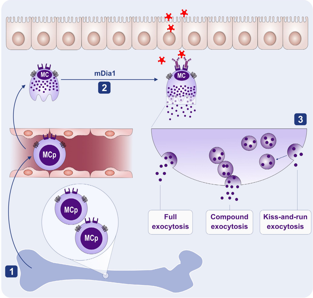


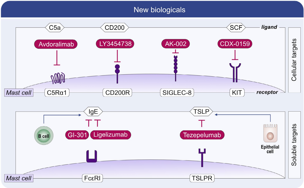
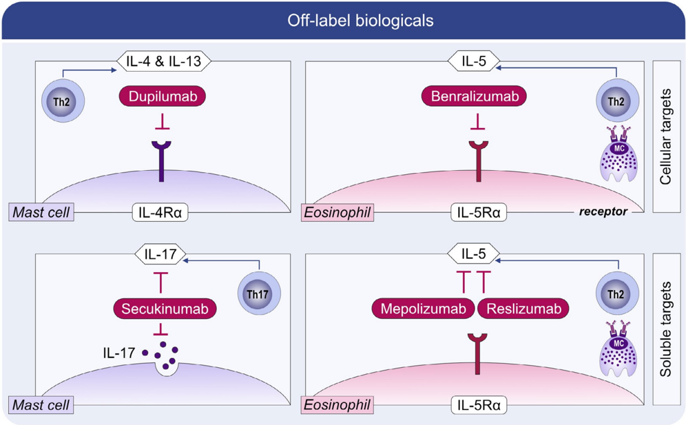

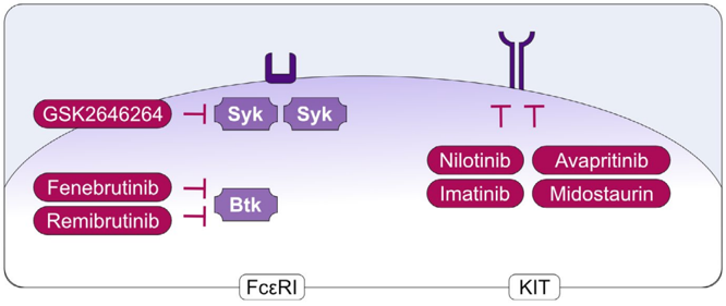


Verification
Claims the paper makes
The claims and statistics of this paper haven’t been checked yet.
Run the check — free, no account needed. It reads the open-access text and checks the sources it cites.
Claims other papers make about this one
Each is checked against this paper’s own text: does it say what the citing paper says it does?
In the citing paper
“Collectively, these Kit‐independent models have allowed researchers to assess mast cell functions in a more specific manner. Indeed, using such models, many of the previous findings from Kit‐dependent models have been challenged. 254”
From Mast cell ontogeny: From fetal development to life‐long health and disease (2023) · cites this paper as [254]
Self-check
Percentages, sums and reported differences re-computed from the paper’s own numbers.
The self-check needs the paper’s full text.
Citations
Cites
Lineage tracing on transcriptional landscapes links state to fate during differentiation
Weinreb, C., Rodriguez-Fraticelli, A., Camargo, F. D., & Klein, A. M. (2020) · Science · doi:10.1126/science.aaw3381 · Cited by 759
Decoding human fetal liver haematopoiesis
Popescu, D.-M., Botting, R. A., Stephenson, E., Green, K., Webb, S., Jardine, L., Calderbank, E. F., Polanski, K., Goh, I., Efremova, M., et al. (2019) · Nature · doi:10.1038/s41586-019-1652-y · Cited by 606
Lyons, J. J., Yu, X., Hughes, J. D., Le, Q. T., Jamil, A., Bai, Y., Ho, N., Zhao, M., Liu, Y., O'Connell, M. P., et al. (2016) · Nature Genetics · doi:10.1038/ng.3696 · Cited by 394
Self-citationPopulation snapshots predict early haematopoietic and erythroid hierarchies
Tusi, B. K., Wolock, S. L., Weinreb, C., Hwang, Y., Hidalgo, D., Zilionis, R., Waisman, A., Huh, J. R., Klein, A. M., & Socolovsky, M. (2018) · Nature · doi:10.1038/nature25741 · Cited by 384
Mast cell activation syndrome: Proposed diagnostic criteria
Akin, C., Valent, P., & Metcalfe, D. D. (2010) · Journal of Allergy and Clinical Immunology · doi:10.1016/j.jaci.2010.08.035 · Cited by 327
Self-citationPavlovian conditioning of rat mucosal mast cells to secrete rat mast cell protease II
MacQueen, G., Marshall, J., Perdue, M., Siegel, S., & Bienenstock, J. (1989) · Science · doi:10.1126/science.2911721 · Cited by 318
Lyons, J. J., Chovanec, J., O’Connell, M. P., Liu, Y., Šelb, J., Zanotti, R., Bai, Y., Kim, J., Le, Q. T., DiMaggio, T., et al. (2021) · Journal of Allergy and Clinical Immunology · doi:10.1016/j.jaci.2020.06.035 · Cited by 230
Self-citationProposed Diagnostic Algorithm for Patients with Suspected Mast Cell Activation Syndrome
Valent, P., Akin, C., Bonadonna, P., Hartmann, K., Brockow, K., Niedoszytko, M., Nedoszytko, B., Siebenhaar, F., Sperr, W. R., Oude Elberink, J. N. G., et al. (2019) · The Journal of Allergy and Clinical Immunology: In Practice · doi:10.1016/j.jaip.2019.01.006 · Cited by 225
Self-citationGreiner, G., Sprinzl, B., Górska, A., Ratzinger, F., Gurbisz, M., Witzeneder, N., Schmetterer, K. G., Gisslinger, B., Uyanik, G., Hadzijusufovic, E., et al. (2021) · Blood · doi:10.1182/blood.2020006157 · Cited by 189
Le, Q. T., Lyons, J. J., Naranjo, A. N., Olivera, A., Lazarus, R. A., Metcalfe, D. D., Milner, J. D., & Schwartz, L. B. (2019) · Journal of Experimental Medicine · doi:10.1084/jem.20190701 · Cited by 126
Research works printed but not linked: 174
- 10.1182/blood-2017-03-773374 · DOI
- 10.1016/j.jaci.2018.01.034 · DOI
- 10.1038/nature14022 · DOI · Paper page
- 10.18176/jiaci.0557 · DOI
- 10.1016/j.jaci.2016.04.051 · DOI
- 10.1016/j.jaci.2020.08.027 · DOI
- 10.1016/j.neuron.2019.01.012 · DOI
- 10.1016/j.immuni.2019.03.013 · DOI
- 10.3389/fimmu.2020.559589 · DOI
- 10.1038/s41590-019-0493-z · DOI
- 10.3389/fimmu.2012.00082 · DOI
- 10.1111/all.12004 · DOI
- 10.1016/j.jid.2019.01.013 · DOI
- 10.3389/fimmu.2019.01361 · DOI
- 10.3390/ijms20174241 · DOI
- 10.1038/nri3690 · DOI · Paper page
- 10.1111/j.1600-065X.2007.00512.x · DOI
- 10.1016/j.ejphar.2015.02.058 · DOI
- 10.1111/imr.12634 · DOI
- 10.3389/fimmu.2014.00569 · DOI
- 10.4049/jimmunol.1003383 · DOI
- 10.1016/j.jaci.2018.06.033 · DOI
- 10.1172/JCI24274 · DOI
- 10.1016/j.jaci.2015.01.042 · DOI
- 10.1111/j.1600-065X.2007.00519.x · DOI
- 10.1016/j.jaci.2021.01.002 · DOI
- 10.4049/jimmunol.166.2.868 · DOI
- 10.1038/ncb1596 · DOI · Paper page
- 10.3390/ijms21155464 · DOI
- 10.1073/pnas.1809938115 · DOI
- 10.1016/j.it.2017.10.005 · DOI
- 10.1111/all.13657 · DOI
- 10.1146/annurev-immunol-071719-094903 · DOI
- 10.1016/j.it.2020.06.006 · DOI
- 10.3390/ijms20184397 · DOI
- 10.1016/j.trsl.2016.01.005 · DOI
- 10.3390/ijms20122851 · DOI
- 10.1111/exd.13907 · DOI
- 10.1016/j.coi.2020.05.003 · DOI
- 10.1038/nature01320 · DOI · Paper page
- 10.1016/j.jaci.2018.10.054 · DOI
- 10.1016/0012-1606(83)90066-0 · DOI · Paper page
- 10.1016/j.immuni.2018.09.023 · DOI · Paper page
- 10.1016/j.immuni.2018.04.025 · DOI · Paper page
- 10.1111/all.13668 · DOI · Paper page
- 10.1038/268442a0 · DOI
- 10.1016/j.jid.2020.03.963 · DOI · Paper page
- 10.1182/blood.V94.7.2333.419k30_2333_2342 · DOI
- 10.1073/pnas.0509148102 · DOI · Paper page
- 10.1016/j.stem.2010.02.013 · DOI
- 10.1016/j.stemcr.2020.06.008 · DOI
- 10.1371/journal.ppat.1008579 · DOI
- 10.1073/pnas.0504197102 · DOI · Paper page
- 10.1182/blood-2013-02-483792 · DOI
- 10.1182/bloodadvances.2018019539 · DOI · Paper page
- 10.1038/nature25022 · DOI
- 10.1182/blood-2017-12-821413 · DOI · Paper page
- 10.1111/all.14633 · DOI
- 10.1016/j.immuni.2013.06.012 · DOI
- 10.1111/all.13981 · DOI
- 10.1126/sciimmunol.aau7148 · DOI
- 10.1038/s41586-020-2316-7 · DOI
- 10.1016/j.jaci.2005.02.030 · DOI
- 10.1182/blood.V80.12.3009.3009 · DOI
- 10.1111/apm.1966.66.3.289 · DOI
- Enerbäck L, 1966, Mast cells in rat gastrointestinal mucosa. II. Dye‐binding and metachromatic properties, Acta Pathol Microbiol Scand, 66, 302
- 10.1073/pnas.83.12.4464 · DOI
- 10.1111/1523-1747.ep12468966 · DOI
- 10.1016/0091-6749(88)90199-6 · DOI
- 10.1038/mi.2014.91 · DOI · Paper page
- 10.1084/jem.20200321 · DOI · Paper page
- 10.1126/sciimmunol.abb7221 · DOI · Paper page
- 10.1136/thx.2008.101683 · DOI
- 10.1164/rccm.200906-0932OC · DOI · Paper page
- 10.1016/j.jaci.2013.02.011 · DOI
- 10.1038/ni.3445 · DOI
- 10.1016/j.immuni.2020.01.012 · DOI
- 10.1182/blood-2015-06-650648 · DOI
- 10.1084/jem.194.9.1243 · DOI · Paper page
- 10.1007/978-1-4419-9533-9_2 · DOI
- 10.3389/fimmu.2020.00321 · DOI
- 10.1159/000087640 · DOI
- 10.3389/fimmu.2012.00119 · DOI
- 10.1182/blood.V93.9.2791 · DOI
- 10.1002/1521-4141(200012)30:12<3614::AID-IMMU3614>3.0.CO;2-B · DOI
- 10.1016/S0006-291X(02)02244-1 · DOI
- Taub D, 1995, Bone marrow‐derived murine mast cells migrate, but do not degranulate, in response to chemokines, J Immunol, 154, 2393, 10.4049/jimmunol.154.5.2393 · DOI
- 10.1002/eji.1830230329 · DOI
- 10.1074/jbc.M408725200 · DOI
- 10.4049/jimmunol.169.8.4586 · DOI
- 10.1016/j.jaci.2019.06.028 · DOI
- 10.1111/j.1600-0854.2005.00372.x · DOI
- 10.1172/JCI85538 · DOI
- 10.1369/jhc.5R6647.2005 · DOI
- 10.1186/1756-0500-4-322 · DOI
- 10.1038/s41598-018-23620-y · DOI
- 10.1080/1042819021000047001 · DOI
- 10.1038/297229a0 · DOI
- 10.1016/0006-2952(85)90675-6 · DOI
- 10.1074/jbc.M412126200 · DOI
- 10.4049/jimmunol.1302196 · DOI
- 10.1038/s41598-017-15047-8 · DOI
- 10.3389/fimmu.2014.00453 · DOI
- 10.1016/j.semcdb.2017.02.002 · DOI
- 10.1158/0008-5472.CAN-16-2234 · DOI
- Metcalfe DD, 2019, Wintrobe’s Clinical Hematology, 1793
- 10.1056/NEJMoa1500611 · DOI
- 10.1111/all.14065 · DOI
- 10.1016/j.jaci.2014.03.007 · DOI
- 10.1159/000359963 · DOI
- 10.1007/164_2016_130 · DOI
- 10.1016/j.cell.2019.09.009 · DOI
- 10.1183/13993003.00685-2019 · DOI
- 10.3389/fimmu.2018.00689 · DOI
- 10.1159/000507218 · DOI
- 10.1111/all.14412 · DOI
- 10.1056/NEJMoa1215372 · DOI
- 10.1056/NEJMoa1900408 · DOI
- 10.1016/j.anai.2019.08.014 · DOI
- 10.1016/j.it.2017.01.008 · DOI
- 10.1097/ACI.0000000000000234 · DOI
- 10.1016/j.jaci.2020.08.001 · DOI
- 10.7150/thno.46719 · DOI
- 10.1182/blood-2018-02-833582 · DOI
- 10.1016/j.immuni.2012.07.007 · DOI · Paper page
- 10.1016/j.it.2012.09.008 · DOI · Paper page
- 10.1016/j.immuni.2011.03.028 · DOI
- 10.1016/j.immuni.2011.09.015 · DOI
- 10.1182/blood-2011-03-343962 · DOI · Paper page
- 10.1371/journal.pone.0025538 · DOI
- 10.1172/JCI75212 · DOI
- 10.3389/fimmu.2019.02399 · DOI
- 10.1016/j.jaci.2020.01.036 · DOI
- 10.18632/oncotarget.11837 · DOI · Paper page
- 10.1182/blood-2012-03-415638 · DOI
- 10.1002/art.38996 · DOI
- 10.1002/jbmr.3234 · DOI · Paper page
- 10.1371/journal.pone.0174396 · DOI
- 10.7554/eLife.39905 · DOI
- 10.1016/j.jaci.2018.04.001 · DOI
- 10.1073/pnas.1908816116 · DOI
- 10.3389/fimmu.2020.00469 · DOI
- 10.1016/j.cmet.2015.04.013 · DOI
- 10.1002/JLB.3MA1219-210RR · DOI
- 10.1016/j.jaci.2016.01.049 · DOI
- 10.1016/j.jaci.2016.04.034 · DOI
- 10.1172/JCI138448 · DOI
- 10.1159/000506985 · DOI
- Ehrlich P, 1878, Beiträge zur Theorie und Praxis der Histologischen Färbung
- Jorpes JE, 1937, Über das Vorkommen von Heparin in den Gefäßwänden und in den Augen, Ztschr mikr‐anat Forsch, 42, 279
- Holmgren H, 1937, Beitrag zur Kenntnis der Chemie und Funktion der Ehrlichschen Mastzellen, Ztschr mikr‐anat Forsch, 42, 242
- Ellis JM, 1949, Urticaria pigmentosa; a report of a case with autopsy, Arch Pathol (Chic), 48, 426
- 10.1113/jphysiol.1953.sp004915 · DOI
- 10.1038/177427a0 · DOI
- Ishizaka K, 1966, Physicochemical properties of reaginic antibody. V. Correlation of reaginic activity with gamma‐E‐globulin antibody, J Immunol, 97, 840
- Johansson SG, 1967, Immunological studies of an atypical (myeloma) immunoglobulin, Immunology, 13, 381
- Bennich HH, 1968, Immunoglobulin E: a new class of human immunoglobulin, Immunology, 15, 323
- Ishizaka T, 1970, The capacity of human immunoglobulin E to mediate the release of histamine and slow reacting substance of anaphylaxis (SRS‐A) from monkey lung, J Immunol, 104, 335, 10.4049/jimmunol.104.2.335 · DOI
- Ishizaka T, 1972, Release of histamine and slow reacting substance of anaphylaxis (SRS‐A) by IgE‐anti‐IgE reactions on monkey mast cells, J Immunol, 108, 513, 10.4049/jimmunol.108.2.513 · DOI
- 10.1016/S0021-9258(19)68496-6 · DOI · Paper page
- 10.4049/jimmunol.126.4.1290 · DOI
- Lewis RA, 1982, Prostaglandin D2 generation after sactivation of rat and human mast cells with anti‐IgE, J Immunol, 129, 1627, 10.4049/jimmunol.129.4.1627 · DOI
- 10.1016/0092-8674(87)90339-4 · DOI
- 10.1073/pnas.84.24.9175 · DOI
- 10.1038/337187a0 · DOI
- 10.1126/science.2523561 · DOI
- 10.1016/0092-8674(90)90280-R · DOI
- Kirshenbaum AS, 1992, Effect of IL‐3 and stem cell factor on the apperance of human basophils and mast cells from CD34+ pluripotent progenitor cells, J Immunol, 148, 772, 10.4049/jimmunol.148.3.772 · DOI
- 10.1182/blood.V80.9.2237.2237 · DOI · Paper page
- 10.1073/pnas.90.2.735 · DOI
- 10.1073/pnas.92.23.10560 · DOI
- 10.1038/nature03085 · DOI · Paper page
- 10.1126/science.1128877 · DOI · Paper page
- 10.1016/j.ejphar.2015.04.047 · DOI
Cited by
Sorted by citations
Mast cell ontogeny: From fetal development to life‐long health and disease
Chia, S. L., Kapoor, S., Carvalho, C., Bajénoff, M., & Gentek, R. (2023) · Immunological Reviews · doi:10.1111/imr.13191 · Cited by 60
Metadata
- Authors
- Dahlin, J. S., Maurer, M., Metcalfe, D. D., Pejler, G., Sagi‐Eisenberg, R., & Nilsson, G.
- Affiliations
- Division of Immunology and Allergy Department of Medicine Karolinska Institutet Karolinska University Hospital Stockholm Sweden
- Department of Dermatology and Allergy Dermatological Allergology Allergie‐Centrum‐Charité Charité—Universitätsmedizin Berlin, corporate member of Freie Universität Berlin, Humboldt‐Universität zu Berlin, Berlin Institute of Health Berlin Germany
- Mast Cell Biology Section Laboratory of Allergic Diseases NIAID, NIH Bethesda MD USA
- Department of Medical Biochemistry and Microbiology Uppsala University Uppsala Sweden
- Department of Cell and Developmental Biology Sackler Faculty of Medicine Tel Aviv University Tel Aviv Israel
- ORCID
- Venue
- Allergy
- Publisher
- Wiley
- Published
- 2021-05-20
- Volume
- 77
- Issue
- 1
- Pages
- 83-99
- ISSN
- 0105-4538
- 1398-9995
- Cited by
- 147
- References
- 11
- Language
- en
- Funders
- Cancerfonden
- Division of Intramural Research, National Institute of Allergy and Infectious Diseases
- Vetenskapsrådet
About these checks
Registry, retraction, Cites and statistics results are computed, not judged, with one exception: a printed reference that no index lists is read once by a model before it counts as a citation error. Claims are extracted by a model and kept only when their quote appears verbatim in the text; each cited source is then checked, with a model’s help, for whether it says what the claim relies on. Those verdicts can be wrong — read the claim’s quote and the cited source before relying on one.
Statistics and arithmetic are recomputed, not judged.
Confidence bands are shown only for fields calibrated on our annotated test set. None is calibrated yet, so no band is shown.
What counts as a claim ›How we measure confidence ›How we check this ›
Are you an author, or do you see a problem with this page? Request a review
