

The DOI matches its Crossref record; 1 of the 1 references it cites were found in a registry or an index.
Selective and hyperactive uptake of foreign DNA by adaptive immune systems of an archaeon via two distinct mechanisms
Abstract
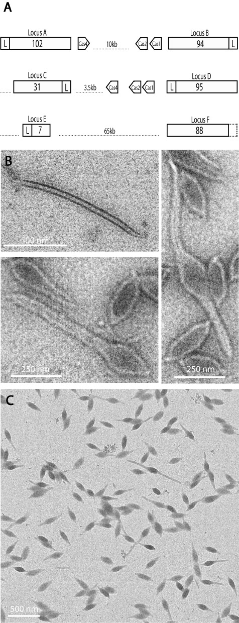
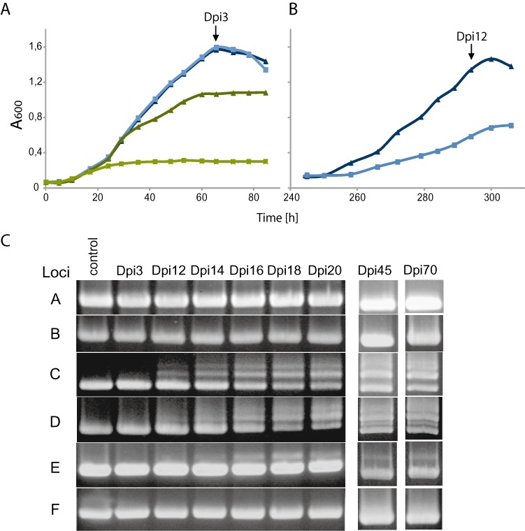
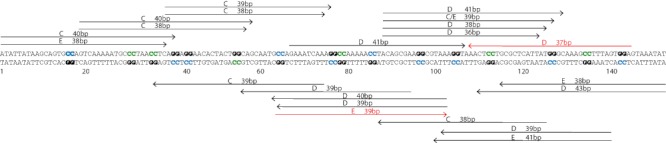
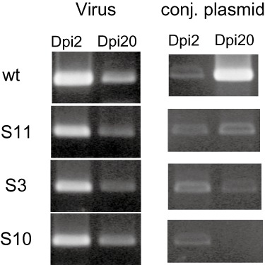
Summary: Central to the disparate adaptive immune systems of archaea and bacteria are clustered regularly interspaced short palindromic repeats (CRISPR). The spacer regions derive from invading genetic elements and, via RNA intermediates and associated proteins, target and cleave nucleic acids of the invader. Here we demonstrate the hyperactive uptake of hundreds of unique spacers within CRISPR loci associated with type I and IIIB immune systems of a hyperthermophilic archaeon. Infection with an environmental virus mixture resulted in the exclusive uptake of protospacers from a co‐infecting putative conjugative plasmid. Spacer uptake occurred by two distinct mechanisms in only one of two CRISPR loci subfamilies present. In two loci, insertions, often multiple, occurred adjacent to the leader while in a third locus single spacers were incorporated throughout the array. Protospacer DNAs were excised from the invading genetic element immediately after CCN motifs, on either strand, with the secondary cut apparently produced by a ruler mechanism. Over a 10‐week period, there was a gradual decrease in the number of wild‐type cells present in the culture but the virus and putative conjugative plasmid were still propagating. The results underline the complex dynamics of CRISPR‐based immune systems within a population infected with genetic elements.
Overview
The claims and statistics of this paper haven’t been checked yet.
Run the check — free, no account needed. It reads the open-access text and checks the sources it cites.
Integrity signals
Registry
Cites
Statistics
Figures

Verification
Claims the paper makes
The claims and statistics of this paper haven’t been checked yet.
Run the check — free, no account needed. It reads the open-access text and checks the sources it cites.
Claims other papers make about this one
Each is checked against this paper’s own text: does it say what the citing paper says it does?
The cited source says what the claim attributes to it.
In the citing paper
“Therefore, spacers are arranged chronologically within the array, with a few exceptions [29,30].”
From CRISPR-Cas: Converting A Bacterial Defence Mechanism into A State-of-the-Art Genetic Manipulation Tool (2019) · cites this paper as [30]
Self-check
Percentages, sums and reported differences re-computed from the paper’s own numbers.
The self-check needs the paper’s full text.
Citations
Cites
CRISPR RNA Maturation by Trans-Encoded Small RNA and Host Factor RNase III
Deltcheva, E., Chylinski, K., Sharma, C. M., Gonzales, K., Chao, Y., Pirzada, Z. A., Eckert, M. R., Vogel, J., & Charpentier, E. (2011) · Nature · doi:10.1038/nature09886 · Cited by 2,563
Research works printed but not linked: 56
- 10.1126/science.1157358 · DOI
- Aranaz A., 1996, Spacer oligonucleotide typing of Mycobacterium bovis strains from cattle and other animals: a tool for studying epidemiology of tuberculosis, J Clin Microbiol, 34, 2734, 10.1128/jcm.34.11.2734-2740.1996 · DOI
- 10.1046/j.1365-2958.1999.01573.x · DOI
- 10.1126/science.1138140 · DOI · Paper page
- 10.1111/j.1365-2958.2008.06488.x · DOI
- 10.1073/pnas.0901238106 · DOI
- 10.1099/mic.0.28048-0 · DOI · Paper page
- 10.1126/science.1159689 · DOI · Paper page
- 10.1093/nar/gkr1111 · DOI
- 10.1128/JB.01412-07 · DOI
- 10.1111/j.1462-2920.2010.02266.x · DOI
- 10.1016/j.tim.2011.08.002 · DOI · Paper page
- 10.1042/BST0390051 · DOI
- 10.1155/2004/151926 · DOI
- 10.1111/j.1365-2958.1993.tb00976.x · DOI · Paper page
- 10.1111/j.1365-2958.2010.07452.x · DOI
- 10.1016/j.cell.2009.07.040 · DOI · Paper page
- 10.1016/j.molcel.2011.10.023 · DOI
- 10.1038/4361101a · DOI
- 10.1073/pnas.1112832108 · DOI · Paper page
- 10.1111/j.1462-2920.2008.01784.x · DOI
- 10.1128/iai.59.8.2695-2705.1991 · DOI
- 10.1128/JB.01415-07 · DOI
- 10.1038/nsmb.2019 · DOI
- 10.1128/JCM.35.4.907-914.1997 · DOI
- 10.1155/2006/542818 · DOI
- 10.1111/j.1365-2958.2009.06641.x · DOI
- 10.1016/j.jmb.2010.11.019 · DOI
- 10.1038/nrmicro2577 · DOI · Paper page
- 10.1111/j.1365-2958.2011.07586.x · DOI
- 10.1126/science.1165771 · DOI · Paper page
- 10.1007/s00239-004-0046-3 · DOI · Paper page
- 10.1099/mic.0.023960-0 · DOI · Paper page
- 10.1111/j.1574-6976.2011.00280.x · DOI
- 10.1016/j.mib.2007.05.017 · DOI
- 10.1099/mic.0.27437-0 · DOI · Paper page
- 10.1006/plas.1998.1363 · DOI
- 10.1038/nrmicro1527 · DOI
- 10.1016/j.jmb.2006.04.027 · DOI
- 10.1093/bioinformatics/16.10.944 · DOI
- 10.1016/j.resmic.2010.09.001 · DOI
- 10.1042/BST0370023 · DOI
- Shah S.A., 2011, Regulatory RNAs in Prokaryotes, 163
- 10.1073/pnas.141222098 · DOI
- 10.1016/j.resmic.2008.04.015 · DOI
- 10.1371/journal.pone.0035888 · DOI
- 10.1073/pnas.112047299 · DOI
- 10.1111/j.1365-2958.2004.04428.x · DOI
- 10.1111/j.1462-2920.2007.01444.x · DOI
- 10.1016/j.virol.2007.01.035 · DOI
- 10.1101/gr.100396.109 · DOI
- 10.1128/JVI.79.14.8677-8686.2005 · DOI
- 10.1093/nar/gks216 · DOI · Paper page
- 10.1016/j.molcel.2011.12.013 · DOI
- Zillig W., 1994, Screening for Sulfolobales, their plasmids and their viruses in Icelandic solfataras, Syst Appl Microbiol, 16, 109
- 10.1007/s007920050052 · DOI
Cited by
Sorted by citations
Loureiro, A., & da Silva, G. J. (2019) · Antibiotics · doi:10.3390/antibiotics8010018 · Cited by 129
CRISPR-Cas systems in multicellular cyanobacteria
Hou, S., Brenes-Álvarez, M., Reimann, V., Alkhnbashi, O. S., Backofen, R., Muro-Pastor, A. M., & Hess, W. R. (2019) · RNA Biology · doi:10.1080/15476286.2018.1493330 · Cited by 47
Metadata
- Authors
- Erdmann, S., & Garrett, R. A.
- Venue
- Molecular Microbiology
- Publisher
- Wiley
- Published
- 2012-07-27
- Volume
- 85
- Issue
- 6
- Pages
- 1044-1056
- ISSN
- 0950-382X
- 1365-2958
- PMC
- PMC3468723
- Cited by
- 138
- References
- 1
- Language
- en
About these checks
Registry, retraction, Cites and statistics results are computed, not judged, with one exception: a printed reference that no index lists is read once by a model before it counts as a citation error. Claims are extracted by a model and kept only when their quote appears verbatim in the text; each cited source is then checked, with a model’s help, for whether it says what the claim relies on. Those verdicts can be wrong — read the claim’s quote and the cited source before relying on one.
Statistics and arithmetic are recomputed, not judged.
Confidence bands are shown only for fields calibrated on our annotated test set. None is calibrated yet, so no band is shown.
What counts as a claim ›How we measure confidence ›How we check this ›
Are you an author, or do you see a problem with this page? Request a review
