

The DOI matches its Crossref record; 23 of the 23 references it cites were found in a registry or an index.
Molecular mechanisms of tumor resistance to radiotherapy
Abstract
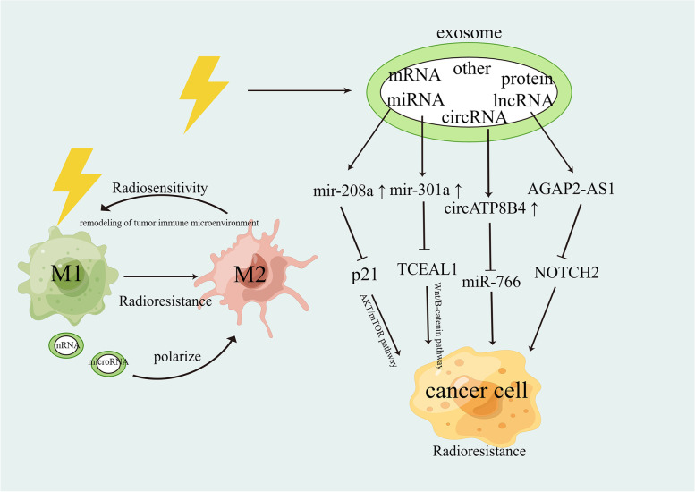
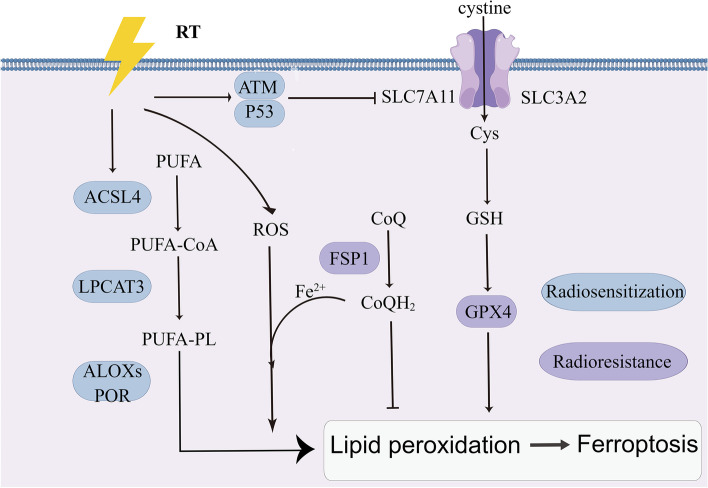
Background: Cancer is the most prevalent cause of death globally, and radiotherapy is considered the standard of care for most solid tumors, including lung, breast, esophageal, and colorectal cancers and glioblastoma. Resistance to radiation can lead to local treatment failure and even cancer recurrence. Main body: In this review, we have extensively discussed several crucial aspects that cause resistance of cancer to radiation therapy, including radiation-induced DNA damage repair, cell cycle arrest, apoptosis escape, abundance of cancer stem cells, modification of cancer cells and their microenvironment, presence of exosomal and non-coding RNA, metabolic reprogramming, and ferroptosis. We aim to focus on the molecular mechanisms of cancer radiotherapy resistance in relation to these aspects and to discuss possible targets to improve treatment outcomes. Conclusions: Studying the molecular mechanisms responsible for radiotherapy resistance and its interactions with the tumor environment will help improve cancer responses to radiotherapy. Our review provides a foundation to identify and overcome the obstacles to effective radiotherapy.
Overview
The claims and statistics of this paper haven’t been checked yet.
Run the check — free, no account needed. It reads the open-access text and checks the sources it cites.
Integrity signals
Registry
Cites
Statistics
Figures
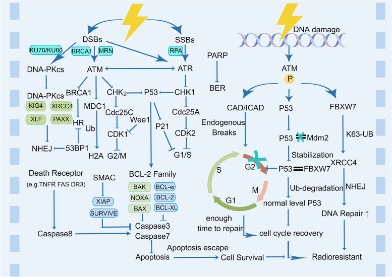
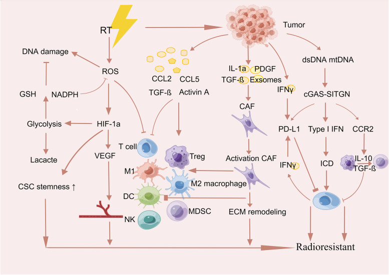
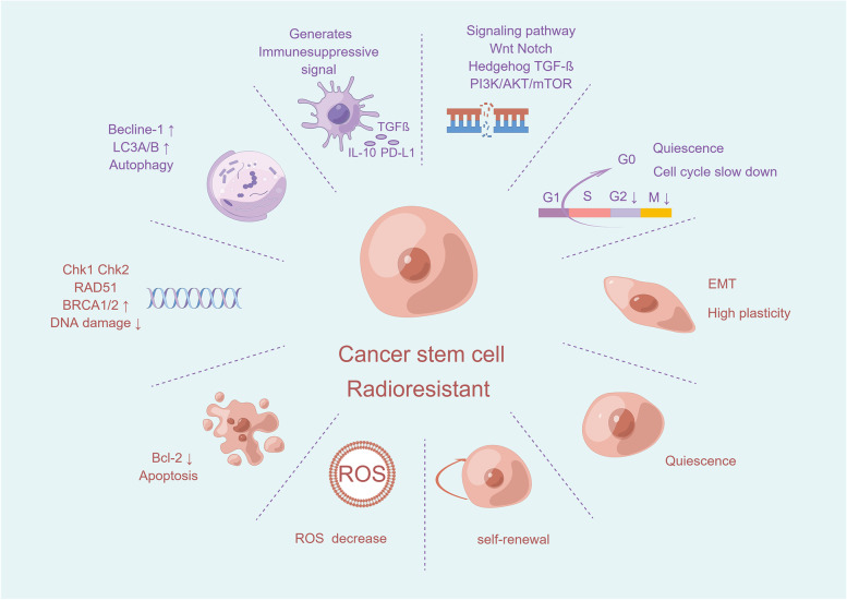
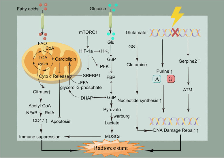
Verification
Claims the paper makes
The claims and statistics of this paper haven’t been checked yet.
Run the check — free, no account needed. It reads the open-access text and checks the sources it cites.
Claims other papers make about this one
Each is checked against this paper’s own text: does it say what the citing paper says it does?
In the citing paper
“Moreover, some tumors are radioresistant and may not respond adequately even at high radiation doses, increasing the risk of recurrence [19].”
From Advances in green-synthesized magnetic nanoparticles for targeted cancer therapy: mechanisms, applications, and future perspectives (2026) · cites this paper as [19]
In the citing paper
“High-energy radiation can damage surrounding healthy tissues and cause long-term side effects, such as fibrosis, secondary malignancies, and organ dysfunction.”
From Advances in green-synthesized magnetic nanoparticles for targeted cancer therapy: mechanisms, applications, and future perspectives (2026) · cites this paper as [19]
Self-check
Percentages, sums and reported differences re-computed from the paper’s own numbers.
The self-check needs the paper’s full text.
Citations
Cites
Sung, H., Ferlay, J., Siegel, R. L., Laversanne, M., Soerjomataram, I., Jemal, A., & Bray, F. (2021) · CA: A Cancer Journal for Clinicians · doi:10.3322/caac.21660 · Cited by 108,699
Siegel, R. L., Miller, K. D., Wagle, N. S., & Jemal, A. (2023) · CA: A Cancer Journal for Clinicians · doi:10.3322/caac.21763 · Cited by 16,303
The biology , function , and biomedical applications of exosomes
Kalluri, R., & LeBleu, V. S. (2020) · Science · doi:10.1126/science.aau6977 · Cited by 10,770
Hallmarks of Cancer: New Dimensions
Hanahan, D. (2022) · Cancer Discovery · doi:10.1158/2159-8290.cd-21-1059 · Cited by 9,887
Autophagy fights disease through cellular self-digestion
Mizushima, N., Levine, B., Cuervo, A. M., & Klionsky, D. J. (2008) · Nature · doi:10.1038/nature06639 · Cited by 6,452
The emerging hallmarks of cancer metabolism
Pavlova, N. N., & Thompson, C. B. (2016) · Cell Metabolism · doi:10.1016/j.cmet.2015.12.006 · Cited by 5,926
Ferroptosis as a p53-mediated activity during tumour suppression
Jiang, L., Kon, N., Li, T., Wang, S.-J., Su, T., Hibshoosh, H., Baer, R., & Gu, W. (2015) · Nature · doi:10.1038/nature14344 · Cited by 3,398
Association of reactive oxygen species levels and radioresistance in cancer stem cells
Diehn, M., Cho, R. W., Lobo, N. A., Kalisky, T., Dorie, M. J., Kulp, A. N., Qian, D., Lam, J. S., Ailles, L. E., Wong, M., et al. (2009) · Nature · doi:10.1038/nature07733 · Cited by 2,555
Radiation and dual checkpoint blockade activate non-redundant immune mechanisms in cancer
Twyman-Saint Victor, C., Rech, A. J., Maity, A., Rengan, R., Pauken, K. E., Stelekati, E., Benci, J. L., Xu, B., Dada, H., Odorizzi, P. M., et al. (2015) · Nature · doi:10.1038/nature14292 · Cited by 2,434
The Tumour Microenvironment after Radiotherapy: Mechanisms of Resistance and Recurrence
Barker, H. E., Paget, J. T. E., Khan, A. A., & Harrington, K. J. (2015) · Nature Reviews Cancer · doi:10.1038/nrc3958 · Cited by 2,144
DNA exonuclease Trex1 regulates radiotherapy-induced tumour immunogenicity
Vanpouille-Box, C., Alard, A., Aryankalayil, M. J., Sarfraz, Y., Diamond, J. M., Schneider, R. J., Inghirami, G., Coleman, C. N., Formenti, S. C., & Demaria, S. (2017) · Nature Communications · doi:10.1038/ncomms15618 · Cited by 1,834
The ATM protein kinase: regulating the cellular response to genotoxic stress, and more
Shiloh, Y., & Ziv, Y. (2013) · Nature Reviews Molecular Cell Biology · doi:10.1038/nrm3546 · Cited by 1,649
Autophagy maintains stemness by preventing senescence
García-Prat, L., Martínez-Vicente, M., Perdiguero, E., Ortet, L., Rodríguez-Ubreva, J., Rebollo, E., Ruiz-Bonilla, V., Gutarra, S., Ballestar, E., Serrano, A. L., et al. (2016) · Nature · doi:10.1038/nature16187 · Cited by 1,355
The role of ferroptosis in ionizing radiation-induced cell death and tumor suppression
Lei, G., Zhang, Y., Koppula, P., Liu, X., Zhang, J., Lin, S. H., Ajani, J. A., Xiao, Q., Liao, Z., Wang, H., et al. (2020) · Cell Research · doi:10.1038/s41422-019-0263-3 · Cited by 1,318
Pancreatic cancer stroma: an update on therapeutic targeting strategies
Hosein, A. N., Brekken, R. A., & Maitra, A. (2020) · Nature Reviews Gastroenterology & Hepatology · doi:10.1038/s41575-020-0300-1 · Cited by 950
Mitotic catastrophe: a mechanism for avoiding genomic instability
Vitale, I., Galluzzi, L., Castedo, M., & Kroemer, G. (2011) · Nature Reviews Molecular Cell Biology · doi:10.1038/nrm3115 · Cited by 834
Gorelik, L., & Flavell, R. A. (2001) · Nature Medicine · doi:10.1038/nm1001-1118 · Cited by 770
Metabolic Reprogramming: the Emerging Concept and Associated Therapeutic Strategies
Yoshida, G. J. (2015) · Journal of Experimental & Clinical Cancer Research · doi:10.1186/s13046-015-0221-y · Cited by 716
Hypoxic Radiosensitization: Adored and Ignored
Overgaard, J. (2007) · Journal of Clinical Oncology · doi:10.1200/jco.2007.12.7878 · Cited by 619
Activation and regulation of ATM kinase activity in response to DNA double-strand breaks
Lee, J.-H., & Paull, T. T. (2007) · Oncogene · doi:10.1038/sj.onc.1210872 · Cited by 545
Fu, S., Li, Z., Xiao, L., Hu, W., Zhang, L., Xie, B., Zhou, Q., He, J., Qiu, Y., Wen, M., et al. (2019) · Cell Reports · doi:10.1016/j.celrep.2019.07.002 · Cited by 169
Peng, Y., Liao, Q., Tan, W., Peng, C., Hu, Z., Chen, Y., Li, Z., Li, J., Zhen, B., Zhu, W., et al. (2019) · Nature Communications · doi:10.1038/s41467-019-09232-8 · Cited by 104
Functional Immune Cell‐Derived Exosomes Engineered for the Trilogy of Radiotherapy Sensitization
Ma, X., Yao, M., Gao, Y., Yue, Y., Li, Y., Zhang, T., Nie, G., Zhao, X., & Liang, X. (2022) · Advanced Science · doi:10.1002/advs.202106031 · Cited by 82
Research works printed but not linked: 232
- Krzyszczyk P, Acevedo A, Davidoff EJ, et al. The growing role of precision and personalized medicine for cancer treatment. Technology (Singap World Sci). 2018;6(3–4):79–100. https://doi.org/10.1142/S2339547818300020. · DOI
- Larionova I, Rakina M, Ivanyuk E, Trushchuk Y, Chernyshova A, Denisov E. Radiotherapy resistance: identifying universal biomarkers for various human cancers. J Cancer Res Clin Oncol. 2022;148(5):1015–31. https://doi.org/10.1007/s00432-022-03923-4. · DOI
- Chandra RA, Keane FK, Voncken FEM, Thomas CR Jr. Contemporary radiotherapy: present and future. Lancet. 2021;398(10295):171–84. https://doi.org/10.1016/S0140-6736(21)00233-6. · DOI
- Kumari N, Raghavan SC. G-quadruplex DNA structures and their relevance in radioprotection. Biochim Biophys Acta Gen Subj. 2021;1865(5):129857. https://doi.org/10.1016/j.bbagen.2021.129857. · DOI
- Balmukhanov SB, Yefimov ML, Kleinbock TS. Acquired radioresistance of tumour cells. Nature. 1967;216(5116):709–11. https://doi.org/10.1038/216709a0. · DOI
- Kim W, Youn H, Kang C, Youn B. Inflammation-induced radioresistance is mediated by ROS-dependent inactivation of protein phosphatase 1 in non-small cell lung cancer cells. Apoptosis. 2015;20(9):1242–52. https://doi.org/10.1007/s10495-015-1141-1. · DOI
- Yard BD, Adams DJ, Chie EK, et al. A genetic basis for the variation in the vulnerability of cancer to DNA damage. Nat Commun. 2016;7:11428. Published 2016 Apr 25. https://doi.org/10.1038/ncomms11428. · DOI
- Olivares-Urbano MA, Griñán-Lisón C, Marchal JA, Núñez MI. CSC Radioresistance: A Therapeutic Challenge to Improve Radiotherapy Effectiveness in Cancer. Cells. 2020;9(7):1651. Published 2020 Jul 9. https://doi.org/10.3390/cells9071651. · DOI
- Kabakov AE, Yakimova AO. Hypoxia-Induced Cancer Cell Responses Driving Radioresistance of Hypoxic Tumors: Approaches to Targeting and Radiosensitizing. Cancers (Basel). 2021;13(5):1102. Published 2021 Mar 4. https://doi.org/10.3390/cancers13051102. · DOI
- Khanna KK, Jackson SP. DNA double-strand breaks: signaling, repair and the cancer connection. Nat Genet. 2001;27(3):247–54. https://doi.org/10.1038/85798. · DOI · Paper page
- Fojta M, Daňhel A, Havran L, Vyskočil V. Recent progress in electrochemical sensors and assays for DNA damage and repair. TrAC Trends Anal Chem. 2016;79:160–7. https://doi.org/10.1016/j.trac.2015.11.018. · DOI
- Blackford AN, Jackson SP. ATM, ATR, and DNA-PK: The Trinity at the Heart of the DNA Damage Response. Mol Cell. 2017;66(6):801–17. https://doi.org/10.1016/j.molcel.2017.05.015. · DOI
- Ho V, Chung L, Singh A, et al. Overexpression of the MRE11-RAD50-NBS1 (MRN) complex in rectal cancer correlates with poor response to neoadjuvant radiotherapy and prognosis. BMC Cancer. 2018;18(1):869. Published 2018 Sep 3. https://doi.org/10.1186/s12885-018-4776-9. · DOI
- Podhorecka M, Skladanowski A, Bozko P. H2AX Phosphorylation: Its Role in DNA Damage Response and Cancer Therapy. J Nucleic Acids. 2010;2010:920161. Published 2010 Aug 3. https://doi.org/10.4061/2010/920161. · DOI
- Borràs M, Armengol G, De Cabo M, Barquinero JF, Barrios L. Comparison of methods to quantify histone H2AX phosphorylation and its usefulness for prediction of radiosensitivity. Int J Radiat Biol. 2015;91(12):915–24. https://doi.org/10.3109/09553002.2015.1101501. · DOI
- Pucci S, Polidoro C, Joubert A, et al. Ku70, Ku80, and sClusterin: A Cluster of Predicting Factors for Response to Neoadjuvant Chemoradiation Therapy in Patients With Locally Advanced Rectal Cancer. Int J Radiat Oncol Biol Phys. 2017;97(2):381–8. https://doi.org/10.1016/j.ijrobp.2016.10.018. · DOI
- Baptistella AR, Landemberger MC, Dias MVS, et al. Rab5C enhances resistance to ionizing radiation in rectal cancer. J Mol Med (Berl). 2019;97(6):855–69. https://doi.org/10.1007/s00109-019-01760-6. · DOI
- Zhang Q, Karnak D, Tan M, Lawrence TS, Morgan MA, Sun Y. FBXW7 Facilitates Nonhomologous End-Joining via K63-Linked Polyubiquitylation of XRCC4. Mol Cell. 2016;61(3):419–33. https://doi.org/10.1016/j.molcel.2015.12.010. · DOI
- Jette N, Lees-Miller SP. The DNA-dependent protein kinase: A multifunctional protein kinase with roles in DNA double strand break repair and mitosis. Prog Biophys Mol Biol. 2015;117(2–3):194–205. https://doi.org/10.1016/j.pbiomolbio.2014.12.003. · DOI
- Ghelli Luserna di Rora' A, Iacobucci I, Martinelli G. The cell cycle checkpoint inhibitors in the treatment of leukemias. J Hematol Oncol. 2017;10(1):77. Published 2017 Mar 29. https://doi.org/10.1186/s13045-017-0443-x. · DOI
- Li J, Yang CX, Mei ZJ, et al. Involvement of cdc25c in cell cycle alteration of a radioresistant lung cancer cell line established with fractionated ionizing radiation. Asian Pac J Cancer Prev. 2013;14(10):5725–30. https://doi.org/10.7314/apjcp.2013.14.10.5725. · DOI
- Halazonetis TD, Gorgoulis VG, Bartek J. An oncogene-induced DNA damage model for cancer development. Science. 2008;319(5868):1352–5. https://doi.org/10.1126/science.1140735. · DOI · Paper page
- Larsen BD, Benada J, Yung PYK, et al. Cancer cells use self-inflicted DNA breaks to evade growth limits imposed by genotoxic stress. Science. 2022;376(6592):476–83. https://doi.org/10.1126/science.abi6378. · DOI
- Paull TT. Mechanisms of ATM Activation. Annu Rev Biochem. 2015;84:711–38. https://doi.org/10.1146/annurev-biochem-060614-034335. · DOI
- Matsuoka S, Ballif BA, Smogorzewska A, et al. ATM and ATR substrate analysis reveals extensive protein networks responsive to DNA damage. Science. 2007;316(5828):1160–6. https://doi.org/10.1126/science.1140321. · DOI · Paper page
- Shiloh Y. The ATM-mediated DNA-damage response: taking shape. Trends Biochem Sci. 2006;31(7):402–10. https://doi.org/10.1016/j.tibs.2006.05.004. · DOI
- Weber AM, Ryan AJ. ATM and ATR as therapeutic targets in cancer. Pharmacol Ther. 2015;149:124–38. https://doi.org/10.1016/j.pharmthera.2014.12.001. · DOI
- Streffer JR, Rimner A, Rieger J, Naumann U, Rodemann HP, Weller M. BCL-2 family proteins modulate radiosensitivity in human malignant glioma cells. J Neurooncol. 2002;56(1):43–9. https://doi.org/10.1023/a:1014448721327. · DOI
- Ma R, Zhao LN, Yang H, et al. RNA binding motif protein 3 (RBM3) drives radioresistance in nasopharyngeal carcinoma by reducing apoptosis via the PI3K/AKT/Bcl-2 signaling pathway. Am J Transl Res. 2018;10(12):4130–4140. Published 2018 Dec 15.
- Ferris RL, Harrington K, Schoenfeld JD, et al. Inhibiting the inhibitors: Development of the IAP inhibitor xevinapant for the treatment of locally advanced squamous cell carcinoma of the head and neck. Cancer Treat Rev. 2023;113:102492. https://doi.org/10.1016/j.ctrv.2022.102492. · DOI
- Lee JU, Hosotani R, Wada M, et al. Role of Bcl-2 family proteins (Bax, Bcl-2 and Bcl-X) on cellular susceptibility to radiation in pancreatic cancer cells. Eur J Cancer. 1999;35(9):1374–80. https://doi.org/10.1016/s0959-8049(99)00134-3. · DOI
- Zhang L, Lu Z, Zhao X. Targeting Bcl-2 for cancer therapy. Biochim Biophys Acta Rev Cancer. 2021;1876(1):188569. https://doi.org/10.1016/j.bbcan.2021.188569. · DOI
- Liang H, Deng L, Hou Y, et al. Host STING-dependent MDSC mobilization drives extrinsic radiation resistance. Nat Commun. 2017;8(1):1736. Published 2017 Nov 23. https://doi.org/10.1038/s41467-017-01566-5. · DOI
- Li JY, Zhao Y, Gong S, et al. TRIM21 inhibits irradiation-induced mitochondrial DNA release and impairs antitumour immunity in nasopharyngeal carcinoma tumour models. Nat Commun. 2023;14(1):865. Published 2023 Feb 16. https://doi.org/10.1038/s41467-023-36523-y. · DOI
- Du SS, Chen GW, Yang P, et al. Radiation Therapy Promotes Hepatocellular Carcinoma Immune Cloaking via PD-L1 Upregulation Induced by cGAS-STING Activation. Int J Radiat Oncol Biol Phys. 2022;112(5):1243–55. https://doi.org/10.1016/j.ijrobp.2021.12.162. · DOI
- Wang NH, Lei Z, Yang HN, et al. Radiation-induced PD-L1 expression in tumor and its microenvironment facilitates cancer-immune escape: a narrative review. Ann Transl Med. 2022;10(24):1406. https://doi.org/10.21037/atm-22-6049. · DOI
- Deng L, Liang H, Burnette B, et al. Irradiation and anti-PD-L1 treatment synergistically promote antitumor immunity in mice. J Clin Invest. 2014;124(2):687–95. https://doi.org/10.1172/JCI67313. · DOI
- Formenti SC, Rudqvist NP, Golden E, et al. Radiotherapy induces responses of lung cancer to CTLA-4 blockade. Nat Med. 2018;24(12):1845–51. https://doi.org/10.1038/s41591-018-0232-2. · DOI · Paper page
- Thomas DA, Massagué J. TGF-beta directly targets cytotoxic T cell functions during tumor evasion of immune surveillance. Cancer Cell. 2005;8(5):369–80. https://doi.org/10.1016/j.ccr.2005.10.012. · DOI · Paper page
- De Martino M, Daviaud C, Diamond JM, et al. Activin A Promotes Regulatory T-cell-Mediated Immunosuppression in Irradiated Breast Cancer. Cancer Immunol Res. 2021;9(1):89–102. https://doi.org/10.1158/2326-6066.CIR-19-0305. · DOI
- Kalbasi A, Komar C, Tooker GM, et al. Tumor-Derived CCL2 Mediates Resistance to Radiotherapy in Pancreatic Ductal Adenocarcinoma. Clin Cancer Res. 2017;23(1):137–48. https://doi.org/10.1158/1078-0432.CCR-16-0870. · DOI
- Vaupel P, Multhoff G. Adenosine can thwart antitumor immune responses elicited by radiotherapy : Therapeutic strategies alleviating protumor ADO activities. Adenosine kann Strahlentherapie-vermittelte Immunantworten gegen Tumore konterkarieren. Strahlenther Onkol. 2016;192(5):279–287. https://doi.org/10.1007/s00066-016-0948-1. · DOI
- Nixon BG, Gao S, Wang X, Li MO. TGFβ control of immune responses in cancer: a holistic immuno-oncology perspective [published online ahead of print, 2022 Nov 15]. Nat Rev Immunol. 2022;https://doi.org/10.1038/s41577-022-00796-z. · DOI
- Hardee ME, Marciscano AE, Medina-Ramirez CM, et al. Resistance of glioblastoma-initiating cells to radiation mediated by the tumor microenvironment can be abolished by inhibiting transforming growth factor-β. Cancer Res. 2012;72(16):4119–29. https://doi.org/10.1158/0008-5472.CAN-12-0546. · DOI
- Paget S. The distribution of secondary growths in cancer of the breast. 1889. Cancer Metastasis Rev. 1989;8(2):98–101.
- Shi R, Tang YQ, Miao H. Metabolism in tumor microenvironment: Implications for cancer immunotherapy. MedComm (2020). 2020;1(1):47–68. Published 2020 Jun 3. https://doi.org/10.1002/mco2.6. · DOI
- Arneth B. Tumor Microenvironment. Medicina (Kaunas). 2019;56(1):15. Published 2019 Dec 30. https://doi.org/10.3390/medicina56010015. · DOI
- Denk D, Greten FR. Inflammation: the incubator of the tumor microenvironment. Trends Cancer. 2022;8(11):901–14. https://doi.org/10.1016/j.trecan.2022.07.002. · DOI
- Chajon E, Castelli J, Marsiglia H, De Crevoisier R. The synergistic effect of radiotherapy and immunotherapy: A promising but not simple partnership. Crit Rev Oncol Hematol. 2017;111:124–32. https://doi.org/10.1016/j.critrevonc.2017.01.017. · DOI
- Shevtsov M, Sato H, Multhoff G, Shibata A. Novel Approaches to Improve the Efficacy of Immuno-Radiotherapy. Front Oncol. 2019;9:156. Published 2019 Mar 19. https://doi.org/10.3389/fonc.2019.00156. · DOI
- Semenza GL. Intratumoral hypoxia, radiation resistance, and HIF-1. Cancer Cell. 2004;5(5):405–6. https://doi.org/10.1016/s1535-6108(04)00118-7. · DOI
- Wang H, Jiang H, Van De Gucht M, De Ridder M. Hypoxic Radioresistance: Can ROS Be the Key to Overcome It?. Cancers (Basel). 2019;11(1):112. Published 2019 Jan 18. https://doi.org/10.3390/cancers11010112. · DOI
- Kotsafti A, Scarpa M, Castagliuolo I, Scarpa M. Reactive Oxygen Species and Antitumor Immunity-From Surveillance to Evasion. Cancers (Basel). 2020;12(7):1748. Published 2020 Jul 1. https://doi.org/10.3390/cancers12071748. · DOI
- Ruan K, Song G, Ouyang G. Role of hypoxia in the hallmarks of human cancer. J Cell Biochem. 2009;107(6):1053–62. https://doi.org/10.1002/jcb.22214. · DOI
- Yoshimura M, Itasaka S, Harada H, Hiraoka M. Microenvironment and radiation therapy. Biomed Res Int. 2013;2013:685308. https://doi.org/10.1155/2013/685308. · DOI
- Saunders M, Dische S. Clinical results of hypoxic cell radiosensitisation from hyperbaric oxygen to accelerated radiotherapy, carbogen and nicotinamide. Br J Cancer Suppl. 1996;27:S271–8.
- Brand A, Singer K, Koehl GE, et al. LDHA-Associated Lactic Acid Production Blunts Tumor Immunosurveillance by T and NK Cells. Cell Metab. 2016;24(5):657–71. https://doi.org/10.1016/j.cmet.2016.08.011. · DOI · Paper page
- Postow MA, Callahan MK, Barker CA, et al. Immunologic correlates of the abscopal effect in a patient with melanoma. N Engl J Med. 2012;366(10):925–31. https://doi.org/10.1056/NEJMoa1112824. · DOI
- Golden EB, Chhabra A, Chachoua A, et al. Local radiotherapy and granulocyte-macrophage colony-stimulating factor to generate abscopal responses in patients with metastatic solid tumours: a proof-of-principle trial. Lancet Oncol. 2015;16(7):795–803. https://doi.org/10.1016/S1470-2045(15)00054-6. · DOI
- Feng H, Wang J, Chen W, et al. Hypoxia-induced autophagy as an additional mechanism in human osteosarcoma radioresistance. J Bone Oncol. 2016;5(2):67–73. Published 2016 Mar 9. https://doi.org/10.1016/j.jbo.2016.03.001. · DOI
- Fiori ME, Di Franco S, Villanova L, Bianca P, Stassi G, De Maria R. Cancer-associated fibroblasts as abettors of tumor progression at the crossroads of EMT and therapy resistance. Mol Cancer. 2019;18(1):70. Published 2019 Mar 30. https://doi.org/10.1186/s12943-019-0994-2. · DOI
- Galluzzi L, Vanpouille-Box C, Bakhoum SF, Demaria S. SnapShot: CGAS-STING Signaling. Cell. 2018;173(1):276-276.e1. https://doi.org/10.1016/j.cell.2018.03.015. · DOI
- Huang Z, Wang Y, Yao D, Wu J, Hu Y, Yuan A. Nanoscale coordination polymers induce immunogenic cell death by amplifying radiation therapy mediated oxidative stress. Nat Commun. 2021;12(1):145. Published 2021 Jan 8. https://doi.org/10.1038/s41467-020-20243-8. · DOI
- O’Brien CA, Kreso A, Dick JE. Cancer stem cells in solid tumors: an overview. Semin Radiat Oncol. 2009;19(2):71–7. https://doi.org/10.1016/j.semradonc.2008.11.001. · DOI
- Hittelman WN, Liao Y, Wang L, Milas L. Are cancer stem cells radioresistant? Future Oncol. 2010;6:1563–76. https://doi.org/10.2217/fon.10.121. · DOI
- Chang JC. Cancer stem cells: Role in tumor growth, recurrence, metastasis, and treatment resistance. Medicine (Baltimore). 2016;95(1 Suppl 1):S20–5. https://doi.org/10.1097/MD.0000000000004766. · DOI · Paper page
- Marie-Egyptienne DT, Lohse I, Hill RP. Cancer stem cells, the epithelial to mesenchymal transition (EMT) and radioresistance: potential role of hypoxia. Cancer Lett. 2013;341(1):63–72. https://doi.org/10.1016/j.canlet.2012.11.019. · DOI
- Skvortsov S, Debbage P, Lukas P, Skvortsova I. Crosstalk between DNA repair and cancer stem cell (CSC) associated intracellular pathways. Semin Cancer Biol. 2015;31:36–42. https://doi.org/10.1016/j.semcancer.2014.06.002. · DOI
- Dando I, Cordani M, Dalla Pozza E, Biondani G, Donadelli M, Palmieri M. Antioxidant Mechanisms and ROS-Related MicroRNAs in Cancer Stem Cells. Oxid Med Cell Longev. 2015;2015:425708. https://doi.org/10.1155/2015/425708. · DOI
- Lei Y, Zhang D, Yu J, Dong H, Zhang J, Yang S. Targeting autophagy in cancer stem cells as an anticancer therapy [published correction appears in Cancer Lett. 2018 Jan 4;:]. Cancer Lett. 2017;393:33–39. https://doi.org/10.1016/j.canlet.2017.02.012. · DOI
- Yang G, Lu C, Mei Z, et al. Association of Cancer Stem Cell Radio-Resistance Under Ultra-High Dose Rate FLASH Irradiation With Lysosome-Mediated Autophagy. Front Cell Dev Biol. 2021;9:672693. Published 2021 Apr 29. https://doi.org/10.3389/fcell.2021.672693. · DOI
- Chen Y, Li D, Wang D, et al. Quiescence and attenuated DNA damage response promote survival of esophageal cancer stem cells [published correction appears in J Cell Biochem. 2021 Feb;122(2):301–302]. J Cell Biochem. 2012;113(12):3643–3652. https://doi.org/10.1002/jcb.24228. · DOI
- Anding AL, Baehrecke EH. Cleaning House: Selective Autophagy of Organelles. Dev Cell. 2017;41(1):10–22. https://doi.org/10.1016/j.devcel.2017.02.016. · DOI · Paper page
- Filomeni G, De Zio D, Cecconi F. Oxidative stress and autophagy: the clash between damage and metabolic needs. Cell Death Differ. 2015;22(3):377–88. https://doi.org/10.1038/cdd.2014.150. · DOI · Paper page
- Hou J, Han ZP, Jing YY, et al. Autophagy prevents irradiation injury and maintains stemness through decreasing ROS generation in mesenchymal stem cells. Cell Death Dis. 2013;4(10):e844. Published 2013 Oct 10. https://doi.org/10.1038/cddis.2013.338. · DOI
- Yang L, Shi P, Zhao G, et al. Targeting cancer stem cell pathways for cancer therapy. Signal Transduct Target Ther. 2020;5(1):8. Published 2020 Feb 7. https://doi.org/10.1038/s41392-020-0110-5. · DOI
- Cojoc M, Peitzsch C, Kurth I, et al. Aldehyde Dehydrogenase Is Regulated by β-Catenin/TCF and Promotes Radioresistance in Prostate Cancer Progenitor Cells. Cancer Res. 2015;75(7):1482–94. https://doi.org/10.1158/0008-5472.CAN-14-1924. · DOI
- Angulo P, Kaushik G, Subramaniam D, et al. Natural compounds targeting major cell signaling pathways: a novel paradigm for osteosarcoma therapy. J Hematol Oncol. 2017;10(1):10. Published 2017 Jan 7. https://doi.org/10.1186/s13045-016-0373-z. · DOI
- Yao Z, Han L, Chen Y, et al. Hedgehog signalling in the tumourigenesis and metastasis of osteosarcoma, and its potential value in the clinical therapy of osteosarcoma. Cell Death Dis. 2018;9(6):701. Published 2018 Jun 13. https://doi.org/10.1038/s41419-018-0647-1. · DOI
- Sharma RA, Plummer R, Stock JK, et al. Clinical development of new drug-radiotherapy combinations. Nat Rev Clin Oncol. 2016;13(10):627–42. https://doi.org/10.1038/nrclinonc.2016.79. · DOI
- Azzam EI, Jay-Gerin JP, Pain D. Ionizing radiation-induced metabolic oxidative stress and prolonged cell injury. Cancer Lett. 2012;327(1–2):48–60. https://doi.org/10.1016/j.canlet.2011.12.012. · DOI
- Li J, Sun H, Huang Y, Wang Y, Liu Y, Chen X. Pathways and assays for DNA double-strand break repair by homologous recombination. Acta Biochim Biophys Sin (Shanghai). 2019;51(9):879–89. https://doi.org/10.1093/abbs/gmz076. · DOI
- Li X, Heyer WD. Homologous recombination in DNA repair and DNA damage tolerance. Cell Res. 2008;18(1):99–113. https://doi.org/10.1038/cr.2008.1. · DOI
- Roos WP, Thomas AD, Kaina B. DNA damage and the balance between survival and death in cancer biology. Nat Rev Cancer. 2016;16(1):20–33. https://doi.org/10.1038/nrc.2015.2. · DOI · Paper page
- Gavande NS, VanderVere-Carozza PS, Hinshaw HD, et al. DNA repair targeted therapy: The past or future of cancer treatment? Pharmacol Ther. 2016;160:65–83. https://doi.org/10.1016/j.pharmthera.2016.02.003. · DOI
- Wang X, Ran T, Zhang X, et al. 3.9 Å structure of the yeast Mec1-Ddc2 complex, a homolog of human ATR-ATRIP. Science. 2017;358(6367):1206–1209. https://doi.org/10.1126/science.aan8414. · DOI
- Bharati BK, Gowder M, Zheng F, et al. Crucial role and mechanism of transcription-coupled DNA repair in bacteria. Nature. 2022;604(7904):152–9. https://doi.org/10.1038/s41586-022-04530-6. · DOI
- Caston RA, Gampala S, Armstrong L, Messmann RA, Fishel ML, Kelley MR. The multifunctional APE1 DNA repair-redox signaling protein as a drug target in human disease. Drug Discov Today. 2021;26(1):218–28. https://doi.org/10.1016/j.drudis.2020.10.015. · DOI
- Lu H, Saha J, Beckmann PJ, Hendrickson EA, Davis AJ. DNA-PKcs promotes chromatin decondensation to facilitate initiation of the DNA damage response. Nucleic Acids Res. 2019;47(18):9467–79. https://doi.org/10.1093/nar/gkz694. · DOI
- Riballo E, Doherty AJ, Dai Y, et al. Cellular and biochemical impact of a mutation in DNA ligase IV conferring clinical radiosensitivity. J Biol Chem. 2001;276(33):31124–32. https://doi.org/10.1074/jbc.M103866200. · DOI
- Herrero AB, San Miguel J, Gutierrez NC. Deregulation of DNA double-strand break repair in multiple myeloma: implications for genome stability. PLoS One. 2015;10(3):e0121581. Published 2015 Mar 19. https://doi.org/10.1371/journal.pone.0121581. · DOI
- Fang X, Huang Z, Zhai K, et al. Inhibiting DNA-PK induces glioma stem cell differentiation and sensitizes glioblastoma to radiation in mice. Sci Transl Med. 2021;13(600):eabc7275. https://doi.org/10.1126/scitranslmed.abc7275. · DOI
- Wang YH, Guo Z, An L, et al. LINC-PINT impedes DNA repair and enhances radiotherapeutic response by targeting DNA-PKcs in nasopharyngeal cancer. Cell Death Dis. 2021;12(5):454. Published 2021 May 7. https://doi.org/10.1038/s41419-021-03728-2. · DOI
- Guo Z, Wang YH, Xu H, et al. LncRNA linc00312 suppresses radiotherapy resistance by targeting DNA-PKcs and impairing DNA damage repair in nasopharyngeal carcinoma. Cell Death Dis. 2021;12(1):69. Published 2021 Jan 4. https://doi.org/10.1038/s41419-020-03302-2. · DOI
- Zenke FT, Zimmermann A, Sirrenberg C, et al. Pharmacologic Inhibitor of DNA-PK, M3814, Potentiates Radiotherapy and Regresses Human Tumors in Mouse Models. Mol Cancer Ther. 2020;19(5):1091–101. https://doi.org/10.1158/1535-7163.MCT-19-0734. · DOI
- Fok JHL, Ramos-Montoya A, Vazquez-Chantada M, et al. AZD7648 is a potent and selective DNA-PK inhibitor that enhances radiation, chemotherapy and olaparib activity. Nat Commun. 2019;10(1):5065. Published 2019 Nov 7. https://doi.org/10.1038/s41467-019-12836-9. · DOI
- Wu C, Williams TM, Robb R, et al. Phase I Trial of Trametinib with Neoadjuvant Chemoradiation in Patients with Locally Advanced Rectal Cancer. Clin Cancer Res. 2020;26(13):3117–25. https://doi.org/10.1158/1078-0432.CCR-19-4193. · DOI
- Estrada-Bernal A, Chatterjee M, Haque SJ, et al. MEK inhibitor GSK1120212-mediated radiosensitization of pancreatic cancer cells involves inhibition of DNA double-strand break repair pathways. Cell Cycle. 2015;14(23):3713–24. https://doi.org/10.1080/15384101.2015.1104437. · DOI
- Wei F, Yan J, Tang D, et al. Inhibition of ERK activation enhances the repair of double-stranded breaks via non-homologous end joining by increasing DNA-PKcs activation. Biochim Biophys Acta. 2013;1833(1):90–100. https://doi.org/10.1016/j.bbamcr.2012.10.016. · DOI
- Ouellette MM, Zhou S, Yan Y. Cell Signaling Pathways That Promote Radioresistance of Cancer Cells. Diagnostics (Basel). 2022;12(3):656. Published 2022 Mar 8. https://doi.org/10.3390/diagnostics12030656. · DOI
- Mansour WY, Rhein T, Dahm-Daphi J. The alternative end-joining pathway for repair of DNA double-strand breaks requires PARP1 but is not dependent upon microhomologies. Nucleic Acids Res. 2010;38(18):6065–77. https://doi.org/10.1093/nar/gkq387. · DOI
- Cheng Q, Barboule N, Frit P, et al. Ku counteracts mobilization of PARP1 and MRN in chromatin damaged with DNA double-strand breaks. Nucleic Acids Res. 2011;39(22):9605–19. https://doi.org/10.1093/nar/gkr656. · DOI
- Curtin NJ, Szabo C. Poly(ADP-ribose) polymerase inhibition: past, present and future. Nat Rev Drug Discov. 2020;19(10):711–36. https://doi.org/10.1038/s41573-020-0076-6. · DOI
- Noordermeer SM, van Attikum H. PARP Inhibitor Resistance: A Tug-of-War in BRCA-Mutated Cells. Trends Cell Biol. 2019;29(10):820–34. https://doi.org/10.1016/j.tcb.2019.07.008. · DOI
- Lee JM, Ledermann JA, Kohn EC. PARP Inhibitors for BRCA1/2 mutation-associated and BRCA-like malignancies. Ann Oncol. 2014;25(1):32–40. https://doi.org/10.1093/annonc/mdt384. · DOI
- Huang A, Garraway LA, Ashworth A, Weber B. Synthetic lethality as an engine for cancer drug target discovery. Nat Rev Drug Discov. 2020;19(1):23–38. https://doi.org/10.1038/s41573-019-0046-z. · DOI
- Gani C, Coackley C, Kumareswaran R, et al. In vivo studies of the PARP inhibitor, AZD-2281, in combination with fractionated radiotherapy: An exploration of the therapeutic ratio. Radiother Oncol. 2015;116(3):486–94. https://doi.org/10.1016/j.radonc.2015.08.003. · DOI
- Pant S, Maitra A, Yap TA. PARP inhibition - opportunities in pancreatic cancer. Nat Rev Clin Oncol. 2019;16(10):595–6. https://doi.org/10.1038/s41571-019-0257-6. · DOI
- Loap P, Loirat D, Berger F, et al. Concurrent Olaparib and Radiotherapy in Patients With Triple-Negative Breast Cancer: The Phase 1 Olaparib and Radiation Therapy for Triple-Negative Breast Cancer Trial. JAMA Oncol. 2022;8(12):1802–8. https://doi.org/10.1001/jamaoncol.2022.5074. · DOI
- Bi Y, Verginadis II, Dey S, et al. Radiosensitization by the PARP inhibitor olaparib in BRCA1-proficient and deficient high-grade serous ovarian carcinomas. Gynecol Oncol. 2018;150(3):534–44. https://doi.org/10.1016/j.ygyno.2018.07.002. · DOI
- Mirza MR, Monk BJ, Herrstedt J, et al. Niraparib Maintenance Therapy in Platinum-Sensitive, Recurrent Ovarian Cancer. N Engl J Med. 2016;375(22):2154–64. https://doi.org/10.1056/NEJMoa1611310. · DOI
- Jannetti SA, Carlucci G, Carney B, et al. PARP-1-Targeted Radiotherapy in Mouse Models of Glioblastoma. J Nucl Med. 2018;59(8):1225–33. https://doi.org/10.2967/jnumed.117.205054. · DOI
- Wang L, Mason KA, Ang KK, et al. MK-4827, a PARP-1/-2 inhibitor, strongly enhances response of human lung and breast cancer xenografts to radiation. Invest New Drugs. 2012;30(6):2113–20. https://doi.org/10.1007/s10637-011-9770-x. · DOI
- Wilson A, Menon V, Khan Z, Alam A, Litovchick L, Yakovlev V. Nitric oxide-donor/PARP-inhibitor combination: A new approach for sensitization to ionizing radiation. Redox Biol. 2019;24:101169. https://doi.org/10.1016/j.redox.2019.101169. · DOI
- Harashima H, Dissmeyer N, Schnittger A. Cell cycle control across the eukaryotic kingdom. Trends Cell Biol. 2013;23(7):345–56. https://doi.org/10.1016/j.tcb.2013.03.002. · DOI
- Fu H, Wu ZX, Lei ZN, et al. The Resistance of Cancer Cells to Palbociclib, a Cyclin-Dependent Kinase 4/6 Inhibitor, is Mediated by the ABCB1 Transporter. Front Pharmacol. 2022;13:861642. Published 2022 Mar 8. https://doi.org/10.3389/fphar.2022.861642. · DOI
- Niida H, Nakanishi M. DNA damage checkpoints in mammals. Mutagenesis. 2006;21(1):3–9. https://doi.org/10.1093/mutage/gei063. · DOI
- Khanna KK, Lavin MF, Jackson SP, Mulhern TD. ATM, a central controller of cellular responses to DNA damage. Cell Death Differ. 2001;8(11):1052–65. https://doi.org/10.1038/sj.cdd.4400874. · DOI
- Kastan MB, Lim DS. The many substrates and functions of ATM. Nat Rev Mol Cell Biol. 2000;1(3):179–86. https://doi.org/10.1038/35043058. · DOI · Paper page
- Hirao A, Cheung A, Duncan G, et al. Chk2 is a tumor suppressor that regulates apoptosis in both an ataxia telangiectasia mutated (ATM)-dependent and an ATM-independent manner. Mol Cell Biol. 2002;22(18):6521–32. https://doi.org/10.1128/MCB.22.18.6521-6532.2002. · DOI
- Jin MH, Oh DY. ATM in DNA repair in cancer. Pharmacol Ther. 2019;203:107391. https://doi.org/10.1016/j.pharmthera.2019.07.002. · DOI
- Stewart-Ornstein J, Iwamoto Y, Miller MA, et al. p53 dynamics vary between tissues and are linked with radiation sensitivity. Nat Commun. 2021;12(1):898. Published 2021 Feb 9. https://doi.org/10.1038/s41467-021-21145-z. · DOI
- Cui D, Xiong X, Shu J, Dai X, Sun Y, Zhao Y. FBXW7 Confers Radiation Survival by Targeting p53 for Degradation. Cell Rep. 2020;30(2):497-509.e4. https://doi.org/10.1016/j.celrep.2019.12.032. · DOI
- Carneiro BA, El-Deiry WS. Targeting apoptosis in cancer therapy. Nat Rev Clin Oncol. 2020;17(7):395–417. https://doi.org/10.1038/s41571-020-0341-y. · DOI
- Shu HK, Kim MM, Chen P, Furman F, Julin CM, Israel MA. The intrinsic radioresistance of glioblastoma-derived cell lines is associated with a failure of p53 to induce p21(BAX) expression. Proc Natl Acad Sci U S A. 1998;95(24):14453–8. https://doi.org/10.1073/pnas.95.24.14453. · DOI
- Condon LT, Ashman JN, Ell SR, Stafford ND, Greenman J, Cawkwell L. Overexpression of Bcl-2 in squamous cell carcinoma of the larynx: a marker of radioresistance. Int J Cancer. 2002;100(4):472–5. https://doi.org/10.1002/ijc.10503. · DOI
- Jackson MR, Ashton M, Koessinger AL, Dick C, Verheij M, Chalmers AJ. Mesothelioma Cells Depend on the Antiapoptotic Protein Bcl-xL for Survival and Are Sensitized to Ionizing Radiation by BH3-Mimetics. Int J Radiat Oncol Biol Phys. 2020;106(4):867–77. https://doi.org/10.1016/j.ijrobp.2019.11.029. · DOI
- Liu Z, Ding Y, Ye N, Wild C, Chen H, Zhou J. Direct Activation of Bax Protein for Cancer Therapy. Med Res Rev. 2016;36(2):313–41. https://doi.org/10.1002/med.21379. · DOI
- Lopez A, Reyna DE, Gitego N, et al. Co-targeting of BAX and BCL-XL proteins broadly overcomes resistance to apoptosis in cancer. Nat Commun. 2022;13(1):1199. Published 2022 Mar 7. https://doi.org/10.1038/s41467-022-28741-7. · DOI
- Lin F, Luo J, Gao W, et al. COX-2 promotes breast cancer cell radioresistance via p38/MAPK-mediated cellular anti-apoptosis and invasiveness. Tumour Biol. 2013;34(5):2817–26. https://doi.org/10.1007/s13277-013-0840-x. · DOI
- Shonai T, Adachi M, Sakata K, et al. MEK/ERK pathway protects ionizing radiation-induced loss of mitochondrial membrane potential and cell death in lymphocytic leukemia cells. Cell Death Differ. 2002;9(9):963–71. https://doi.org/10.1038/sj.cdd.4401050. · DOI
- Liao J, Jin H, Li S, et al. Apatinib potentiates irradiation effect via suppressing PI3K/AKT signaling pathway in hepatocellular carcinoma. J Exp Clin Cancer Res. 2019;38(1):454. Published 2019 Nov 6. https://doi.org/10.1186/s13046-019-1419-1. · DOI
- Ciccarelli C, Di Rocco A, Gravina GL, et al. Disruption of MEK/ERK/c-Myc signaling radiosensitizes prostate cancer cells in vitro and in vivo. J Cancer Res Clin Oncol. 2018;144(9):1685–99. https://doi.org/10.1007/s00432-018-2696-3. · DOI
- Zhou W, Xu J, Li H, et al. Neddylation E2 UBE2F Promotes the Survival of Lung Cancer Cells by Activating CRL5 to Degrade NOXA via the K11 Linkage. Clin Cancer Res. 2017;23(4):1104–16. https://doi.org/10.1158/1078-0432.CCR-16-1585. · DOI
- Xu T, Ma Q, Li Y, et al. A small molecule inhibitor of the UBE2F-CRL5 axis induces apoptosis and radiosensitization in lung cancer. Signal Transduct Target Ther. 2022;7(1):354. Published 2022 Oct 17. https://doi.org/10.1038/s41392-022-01182-w. · DOI
- Hrdinka M, Yabal M. Inhibitor of apoptosis proteins in human health and disease. Genes Immun. 2019;20(8):641–50. https://doi.org/10.1038/s41435-019-0078-8. · DOI
- Rödel F, Hoffmann J, Distel L, et al. Survivin as a radioresistance factor, and prognostic and therapeutic target for radiotherapy in rectal cancer. Cancer Res. 2005;65(11):4881–7. https://doi.org/10.1158/0008-5472.CAN-04-3028. · DOI
- Sun XS, Tao Y, Le Tourneau C, et al. Debio 1143 and high-dose cisplatin chemoradiotherapy in high-risk locoregionally advanced squamous cell carcinoma of the head and neck: a double-blind, multicentre, randomised, phase 2 study. Lancet Oncol. 2020;21(9):1173–87. https://doi.org/10.1016/S1470-2045(20)30327-2. · DOI
- Bourhis J, Burtness B, Licitra LF, et al. Xevinapant or placebo plus chemoradiotherapy in locally advanced squamous cell carcinoma of the head and neck: TrilynX phase III study design. Future Oncol. 2022;18(14):1669–78. https://doi.org/10.2217/fon-2021-1634. · DOI
- Zhou S, Zhang M, Zhou C, Wang W, Yang H, Ye W. The role of epithelial-mesenchymal transition in regulating radioresistance. Crit Rev Oncol Hematol. 2020;150:102961. https://doi.org/10.1016/j.critrevonc.2020.102961. · DOI
- Takala H, Saarnio J, Wiik H, Ohtonen P, Soini Y. HIF-1α and VEGF are associated with disease progression in esophageal carcinoma. J Surg Res. 2011;167(1):41–8. https://doi.org/10.1016/j.jss.2009.11.725. · DOI
- Yi M, Ban Y, Tan Y, Xiong W, Li G, Xiang B. 6-Phosphofructo-2-kinase/fructose-2,6-biphosphatase 3 and 4: A pair of valves for fine-tuning of glucose metabolism in human cancer. Mol Metab. 2019;20:1–13. https://doi.org/10.1016/j.molmet.2018.11.013. · DOI
- Huang R, & Zhou PK. HIF-1 signaling: A key orchestrator of cancer radioresistance. Radiation Medicine and Protection. https://doi.org/10.1016/j.radmp.2020.01.006. · DOI
- Yang X, Lu Y, Hang J, et al. Lactate-Modulated Immunosuppression of Myeloid-Derived Suppressor Cells Contributes to the Radioresistance of Pancreatic Cancer. Cancer Immunol Res. 2020;8(11):1440–51. https://doi.org/10.1158/2326-6066.CIR-20-0111. · DOI
- Colwell N, Larion M, Giles AJ, et al. Hypoxia in the glioblastoma microenvironment: shaping the phenotype of cancer stem-like cells. Neuro Oncol. 2017;19(7):887–96. https://doi.org/10.1093/neuonc/now258. · DOI
- Zhang Y, Liu X, Zeng L, et al. Exosomal protein angiopoietin-like 4 mediated radioresistance of lung cancer by inhibiting ferroptosis under hypoxic microenvironment. Br J Cancer. 2022;127(10):1760–72. https://doi.org/10.1038/s41416-022-01956-7. · DOI
- Mayer R, Hamilton-Farrell MR, van der Kleij AJ, et al. Hyperbaric oxygen and radiotherapy. Strahlenther Onkol. 2005;181(2):113–23. https://doi.org/10.1007/s00066-005-1277-y. · DOI
- Janssens GO, Rademakers SE, Terhaard CH, et al. Accelerated radiotherapy with carbogen and nicotinamide for laryngeal cancer: results of a phase III randomized trial. J Clin Oncol. 2012;30(15):1777–83. https://doi.org/10.1200/JCO.2011.35.9315. · DOI
- Bennett MH, Feldmeier J, Smee R, Milross C. Hyperbaric oxygenation for tumour sensitisation to radiotherapy. Cochrane Database Syst Rev. 2018;4(4):CD005007. Published 2018 Apr 11. https://doi.org/10.1002/14651858.CD005007.pub4. · DOI
- Urtasun R, Feldstein ML, Partington J, et al. Radiation and nitroimidazoles in supratentorial high grade gliomas: a second clinical trial. Br J Cancer. 1982;46(1):101–8. https://doi.org/10.1038/bjc.1982.171. · DOI
- Thomson D, Yang H, Baines H, et al. NIMRAD - a phase III trial to investigate the use of nimorazole hypoxia modification with intensity-modulated radiotherapy in head and neck cancer. Clin Oncol (R Coll Radiol). 2014;26(6):344–7. https://doi.org/10.1016/j.clon.2014.03.003. · DOI
- Meng L, Cheng Y, Tong X, et al. Tumor Oxygenation and Hypoxia Inducible Factor-1 Functional Inhibition via a Reactive Oxygen Species Responsive Nanoplatform for Enhancing Radiation Therapy and Abscopal Effects. ACS Nano. 2018;12(8):8308–22. https://doi.org/10.1021/acsnano.8b03590. · DOI
- Kuzet SE, Gaggioli C. Fibroblast activation in cancer: when seed fertilizes soil. Cell Tissue Res. 2016;365(3):607–19. https://doi.org/10.1007/s00441-016-2467-x. · DOI
- Chen X, Song E. Turning foes to friends: targeting cancer-associated fibroblasts. Nat Rev Drug Discov. 2019;18(2):99–115. https://doi.org/10.1038/s41573-018-0004-1. · DOI
- Biffi G, Tuveson DA. Diversity and Biology of Cancer-Associated Fibroblasts. Physiol Rev. 2021;101(1):147–76. https://doi.org/10.1152/physrev.00048.2019. · DOI · Paper page
- Goulet CR, Champagne A, Bernard G, et al. Cancer-associated fibroblasts induce epithelial-mesenchymal transition of bladder cancer cells through paracrine IL-6 signalling. BMC Cancer. 2019;19(1):137. Published 2019 Feb 11. https://doi.org/10.1186/s12885-019-5353-6. · DOI
- Zhan Y, Du J, Min Z, et al. Carcinoma-associated fibroblasts derived exosomes modulate breast cancer cell stemness through exonic circHIF1A by miR-580–5p in hypoxic stress. Cell Death Discov. 2021;7(1):141. Published 2021 Jun 12. https://doi.org/10.1038/s41420-021-00506-z. · DOI
- Ren J, Ding L, Zhang D, et al. Carcinoma-associated fibroblasts promote the stemness and chemoresistance of colorectal cancer by transferring exosomal lncRNA H19. Theranostics. 2018;8(14):3932–3948. Published 2018 Jun 24. https://doi.org/10.7150/thno.25541. · DOI · Paper page
- Zhang H, Yue J, Jiang Z, et al. CAF-secreted CXCL1 conferred radioresistance by regulating DNA damage response in a ROS-dependent manner in esophageal squamous cell carcinoma. Cell Death Dis. 2017;8(5):e2790. Published 2017 May 18. https://doi.org/10.1038/cddis.2017.180. · DOI
- Zhang H, Hua Y, Jiang Z, et al. Cancer-associated Fibroblast-promoted LncRNA DNM3OS Confers Radioresistance by Regulating DNA Damage Response in Esophageal Squamous Cell Carcinoma. Clin Cancer Res. 2019;25(6):1989–2000. https://doi.org/10.1158/1078-0432.CCR-18-0773. · DOI
- Nicolas AM, Pesic M, Engel E, et al. Inflammatory fibroblasts mediate resistance to neoadjuvant therapy in rectal cancer. Cancer Cell. 2022;40(2):168-184.e13. https://doi.org/10.1016/j.ccell.2022.01.004. · DOI · Paper page
- Pavlides S, Whitaker-Menezes D, Castello-Cros R, et al. The reverse Warburg effect: aerobic glycolysis in cancer associated fibroblasts and the tumor stroma. Cell Cycle. 2009;8(23):3984–4001. https://doi.org/10.4161/cc.8.23.10238. · DOI · Paper page
- Mao X, Xu J, Wang W, et al. Crosstalk between cancer-associated fibroblasts and immune cells in the tumor microenvironment: new findings and future perspectives. Mol Cancer. 2021;20(1):131. Published 2021 Oct 11. https://doi.org/10.1186/s12943-021-01428-1. · DOI · Paper page
- Zhang R, Qi F, Zhao F, et al. Cancer-associated fibroblasts enhance tumor-associated macrophages enrichment and suppress NK cells function in colorectal cancer. Cell Death Dis. 2019;10(4):273. Published 2019 Mar 20. https://doi.org/10.1038/s41419-019-1435-2. · DOI · Paper page
- Gunaydin G, Kesikli SA, Guc D. Cancer associated fibroblasts have phenotypic and functional characteristics similar to the fibrocytes that represent a novel MDSC subset. Oncoimmunology. 2015;4(9):e1034918. Published 2015 May 27. https://doi.org/10.1080/2162402X.2015.1034918. · DOI
- Li MO, Wolf N, Raulet DH, et al. Innate immune cells in the tumor microenvironment. Cancer Cell. 2021;39(6):725–9. https://doi.org/10.1016/j.ccell.2021.05.016. · DOI
- Lan Y, Moustafa M, Knoll M, et al. Simultaneous targeting of TGF-β/PD-L1 synergizes with radiotherapy by reprogramming the tumor microenvironment to overcome immune evasion. Cancer Cell. 2021;39(10):1388-1403.e10. https://doi.org/10.1016/j.ccell.2021.08.008. · DOI
- Peitzsch C, Kurth I, Kunz-Schughart L, Baumann M, Dubrovska A. Discovery of the cancer stem cell related determinants of radioresistance. Radiother Oncol. 2013;108(3):378–87. https://doi.org/10.1016/j.radonc.2013.06.003. · DOI
- Batlle E, Clevers H. Cancer stem cells revisited. Nat Med. 2017;23(10):1124–34. https://doi.org/10.1038/nm.4409. · DOI · Paper page
- Liu Z, Wu K, Gu S, et al. A methyltransferase-like 14/miR-99a-5p/tribble 2 positive feedback circuit promotes cancer stem cell persistence and radioresistance via histone deacetylase 2-mediated epigenetic modulation in esophageal squamous cell carcinoma [published correction appears in Clin Transl Med. 2022 Jun;12(6):e741]. Clin Transl Med. 2021;11(9):e545. https://doi.org/10.1002/ctm2.545. · DOI
- Schulz A, Meyer F, Dubrovska A, Borgmann K. Cancer Stem Cells and Radioresistance: DNA Repair and Beyond. Cancers (Basel). 2019;11(6):862. Published 2019 Jun 21. https://doi.org/10.3390/cancers11060862. · DOI
- Desai A, Webb B, Gerson SL. CD133+ cells contribute to radioresistance via altered regulation of DNA repair genes in human lung cancer cells. Radiother Oncol. 2014;110(3):538–45. https://doi.org/10.1016/j.radonc.2013.10.040. · DOI
- Zhang P, Wei Y, Wang L, et al. ATM-mediated stabilization of ZEB1 promotes DNA damage response and radioresistance through CHK1. Nat Cell Biol. 2014;16(9):864–75. https://doi.org/10.1038/ncb3013. · DOI
- Wang X, Ma Z, Xiao Z, et al. Chk1 knockdown confers radiosensitization in prostate cancer stem cells. Oncol Rep. 2012;28(6):2247–54. https://doi.org/10.3892/or.2012.2068. · DOI
- Carruthers R, Ahmed SU, Strathdee K, et al. Abrogation of radioresistance in glioblastoma stem-like cells by inhibition of ATM kinase. Mol Oncol. 2015;9(1):192–203. https://doi.org/10.1016/j.molonc.2014.08.003. · DOI
- Driessens G, Beck B, Caauwe A, Simons BD, Blanpain C. Defining the mode of tumour growth by clonal analysis. Nature. 2012;488(7412):527–30. https://doi.org/10.1038/nature11344. · DOI
- Bao S, Wu Q, McLendon RE, et al. Glioma stem cells promote radioresistance by preferential activation of the DNA damage response. Nature. 2006;444(7120):756–60. https://doi.org/10.1038/nature05236. · DOI
- Balbous A, Cortes U, Guilloteau K, et al. A radiosensitizing effect of RAD51 inhibition in glioblastoma stem-like cells. BMC Cancer. 2016;16:604. Published 2016 Aug 5. https://doi.org/10.1186/s12885-016-2647-9. · DOI
- Szumiel I. Ionizing radiation-induced oxidative stress, epigenetic changes and genomic instability: the pivotal role of mitochondria. Int J Radiat Biol. 2015;91(1):1–12. https://doi.org/10.3109/09553002.2014.934929. · DOI
- Rothe K, Porter V, Jiang X. Current Outlook on Autophagy in Human Leukemia: Foe in Cancer Stem Cells and Drug Resistance, Friend in New Therapeutic Interventions. Int J Mol Sci. 2019;20(3):461. Published 2019 Jan 22. https://doi.org/10.3390/ijms20030461. · DOI
- Digomann D, Linge A, Dubrovska A. SLC3A2/CD98hc, autophagy and tumor radioresistance: a link confirmed. Autophagy. 2019;15(10):1850–1. https://doi.org/10.1080/15548627.2019.1639302. · DOI
- Tsai CY, Ko HJ, Huang CF, et al. Ionizing Radiation Induces Resistant Glioblastoma Stem-Like Cells by Promoting Autophagy via the Wnt/β-Catenin Pathway. Life (Basel). 2021;11(5):451. Published 2021 May 18. https://doi.org/10.3390/life11050451. · DOI
- Borovski T, De Sousa E Melo F, Vermeulen L, Medema JP. Cancer stem cell niche: the place to be. Cancer Res. 2011;71(3):634–639. https://doi.org/10.1158/0008-5472.CAN-10-3220. · DOI · Paper page
- Mohyeldin A, Garzón-Muvdi T, Quiñones-Hinojosa A. Oxygen in stem cell biology: a critical component of the stem cell niche. Cell Stem Cell. 2010;7(2):150–61. https://doi.org/10.1016/j.stem.2010.07.007. · DOI
- Schöning JP, Monteiro M, Gu W. Drug resistance and cancer stem cells: the shared but distinct roles of hypoxia-inducible factors HIF1α and HIF2α. Clin Exp Pharmacol Physiol. 2017;44(2):153–61. https://doi.org/10.1111/1440-1681.12693. · DOI
- Peitzsch C, Perrin R, Hill RP, Dubrovska A, Kurth I. Hypoxia as a biomarker for radioresistant cancer stem cells. Int J Radiat Biol. 2014;90(8):636–52. https://doi.org/10.3109/09553002.2014.916841. · DOI
- Wang J, Wakeman TP, Lathia JD, et al. Notch promotes radioresistance of glioma stem cells. Stem Cells. 2010;28(1):17–28. https://doi.org/10.1002/stem.261. · DOI
- Oren O, Smith BD. Eliminating Cancer Stem Cells by Targeting Embryonic Signaling Pathways. Stem Cell Rev Rep. 2017;13(1):17–23. https://doi.org/10.1007/s12015-016-9691-3. · DOI
- Nwabo Kamdje AH, Takam Kamga P, Tagne Simo R, et al. Developmental pathways associated with cancer metastasis: Notch, Wnt, and Hedgehog. Cancer Biol Med. 2017;14(2):109–20. https://doi.org/10.20892/j.issn.2095-3941.2016.0032. · DOI
- Chatterjee S, Sil PC. Targeting the crosstalks of Wnt pathway with Hedgehog and Notch for cancer therapy. Pharmacol Res. 2019;142:251–61. https://doi.org/10.1016/j.phrs.2019.02.027. · DOI
- Weinberg F, Ramnath N, Nagrath D. Reactive Oxygen Species in the Tumor Microenvironment: An Overview. Cancers (Basel). 2019;11(8):1191. Published 2019 Aug 16. https://doi.org/10.3390/cancers11081191. · DOI · Paper page
- Martínez-Reyes I, Chandel NS. Cancer metabolism: looking forward. Nat Rev Cancer. 2021;21(10):669–80. https://doi.org/10.1038/s41568-021-00378-6. · DOI
- Li Z, Zhang H. Reprogramming of glucose, fatty acid and amino acid metabolism for cancer progression. Cell Mol Life Sci. 2016;73(2):377–92. https://doi.org/10.1007/s00018-015-2070-4. · DOI
- Shimura T, Noma N, Sano Y, et al. AKT-mediated enhanced aerobic glycolysis causes acquired radioresistance by human tumor cells. Radiother Oncol. 2014;112(2):302–7. https://doi.org/10.1016/j.radonc.2014.07.015. · DOI
- Li L, Liu H, Du L, et al. miR-449a Suppresses LDHA-Mediated Glycolysis to Enhance the Sensitivity of Non-Small Cell Lung Cancer Cells to Ionizing Radiation. Oncol Res. 2018;26(4):547–56. https://doi.org/10.3727/096504017X15016337254605. · DOI
- Lu J, Tan M, Cai Q. The Warburg effect in tumor progression: mitochondrial oxidative metabolism as an anti-metastasis mechanism. Cancer Lett. 2015;356(2 Pt A):156–164. https://doi.org/10.1016/j.canlet.2014.04.001. · DOI
- Fang Y, Zhan Y, Xie Y, et al. Integration of glucose and cardiolipin anabolism confers radiation resistance of HCC. Hepatology. 2022;75(6):1386–401. https://doi.org/10.1002/hep.32177. · DOI
- Sunami Y, Rebelo A, Kleeff J. Lipid Metabolism and Lipid Droplets in Pancreatic Cancer and Stellate Cells. Cancers (Basel). 2017;10(1):3. Published 2017 Dec 23. https://doi.org/10.3390/cancers10010003. · DOI
- Bacci M, Lorito N, Smiriglia A, Morandi A. Fat and Furious: Lipid Metabolism in Antitumoral Therapy Response and Resistance. Trends Cancer. 2021;7(3):198–213. https://doi.org/10.1016/j.trecan.2020.10.004. · DOI
- Du Q, Tan Z, Shi F, et al. PGC1α/CEBPB/CPT1A axis promotes radiation resistance of nasopharyngeal carcinoma through activating fatty acid oxidation. Cancer Sci. 2019;110(6):2050–62. https://doi.org/10.1111/cas.14011. · DOI
200 of 232 shown
Cited by
Sorted by citations
Hu, H., Zheng, S., He, C., Zheng, Y., Wei, Q., Chen, S., Wu, Z., Xu, Y., Zhao, B., & Yan, C. (2024) · Journal of Nanobiotechnology · doi:10.1186/s12951-024-02502-8 · Cited by 43
El-khawaga, A. M., Youssef, O. I., El-dydamoni, O. A., Ragab, W. M., & Mohammed, R. A. E. (2026) · European Journal of Medical Research · doi:10.1186/s40001-025-03804-9 · Cited by 14
Zetrini, A. E., Abbasi, A. Z., He, C., Lip, H., Alradwan, I., Rauth, A. M., Henderson, J. T., & Wu, X. Y. (2024) · Materials Today Bio · doi:10.1016/j.mtbio.2024.101206 · Cited by 12
Mageau, E., Derbowka, R., Dickinson, N., Lefort, N., Kovala, A. T., Boreham, D. R., Tai, T. C., Thome, C., & Tharmalingam, S. (2025) · Current Issues in Molecular Biology · doi:10.3390/cimb47080589 · Cited by 12
Uti, D. E., Egbung, J. E., Anthony, B. O., Omari, O. R., Omang, W. A., Aggad, W. S., Almohaimeed, H. M., Bainsal, N., Shukla, S. K., Ealla, K. K. R., et al. (2026) · Discover Nano · doi:10.1186/s11671-026-04681-9 · Cited by 3
Metadata
- Authors
- Wu, Y., Song, Y., Wang, R., & Wang, T.
- Venue
- Molecular Cancer
- Published
- 2023-06-15
- Volume
- 22
- Issue
- 1
- ISSN
- 1476-4598
- PMC
- PMC10268375
- Cited by
- 527
- References
- 23
- Language
- en
About these checks
Registry, retraction, Cites and statistics results are computed, not judged, with one exception: a printed reference that no index lists is read once by a model before it counts as a citation error. Claims are extracted by a model and kept only when their quote appears verbatim in the text; each cited source is then checked, with a model’s help, for whether it says what the claim relies on. Those verdicts can be wrong — read the claim’s quote and the cited source before relying on one.
Statistics and arithmetic are recomputed, not judged.
Confidence bands are shown only for fields calibrated on our annotated test set. None is calibrated yet, so no band is shown.
What counts as a claim ›How we measure confidence ›How we check this ›
Are you an author, or do you see a problem with this page? Request a review
