

The DOI matches its Crossref record; 36 of the 36 references it cites were found in a registry or an index.
Nanoparticles in tumor microenvironment remodeling and cancer immunotherapy
Abstract
Cancer immunotherapy and vaccine development have significantly improved the fight against cancers. Despite these advancements, challenges remain, particularly in the clinical delivery of immunomodulatory compounds. The tumor microenvironment (TME), comprising macrophages, fibroblasts, and immune cells, plays a crucial role in immune response modulation. Nanoparticles, engineered to reshape the TME, have shown promising results in enhancing immunotherapy by facilitating targeted delivery and immune modulation. These nanoparticles can suppress fibroblast activation, promote M1 macrophage polarization, aid dendritic cell maturation, and encourage T cell infiltration. Biomimetic nanoparticles further enhance immunotherapy by increasing the internalization of immunomodulatory agents in immune cells such as dendritic cells. Moreover, exosomes, whether naturally secreted by cells in the body or bioengineered, have been explored to regulate the TME and immune-related cells to affect cancer immunotherapy. Stimuli-responsive nanocarriers, activated by pH, redox, and light conditions, exhibit the potential to accelerate immunotherapy. The co-application of nanoparticles with immune checkpoint inhibitors is an emerging strategy to boost anti-tumor immunity. With their ability to induce long-term immunity, nanoarchitectures are promising structures in vaccine development. This review underscores the critical role of nanoparticles in overcoming current challenges and driving the advancement of cancer immunotherapy and TME modification.
Overview
The claims and statistics of this paper haven’t been checked yet.
Run the check — free, no account needed. It reads the open-access text and checks the sources it cites.
Integrity signals
Registry
Cites
Statistics
Figures
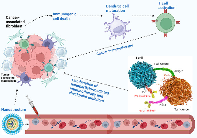
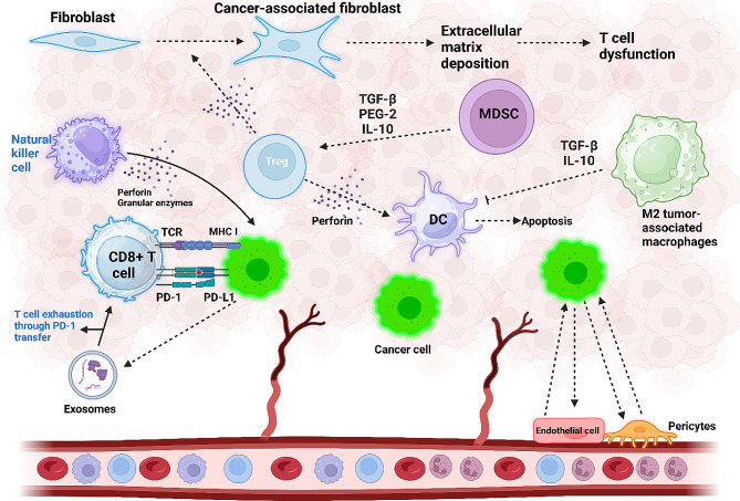
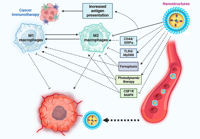
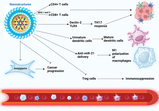
Verification
Claims the paper makes
The claims and statistics of this paper haven’t been checked yet.
Run the check — free, no account needed. It reads the open-access text and checks the sources it cites.
Claims other papers make about this one
Each is checked against this paper’s own text: does it say what the citing paper says it does?
In the citing paper
“These combined strategies help overcome the physiological and immune barriers in musculoskeletal tumors, improving the therapeutic efficacy of exosome-based treatments [285].”
From Emerging role of exosomes in cancer therapy: progress and challenges (2025) · cites this paper as [285]
Self-check
Percentages, sums and reported differences re-computed from the paper’s own numbers.
The self-check needs the paper’s full text.
Citations
Cites
Myeloid-derived-suppressor cells as regulators of the immune system
Gabrilovich, D. I., & Nagaraj, S. (2009) · Nature Reviews Immunology · doi:10.1038/nri2506 · Cited by 6,753 · cited 3×
“In the third and final way, MDSCs are able to enhance Treg cell differentiation to disrupt anti-cancer immunity [172, 351 T c e ll s [ 316 ] T a b l e 2 N a n o p a r t i c l e - m e d i a t e d T c e ll r e g u l a t i o n i n c a n c e r t h e r a p y function of the immune system [352, 353].”
BackgroundAcute-Phase Proteins and Other Systemic Responses to Inflammation
Gabay, C., & Kushner, I. (1999) · New England Journal of Medicine · doi:10.1056/nejm199902113400607 · Cited by 6,734
Hypoxia — a key regulatory factor in tumour growth
Harris, A. L. (2002) · Nature Reviews Cancer · doi:10.1038/nrc704 · Cited by 5,177
Functions of natural killer cells
Vivier, E., Tomasello, E., Baratin, M., Walzer, T., & Ugolini, S. (2008) · Nature Immunology · doi:10.1038/ni1582 · Cited by 3,943
Coordinated regulation of myeloid cells by tumours
Gabrilovich, D. I., Ostrand-Rosenberg, S., & Bronte, V. (2012) · Nature Reviews Immunology · doi:10.1038/nri3175 · Cited by 3,544
Targeting hypoxia in cancer therapy
Wilson, W. R., & Hay, M. P. (2011) · Nature Reviews Cancer · doi:10.1038/nrc3064 · Cited by 3,172
Recommendations for myeloid-derived suppressor cell nomenclature and characterization standards
Bronte, V., Brandau, S., Chen, S.-H., Colombo, M. P., Frey, A. B., Greten, T. F., Mandruzzato, S., Murray, P. J., Ochoa, A., Ostrand-Rosenberg, S., et al. (2016) · Nature Communications · doi:10.1038/ncomms12150 · Cited by 2,751
Distinct populations of inflammatory fibroblasts and myofibroblasts in pancreatic cancer
Öhlund, D., Handly-Santana, A., Biffi, G., Elyada, E., Almeida, A. S., Ponz-Sarvise, M., Corbo, V., Oni, T. E., Hearn, S. A., Lee, E. J., et al. (2017) · Journal of Experimental Medicine · doi:10.1084/jem.20162024 · Cited by 2,590
A Lineage of Myeloid Cells Independent of Myb and Hematopoietic Stem Cells
Schulz, C., Perdiguero, E. G., Chorro, L., Szabo-Rogers, H., Cagnard, N., Kierdorf, K., Prinz, M., Wu, B., Jacobsen, S. E. W., Pollard, J. W., et al. (2012) · Science · doi:10.1126/science.1219179 · Cited by 2,578
PD-1 expression by tumour-associated macrophages inhibits phagocytosis and tumour immunity
Gordon, S. R., Maute, R. L., Dulken, B. W., Hutter, G., George, B. M., McCracken, M. N., Gupta, R., Tsai, J. M., Sinha, R., Corey, D., et al. (2017) · Nature · doi:10.1038/nature22396 · Cited by 2,247
Top 10 Challenges in Cancer Immunotherapy
Hegde, P. S., & Chen, D. S. (2020) · Immunity · doi:10.1016/j.immuni.2019.12.011 · Cited by 2,238
Batf3 Deficiency Reveals a Critical Role for CD8α + Dendritic Cells in Cytotoxic T Cell Immunity
Hildner, K., Edelson, B. T., Purtha, W. E., Diamond, M., Matsushita, H., Kohyama, M., Calderon, B., Schraml, B. U., Unanue, E. R., Diamond, M. S., et al. (2008) · Science · doi:10.1126/science.1164206 · Cited by 2,081
Macrophages and metabolism in the tumor microenvironment
Vitale, I., Manic, G., Coussens, L. M., Kroemer, G., & Galluzzi, L. (2019) · Cell Metabolism · doi:10.1016/j.cmet.2019.06.001 · Cited by 1,837
Systemic RNA delivery to dendritic cells exploits antiviral defence for cancer immunotherapy
Kranz, L. M., Diken, M., Haas, H., Kreiter, S., Loquai, C., Reuter, K. C., Meng, M., Fritz, D., Vascotto, F., Hefesha, H., et al. (2016) · Nature · doi:10.1038/nature18300 · Cited by 1,819
Tumor microenvironment and therapeutic response
Wu, T., & Dai, Y. (2017) · Cancer Letters · doi:10.1016/j.canlet.2016.01.043 · Cited by 1,802
Regulation of immune responses by L-arginine metabolism
Bronte, V., & Zanovello, P. (2005) · Nature Reviews Immunology · doi:10.1038/nri1668 · Cited by 1,775
Majeti, R., Chao, M. P., Alizadeh, A. A., Pang, W. W., Jaiswal, S., Gibbs, K. D., van Rooijen, N., & Weissman, I. L. (2009) · Cell · doi:10.1016/j.cell.2009.05.045 · Cited by 1,752
Immune evasion in cancer: mechanistic basis and therapeutic strategies
Vinay, D. S., Ryan, E. P., Pawelec, G., Talib, W. H., Stagg, J., Elkord, E., Lichtor, T., Decker, W. K., Whelan, R. L., Kumara, H. M. C. S., et al. (2015) · Seminars in Cancer Biology · doi:10.1016/j.semcancer.2015.03.004 · Cited by 1,625 · cited 1×
“The immune cells have shown potential in the identification and recognition of neoplastic cells possessing initiation mutations to suppress tumorigenesis [238].”
BackgroundOrigin and functions of tissue macrophages
Epelman, S., Lavine, K. J., & Randolph, G. J. (2014) · Immunity · doi:10.1016/j.immuni.2014.06.013 · Cited by 1,585
Spranger, S., Dai, D., Horton, B., & Gajewski, T. F. (2017) · Cancer Cell · doi:10.1016/j.ccell.2017.04.003 · Cited by 1,441
Tumour hypoxia promotes tolerance and angiogenesis via CCL28 and Treg cells
Facciabene, A., Peng, X., Hagemann, I. S., Balint, K., Barchetti, A., Wang, L.-P., Gimotty, P. A., Gilks, C. B., Lal, P., Zhang, L., et al. (2011) · Nature · doi:10.1038/nature10169 · Cited by 1,376
Self-citationBlockade of B7-H1 improves myeloid dendritic cell-mediated antitumor immunity
Curiel, T. J., Wei, S., Dong, H., Alvarez, X., Cheng, P., Mottram, P., Krzysiek, R., Knutson, K. L., Daniel, B., Zimmermann, M. C., et al. (2003) · Nature Medicine · doi:10.1038/nm863 · Cited by 1,314
Combination cancer immunotherapy and new immunomodulatory targets
Mahoney, K. M., Rennert, P. D., & Freeman, G. J. (2015) · Nature Reviews Drug Discovery · doi:10.1038/nrd4591 · Cited by 1,294
Corzo, C. A., Condamine, T., Lu, L., Cotter, M. J., Youn, J.-I., Cheng, P., Cho, H.-I., Celis, E., Quiceno, D. G., Padhya, T., et al. (2010) · Journal of Experimental Medicine · doi:10.1084/jem.20100587 · Cited by 1,173
Garris, C. S., Arlauckas, S. P., Kohler, R. H., Trefny, M. P., Garren, S., Piot, C., Engblom, C., Pfirschke, C., Siwicki, M., Gungabeesoon, J., et al. (2018) · Immunity · doi:10.1016/j.immuni.2018.09.024 · Cited by 966
Munn, D. H., Sharma, M. D., Hou, D., Baban, B., Lee, J. R., Antonia, S. J., Messina, J. L., Chandler, P., Koni, P. A., & Mellor, A. L. (2004) · Journal of Clinical Investigation · doi:10.1172/jci21583 · Cited by 852
A mechanism of hypoxia-mediated escape from adaptive immunity in cancer cells
Barsoum, I. B., Smallwood, C. A., Siemens, D. R., & Graham, C. H. (2014) · Cancer Research · doi:10.1158/0008-5472.can-13-0992 · Cited by 767
Chow, M. T., Ozga, A. J., Servis, R. L., Frederick, D. T., Lo, J. A., Fisher, D. E., Freeman, G. J., Boland, G. M., & Luster, A. D. (2019) · Immunity · doi:10.1016/j.immuni.2019.04.010 · Cited by 716
Regulation of Immunity and Inflammation by Hypoxia in Immunological Niches
Taylor, C. T., & Colgan, S. P. (2017) · Nature reviews. Immunology · doi:10.1038/nri.2017.103 · Cited by 713
Beyond stem cells: self-renewal of differentiated macrophages
Sieweke, M. H., & Allen, J. E. (2013) · Science · doi:10.1126/science.1242974 · Cited by 503
Lactic Acid and an Acidic Tumor Microenvironment suppress Anticancer Immunity
Wang, J. X., Choi, S. Y. C., Niu, X., Kang, N., Xue, H., Killam, J., & Wang, Y. (2020) · International Journal of Molecular Sciences · doi:10.3390/ijms21218363 · Cited by 460
Self-citationOjha, T., Pathak, V., Shi, Y., Hennink, W. E., Moonen, C. T. W., Storm, G., Kiessling, F., & Lammers, T. (2017) · Advanced Drug Delivery Reviews · doi:10.1016/j.addr.2017.07.007 · Cited by 260
Cancer-Associated Fibroblasts and Tumor-Associated Macrophages in Cancer and Cancer Immunotherapy
Raskov, H., Orhan, A., Gaggar, S., & Gögenur, I. (2021) · Frontiers in Oncology · doi:10.3389/fonc.2021.668731 · Cited by 150
Barriga, F. M., Tsanov, K. M., Ho, Y.-J., Sohail, N., Zhang, A., Baslan, T., Wuest, A. N., Del Priore, I., Meškauskaitė, B., Livshits, G., et al. (2022) · Nature Cancer · doi:10.1038/s43018-022-00443-5 · Cited by 125 · cited 1×
“3, increasing the escape of tumor cells from CD 8 + T cell surveillance [235].”
BackgroundLi, Q., Zhang, L., You, W., Xu, J., Dai, J., Hua, D., Zhang, R., Yao, F., Zhou, S., Huang, W., et al. (2022) · Nature Communications · doi:10.1038/s41467-022-35469-x · Cited by 120 · cited 1×
“This, in turn, results in increased PD-L1 expression, promoting immune evasion [227].”
BackgroundLee, H., Quek, C., Silva, I., Tasker, A., Batten, M., Rizos, H., Lim, S. Y., Nur Gide, T., Shang, P., Attrill, G. H., et al. (2019) · OncoImmunology · doi:10.1080/2162402x.2018.1537581 · Cited by 115
Research works printed but not linked: 575
- Liu Y, et al. Emerging phagocytosis checkpoints in cancer immunotherapy. Signal Transduct Target Ther. 2023;8(1):104. · DOI
- Kennedy LB, Salama AKS. A review of cancer immunotherapy toxicity. CA Cancer J Clin. 2020;70(2):86–104. · DOI
- Webster RM. The immune checkpoint inhibitors: where are we now? Nat Rev Drug Discov. 2014;13(12):883–4. · DOI
- Zhang Q, et al. Immune and Clinical features of CD96 expression in Glioma by in silico analysis. Front Bioeng Biotechnol. 2020;8:592. · DOI
- Galipeau J, Sensébé L. Mesenchymal stromal cells: Clinical challenges and Therapeutic opportunities. Cell Stem Cell. 2018;22(6):824–33. · DOI · Paper page
- Liotta F, et al. Toll-like receptors 3 and 4 are expressed by human bone marrow-derived mesenchymal stem cells and can inhibit their T-cell modulatory activity by impairing notch signaling. Stem Cells. 2008;26(1):279–89. · DOI
- Barkal AA, et al. CD24 signalling through macrophage Siglec-10 is a target for cancer immunotherapy. Nature. 2019;572(7769):392–6. · DOI
- Barkal AA, et al. Engagement of MHC class I by the inhibitory receptor LILRB1 suppresses macrophages and is a target of cancer immunotherapy. Nat Immunol. 2018;19(1):76–84. · DOI
- Lin H, et al. Stanniocalcin 1 is a phagocytosis checkpoint driving tumor immune resistance. Cancer Cell. 2021;39(4):480–e4936. · DOI
- Theruvath J, et al. Anti-GD2 synergizes with CD47 blockade to mediate tumor eradication. Nat Med. 2022;28(2):333–44. · DOI
- Gardner A, de Mingo Á, Pulido, Ruffell B. Dendritic cells and their role in Immunotherapy. Front Immunol. 2020;11:924. · DOI
- Chen DS, Mellman I. Oncology meets immunology: the cancer-immunity cycle. Immunity. 2013;39(1):1–10. · DOI · Paper page
- Nakayama M. Antigen Presentation by MHC-Dressed cells. Front Immunol. 2014;5:672.
- Lin A, Loré K. Granulocytes: New members of the Antigen-presenting cell family. Front Immunol. 2017;8:1781. · DOI
- Broz ML, et al. Dissecting the tumor myeloid compartment reveals rare activating antigen-presenting cells critical for T cell immunity. Cancer Cell. 2014;26(5):638–52. · DOI · Paper page
- DeNardo DG, Ruffell B. Macrophages as regulators of tumour immunity and immunotherapy. Nat Rev Immunol. 2019;19(6):369–82. · DOI
- Ruffell B, Coussens LM. Macrophages and therapeutic resistance in cancer. Cancer Cell. 2015;27(4):462–72. · DOI · Paper page
- Sánchez-Paulete AR, et al. Cancer Immunotherapy with Immunomodulatory Anti-CD137 and Anti-PD-1 monoclonal antibodies requires BATF3-Dependent dendritic cells. Cancer Discov. 2016;6(1):71–9. · DOI
- Roberts EW, et al. Critical role for CD103(+)/CD141(+) dendritic cells bearing CCR7 for Tumor Antigen Trafficking and priming of T cell immunity in Melanoma. Cancer Cell. 2016;30(2):324–36. · DOI
- Salmon H, et al. Expansion and activation of CD103(+) dendritic cell progenitors at the Tumor Site enhances tumor responses to therapeutic PD-L1 and BRAF inhibition. Immunity. 2016;44(4):924–38. · DOI
- de Pulido M. TIM-3 regulates CD103(+) dendritic cell function and response to chemotherapy in breast Cancer. Cancer Cell. 2018;33(1):60–74.e6. · DOI · Paper page
- Xia Y, et al. Engineering macrophages for Cancer Immunotherapy and Drug Delivery. Adv Mater. 2020;32(40):e2002054. · DOI · Paper page
- Gao J, Liang Y, Wang L. Shap Polarization Tumor-Associated Macrophages Cancer Immunotherapy Front Immunol. 2022;13:888713.
- Kumari N, Choi SH. Tumor-associated macrophages in cancer: recent advancements in cancer nanoimmunotherapies. J Exp Clin Cancer Res. 2022;41(1):68. · DOI
- Li L, Ma SR, Yu ZL. Targeting the lipid metabolic reprogramming of tumor-associated macrophages: a novel insight into cancer immunotherapy. Cell Oncol (Dordr); 2023. · DOI
- Pei L, et al. Roles of cancer-associated fibroblasts (CAFs) in anti- PD-1/PD-L1 immunotherapy for solid cancers. Mol Cancer. 2023;22(1):29. · DOI
- Zhang H, et al. Define cancer-associated fibroblasts (CAFs) in the tumor microenvironment: new opportunities in cancer immunotherapy and advances in clinical trials. Mol Cancer. 2023;22(1):159. · DOI
- Thiery J. Modulation of the antitumor immune response by cancer-associated fibroblasts: mechanisms and targeting strategies to hamper their immunosuppressive functions. Explor Target Antitumor Ther. 2022;3(5):598–629. · DOI
- Wang Q, et al. Role of tumor microenvironment in cancer progression and therapeutic strategy. Cancer Med. 2023;12(10):11149–65. · DOI
- Han S et al. Tumor Microenvironment Regulation and Cancer Targeting Therapy based on nanoparticles. J Funct Biomater, 2023. 14(3). · DOI
- Li Z, Yin P. Tumor microenvironment diversity and plasticity in cancer multidrug resistance. Biochim Biophys Acta Rev Cancer. 2023;1878(6):188997. · DOI
- Yang R, et al. Tumor microenvironment responsive metal nanoparticles in cancer immunotherapy. Front Immunol. 2023;14:1237361. · DOI
- Zhang Z, et al. Involvement of inflammasomes in tumor microenvironment and tumor therapies. J Hematol Oncol. 2023;16(1):24. · DOI
- Jin HR, et al. Lipid metabolic reprogramming in tumor microenvironment: from mechanisms to therapeutics. J Hematol Oncol. 2023;16(1):103. · DOI
- Arner EN, Rathmell JC. Metabolic programming and immune suppression in the tumor microenvironment. Cancer Cell. 2023;41(3):421–33. · DOI
- Lin MJ, et al. Cancer vaccines: the next immunotherapy frontier. Nat Cancer. 2022;3(8):911–26. · DOI
- Beck JD, et al. mRNA therapeutics in cancer immunotherapy. Mol Cancer. 2021;20(1):69. · DOI · Paper page
- Siminiak N, et al. Immunotherapy in Ovarian Cancer. Arch Immunol Ther Exp (Warsz). 2022;70(1):19. · DOI
- Mauricio D, et al. Immunotherapy in Cervical Cancer. Curr Oncol Rep. 2021;23(6):61. · DOI
- Bockorny B, Grossman JE, Hidalgo M. Facts and hopes in Immunotherapy of Pancreatic Cancer. Clin Cancer Res. 2022;28(21):4606–17. · DOI
- Yang M, et al. The application of nanoparticles in cancer immunotherapy: targeting tumor microenvironment. Bioact Mater. 2021;6(7):1973–87.
- Overchuk M, Zheng G. Overcoming obstacles in the tumor microenvironment: recent advancements in nanoparticle delivery for cancer theranostics. Biomaterials. 2018;156:217–37. · DOI
- Matsumura Y, Maeda H. A new concept for macromolecular therapeutics in cancer chemotherapy: mechanism of tumoritropic accumulation of proteins and the antitumor agent smancs. Cancer Res. 1986;46(12 Pt 1):6387–92.
- Prabhakar U, et al. Challenges and key considerations of the enhanced permeability and retention effect for nanomedicine drug delivery in oncology. Cancer Res. 2013;73(8):2412–7. · DOI
- Yang S, Gao H. Nanoparticles for modulating tumor microenvironment to improve drug delivery and tumor therapy. Pharmacol Res. 2017;126:97–108. · DOI · Paper page
- Chen Q, et al. Remodeling the Tumor Microenvironment with Emerging Nanotherapeutics. Trends Pharmacol Sci. 2018;39(1):59–74. · DOI
- Li M, et al. Nanoparticles designed to regulate tumor microenvironment for cancer therapy. Life Sci. 2018;201:37–44. · DOI
- Guo B, et al. Cuproptosis Induced by ROS responsive nanoparticles with Elesclomol and Copper Combined with αPD-L1 for enhanced Cancer Immunotherapy. Adv Mater. 2023;35(22):e2212267. · DOI
- Wang-Bishop L, et al. STING-activating nanoparticles normalize the vascular-immune interface to potentiate cancer immunotherapy. Sci Immunol. 2023;8(83):eadd1153. · DOI
- Ding B, et al. ZIF-8 nanoparticles evoke pyroptosis for high-efficiency Cancer Immunotherapy. Angew Chem Int Ed Engl. 2023;62(10):e202215307. · DOI
- Liu J et al. Ultrathin clay nanoparticles-mediated mutual reinforcement of ferroptosis and Cancer immunotherapy. Adv Mater, 2023: p. e2309562. · DOI
- Huang A, et al. Engineered apoptosis-bioinspired nanoparticles initiate Immune Cascade for Cancer Immunotherapy of Malignant ascites. ACS Appl Mater Interfaces. 2023;15(8):10371–82. · DOI
- Liu D et al. Tumor Microenvironment-Responsive nanoparticles amplifying STING Signaling Pathway for Cancer Immunotherapy. Adv Mater, 2023: p. e2304845. · DOI
- Sun Y, et al. Sialic acid-targeted cyclodextrin-based nanoparticles deliver CSF-1R siRNA and reprogram tumour-associated macrophages for immunotherapy of prostate cancer. Eur J Pharm Sci. 2023;185:106427. · DOI
- Liu C, et al. Microfluidic sonication to assemble Exosome membrane-coated nanoparticles for Immune evasion-mediated targeting. Nano Lett. 2019;19(11):7836–44. · DOI
- Li M et al. Signaling pathways in macrophages: molecular mechanisms and therapeutic targets MedComm (2020), 2023;4(5):e349. · DOI
- Ginhoux F, Guilliams M. Tissue-Resident Macrophage Ontogeny and Homeostasis. Immunity. 2016;44(3):439–49. · DOI · Paper page
- Italiani P, Boraschi D. From monocytes to M1/M2 macrophages: phenotypical vs. functional differentiation. Front Immunol. 2014;5:514. · DOI · Paper page
- Zhan C et al. Antitumor therapy for breast cancer: focus on tumor-associated macrophages and nanosized drug delivery systems. Cancer Med, 2023. · DOI
- Wang N, et al. Research trends in pharmacological modulation of tumor-associated macrophages. Clin Transl Med. 2021;11(1):e288. · DOI
- Wang J, et al. Crosstalk between cancer and immune cells: role of tumor-associated macrophages in the tumor microenvironment. Cancer Med. 2019;8(10):4709–21. · DOI
- Munir MT et al. Tumor-Associated macrophages as multifaceted regulators of breast tumor growth. Int J Mol Sci, 2021. 22(12). · DOI
- Qiu SQ, et al. Tumor-associated macrophages in breast cancer: innocent bystander or important player? Cancer Treat Rev. 2018;70:178–89. · DOI
- Wang C, et al. Macrophage balance fraction determines the degree of immunosuppression and metastatic ability of breast cancer. Int Immunopharmacol. 2021;97:107682. · DOI
- Steenbrugge J, et al. Anti-inflammatory signaling by mammary tumor cells mediates prometastatic macrophage polarization in an innovative intraductal mouse model for triple-negative breast cancer. J Exp Clin Cancer Res. 2018;37(1):191. · DOI
- Shu Y, Cheng P. Targeting tumor-associated macrophages for cancer immunotherapy Biochimica et Biophysica Acta (BBA) - Reviews on Cancer, 2020;1874(2):188434. · DOI
- Nascimento C, et al. Reprogramming of tumor-associated macrophages by polyaniline-coated iron oxide nanoparticles applied to treatment of breast cancer. Int J Pharm. 2023;636:122866. · DOI
- Shang Y, et al. Bioengineered Nanospores selectively blocking LC3-Associated phagocytosis in Tumor-Associated macrophages Potentiate Antitumor Immunity. ACS Nano. 2023;17(11):10872–87. · DOI
- Liu Y, et al. Multifunctional nanoparticles inhibit tumor and tumor-associated macrophages for triple-negative breast cancer therapy. J Colloid Interface Sci. 2024;657:598–610. · DOI
- Liang DS, et al. Targeted delivery of pexidartinib to tumor-associated macrophages via legumain-sensitive dual-coating nanoparticles for cancer immunotherapy. Colloids Surf B Biointerfaces. 2023;226:113283. · DOI
- Sun H, et al. The role of cancer-associated fibroblasts in tumorigenesis of gastric cancer. Cell Death Dis. 2022;13(10):874. · DOI
- Gu J, et al. The role of PKM2 nuclear translocation in the constant activation of the NF-κB signaling pathway in cancer-associated fibroblasts. Cell Death Dis. 2021;12(4):291. · DOI
- Bu L, et al. Biological heterogeneity and versatility of cancer-associated fibroblasts in the tumor microenvironment. Oncogene. 2019;38(25):4887–901. · DOI
- Kalluri R. The biology and function of fibroblasts in cancer. Nat Rev Cancer. 2016;16(9):582–98. · DOI · Paper page
- Kobayashi H, et al. Cancer-associated fibroblasts in gastrointestinal cancer. Nat Rev Gastroenterol Hepatol. 2019;16(5):282–95. · DOI
- Zhang T, et al. Cancer-associated fibroblasts in pancreatic ductal adenocarcinoma. Cell Death Dis. 2022;13(10):897. · DOI
- Biffi G, et al. IL1-Induced JAK/STAT signaling is antagonized by TGFβ to shape CAF heterogeneity in pancreatic ductal adenocarcinoma. Cancer Discov. 2019;9(2):282–301. · DOI · Paper page
- Elyada E, et al. Cross-species single-cell analysis of pancreatic ductal adenocarcinoma reveals Antigen-Presenting Cancer-Associated fibroblasts. Cancer Discov. 2019;9(8):1102–23. · DOI · Paper page
- Fang Z, et al. Signaling pathways in cancer-associated fibroblasts: recent advances and future perspectives. Cancer Commun. 2023;43(1):3–41. · DOI
- Ai W, et al. Modulation of cancer-associated fibroblasts by nanodelivery system to enhance efficacy of tumor therapy. Nanomed (Lond). 2023;18(15):1025–39. · DOI
- Geng S, et al. Safe engineering of cancer-associated fibroblasts enhances checkpoint blockade immunotherapy. J Control Release. 2023;356:272–87. · DOI
- Jian C et al. Biomimetic Nanoplatform for Dual-Targeted Clearance of Activated and Senescent Cancer-Associated Fibroblasts to Improve Radiation Resistance in Breast Cancer Small, 2024: p. e2309279. · DOI · Paper page
- Hu M, et al. Salvianolic acid B-loaded polydopamine-modified hollow mesoporous organic silica nanoparticles for treatment of breast cancer metastasis via suppressing cancer-associated fibroblasts. Eur J Pharm Sci. 2024;192:106641. · DOI
- Hinshaw DC, Shevde LA. The Tumor Microenvironment innately modulates Cancer Progression. Cancer Res. 2019;79(18):4557–66. · DOI · Paper page
- Wang J, et al. The clinical significance of tumor-infiltrating neutrophils and neutrophil-to-CD8 + lymphocyte ratio in patients with resectable esophageal squamous cell carcinoma. J Transl Med. 2014;12:7. · DOI
- Deniset JF, Kubes P. Recent Adv Underst Neutrophils F1000Res. 2016;5:2912.
- Uribe-Querol E, Rosales C. Neutrophils in Cancer: two sides of the same Coin. J Immunol Res. 2015;2015:983698. · DOI
- Cools-Lartigue J, et al. Neutrophil extracellular traps in cancer progression. Cell Mol Life Sci. 2014;71(21):4179–94. · DOI
- Jin W, et al. Tumor-infiltrating NETs predict postsurgical survival in patients with pancreatic ductal adenocarcinoma. Ann Surg Oncol. 2019;26(2):635–43. · DOI
- Fridlender ZG, Albelda SM. Tumor-associated neutrophils: friend or foe? Carcinogenesis. 2012;33(5):949–55. · DOI
- Demers M, et al. Priming of neutrophils toward NETosis promotes tumor growth. Oncoimmunology. 2016;5(5):e1134073. · DOI · Paper page
- He M, et al. Peritumoral stromal neutrophils are essential for c-Met-elicited metastasis in human hepatocellular carcinoma. Oncoimmunology. 2016;5(10):e1219828. · DOI
- Schauer C, et al. Aggregated neutrophil extracellular traps limit inflammation by degrading cytokines and chemokines. Nat Med. 2014;20(5):511–7. · DOI · Paper page
- Ralph SJ, Reynolds MJ. Intratumoral pro-oxidants promote cancer immunotherapy by recruiting and reprogramming neutrophils to eliminate tumors. Cancer Immunol Immunother. 2023;72(3):527–42. · DOI
- Huntington ND, Cursons J, Rautela J. The cancer–natural killer cell immunity cycle. Nat Rev Cancer. 2020;20(8):437–54. · DOI
- Huntington ND, Vosshenrich CA, Di Santo JP. Developmental pathways that generate natural-killer-cell diversity in mice and humans. Nat Rev Immunol. 2007;7(9):703–14. · DOI
- Pfefferle A, et al. Intra-lineage plasticity and functional reprogramming maintain natural killer cell repertoire diversity. Cell Rep. 2019;29(8):2284–e22944. · DOI
- Freud AG, Caligiuri MA. Human natural killer cell development. Immunol Rev. 2006;214:56–72. · DOI
- Gil M, Kim KE. Interleukin-18 is a prognostic biomarker correlated with CD8 + T cell and natural killer cell infiltration in skin cutaneous melanoma Journal of clinical medicine, 2019;8(11):1993. · DOI
- Cursons J, et al. A gene signature Predicting Natural Killer Cell Infiltration and Improved Survival in Melanoma patients. Cancer Immunol Res. 2019;7(7):1162–74. · DOI
- Li B et al. Natural killer cell and stroma abundance are independently prognostic and predict gastric cancer chemotherapy benefit. JCI Insight, 2020. 5(9). · DOI
- Ran GH, et al. Natural killer cell homing and trafficking in tissues and tumors: from biology to application. Signal Transduct Target Therapy. 2022;7(1):205. · DOI
- Scoville SD, Freud AG, Caligiuri MA. Model Hum Nat Killer Cell Dev Era Innate Lymphoid Cells Front Immunol. 2017;8:360.
- Stokic-Trtica V, Diefenbach A, Klose CSN. NK Cell Dev Times Innate Lymphoid Cell Divers Front Immunol. 2020;11:813.
- Vivier E, et al. Innate lymphoid cells: 10 years on. Cell. 2018;174(5):1054–66. · DOI
- Freud AG, et al. The Broad Spectrum of Human Natural Killer Cell Diversity. Immunity. 2017;47(5):820–33. · DOI
- Björkström NK, Ljunggren HG, Michaëlsson J. Emerging insights into natural killer cells in human peripheral tissues. Nat Rev Immunol. 2016;16(5):310–20. · DOI
- Yu J, Freud AG, Caligiuri MA. Location and cellular stages of natural killer cell development. Trends Immunol. 2013;34(12):573–82. · DOI
- Fang F, Xiao W, Tian Z. NK cell-based immunotherapy for cancer. Semin Immunol. 2017;31:37–54. · DOI
- Hammer Q, Rückert T, Romagnani C. Natural killer cell specificity for viral infections. Nat Immunol. 2018;19(8):800–8. · DOI
- Björkström NK, Strunz B, Ljunggren HG. Natural killer cells in antiviral immunity. Nat Rev Immunol. 2022;22(2):112–23. · DOI
- Li Y, et al. Natural killer cells: friend or foe in metabolic diseases? Front Immunol. 2021;12:614429. · DOI
- Shimasaki N, Jain A, Campana D. NK cells for cancer immunotherapy. Nat Rev Drug Discov. 2020;19(3):200–18. · DOI
- Maskalenko NA, Zhigarev D, Campbell KS. Harnessing natural killer cells for cancer immunotherapy: dispatching the first responders. Nat Rev Drug Discov. 2022;21(8):559–77. · DOI
- Pockley AG, Vaupel P, Multhoff G. NK cell-based therapeutics for lung cancer. Expert Opin Biol Ther. 2020;20(1):23–33. · DOI
- Muntasell A, et al. NK Cell infiltrates and HLA Class I expression in primary HER2(+) breast Cancer Predict and Uncouple Pathological Response and Disease-free survival. Clin Cancer Res. 2019;25(5):1535–45. · DOI
- Liu P, Chen L, Zhang H. Natural killer cells in Liver Disease and Hepatocellular Carcinoma and the NK cell-based immunotherapy. J Immunol Res. 2018;2018:p1206737. · DOI
- Ali TH, et al. Enrichment of CD56(dim)KIR + CD57 + highly cytotoxic NK cells in tumour-infiltrated lymph nodes of melanoma patients. Nat Commun. 2014;5:5639. · DOI
- Terrén I et al. NK Cell-based immunotherapy in renal cell carcinoma. Cancers (Basel), 2020. 12(2). · DOI
- Lim SA, et al. Lipid metabolism in T cell signaling and function. Nat Chem Biol. 2022;18(5):470–81. · DOI
- Ando M, et al. Memory T cell, exhaustion, and tumor immunity. Immunological Med. 2020;43(1):1–9. · DOI
- Franco F, et al. Metabolic and epigenetic regulation of T-cell exhaustion. Nat Metabolism. 2020;2(10):1001–12. · DOI
- Douka S, et al. Lipid nanoparticle-mediated messenger RNA delivery for ex vivo engineering of natural killer cells. J Control Release. 2023;361:455–69. · DOI
- Shamalov K, et al. Noninvasive Tracking of Natural Killer cells using gold nanoparticles. ACS Omega. 2021;6(43):28507–14. · DOI
- Kim KS, et al. Cationic nanoparticle-mediated activation of natural killer cells for Effective Cancer Immunotherapy. ACS Appl Mater Interfaces. 2020;12(51):56731–40. · DOI
- Sun X, et al. Acid-switchable nanoparticles induce self-adaptive aggregation for enhancing antitumor immunity of natural killer cells. Acta Pharm Sin B. 2023;13(7):3093–105. · DOI
- Liu GW, et al. Lipid nanoparticles for nucleic acid delivery to endothelial cells. Pharm Res. 2023;40(1):3–25. · DOI
- Félétou M. Integrated Systems Physiology: from molecule to function to Disease, in The endothelium: part 1: multiple functions of the endothelial cells—focus on endothelium-derived Vasoactive mediators. 2011, Morgan & Claypool Life Sciences copyright © 2011 by Morgan & Claypool Life Sciences Publishers.: San Rafael (CA). · DOI
- Stoltz JF, et al. Introduction to endothelial cell biology. Clin Hemorheol Microcirc. 2007;37(1–2):5–8.
- Martin FA, Murphy RP, Cummins PM. Thrombomodulin and the vascular endothelium: insights into functional, regulatory, and therapeutic aspects. Am J Physiol Heart Circ Physiol. 2013;304(12):H1585–97. · DOI
- Maroney SA, Mast AE. Expression of tissue factor pathway inhibitor by endothelial cells and platelets. Transfus Apher Sci. 2008;38(1):9–14. · DOI
- Mohan Rao LV, Esmon CT, Pendurthi UR. Endothelial cell protein C receptor: a multiliganded and multifunctional receptor. Blood. 2014;124(10):1553–62. · DOI · Paper page
- Bremnes RM, et al. The role of tumor stroma in cancer progression and prognosis: emphasis on carcinoma-associated fibroblasts and non-small cell lung cancer. J Thorac Oncol. 2011;6(1):209–17. · DOI · Paper page
- Pietras K, Ostman A. Hallmarks of cancer: interactions with the tumor stroma. Exp Cell Res. 2010;316(8):1324–31. · DOI
- McAllister SS, Weinberg RA. The tumour-induced systemic environment as a critical regulator of cancer progression and metastasis. Nat Cell Biol. 2014;16(8):717–27. · DOI
- Dillekås H, Rogers MS, Straume O. Are 90% of deaths from cancer caused by metastases? Cancer Med. 2019;8(12):5574–6. · DOI
- Khorana AA, et al. Thromboembolism is a leading cause of death in cancer patients receiving outpatient chemotherapy. J Thromb Haemost. 2007;5(3):632–4. · DOI
- Tisdale MJ. Cachexia in cancer patients. Nat Rev Cancer. 2002;2(11):862–71. · DOI
- Preuss SF, Grieshober D, Augustin HG. Systemic Reprogramming Endothelial cell Signal Metastasis cachexia Physiol. 2023;38(4):189–202.
- Eilken HM, et al. Pericytes regulate VEGF-induced endothelial sprouting through VEGFR1. Nat Commun. 2017;8(1):1574. · DOI
- Zimmermann KW. Der Feinere Bau Der Blutcapillaren. Z für Anatomie und Entwicklungsgeschichte. 1923;68:29–109. · DOI
- Schrimpf C, et al. The role of pericyte detachment in vascular rarefaction. J Vasc Res. 2014;51(4):247–58. · DOI
- Hirschi KK, D’Amore PA. Pericytes in the microvasculature. Cardiovascular Res. 1996;32(4):687–98. · DOI
- Díaz-Flores L, et al. Microvascular pericytes, a review of their morphological and functional characteristics. Histology and histopathology; 1991.
- Birbrair A, et al. How plastic are pericytes? Stem Cells Dev. 2017;26(14):1013–9. · DOI
- Birbrair A, et al. Pericytes at the intersection between tissue regeneration and pathology. Clin Sci. 2015;128(2):81–93. · DOI
- Picoli CC et al. Pericytes cross-talks within the tumor microenvironment Biochimica et Biophysica Acta (BBA)-Reviews on Cancer, 2021;1876(2):188608. · DOI
- Jiang Z et al. Pericytes in the tumor microenvironment. Cancer Lett, 2023: p. 216074. · DOI
- Chen Z, Xu XH, Hu J. Role of pericytes in angiogenesis: focus on cancer angiogenesis and anti-angiogenic therapy. Neoplasma. 2016;63(2):173–82.
- Hong CL, et al. CD248 regulates wnt signaling in Pericytes to Promote Angiogenesis and Tumor Growth in Lung Cancer. Cancer Res. 2022;82(20):3734–50. · DOI
- Meng YM, et al. Hexokinase 2-driven glycolysis in pericytes activates their contractility leading to tumor blood vessel abnormalities. Nat Commun. 2021;12(1):6011. · DOI
- Dasgupta S, et al. RGS5-TGFβ-Smad2/3 axis switches pro- to anti-apoptotic signaling in tumor-residing pericytes, assisting tumor growth. Cell Death Differ. 2021;28(11):3052–76. · DOI
- Pillay J, et al. Immune suppression by neutrophils and granulocytic myeloid-derived suppressor cells: similarities and differences. Cell Mol Life Sci. 2013;70(20):3813–27. · DOI
- Rodriguez PC, et al. Arginase I-producing myeloid-derived suppressor cells in renal cell carcinoma are a subpopulation of activated granulocytes. Cancer Res. 2009;69(4):1553–60. · DOI
- Youn JI, et al. Characterization of the nature of granulocytic myeloid-derived suppressor cells in tumor-bearing mice. J Leukoc Biol. 2012;91(1):167–81. · DOI
- Kumar V, et al. The nature of myeloid-derived suppressor cells in the Tumor Microenvironment. Trends Immunol. 2016;37(3):208–20. · DOI · Paper page
- Condamine T, et al. Regulation of tumor metastasis by myeloid-derived suppressor cells. Annu Rev Med. 2015;66:97–110. · DOI
- Condamine T et al. Lectin-type oxidized LDL receptor-1 distinguishes population of human polymorphonuclear myeloid-derived suppressor cells in cancer patients. Sci Immunol, 2016. 1(2). · DOI
- Hossain F, et al. Inhibition of fatty acid oxidation modulates immunosuppressive functions of myeloid-derived suppressor cells and enhances Cancer therapies. Cancer Immunol Res. 2015;3(11):1236–47. · DOI
- Zhou J, et al. Hepatoma-intrinsic CCRK inhibition diminishes myeloid-derived suppressor cell immunosuppression and enhances immune-checkpoint blockade efficacy. Gut. 2018;67(5):931–44. · DOI
- Wu C, et al. Repolarization of myeloid derived suppressor cells via magnetic nanoparticles to promote radiotherapy for glioma treatment. Nanomedicine. 2019;16:126–37. · DOI
- Ding M, et al. Augmenting immunogenic cell death and alleviating myeloid-derived suppressor cells by Sono-Activatable Semiconducting Polymer Nanopartners for Immunotherapy. Adv Mater. 2023;35(33):e2302508. · DOI
- Zuo H, et al. Circumventing myeloid-derived suppressor cell-mediated immunosuppression using an oxygen-generated and -economized nanoplatform. ACS Appl Mater Interfaces. 2020;12(50):55723–36. · DOI
- Habanjar O et al. Crosstalk of inflammatory cytokines within the breast Tumor Microenvironment. Int J Mol Sci, 2023. 24(4). · DOI
- Zhang JM, An J. Cytokines, inflammation, and pain. Int Anesthesiol Clin. 2007;45(2):27–37. · DOI
- Raman D, Sobolik-Delmaire T, Richmond A. Chemokines in health and disease. Exp Cell Res. 2011;317(5):575–89. · DOI
- Bachelerie F, et al. International Union of Basic and Clinical Pharmacology. [corrected]. LXXXIX. Update on the extended family of chemokine receptors and introducing a new nomenclature for atypical chemokine receptors. Pharmacol Rev. 2014;66(1):1–79. · DOI
- Mélik-Parsadaniantz S, Rostène W. Chemokines and neuromodulation. J Neuroimmunol. 2008;198(1–2):62–8. · DOI
- Zlotnik A, Yoshie O. The chemokine superfamily revisited. Immunity. 2012;36(5):705–16. · DOI
- Kaneko N, et al. The role of interleukin-1 in general pathology. Inflamm Regen. 2019;39:12. · DOI
- Akdis M et al. Interleukins, from 1 to 37, and interferon-γ: receptors, functions, and roles in diseases J Allergy Clin Immunol, 2011;127(3):701 – 21.e1-70. · DOI
- Shin E, Koo JS. The role of Adipokines and bone marrow adipocytes in breast Cancer bone metastasis. Int J Mol Sci, 2020. 21(14). · DOI
- Blüher M, Mantzoros CS. From leptin to other adipokines in health and disease: facts and expectations at the beginning of the 21st century. Metabolism. 2015;64(1):131–45. · DOI
- Mancuso P. The role of adipokines in chronic inflammation. Immunotargets Ther. 2016;5:47–56. · DOI
- Barchetta I, et al. Sick fat: the good and the bad of old and new circulating markers of adipose tissue inflammation. J Endocrinol Invest. 2019;42(11):1257–72. · DOI · Paper page
- Nehme R et al. Targeting adiponectin in breast Cancer. Biomedicines, 2022. 10(11). · DOI
- Artac M, Altundag K. Leptin and breast cancer: an overview. Med Oncol. 2012;29(3):1510–4. · DOI
- Ferrer I, et al. Transforming growth factor-alpha (TGF-alpha) and epidermal growth factor-receptor (EGF-R) immunoreactivity in normal and pathologic brain. Prog Neurobiol. 1996;49(2):99–123. · DOI
- MaruYama T, Chen W, Shibata H. TGF-β Cancer Immunotherapy Biol Pharm Bull. 2022;45(2):155–61. · DOI
- Isaacs A, Lindenmann J. Virus interference. I. The interferon. Proc R Soc Lond B Biol Sci. 1957;147(927):258–67. · DOI · Paper page
- Schroder K, et al. Interferon-gamma: an overview of signals, mechanisms and functions. J Leukoc Biol. 2004;75(2):163–89. · DOI · Paper page
- Peng S, et al. Tumor-microenvironment-responsive nanomedicine for enhanced Cancer Immunotherapy. Adv Sci (Weinh). 2022;9(1):e2103836. · DOI
- Mu J, et al. Development of endogenous enzyme-responsive nanomaterials for theranostics. Chem Soc Rev. 2018;47(15):5554–73. · DOI
- Hu J, Zhang G, Liu S. Enzyme-responsive polymeric assemblies, nanoparticles and hydrogels. Chem Soc Rev. 2012;41(18):5933–49. · DOI
- Shahriari M, et al. Enzyme responsive drug delivery systems in cancer treatment. J Control Release. 2019;308:172–89. · DOI
- Qiu N, et al. Esterase-activated charge-reversal polymer for fibroblast-exempt Cancer Gene Therapy. Adv Mater. 2016;28(48):10613–22. · DOI
- Luo Z, Dai Y, Gao H. Development and application of hyaluronic acid in tumor targeting drug delivery. Acta Pharm Sin B. 2019;9(6):1099–112. · DOI · Paper page
- Li H, et al. The relationship between MMP-2 and MMP-9 expression levels with breast cancer incidence and prognosis. Oncol Lett. 2017;14(5):5865–70.
- Pham HT, Block NL, Lokeshwar VB. Tumor-derived hyaluronidase: a diagnostic urine marker for high-grade bladder cancer. Cancer Res. 1997;57(4):778–83.
- Moreira AM et al. The Extracellular Matrix: an accomplice in gastric Cancer Development and Progression. Cells, 2020. 9(2). · DOI
- Liu Y, et al. Tumor microenvironment-mediated immune tolerance in development and treatment of gastric cancer. Front Immunol. 2022;13:1016817. · DOI
- Mohan V, Das A, Sagi I. Emerging roles of ECM remodeling processes in cancer. Semin Cancer Biol. 2020;62:192–200. · DOI
- Sung JY, Cheong JH. The Matrisome is Associated with metabolic reprograming in stem-like phenotypes of gastric Cancer. Cancers (Basel), 2022. 14(6). · DOI
- Moreira AM, et al. Proteomic identification of a gastric tumor ECM signature Associated with Cancer Progression. Front Mol Biosci. 2022;9:818552. · DOI · Paper page
- Jang M, et al. Increased extracellular matrix density disrupts E-cadherin/β-catenin complex in gastric cancer cells. Biomater Sci. 2018;6(10):2704–13. · DOI
- Acerbi I, et al. Human breast cancer invasion and aggression correlates with ECM stiffening and immune cell infiltration. Integr Biol (Camb). 2015;7(10):1120–34. · DOI
- Kuczek DE, et al. Collagen density regulates the activity of tumor-infiltrating T cells. J Immunother Cancer. 2019;7(1):68. · DOI
- Patel A, Sant S. Hypoxic tumor microenvironment: opportunities to develop targeted therapies. Biotechnol Adv. 2016;34(5):803–12. · DOI
- Nelson DA, et al. Hypoxia and defective apoptosis drive genomic instability and tumorigenesis. Genes Dev. 2004;18(17):2095–107. · DOI
- Saito G, Swanson JA, Lee K-D. Drug delivery strategy utilizing conjugation via reversible disulfide linkages: role and site of cellular reducing activities. Adv Drug Deliv Rev. 2003;55(2):199–215. · DOI · Paper page
200 of 575 shown
Cited by
Sorted by citations
Emerging role of exosomes in cancer therapy: progress and challenges
Li, J., Wang, J., & Chen, Z. (2025) · Molecular Cancer · doi:10.1186/s12943-024-02215-4 · Cited by 297
Metadata
- Authors
- Lu, Q., Kou, D., Lou, S., Ashrafizadeh, M., Aref, A. R., Canadas, I., Tian, Y., Niu, X., Wang, Y., Torabian, P., Wang, L., Sethi, G., Tergaonkar, V., Tay, F., Yuan, Z., & Han, P.
- Venue
- Journal of Hematology & Oncology
- Published
- 2024-04-02
- Volume
- 17
- Issue
- 1
- ISSN
- 1756-8722
- PMC
- PMC10986145
- Cited by
- 373
- References
- 36
- Language
- en
About these checks
Registry, retraction, Cites and statistics results are computed, not judged, with one exception: a printed reference that no index lists is read once by a model before it counts as a citation error. Claims are extracted by a model and kept only when their quote appears verbatim in the text; each cited source is then checked, with a model’s help, for whether it says what the claim relies on. Those verdicts can be wrong — read the claim’s quote and the cited source before relying on one.
Statistics and arithmetic are recomputed, not judged.
Confidence bands are shown only for fields calibrated on our annotated test set. None is calibrated yet, so no band is shown.
What counts as a claim ›How we measure confidence ›How we check this ›
Are you an author, or do you see a problem with this page? Request a review
