

The DOI matches its Crossref record; 74 of the 74 references it cites were found in a registry or an index.
β-glucan attenuates cognitive impairment via the gut-brain axis in diet-induced obese mice
Open accessPublished version of a preprint
Abstract
Background: “Western” style dietary patterns are characterized by a high proportion of highly processed foods rich in fat and low in fiber. This diet pattern is associated with a myriad of metabolic dysfunctions, including neuroinflammation and cognitive impairment. β-glucan, the major soluble fiber in oat and barley grains, is fermented in the lower gastrointestinal tract, potentially impacting the microbial ecosystem and thus may improve elements of cognition and brain function via the gut-brain axis. The present study aimed to evaluate the effect of β-glucan on the microbiota gut-brain axis and cognitive function in an obese mouse model induced by a high-fat and fiber-deficient diet (HFFD). Results: After long-term supplementation for 15 weeks, β-glucan prevented HFFD-induced cognitive impairment assessed behaviorally by object location, novel object recognition, and nesting building tests. In the hippocampus, β-glucan countered the HFFD-induced microglia activation and its engulfment of synaptic puncta, and upregulation of proinflammatory cytokine (TNF-α, IL-1β, and IL-6) mRNA expression. Also, in the hippocampus, β-glucan significantly promoted PTP1B-IRS-pAKT-pGSK3β-pTau signaling for synaptogenesis, improved the synaptic ultrastructure examined by transmission electron microscopy, and increased both pre- and postsynaptic protein levels compared to the HFFD-treated group. In the colon, β-glucan reversed HFFD-induced gut barrier dysfunction increased the thickness of colonic mucus (Alcian blue and mucin-2 glycoprotein immunofluorescence staining), increased the levels of tight junction proteins occludin and zonula occludens-1, and attenuated bacterial endotoxin translocation. The HFFD resulted in microbiota alteration, effects abrogated by long-term β-glucan supplementation, with the β-glucan effects on Bacteroidetes and its lower taxa particularly striking. Importantly, the study of short-term β-glucan supplementation for 7 days demonstrated pronounced, rapid differentiating microbiota changes before the cognitive improvement, suggesting the possible causality of gut microbiota profile on cognition. In support, broad-spectrum antibiotic intervention abrogated β-glucan’s effects on improving cognition, highlighting the role of gut microbiota to mediate cognitive behavior. Conclusion: This study provides the first evidence that β-glucan improves indices of cognition and brain function with major beneficial effects all along the gut microbiota-brain axis. Our data suggest that elevating consumption of β-glucan-rich foods is an easily implementable nutritional strategy to alleviate detrimental features of gut-brain dysregulation and prevent neurodegenerative diseases associated with Westernized dietary patterns.
Overview
The claims and statistics of this paper haven’t been checked yet.
Run the check — free, no account needed. It reads the open-access text and checks the sources it cites.
Integrity signals
Registry
Cites
Statistics
Figures
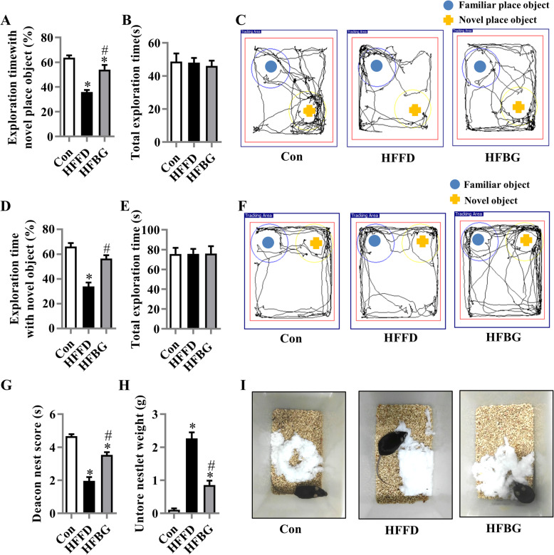
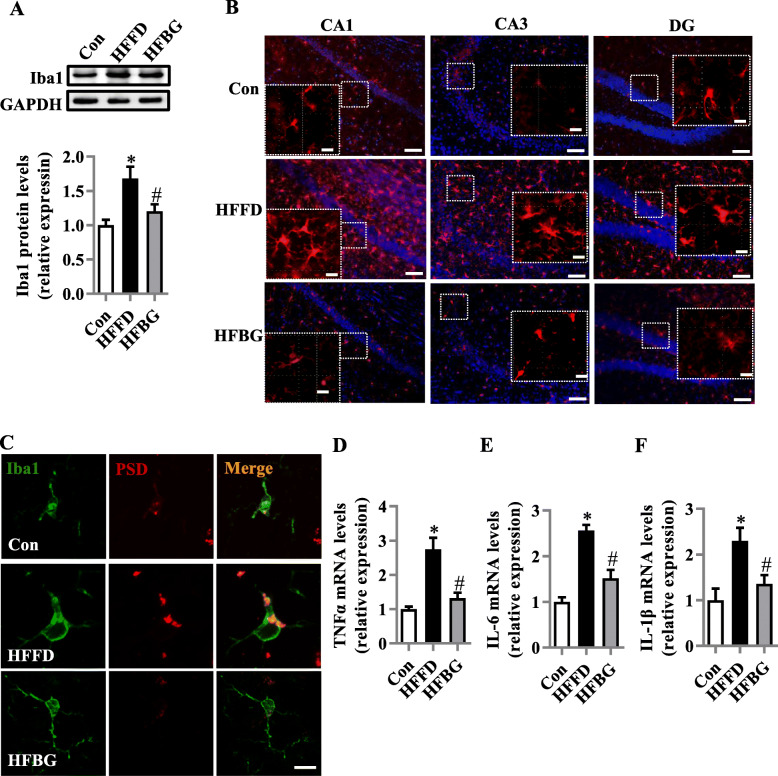
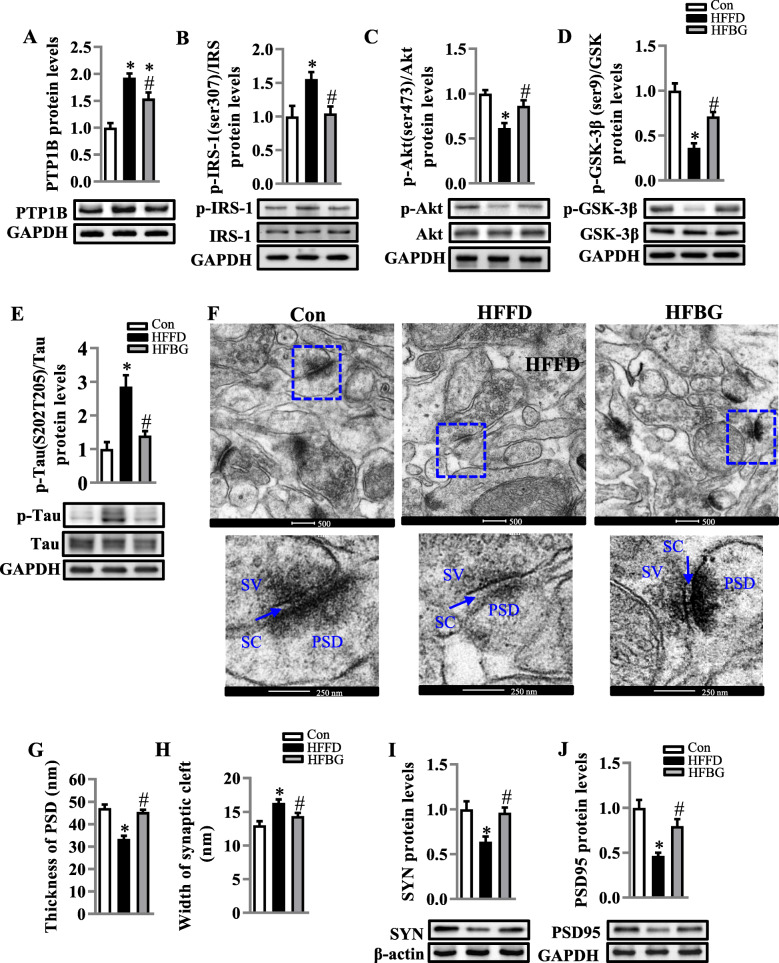
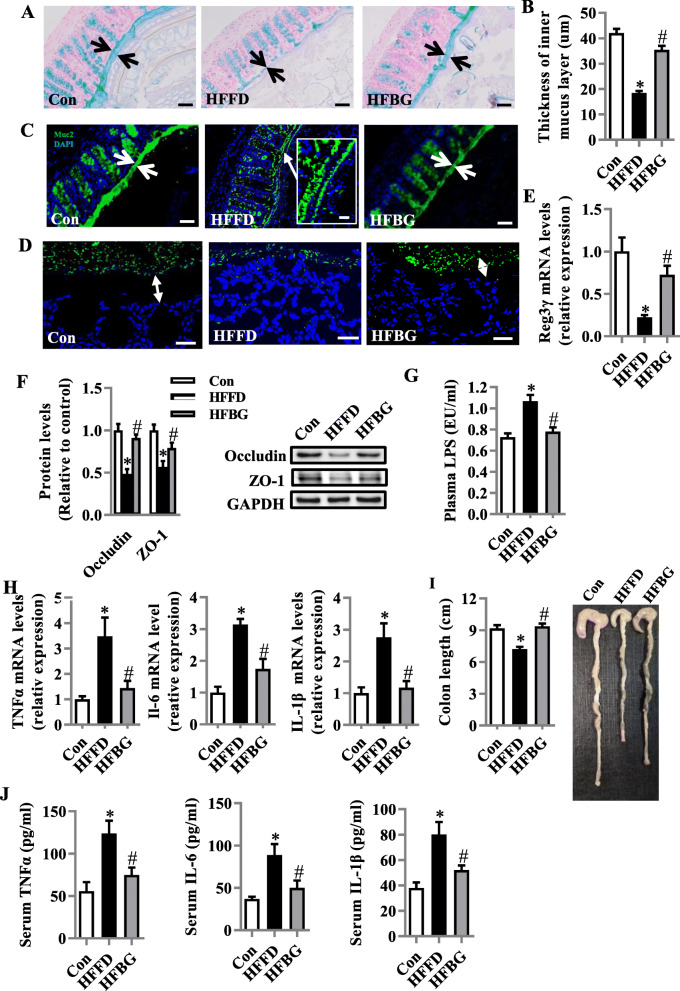
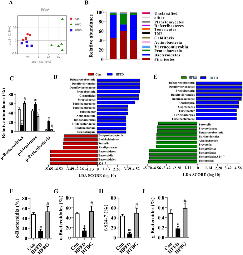
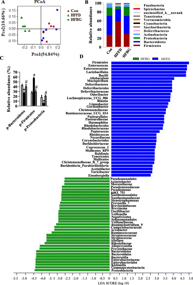
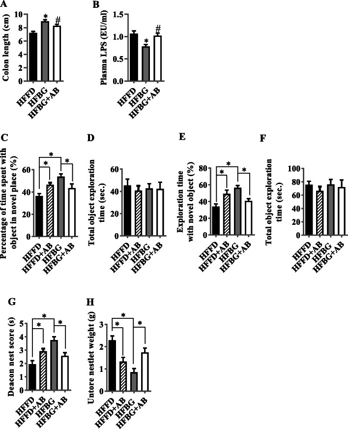
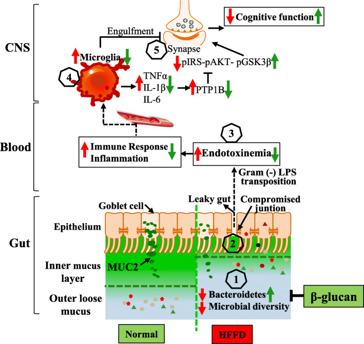
Verification
Claims the paper makes
The claims and statistics of this paper haven’t been checked yet.
Run the check — free, no account needed. It reads the open-access text and checks the sources it cites.
Claims other papers make about this one
Each is checked against this paper’s own text: does it say what the citing paper says it does?
The cited source says what the claim attributes to it.
In the citing paper
“β-glucan supplementation attenuated microglial activation and cognitive impairment in the obese mice.”
From Microbiota–gut–brain axis and its therapeutic applications in neurodegenerative diseases (2024) · cites this paper as [644]
In the citing paper
“Restoring the Bacteroidetes of obese mice fed a high-fat and fiber-deficient diet through oat β-glucan supplementation successfully restored the intestinal barrier integrity, including mucus thickness and levels of tight junction (TJ) proteins.”
From Microbiota–gut–brain axis and its therapeutic applications in neurodegenerative diseases (2024) · cites this paper as [644]
In the citing paper
“the beneficial effects of β-glucan on cognitive function were lost upon antibiotics administration, indicating that the gut microbiota is a vital mediator of cognitive function.”
From Microbiota–gut–brain axis and its therapeutic applications in neurodegenerative diseases (2024) · cites this paper as [644]
In the citing paper
“In another recent animal study, supplementation with the prebiotic β-glucan abrogated microbiota alterations and cognitive impairment as well as microglia activation and neuroinflammation induced by a high-fat, fiber-deficient diet (321).”
From Diet and the Microbiota–Gut–Brain Axis: Sowing the Seeds of Good Mental Health (2021) · cites this paper as [321]
In the citing paper
“Importantly, the microbiota composition was rescued prior to cognitive changes and the positive effects of β-glucan were eliminated with antibiotic treatment (321), suggesting a potential causal relation between diet-induced microbial alterations and cognitive function.”
From Diet and the Microbiota–Gut–Brain Axis: Sowing the Seeds of Good Mental Health (2021) · cites this paper as [321]
Self-check
Percentages, sums and reported differences re-computed from the paper’s own numbers.
The self-check needs the paper’s full text.
Citations
Cites
QIIME allows analysis of high-throughput community sequencing data
Caporaso, J. G., Kuczynski, J., Stombaugh, J., Bittinger, K., Bushman, F. D., Costello, E. K., Fierer, N., Peña, A. G., Goodrich, J. K., Gordon, J. I., et al. (2010) · Nature Methods · doi:10.1038/nmeth.f.303 · Cited by 34,887
Predictive functional profiling of microbial communities using 16S rRNA marker gene sequences
Langille, M. G. I., Zaneveld, J., Caporaso, J. G., McDonald, D., Knights, D., Reyes, J. A., Clemente, J. C., Burkepile, D. E., Vega Thurber, R. L., Knight, R., et al. (2013) · Nature Biotechnology · doi:10.1038/nbt.2676 · Cited by 9,116
Dementia prevention, intervention, and care
Livingston, G., Sommerlad, A., Orgeta, V., Costafreda, S. G., Huntley, J., Ames, D., Ballard, C., Banerjee, S., Burns, A., Cohen-Mansfield, J., et al. (2017) · The Lancet · doi:10.1016/s0140-6736(17)31363-6 · Cited by 6,072
Host-Bacterial Mutualism in the Human Intestine
Bäckhed, F., Ley, R. E., Sonnenburg, J. L., Peterson, D. A., & Gordon, J. I. (2005) · Science · doi:10.1126/science.1104816 · Cited by 5,277
Cross-talk between Akkermansia muciniphila and intestinal epithelium controls diet-induced obesity
Everard, A., Belzer, C., Geurts, L., Ouwerkerk, J. P., Druart, C., Bindels, L. B., Guiot, Y., Derrien, M., Muccioli, G. G., Delzenne, N. M., et al. (2013) · Proceedings of the National Academy of Sciences · doi:10.1073/pnas.1219451110 · Cited by 4,511
Gut microbiota, metabolites and host immunity
Rooks, M. G., & Garrett, W. S. (2016) · Nature reviews. Immunology · doi:10.1038/nri.2016.42 · Cited by 3,409
Complement and microglia mediate early synapse loss in Alzheimer mouse models
Hong, S., Beja-Glasser, V. F., Nfonoyim, B. M., Frouin, A., Li, S., Ramakrishnan, S., Merry, K. M., Shi, Q., Rosenthal, A., Barres, B. A., et al. (2016) · Science · doi:10.1126/science.aad8373 · Cited by 3,343 · cited 1×
“as exemplified by synaptic engulfment and pruning [49].”
BackgroundHsiao, E. Y., McBride, S. W., Hsien, S., Sharon, G., Hyde, E. R., McCue, T., Codelli, J. A., Chow, J., Reisman, S. E., Petrosino, J. F., et al. (2013) · Cell · doi:10.1016/j.cell.2013.11.024 · Cited by 3,283
Desai, M. S., Seekatz, A. M., Koropatkin, N. M., Kamada, N., Hickey, C. A., Wolter, M., Pudlo, N. A., Kitamoto, S., Terrapon, N., Muller, A., et al. (2016) · Cell · doi:10.1016/j.cell.2016.10.043 · Cited by 2,805
Systemic LPS causes chronic neuroinflammation and progressive neurodegeneration
Qin, L., Wu, X., Block, M. L., Liu, Y., Breese, G. R., Hong, J., Knapp, D. J., & Crews, F. T. (2007) · Glia · doi:10.1002/glia.20467 · Cited by 2,144
Environmental and Gut Bacteroidetes: The Food Connection
Thomas, F., Hehemann, J., Rebuffet, E., Czjzek, M., & Michel, G. (2011) · Frontiers in Microbiology · doi:10.3389/fmicb.2011.00093 · Cited by 1,398
Cattaneo, A., Cattane, N., Galluzzi, S., Provasi, S., Lopizzo, N., Festari, C., Ferrari, C., Guerra, U. P., Paghera, B., Muscio, C., et al. (2017) · Neurobiology of Aging · doi:10.1016/j.neurobiolaging.2016.08.019 · Cited by 1,382
The Intersection of Amyloid Beta and Tau at Synapses in Alzheimer’s Disease
Spires-Jones, T. L., & Hyman, B. T. (2014) · Neuron · doi:10.1016/j.neuron.2014.05.004 · Cited by 1,201
Assessing nest building in mice
Deacon, R. M. J. (2006) · Nature Protocols · doi:10.1038/nprot.2006.170 · Cited by 1,115
Spatial memory, recognition memory, and the hippocampus
Broadbent, N. J., Squire, L. R., & Clark, R. E. (2004) · Proceedings of the National Academy of Sciences · doi:10.1073/pnas.0406344101 · Cited by 985
Microglia-mediated neuroinflammation in neurodegenerative diseases
Subhramanyam, C. S., Wang, C., Hu, Q., & Dheen, S. T. (2019) · Seminars in Cell and Developmental Biology · doi:10.1016/j.semcdb.2019.05.004 · Cited by 897
Characterizing a model human gut microbiota composed of members of its two dominant bacterial phyla
Mahowald, M. A., Rey, F. E., Seedorf, H., Turnbaugh, P. J., Fulton, R. S., Wollam, A., Shah, N., Wang, C., Magrini, V. J., Wilson, R. K., et al. (2009) · Proceedings of the National Academy of Sciences · doi:10.1073/pnas.0901529106 · Cited by 752
Fröhlich, E. E., Farzi, A., Mayerhofer, R., Reichmann, F., Jačan, A., Wagner, B., Zinser, E., Bordag, N., Magnes, C., Fröhlich, E., et al. (2016) · Brain Behavior and Immunity · doi:10.1016/j.bbi.2016.02.020 · Cited by 720
Liu, P., Wu, L., Peng, G., Han, Y., Tang, R., Ge, J., Zhang, L., Jia, L., Yue, S., Zhou, K., et al. (2019) · Brain, Behavior, and Immunity · doi:10.1016/j.bbi.2019.05.008 · Cited by 669
Self-citationRegulation of prefrontal cortex myelination by the microbiota
Hoban, A. E., Stilling, R. M., Ryan, F. J., Shanahan, F., Dinan, T. G., Claesson, M. J., Clarke, G., & Cryan, J. F. (2016) · Translational Psychiatry · doi:10.1038/tp.2016.42 · Cited by 635
Kennedy, E. A., King, K. Y., & Baldridge, M. T. (2018) · Frontiers in Physiology · doi:10.3389/fphys.2018.01534 · Cited by 602
Zou, J., Chassaing, B., Singh, V., Pellizzon, M., Ricci, M. R., Flythe, M. D. D., Kumar, M. V., & Gewirtz, A. T. (2017) · Cell Host & Microbe · doi:10.1016/j.chom.2017.11.003 · Cited by 570
Obese-type gut microbiota induce neurobehavioral changes in the absence of obesity
Bruce-Keller, A. J., Salbaum, J. M., Luo, M., Blanchard, E., Taylor, C. M., Welsh, D. A., & Berthoud, H.-R. (2015) · Biological Psychiatry · doi:10.1016/j.biopsych.2014.07.012 · Cited by 563
Butt, M. S., Tahir-Nadeem, M., Khan, M. K. I., Shabir, R., & Butt, M. S. (2008) · European Journal of Nutrition · doi:10.1007/s00394-008-0698-7 · Cited by 559
Infant gut microbiome associated with cognitive development
Carlson, A. L., Xia, K., Azcarate-Peril, M. A., Goldman, B. D., Ahn, M., Styner, M. A., Thompson, A. L., Geng, X., Gilmore, J. H., & Knickmeyer, R. C. (2018) · Biological Psychiatry · doi:10.1016/j.biopsych.2017.06.021 · Cited by 541
Gram-negative bacterial molecules associate with Alzheimer disease pathology
Zhan, X., Stamova, B., Jin, L.-W., DeCarli, C., Phinney, B., & Sharp, F. R. (2016) · Neurology · doi:10.1212/wnl.0000000000003391 · Cited by 521
SCFAs strongly stimulate PYY production in human enteroendocrine cells
Larraufie, P., Martin‐Gallausiaux, C., Lapaque, N., Dore, J. M., Gribble, F. M., Reimann, F. M., & Blottière, H. M. (2018) · Scientific Reports · doi:10.1038/s41598-017-18259-0 · Cited by 484
Yadav, M., & Schorey, J. S. (2006) · Blood · doi:10.1182/blood-2006-05-024406 · Cited by 422
Protein-tyrosine phosphatase 1B expression is induced by inflammation in vivo.
Zabolotny, J. M., Kim, Y.-B., Welsh, L. A., Kershaw, E. E., Neel, B. G., & Kahn, B. B. (2008) · Journal of Biological Chemistry · doi:10.1074/jbc.m800061200 · Cited by 404
The longer-term impacts of Western diet on human cognition and the brain
Francis, H., & Stevenson, R. J. (2013) · Appetite · doi:10.1016/j.appet.2012.12.018 · Cited by 368
Chassaing, B., Ley, R. E., & Gewirtz, A. T. (2014) · Gastroenterology · doi:10.1053/j.gastro.2014.08.033 · Cited by 326
Zhang, R., Miller, R. G., Gascon, R., Champion, S., Katz, J., Lancero, M., Narvaez, A., Honrada, R., Ruvalcaba, D., & McGrath, M. S. (2009) · Journal of Neuroimmunology · doi:10.1016/j.jneuroim.2008.09.017 · Cited by 319
Psychobiotics in mental health, neurodegenerative and neurodevelopmental disorders
Cheng, L.-H., Liu, Y.-W., Wu, C.-C., Wang, S. Z., & Tsai, Y.-C. (2019) · Journal of Food and Drug Analysis · doi:10.1016/j.jfda.2019.01.002 · Cited by 312
Interplay between the gut-brain axis, obesity and cognitive function
Agustí, A., García-Pardo, M. P., López-Almela, I., Campillo, I., Maes, M., Romaní-Pérez, M., & Sanz, Y. (2018) · Frontiers in Neuroscience · doi:10.3389/fnins.2018.00155 · Cited by 294
Saji, N., Niida, S., Murotani, K., Hisada, T., Tsuduki, T., Sugimoto, T., Kimura, A., Toba, K., & Sakurai, T. (2019) · Scientific Reports · doi:10.1038/s41598-018-38218-7 · Cited by 224
Microglia Play an Active Role in Obesity-Associated Cognitive Decline
Cope, E. C., LaMarca, E. A., Monari, P. K., Olson, L. B., Martinez, S., Zych, A. D., Katchur, N. J., & Gould, E. (2018) · The Journal of Neuroscience · doi:10.1523/jneurosci.0789-18.2018 · Cited by 219
Gut microbiota modulate neurobehavior through changes in brain insulin sensitivity and metabolism
Soto, M., Herzog, C., Pacheco, J. A., Fujisaka, S., Bullock, K., Clish, C. B., & Kahn, C. R. (2018) · Molecular Psychiatry · doi:10.1038/s41380-018-0086-5 · Cited by 216
Wang, Y., Ames, N. P., Tun, H. M., Tosh, S. M., Jones, P. J. H., & Khafipour, E. (2016) · Frontiers in Microbiology · doi:10.3389/fmicb.2016.00129 · Cited by 211
Dysbiosis of small intestinal microbiota in liver cirrhosis and its association with etiology
Chen, Y., Ji, F., Guo, J., Shi, D., Fang, D., & Li, L. (2016) · Scientific Reports · doi:10.1038/srep34055 · Cited by 205
Luteolin Protects against High Fat Diet-Induced Cognitive Deficits in Obesity Mice
Liu, Y., Fu, X., Lan, N., Li, S., Zhang, J., Wang, S., Li, C., Shang, Y., Huang, T., & Zhang, L. (2014) · Behavioural Brain Research · doi:10.1016/j.bbr.2014.02.040 · Cited by 194
Ropelle, E. R., Pauli, J. R., Prada, P. O., de Souza, C. T., Picardi, P. K., Faria, M. C., Cintra, D. E., Fernandes, M. F. D. A., Flores, M. B. S., Velloso, L. A., et al. (2006) · The Journal of Physiology · doi:10.1113/jphysiol.2006.120006 · Cited by 187
Tamura, K., Hemsworth, G. R., Déjean, G., Rogers, T. E., Pudlo, N. A., Urs, K., Jain, N., Davies, G. J., Martens, E. C., & Brumer, H. (2017) · Cell Reports · doi:10.1016/j.celrep.2017.09.049 · Cited by 178
Synaptic proteins, neuropathology and cognitive status in the oldest-old
Head, E., Corrada, M. M., Kahle-Wrobleski, K., Kim, R. C., Sarsoza, F., Goodus, M., & Kawas, C. H. (2009) · Neurobiology of Aging · doi:10.1016/j.neurobiolaging.2007.10.001 · Cited by 145
Hattiangady, B., Mishra, V., Kodali, M., Shuai, B., Rao, X., & Shetty, A. K. (2014) · Frontiers in Behavioral Neuroscience · doi:10.3389/fnbeh.2014.00078 · Cited by 145
Whitfield, D., Vallortigara, J., Alghamdi, A., Howlett, D. R., Hortobágyi, T., Johnson, M., Attems, J., Newhouse, S., Ballard, C. G., Thomas, A. J., et al. (2014) · Neurobiology of Aging · doi:10.1016/j.neurobiolaging.2014.06.015 · Cited by 144
Relationships between dietary intake and cognitive function level in Korean elderly people
Lee, L., Kang, S., Lee, H. O., Lee, B.-H., Park, J., Kim, J.-H., Jung, I.-K., Park, Y., & Lee, J. (2001) · Public Health · doi:10.1038/sj.ph.1900729 · Cited by 138
Clemens, R., Kranz, S., Mobley, A. R., Nicklas, T. A., Raimondi, M. P., Rodriguez, J. C., Slavin, J. L., & Warshaw, H. S. (2012) · Journal of Nutrition · doi:10.3945/jn.112.160176 · Cited by 125
Dietary Fiber Is Positively Associated with Cognitive Control among Prepubertal Children ,
Khan, N. A., Raine, L. B., Drollette, E. S., Scudder, M. R., Kramer, A. F., & Hillman, C. H. (2014) · Journal of Nutrition · doi:10.3945/jn.114.198457 · Cited by 123
Protein Tyrosine Phosphatase 1B (PTP1B): A Potential Target for Alzheimer’s Therapy?
Vieira, M. N. N., Silva, N. M. L. E., Ferreira, S. T., & Felice, F. G. D. (2017) · Frontiers in Aging Neuroscience · doi:10.3389/fnagi.2017.00007 · Cited by 123
Obesity Weighs down Memory through a Mechanism Involving the Neuroepigenetic Dysregulation of Sirt1
Heyward, F. D., Gilliam, D., Coleman, M. A., Gavin, C. F., Wang, J., Kaas, G. A., Trieu, R. J., Lewis, J., Moulden, J., & Sweatt, J. D. (2016) · Journal of Neuroscience · doi:10.1523/jneurosci.1934-15.2016 · Cited by 97
Inhibition of inflammation by astaxanthin alleviates cognition deficits in diabetic mice
Zhou, X., Zhang, F., Hu, X., Chen, J., Wen, X., Sun, Y., Liu, Y., Tang, R., Zheng, K., & Song, Y. (2015) · Physiology & Behavior · doi:10.1016/j.physbeh.2015.08.015 · Cited by 94
Self-citationZhang, P., Yu, Y., Qin, Y., Zhou, Y., Tang, R., Wang, Q., Li, X., Wang, H., Weston–Green, K., Huang, X., et al. (2018) · The Journal of Nutritional Biochemistry · doi:10.1016/j.jnutbio.2018.08.016 · Cited by 92
Self-citationKang, S., Kim, C. H., Jung, H., Kim, E., Song, H., & Lee, J. E. (2016) · Neuropharmacology · doi:10.1016/j.neuropharm.2016.10.029 · Cited by 92
The impact of dietary energy intake on cognitive aging
Mattson, M. P. (2010) · Frontiers in Aging Neuroscience · doi:10.3389/neuro.24.005.2010 · Cited by 91
Beck, E. J., Tapsell, L. C., Batterham, M. J., Tosh, S. M., & Huang, X. (2009) · Nutrition Research · doi:10.1016/j.nutres.2009.09.012 · Cited by 86
Self-citationKarumuthil‐Melethil, S., Gudi, R. R., Johnson, B. M., Perez, N., & Vasu, C. (2014) · The Journal of Immunology · doi:10.4049/jimmunol.1400186 · Cited by 79
Wang, H., Sun, X., Zhang, N., Ji, Z., Ma, Z., Fu, Q., Qu, R., & Ma, S. (2017) · Physiology & Behavior · doi:10.1016/j.physbeh.2017.10.001 · Cited by 78
Jiang, Y., Liu, Y., Zhu, C., Ma, X., Ma, L., Zhou, L., Huang, Q., Lei, C., Pi, R., & Chen, X. (2015) · Neurobiology of Learning and Memory · doi:10.1016/j.nlm.2015.03.003 · Cited by 75
Role of microtubule-associated protein tau phosphorylation in Alzheimer’s disease
Ma, R.-H., Zhang, Y., Hong, X.-Y., Zhang, J., Wang, J., & Liu, G. (2017) · Journal of Huazhong University of Science and Technology [Medical Sciences] · doi:10.1007/s11596-017-1732-x · Cited by 66
Camer, D., Yu, Y., Szabo, A., Fernandez, F., Dinh, C. H. L., & Huang, X. (2015) · Progress in Neuro-Psychopharmacology and Biological Psychiatry · doi:10.1016/j.pnpbp.2015.01.004 · Cited by 63
Self-citationProtein Tyrosine Phosphatase PTP1B Is Involved in Hippocampal Synapse Formation and Learning
Fuentes, F., Zimmer, D. J., Atienza, M., Schottenfeld, J., Penkala, I. J., Bale, T. L., Bence, K. K., & Arregui, C. O. (2012) · PLoS ONE · doi:10.1371/journal.pone.0041536 · Cited by 62
Huang, X., Yu, Y., Beck, E. J., South, T., Li, Y., Batterham, M. J., Tapsell, L. C., & Chen, J. (2011) · Molecular Nutrition & Food Research · doi:10.1002/mnfr.201100095 · Cited by 58
Self-citationXu, N., Fan, W., Zhou, X., Liu, Y., Ma, P., Qi, S., & Gu, B. (2018) · Metabolic Brain Disease · doi:10.1007/s11011-018-0269-4 · Cited by 55
Wang, S., Huang, X., Zhang, P., Newell, K. A., Wang, H., Zheng, K., & Yu, Y. (2017) · Scientific Reports · doi:10.1038/s41598-017-12156-2 · Cited by 53
Self-citationNeuron‐microglia interaction induced bi‐directional cytotoxicity associated with calpain activation
Podbielska, M., Das, A., Smith, A. W., Chauhan, A., Ray, S. K., Inoue, J., Azuma, M., Nozaki, K., Hogan, E. L., & Banik, N. L. (2016) · Journal of Neurochemistry · doi:10.1111/jnc.13774 · Cited by 50
Trends in dietary fiber intake in Chinese aged 45 years and above, 1991–2011
Wang, H. J., Wang, Z. H., Zhang, J. G., Du, W., Su, C., Zhang, J., Zhai, F., & Zhang, B. (2014) · European Journal of Clinical Nutrition · doi:10.1038/ejcn.2014.24 · Cited by 49
Zhou, X., Wang, S., Ding, X., Li, Q., Mao, Y., Chen, L., Li, W., & Ying, C. (2017) · Brain Research Bulletin · doi:10.1016/j.brainresbull.2017.06.001 · Cited by 45
Self-citationMechanism of macrophage activation induced by β-glucan produced from Paenibacillus polymyxa JB115
Chang, Z., Lee, J., Gebru, E., Hong, J., Jung, H.-K., Jo, W.-S., & Park, S. (2009) · Biochemical and Biophysical Research Communications · doi:10.1016/j.bbrc.2009.12.064 · Cited by 38
Hynönen, U., Rasinkangas, P., Satokari, R. M., Paulin, L. G., de Vos, W. M., Pietilä, T. E., Kant, R., & Palva, A. (2016) · Anaerobe · doi:10.1016/j.anaerobe.2016.03.001 · Cited by 34
Camer, D., Yu, Y., Szabo, A., Dinh, C. H. L., Wang, H., Cheng, L., & Huang, X. (2015) · Molecular and Cellular Endocrinology · doi:10.1016/j.mce.2015.05.018 · Cited by 34
Self-citationPathak, N. M., Pathak, V., Lynch, A. M., Irwin, N., Gault, V. A., & Flatt, P. R. (2015) · Molecular and Cellular Endocrinology · doi:10.1016/j.mce.2015.05.035 · Cited by 29
Temporal order memory differences in Alzheimer's disease and vascular dementia
Hampstead, B. M., Libon, D., Moelter, S., Swirsky-Sacchetti, T., Scheffer, L., Platek, S., & Chute, D. (2010) · Journal of Clinical and Experimental Neuropsychology · doi:10.1080/13803390903418918 · Cited by 23
The effect of dietary fat on behavior in mice
Keleher, M. R., Zaidi, R., Patel, K., Ahmed, A. A. B., Bettler, C., Pavlatos, C., Shah, S., & Cheverud, J. M. (2018) · Journal of Diabetes & Metabolic Disorders · doi:10.1007/s40200-018-0373-3 · Cited by 19
Is Dietary Fiber Considered an Essential Nutrient?
Kohn, J. B. (2016) · Journal of the Academy of Nutrition and Dietetics · doi:10.1016/j.jand.2015.12.004 · Cited by 18
Research works printed but not linked: 2
- Huang XF, et al. Diet high in oat β-glucan activates the gut-hypothalamic (PYY3-36-NPY) axis and increases satiety in diet-induced obesity in mice. Mol Nutr Food Res. 2011;55(7):1118–21. · DOI
- Paxinos G, Franklin KBJ. The mouse brain in stereotaxic coordinates, 1st edn. San Diego: Academic Press; 2002.
Cited by
Sorted by citations
Microbiota–gut–brain axis and its therapeutic applications in neurodegenerative diseases
Loh, J. S., Mak, W. Q., Tan, L. K. S., Ng, C. X., Chan, H. H., Yeow, S. H., Foo, J. B., Ong, Y. S., How, C. W., & Khaw, K. Y. (2024) · Signal Transduction and Targeted Therapy · doi:10.1038/s41392-024-01743-1 · Cited by 1,193
Diet and the Microbiota–Gut–Brain Axis: Sowing the Seeds of Good Mental Health
Berding, K., Vlckova, K., Marx, W., Schellekens, H., Stanton, C., Clarke, G., Jacka, F., Dinan, T. G., & Cryan, J. F. (2021) · Advances in Nutrition · doi:10.1093/advances/nmaa181 · Cited by 451
Pediatric obesity—Long‐term consequences and effect of weight loss
Marcus, C., Danielsson, P., & Hagman, E. (2022) · Journal of Internal Medicine · doi:10.1111/joim.13547 · Cited by 267
Highland barley: Chemical composition, bioactive compounds, health effects, and applications
Obadi, M., Sun, J. S., & Xu, B. (2021) · Food Research International · doi:10.1016/j.foodres.2020.110065 · Cited by 253
Hao, W., Ma, Q., Wang, L., Yuan, N., Gan, H. T., He, L., Li, X., Huang, J., & Chen, J. (2024) · Microbiome · doi:10.1186/s40168-024-01756-6 · Cited by 163
Metadata
- Authors
- Shi, H., Yu, Y., Lin, D., Zheng, P., Zhang, P., Hu, M., Wang, Q., Pan, W., Yang, X., Hu, T., Li, Q., Tang, R., Zhou, F., Zheng, K., & Huang, X.-F.
- Affiliations
- Xuzhou Medical College (CN)
- University of Wollongong (AU)
- Corresponding author
- Yu, Yinghua
- Zheng, Kuiyang
- Huang, Xu-Feng
- ORCID
- Venue
- Microbiome
- Published
- 2020-10-02
- Volume
- 8
- Issue
- 1
- Pages
- 143-143
- ISSN
- 2049-2618 (ISSN-L)
- PMC
- PMC7532656
- OpenAlex
- W3090508437
- Cited by
- 264
- References
- 74
- Citations by year
- 2026: 23 · 2025: 56 · 2024: 64 · 2023: 43 · 2022: 53 · 2021: 22 · 2020: 2
- FWCI
- 9.72
- Type
- journal article
- Language
- en
- Open access
- gold
- doi.org
- Locations
- Microbiome (publishedVersion, cc-by)
- Research Square (acceptedVersion, cc-by)
- PubMed Central (publishedVersion, cc-by)
- PubMed (publishedVersion)
- Research Online (University of Wollongong) (submittedVersion)
- DOAJ (DOAJ: Directory of Open Access Journals) (submittedVersion, cc-by-sa)
- Figshare (submittedVersion)
- Funders
- National Natural Science Foundation of China (National Natural Science Foundation of China)
- Natural Science Foundation of the Jiangsu Higher Education Institutions of China (19KJA560003)
- the Starting Foundation for Talents of Xuzhou Medical University (D2018003)
- the Priority Academic Program Development of Jiangsu Higher Education Institutions ((PAPD) in 2014)
- Topics
- Life Sciences › Biochemistry, Genetics and Molecular Biology › Molecular Biology › Gut microbiota and health
- Health Sciences › Nursing › Nutrition and Dietetics › Microbial Metabolites in Food Biotechnology
- Life Sciences › Neuroscience › Neurology › Neuroinflammation and Neurodegeneration Mechanisms
- Keywords
- gut-brain axis, cognitive impairment, diet-induced obesity, gut microbiota, hippocampus, neuroinflammation, microglial activation, synaptic ultrastructure, tau phosphorylation, intestinal barrier dysfunction, colonic mucus, tight junctions, endotoxin translocation, Bacteroidetes, dietary fiber supplementation
- MeSH terms
- Gastrointestinal Microbiome, Animals, Brain, Dietary Fiber, Male, Mice, Inbred C57BL, Obesity, beta-Glucans, Mice, Diet, High-Fat, Cognitive Dysfunction
About these checks
Registry, retraction, Cites and statistics results are computed, not judged, with one exception: a printed reference that no index lists is read once by a model before it counts as a citation error. Claims are extracted by a model and kept only when their quote appears verbatim in the text; each cited source is then checked, with a model’s help, for whether it says what the claim relies on. Those verdicts can be wrong — read the claim’s quote and the cited source before relying on one.
Statistics and arithmetic are recomputed, not judged.
Confidence bands are shown only for fields calibrated on our annotated test set. None is calibrated yet, so no band is shown.
What counts as a claim ›How we measure confidence ›How we check this ›
Are you an author, or do you see a problem with this page? Request a review
