

The DOI matches its Crossref record; 56 of the 60 references it cites were found in a registry or an index; 6 of the 27 sources its claims cite were checked for support.
An mRNA Vaccine Encoding Rabies Virus Glycoprotein Induces Protection against Lethal Infection in Mice and Correlates of Protection in Adult and Newborn Pigs
Open accessAbstract
Rabies is a zoonotic infectious disease of the central nervous system (CNS). In unvaccinated or untreated subjects, rabies virus infection causes severe neurological symptoms and is invariably fatal. Despite the long-standing existence of effective vaccines, vaccine availability remains insufficient, with high numbers of fatal infections mostly in developing countries. Nucleic acid based vaccines have proven convincingly as a new technology for the fast development of vaccines against newly emerging pathogens, diseases where no vaccine exists or for replacing already existing vaccines. We used an optimized non-replicating rabies virus glycoprotein (RABV-G) encoding messenger RNA (mRNA) to induce potent neutralizing antibodies (VN titers) in mice and domestic pigs. Functional antibody titers were followed in mice for up to one year and titers remained stable for the entire observation period in all dose groups. T cell analysis revealed the induction of both, specific CD4+ as well as CD8+ T cells by RABV-G mRNA, with the induced CD4+ T cells being higher than those induced by a licensed vaccine. Notably, RABV-G mRNA vaccinated mice were protected against lethal intracerebral challenge infection. Inhibition of viral replication by vaccination was verified by qRT-PCR. Furthermore, we demonstrate that CD4+ T cells are crucial for the generation of neutralizing antibodies. In domestic pigs we were able to induce VN titers that correlate with protection in adult and newborn pigs. This study demonstrates the feasibility of a non-replicating mRNA rabies vaccine in small and large animals and highlights the promises of mRNA vaccines for the prevention of infectious diseases.
Overview
AI snapshot
Written from the paper’s full text.
TL;DR
A non-replicating, protamine-complexed mRNA vaccine encoding rabies virus glycoprotein induced neutralizing antibodies and T cells in mice and pigs, and protected mice against lethal intracerebral rabies challenge. Protection required CD4+ T cells during immunization, and adult and newborn pigs developed neutralizing titers above the WHO correlate of 0.5 IU/ml.
Key findings
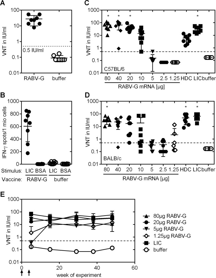
- Two 80 μg RABV-G mRNA vaccinations induced VN titers ≥0.5 IU/ml in all immunized mice (range 5.9–70.2 IU/ml); after doses ≥10 μg, 100% of C57BL/6 and 90% (29/32) of BALB/c mice seroconverted. Titers remained stable for about one year, with mean titers between 17 and 65 IU/ml for 20 μg, 80 μg, and Rabipur groups.
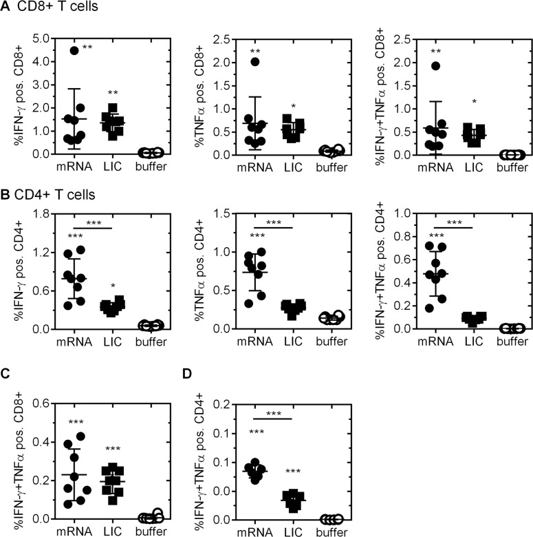
- RABV-G mRNA induced antigen-specific CD4+ and CD8+ T cells. IFN-γ/TNFα-double-positive CD4+ T cells were higher after mRNA than Rabipur at 6 days after boost (mean 0.48% vs 0.091%) and at day 91 (mean 0.084% vs 0.034%).
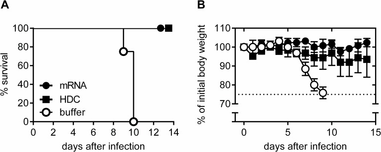
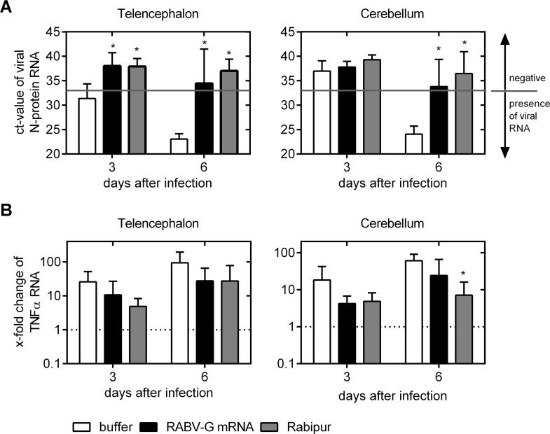
- All RABV-G mRNA and HDC vaccinated mice survived lethal intracerebral challenge with 40-fold LD50 CVS-11, while none of the buffer-treated mice survived. qRT-PCR showed viral N-protein RNA was suppressed below detection in all but one mRNA-vaccinated mouse.
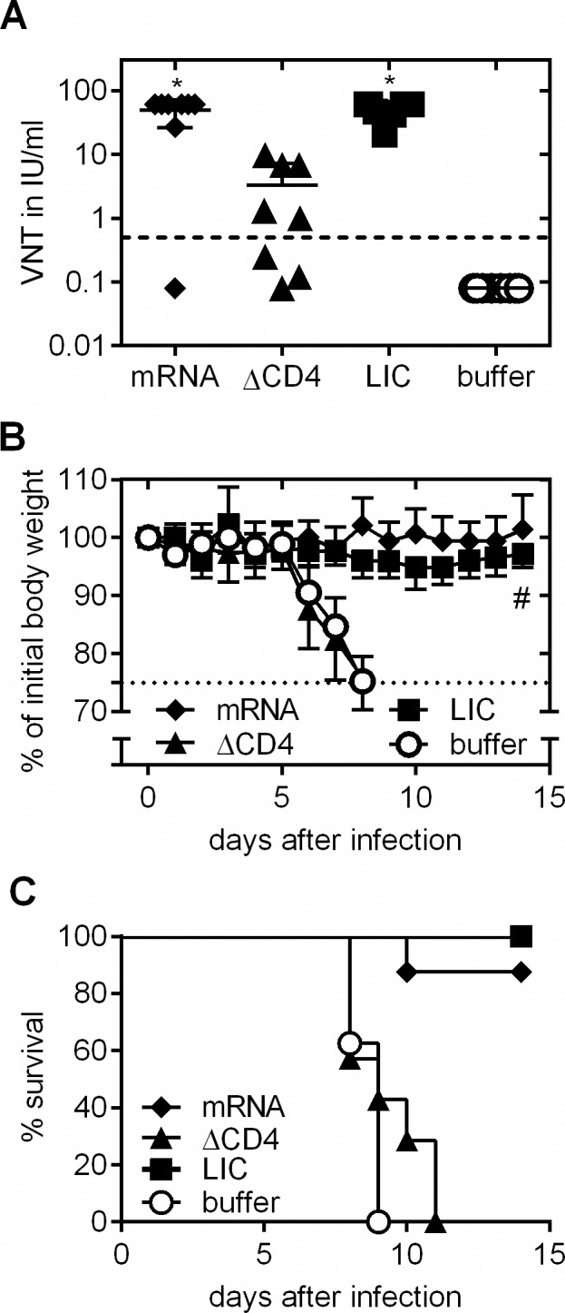
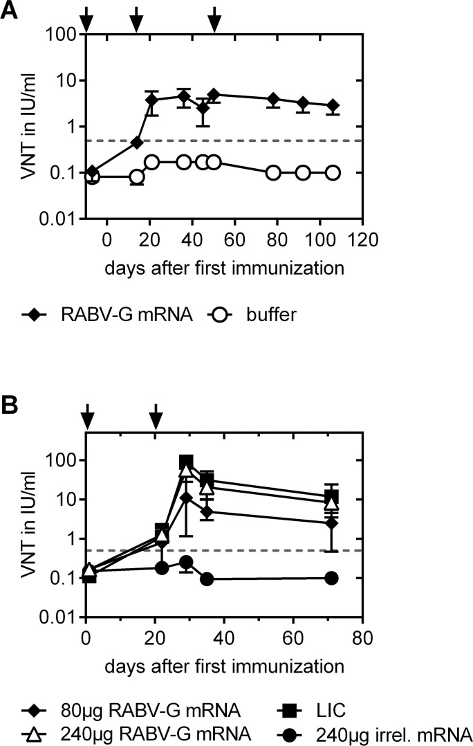
- CD4+ T cell depletion during immunization reduced mean VN titers to about 3 IU/ml versus about 50 IU/ml in CD4-intact mRNA-vaccinated mice, and all CD4-depleted mRNA-vaccinated mice died within 11 days after challenge. Adult and newborn pigs seroconverted above 0.5 IU/ml; newborn piglets given 80 μg had mean titers of 11.0 IU/ml at day 28 and 2.5 IU/ml at day 70, while 240 μg gave 56.8 IU/ml and 8.3 IU/ml, comparable to full-dose Rabipur.
Methods
- Mice (BALB/c and C57BL/6, 6–8 weeks old) and domestic pigs (adult and newborn) were immunized intradermally with optimized non-replicating RABV-G mRNA complexed with protamine; controls included buffer, irrelevant mRNA, and licensed vaccines Rabipur or HDC.
- Antibody responses were measured by FAVN test; T cell responses by intracellular cytokine staining and ELISPOT; CD4+ T cells were depleted with anti-CD4 monoclonal antibody; mice were challenged intracerebrally with 40-fold LD50 CVS-11; viral N-protein RNA and TNFα mRNA in brains were quantified by qRT-PCR.
- Statistical comparisons used Mann Whitney, Kruskal-Wallis, one-way ANOVA with Tukey’s or Dunnett’s multiple comparisons tests.
Limitations
- The paper states that one mRNA-vaccinated mouse had detectable viral N-protein RNA at day 6 and was non-protected, and that reduced TNFα mRNA levels in vaccinated brains were not statistically significant.
- The paper notes that the 0.5 IU/ml threshold may be a surrogate for protection against natural peripheral rabies infection rather than the laboratory intracerebral challenge used here.
- Evident from the text: this is a preclinical animal study; pigs were assessed for immunogenicity only, not challenge protection; group sizes were small; and no human efficacy or safety data are reported, although a phase I clinical study is mentioned as initiated.
Model-written summary. Check it against the paper before relying on it.
Integrity signals
Registry
Cites
Statistics
Figures
Verification
Claims the paper makes
53 Own result · 20 Own interpretation · 9 No full text of the sources · 4 Own method · 2 Supported so far · 2 Partly supported · 2 Sources not found · 2 No basis given · 1 Supported · 1 Not checked yet · accuracy not yet measured
The cited sources support only part of this claim — read each source’s verdict below.
In the paper
“Concerning the induction of neutralizing antibodies, this finding indicates a comparable mode of action for the mRNA vaccine format as seen for cell based vaccines and other vaccine formats [3,13].”
§ An mRNA Vaccine Encoding Rabies Virus Glycoprotein Induces Protection against Lethal Infection in Mice and Correlates of Protection in Adult and Newborn Pigs > Discussion
What the sources say
- Partly supportedcites [3] HCJ Ertl, 2009, Novel vaccines to human rabies, PLoS neglected tropical diseases sourceExperimental — accuracy not yet measured
The Introduction and “Vaccines to Rabies Virus” section identify neutralizing antibodies against rabies glycoprotein as the key correlate of protection, and describe this principle across several vaccine formats. The paper does not discuss mRNA vaccines or establish that their mode of action is comparable to that of cell-based vaccines. (LLM, conf=0.86)
- Not checked — no full text of the sourcecites [13] R Amann, 2013, A new rabies vaccine based on a recombinant ORF virus (parapoxvirus) expressing the rabies virus glycoprotein, Journal of Virology source
hedgedcorrelational- Asserted: passes — The authors put it forward as true.
- Checkable: passes — It can be checked against evidence.
- Load-bearing: unclear — It is unclear whether the paper’s conclusions depend on it.
- Based: passes — The paper gives a reason why it holds.
- Partly supported
Every source the paper cites for this claim was checked and supports it.
In the paper
“We next analyzed immunogenicity in domestic pigs, a large animal model that exhibits relevant physiological and anatomical similarities to humans [44,45].”
§ An mRNA Vaccine Encoding Rabies Virus Glycoprotein Induces Protection against Lethal Infection in Mice and Correlates of Protection in Adult and Newborn Pigs > Results > Seroconversion in newborn and adult pigs
What the sources say
- Supportedcites [44] F Meurens, 2012, The pig: a model for human infectious diseases, Trends Microbiol source
The “Pigs as a model for humans” section states that pigs are very similar to humans in anatomy and physiology, and describes breeds ranging from miniature to large. Its discussion of the porcine model also lists human-like size as an advantage. This supports the cited description of domestic pigs as a large animal model with relevant physiological and anatomical similarities to humans. (LLM, conf=0.94)
- Supportedcites [45] KH Mair, 2014, The porcine innate immune system: an update, Dev Comp Immunol source
The section “Relevance of the pig as a large animal model to study the innate immune system” explicitly describes pigs as an animal model with anatomical and physiological characteristics comparable to humans. Its table also lists their size as similar to humans. Page numbers are not provided in the supplied copy. (LLM, conf=0.94)
plain- Asserted: passes — The authors put it forward as true.
- Checkable: passes — It can be checked against evidence.
- Load-bearing: passes — The paper’s conclusions depend on it.
- Based: passes — The paper gives a reason why it holds.
- Supported
A result the paper reports from its own data — it cites no other source for it, so there is none to check. The quote shows it is in the paper.
In the paper
“with the induced CD4+ T cells being higher than those induced by a licensed vaccine.”
§ An mRNA Vaccine Encoding Rabies Virus Glycoprotein Induces Protection against Lethal Infection in Mice and Correlates of Protection in Adult and Newborn Pigs > Abstract
plain- Asserted: passes — The authors put it forward as true.
- Checkable: passes — It can be checked against evidence.
- Load-bearing: unclear — It is unclear whether the paper’s conclusions depend on it.
- Based: passes — The paper gives a reason why it holds.
A result the paper reports from its own data — it cites no other source for it, so there is none to check. The quote shows it is in the paper.
In the paper
“Notably, RABV-G mRNA vaccinated mice were protected against lethal intracerebral challenge infection.”
- Also stated in: § Introduction · plain
“The format conferred full protection in a stringent murine challenge model of intracerebral inoculation with rabies virus.”
- Also stated in: § Protection against lethal intracerebral challenge in mice · plain
“All RABV-G mRNA and HDC vaccinated mice were protected against lethal rabies virus challenge (Fig 3A).”
§ An mRNA Vaccine Encoding Rabies Virus Glycoprotein Induces Protection against Lethal Infection in Mice and Correlates of Protection in Adult and Newborn Pigs > Abstract
plaindirect- Asserted: passes — The authors put it forward as true.
- Checkable: passes — It can be checked against evidence.
- Load-bearing: passes — The paper’s conclusions depend on it.
- Based: passes — The paper gives a reason why it holds.
- Also stated in: § Introduction · plain
A result the paper reports from its own data — it cites no other source for it, so there is none to check. The quote shows it is in the paper.
In the paper
“Furthermore, we demonstrate that CD4+ T cells are crucial for the generation of neutralizing antibodies.”
§ An mRNA Vaccine Encoding Rabies Virus Glycoprotein Induces Protection against Lethal Infection in Mice and Correlates of Protection in Adult and Newborn Pigs > Abstract
boosteddirect- Asserted: passes — The authors put it forward as true.
- Checkable: passes — It can be checked against evidence.
- Load-bearing: passes — The paper’s conclusions depend on it.
- Based: passes — The paper gives a reason why it holds.
A result the paper reports from its own data — it cites no other source for it, so there is none to check. The quote shows it is in the paper.
In the paper
“T cell analysis revealed the induction of both, specific CD4+ as well as CD8+ T cells by RABV-G mRNA”
§ An mRNA Vaccine Encoding Rabies Virus Glycoprotein Induces Protection against Lethal Infection in Mice and Correlates of Protection in Adult and Newborn Pigs > Abstract
plaindirect- Asserted: passes — The authors put it forward as true.
- Checkable: passes — It can be checked against evidence.
- Load-bearing: unclear — It is unclear whether the paper’s conclusions depend on it.
- Based: passes — The paper gives a reason why it holds.
A result the paper reports from its own data — it cites no other source for it, so there is none to check. The quote shows it is in the paper.
In the paper
“Functional antibody titers were followed in mice for up to one year and titers remained stable for the entire observation period in all dose groups.”
§ An mRNA Vaccine Encoding Rabies Virus Glycoprotein Induces Protection against Lethal Infection in Mice and Correlates of Protection in Adult and Newborn Pigs > Abstract
plain- Asserted: passes — The authors put it forward as true.
- Checkable: passes — It can be checked against evidence.
- Load-bearing: unclear — It is unclear whether the paper’s conclusions depend on it.
- Based: passes — The paper gives a reason why it holds.
A result the paper reports from its own data — it cites no other source for it, so there is none to check. The quote shows it is in the paper.
In the paper
“We used an optimized non-replicating rabies virus glycoprotein (RABV-G) encoding messenger RNA (mRNA) to induce potent neutralizing antibodies (VN titers) in mice and domestic pigs.”
§ An mRNA Vaccine Encoding Rabies Virus Glycoprotein Induces Protection against Lethal Infection in Mice and Correlates of Protection in Adult and Newborn Pigs > Abstract
plaindirect- Asserted: passes — The authors put it forward as true.
- Checkable: passes — It can be checked against evidence.
- Load-bearing: passes — The paper’s conclusions depend on it.
- Based: passes — The paper gives a reason why it holds.
A result the paper reports from its own data — it cites no other source for it, so there is none to check. The quote shows it is in the paper.
In the paper
“Inhibition of viral replication by vaccination was verified by qRT-PCR.”
§ An mRNA Vaccine Encoding Rabies Virus Glycoprotein Induces Protection against Lethal Infection in Mice and Correlates of Protection in Adult and Newborn Pigs > Abstract
plaindirect- Asserted: passes — The authors put it forward as true.
- Checkable: passes — It can be checked against evidence.
- Load-bearing: passes — The paper’s conclusions depend on it.
- Based: passes — The paper gives a reason why it holds.
A result the paper reports from its own data — it cites no other source for it, so there is none to check. The quote shows it is in the paper.
In the paper
“Finally, immunogenicity was shown in domestic pigs”
§ An mRNA Vaccine Encoding Rabies Virus Glycoprotein Induces Protection against Lethal Infection in Mice and Correlates of Protection in Adult and Newborn Pigs > Introduction
plain- Asserted: passes — The authors put it forward as true.
- Checkable: passes — It can be checked against evidence.
- Load-bearing: passes — The paper’s conclusions depend on it.
- Based: passes — The paper gives a reason why it holds.
- Probably not a substantial claim. 17
- It may state two separate claims as one. 36
- The quote only partly states this claim. 1
The cited sources support only part of this claim — read each source’s verdict below.
In the paper
“While incubation phases vary, death commonly follows within an average survival time between 6 and 11 days after first symptomatic onset for furious or paralytic forms, respectively, thus leaving little time and extremely limited therapeutic options [1,2].”
§ An mRNA Vaccine Encoding Rabies Virus Glycoprotein Induces Protection against Lethal Infection in Mice and Correlates of Protection in Adult and Newborn Pigs > Introduction
What the sources say
- Not checked — no full text of the sourcecites [1] T Hemachudha, 2013, Human rabies: neuropathogenesis, diagnosis, and management, Lancet Neurology source
- Partly supportedcites [2] S Pathak, 2014, Diagnosis, management and post-mortem findings of a human case of rabies imported into the United Kingdom from India: a case report, Virology Journal sourceExperimental — accuracy not yet measured
The Background says clinical rabies is invariably fatal, and the Conclusion describes limited evidence for the Milwaukee Protocol’s efficacy. The Case presentation records one patient’s deterioration through day 10, but the paper does not give average survival times of 6 and 11 days after symptom onset for furious and paralytic rabies, respectively. (LLM, conf=0.88)
plaindirect- Asserted: passes — The authors put it forward as true.
- Checkable: passes — It can be checked against evidence.
- Load-bearing: fails — The paper’s conclusions may not depend on it.
- Based: passes — The paper gives a reason why it holds.
Probably not a substantial claim. The paper’s conclusions may not depend on it. It may state two separate claims as one.
The sources with a verdict support this claim; the rest weren’t checked yet, have no full text we could read, or their verdicts aren’t rated yet.
In the paper
“Novel vaccine formats such as subunit, DNA or viral vector vaccines have been assessed successfully for their protective capacity against rabies infection in preclinical settings [3,13] but none of these resulted in a licensed product for human use yet.”
§ An mRNA Vaccine Encoding Rabies Virus Glycoprotein Induces Protection against Lethal Infection in Mice and Correlates of Protection in Adult and Newborn Pigs > Introduction
What the sources say
- Supportedcites [3] HCJ Ertl, 2009, Novel vaccines to human rabies, PLoS neglected tropical diseases source
The Protein Subunit Vaccines and DNA Vaccines sections report protection against rabies challenge in animals, and the AdHu5 vector section reports full protection in a mouse model. The Introduction identifies commercially available human vaccines as inactivated-virus vaccines, while the Summary describes novel candidates as largely awaiting extensive preclinical or clinical testing, consistent with none of these formats having yielded a licensed human product at the time of the review. (LLM, conf=0.89)
- Not checked — no full text of the sourcecites [13] R Amann, 2013, A new rabies vaccine based on a recombinant ORF virus (parapoxvirus) expressing the rabies virus glycoprotein, Journal of Virology source
plain- Asserted: passes — The authors put it forward as true.
- Checkable: passes — It can be checked against evidence.
- Load-bearing: fails — The paper’s conclusions may not depend on it.
- Based: passes — The paper gives a reason why it holds.
Probably not a substantial claim. The paper’s conclusions may not depend on it. It may state two separate claims as one.
- Supported
The sources with a verdict support this claim; the rest weren’t checked yet, have no full text we could read, or their verdicts aren’t rated yet.
In the paper
“Benefits are the induction of balanced and enduring immunity as demonstrated for anti-tumor and prophylactic vaccination [14–16], simple supply, and storage at elevated temperature, as reviewed elsewhere [17,18].”
§ An mRNA Vaccine Encoding Rabies Virus Glycoprotein Induces Protection against Lethal Infection in Mice and Correlates of Protection in Adult and Newborn Pigs > Introduction
What the sources say
- Not checked — no full text of the sourcecites [14] I Hoerr, 2000, In vivo application of RNA leads to induction of specific cytotoxic T lymphocytes and antibodies, Eur J Immunol source
- Not checked — no full text of the sourcecites [15] M Fotin-Mleczek, 2011, Messenger RNA-based vaccines with dual activity induce balanced TLR-7 dependent adaptive immune responses and provide antitumor activity, J Immunother source
- Not checked — no full text of the sourcecites [16] EJ Solari, 2012, Differentiating Literacy Growth of ELL Students With LD From Other High-Risk Subgroups and General Education Peers: Evidence From Grades 3–10, J Learn Disabil source
- Supportedcites [17] K-J Kallen, 2013, A novel, disruptive vaccination technology: self-adjuvanted RNActive vaccines, Human Vaccines & Immunotherapeutics source
The Conclusion (page unavailable) describes RNActive® vaccines as inducing balanced effector and memory responses and says RNA vaccines can be produced flexibly at scale without a cold chain. The prophylaxis section (page unavailable) reports that an RNActive® vaccine stored at 37°C protected mice as effectively as one stored at −20°C. These benefits are demonstrated for the RNActive® format discussed in the paper. (LLM, conf=0.90)
- Not checked — no full text of the sourcecites [18] T Kramps, 2013, Messenger RNA-based vaccines: progress, challenges, applications, Wiley interdisciplinary reviews RNA source
boosteddirect- Asserted: passes — The authors put it forward as true.
- Checkable: passes — It can be checked against evidence.
- Load-bearing: fails — The paper’s conclusions may not depend on it.
- Based: passes — The paper gives a reason why it holds.
Probably not a substantial claim. The paper’s conclusions may not depend on it. It may state two separate claims as one.
A result the paper reports from its own data — it cites no other source for it, so there is none to check. The quote shows it is in the paper.
In the paper
“We found that after vaccination with 20 μg and 80 μg RABV-G mRNA or with Rabipur antibody titers remained stable throughout the observation period with mean titers between 17 and 65 IU/ml (Fig 1E).”
§ An mRNA Vaccine Encoding Rabies Virus Glycoprotein Induces Protection against Lethal Infection in Mice and Correlates of Protection in Adult and Newborn Pigs > Results > Induction of long-lasting virus neutralizing titers in mice
plain- Asserted: passes — The authors put it forward as true.
- Checkable: passes — It can be checked against evidence.
- Load-bearing: passes — The paper’s conclusions depend on it.
- Based: passes — The paper gives a reason why it holds.
It may state two separate claims as one.
A result the paper reports from its own data — it cites no other source for it, so there is none to check. The quote shows it is in the paper.
In the paper
“In C57BL/6 mice immunization with a dose of 10 μg RABV-G mRNA vaccine (range: 0.5 to 40.6 IU/ml) revealed comparable immunogenicity to HDC (range: 1.5 to 13.5 IU/ml) and significantly higher titers were achieved at mRNA doses of ≥20 μg (range: 17.8 to 92.4 IU/ml).”
§ An mRNA Vaccine Encoding Rabies Virus Glycoprotein Induces Protection against Lethal Infection in Mice and Correlates of Protection in Adult and Newborn Pigs > Results > Induction of long-lasting virus neutralizing titers in mice
plaindirect- Asserted: passes — The authors put it forward as true.
- Checkable: passes — It can be checked against evidence.
- Load-bearing: passes — The paper’s conclusions depend on it.
- Based: passes — The paper gives a reason why it holds.
It may state two separate claims as one.
A result the paper reports from its own data — it cites no other source for it, so there is none to check. The quote shows it is in the paper.
In the paper
“Thus, in both mouse strains, a dose-response correlation was seen and high neutralizing antibody titers were induced at doses ≥10 μg mRNA.”
§ An mRNA Vaccine Encoding Rabies Virus Glycoprotein Induces Protection against Lethal Infection in Mice and Correlates of Protection in Adult and Newborn Pigs > Results > Induction of long-lasting virus neutralizing titers in mice
plaincorrelational- Asserted: passes — The authors put it forward as true.
- Checkable: passes — It can be checked against evidence.
- Load-bearing: passes — The paper’s conclusions depend on it.
- Based: passes — The paper gives a reason why it holds.
It may state two separate claims as one.
A result the paper reports from its own data — it cites no other source for it, so there is none to check. The quote shows it is in the paper.
In the paper
“Moreover, we demonstrate the early, efficient viral clearance and brain homeostasis by mRNA vaccination upon intracerebral infection.”
§ An mRNA Vaccine Encoding Rabies Virus Glycoprotein Induces Protection against Lethal Infection in Mice and Correlates of Protection in Adult and Newborn Pigs > Introduction
boosteddirect- Asserted: passes — The authors put it forward as true.
- Checkable: passes — It can be checked against evidence.
- Load-bearing: passes — The paper’s conclusions depend on it.
- Based: passes — The paper gives a reason why it holds.
It may state two separate claims as one.
A result the paper reports from its own data — it cites no other source for it, so there is none to check. The quote shows it is in the paper.
In the paper
“This analysis revealed that in splenocytes of RABV-G mRNA vaccinated mice, IFN-γ secreting cells were detected upon Rabipur stimulation, but not after stimulation with BSA (negative control) or in splenocytes of buffer treated control mice (Fig 1B).”
§ An mRNA Vaccine Encoding Rabies Virus Glycoprotein Induces Protection against Lethal Infection in Mice and Correlates of Protection in Adult and Newborn Pigs > Results > Induction of long-lasting virus neutralizing titers in mice
plain- Asserted: passes — The authors put it forward as true.
- Checkable: passes — It can be checked against evidence.
- Load-bearing: passes — The paper’s conclusions depend on it.
- Based: passes — The paper gives a reason why it holds.
It may state two separate claims as one.
A result the paper reports from its own data — it cites no other source for it, so there is none to check. The quote shows it is in the paper.
In the paper
“For the 1.25 μg and 5 μg mRNA vaccine doses, response onset was delayed, but caught up by week 12 of the experiment and titers remained at a high level of ≥ 10 IU/ml.”
§ An mRNA Vaccine Encoding Rabies Virus Glycoprotein Induces Protection against Lethal Infection in Mice and Correlates of Protection in Adult and Newborn Pigs > Results > Induction of long-lasting virus neutralizing titers in mice
plaindirect- Asserted: passes — The authors put it forward as true.
- Checkable: passes — It can be checked against evidence.
- Load-bearing: passes — The paper’s conclusions depend on it.
- Based: passes — The paper gives a reason why it holds.
It may state two separate claims as one.
A result the paper reports from its own data — it cites no other source for it, so there is none to check. The quote shows it is in the paper.
In the paper
“In the CD4-depleted, mRNA vaccinated group, all mice had to be sacrificed within 11 days after infection with similar kinetics as in the negative control group”
§ An mRNA Vaccine Encoding Rabies Virus Glycoprotein Induces Protection against Lethal Infection in Mice and Correlates of Protection in Adult and Newborn Pigs > Results > Importance of CD4+ T cells for the induction of a protective immune response
plain- Asserted: passes — The authors put it forward as true.
- Checkable: passes — It can be checked against evidence.
- Load-bearing: passes — The paper’s conclusions depend on it.
- Based: passes — The paper gives a reason why it holds.
It may state two separate claims as one.
A result the paper reports from its own data — it cites no other source for it, so there is none to check. The quote shows it is in the paper.
In the paper
“All mice vaccinated with Rabipur (undepleted positive control) survived the challenge infection while buffer injected mice (negative controls) had to be sacrificed by day nine post challenge due to dramatic weight loss”
§ An mRNA Vaccine Encoding Rabies Virus Glycoprotein Induces Protection against Lethal Infection in Mice and Correlates of Protection in Adult and Newborn Pigs > Results > Importance of CD4+ T cells for the induction of a protective immune response
plain- Asserted: passes — The authors put it forward as true.
- Checkable: passes — It can be checked against evidence.
- Load-bearing: passes — The paper’s conclusions depend on it.
- Based: passes — The paper gives a reason why it holds.
It may state two separate claims as one.
A result the paper reports from its own data — it cites no other source for it, so there is none to check. The quote shows it is in the paper.
In the paper
“At day 3 after infection a low quantity of N-protein mRNA was detectable in the telencephalon of animals treated with buffer (mean: 31.4 cycle threshold (ct)-value) whereas the cerebellum was negative for viral N-protein mRNA (mean above detection limit). Ct-values, however, decreased over the period of infection as at day 6 high viral loads were detected in both analyzed parts of the brains (mean”
§ An mRNA Vaccine Encoding Rabies Virus Glycoprotein Induces Protection against Lethal Infection in Mice and Correlates of Protection in Adult and Newborn Pigs > Results > Protection against lethal intracerebral challenge in mice
plainn = 6- Asserted: passes — The authors put it forward as true.
- Checkable: passes — It can be checked against evidence.
- Load-bearing: passes — The paper’s conclusions depend on it.
- Based: passes — The paper gives a reason why it holds.
It may state two separate claims as one.
A result the paper reports from its own data — it cites no other source for it, so there is none to check. The quote shows it is in the paper.
In the paper
“In buffer-treated mice we found viral replication in the telencephalon (the site of injection) and spreading to the cerebellum. In addition, both telencephalon and cerebellum of these animals showed increased TNFα expression”
§ An mRNA Vaccine Encoding Rabies Virus Glycoprotein Induces Protection against Lethal Infection in Mice and Correlates of Protection in Adult and Newborn Pigs > Discussion
plaincorrelational- Asserted: passes — The authors put it forward as true.
- Checkable: passes — It can be checked against evidence.
- Load-bearing: passes — The paper’s conclusions depend on it.
- Based: passes — The paper gives a reason why it holds.
It may state two separate claims as one.
A result the paper reports from its own data — it cites no other source for it, so there is none to check. The quote shows it is in the paper.
In the paper
“Inter-individual variability was low in all test groups.”
§ An mRNA Vaccine Encoding Rabies Virus Glycoprotein Induces Protection against Lethal Infection in Mice and Correlates of Protection in Adult and Newborn Pigs > Results > Induction of long-lasting virus neutralizing titers in mice
plain- Asserted: passes — The authors put it forward as true.
- Checkable: passes — It can be checked against evidence.
- Load-bearing: fails — The paper’s conclusions may not depend on it.
- Based: passes — The paper gives a reason why it holds.
Probably not a substantial claim. The paper’s conclusions may not depend on it.
A result the paper reports from its own data — it cites no other source for it, so there is none to check. The quote shows it is in the paper.
In the paper
“In contrast, at day 9 after infection, first buffer treated mice had to be sacrificed due to pronounced weight loss (25% of initial body weight) and none in this group survived the infection.”
§ An mRNA Vaccine Encoding Rabies Virus Glycoprotein Induces Protection against Lethal Infection in Mice and Correlates of Protection in Adult and Newborn Pigs > Results > Protection against lethal intracerebral challenge in mice
plain- Asserted: passes — The authors put it forward as true.
- Checkable: passes — It can be checked against evidence.
- Load-bearing: passes — The paper’s conclusions depend on it.
- Based: passes — The paper gives a reason why it holds.
It may state two separate claims as one.
A result the paper reports from its own data — it cites no other source for it, so there is none to check. The quote shows it is in the paper.
In the paper
“In the CD4-depleted, mRNA immunized group VN titers were strongly reduced: 3 out of 8 mice showed VN titers below 0.5 IU/ml and the mean titer of the groups was about 3 IU/ml, i.e. more than 15-fold reduced compared to the CD4-intact, mRNA-vaccinated group.”
§ An mRNA Vaccine Encoding Rabies Virus Glycoprotein Induces Protection against Lethal Infection in Mice and Correlates of Protection in Adult and Newborn Pigs > Results > Importance of CD4+ T cells for the induction of a protective immune response
boosteddirectn = 8- Asserted: passes — The authors put it forward as true.
- Checkable: passes — It can be checked against evidence.
- Load-bearing: passes — The paper’s conclusions depend on it.
- Based: passes — The paper gives a reason why it holds.
It may state two separate claims as one.
A result the paper reports from its own data — it cites no other source for it, so there is none to check. The quote shows it is in the paper.
In the paper
“In mRNA and Rabipur vaccinated mice TNFα mRNA transcript level was reduced compared to mock vaccinated control group at both analysis time points and different brain sections”
- Also stated in: § Discussion · hedged
“Though not statistically significant, the data indicate a reduced innate inflammatory reaction”
§ An mRNA Vaccine Encoding Rabies Virus Glycoprotein Induces Protection against Lethal Infection in Mice and Correlates of Protection in Adult and Newborn Pigs > Results > Protection against lethal intracerebral challenge in mice
plaindirect- Asserted: passes — The authors put it forward as true.
- Checkable: passes — It can be checked against evidence.
- Load-bearing: unclear — It is unclear whether the paper’s conclusions depend on it.
- Based: passes — The paper gives a reason why it holds.
It may state two separate claims as one. The quote only partly states this claim.
- Also stated in: § Discussion · hedged
A result the paper reports from its own data — it cites no other source for it, so there is none to check. The quote shows it is in the paper.
In the paper
“In groups with an intact CD4+ T cell compartment during prophylactic immunization, more than 87% of mRNA-vaccinated mice survived infection (one out of eight mRNA-immunized mice without detectable virus neutralizing antibodies after immunization had to be sacrificed on day ten due to pronounced weight loss)”
§ An mRNA Vaccine Encoding Rabies Virus Glycoprotein Induces Protection against Lethal Infection in Mice and Correlates of Protection in Adult and Newborn Pigs > Results > Importance of CD4+ T cells for the induction of a protective immune response
plainn = 8- Asserted: passes — The authors put it forward as true.
- Checkable: passes — It can be checked against evidence.
- Load-bearing: passes — The paper’s conclusions depend on it.
- Based: passes — The paper gives a reason why it holds.
It may state two separate claims as one.
A result the paper reports from its own data — it cites no other source for it, so there is none to check. The quote shows it is in the paper.
In the paper
“Two mice treated with HDC showed a transient weight loss (10–20% of initial body weight), but survived the experiment (Fig 3B).”
§ An mRNA Vaccine Encoding Rabies Virus Glycoprotein Induces Protection against Lethal Infection in Mice and Correlates of Protection in Adult and Newborn Pigs > Results > Protection against lethal intracerebral challenge in mice
plainn = 2- Asserted: passes — The authors put it forward as true.
- Checkable: passes — It can be checked against evidence.
- Load-bearing: fails — The paper’s conclusions may not depend on it.
- Based: passes — The paper gives a reason why it holds.
Probably not a substantial claim. The paper’s conclusions may not depend on it. It may state two separate claims as one.
A result the paper reports from its own data — it cites no other source for it, so there is none to check. The quote shows it is in the paper.
In the paper
“Eight weeks post second boost RABV-G specific neutralizing antibodies had a mean titer of 2.9 IU/ml and were still above the 0.5 IU/ml limit.”
§ An mRNA Vaccine Encoding Rabies Virus Glycoprotein Induces Protection against Lethal Infection in Mice and Correlates of Protection in Adult and Newborn Pigs > Results > Seroconversion in newborn and adult pigs
plain- Asserted: passes — The authors put it forward as true.
- Checkable: passes — It can be checked against evidence.
- Load-bearing: passes — The paper’s conclusions depend on it.
- Based: passes — The paper gives a reason why it holds.
It may state two separate claims as one.
A result the paper reports from its own data — it cites no other source for it, so there is none to check. The quote shows it is in the paper.
In the paper
“Furthermore, immunogenicity of the RABV-G mRNA was also investigated in newborn piglets, again showing neutralizing antibody titers after the first vaccination in the protective range that were further increased after the booster vaccination.”
§ An mRNA Vaccine Encoding Rabies Virus Glycoprotein Induces Protection against Lethal Infection in Mice and Correlates of Protection in Adult and Newborn Pigs > Discussion
plaindirect- Asserted: passes — The authors put it forward as true.
- Checkable: passes — It can be checked against evidence.
- Load-bearing: passes — The paper’s conclusions depend on it.
- Based: passes — The paper gives a reason why it holds.
It may state two separate claims as one.
A result the paper reports from its own data — it cites no other source for it, so there is none to check. The quote shows it is in the paper.
In the paper
“In adult pigs, titers reached the protective limit of ≥0.5 IU/ml after the first immunization and could be further increased by booster vaccination, then remaining stable throughout the experiment.”
§ An mRNA Vaccine Encoding Rabies Virus Glycoprotein Induces Protection against Lethal Infection in Mice and Correlates of Protection in Adult and Newborn Pigs > Discussion
plaindirect- Asserted: passes — The authors put it forward as true.
- Checkable: passes — It can be checked against evidence.
- Load-bearing: passes — The paper’s conclusions depend on it.
- Based: passes — The paper gives a reason why it holds.
It may state two separate claims as one.
A result the paper reports from its own data — it cites no other source for it, so there is none to check. The quote shows it is in the paper.
In the paper
“This mRNA vaccine encoding the Rabies virus glycoprotein was immunogenic upon two injections at microgram doses and resulted in the induction of virus neutralizing antibodies, antigen-specific CD4+ T cells and CD8+ T cells (Fig 1).”
§ An mRNA Vaccine Encoding Rabies Virus Glycoprotein Induces Protection against Lethal Infection in Mice and Correlates of Protection in Adult and Newborn Pigs > Discussion
plaindirect- Asserted: passes — The authors put it forward as true.
- Checkable: passes — It can be checked against evidence.
- Load-bearing: passes — The paper’s conclusions depend on it.
- Based: passes — The paper gives a reason why it holds.
It may state two separate claims as one.
A result the paper reports from its own data — it cites no other source for it, so there is none to check. The quote shows it is in the paper.
In the paper
“Piglets receiving 80 μg RABV-G mRNA developed mean protective titers of 11.0 IU/ml at day 28 and 2.5 IU/ml at day 70, while piglets immunized with the 240 μg dose revealed higher mean antibody titers of 56.8 IU/ml at day 28 and 8.3 IU/ml at day 70 that were comparable to those induced by full doses of the licensed human vaccine”
§ An mRNA Vaccine Encoding Rabies Virus Glycoprotein Induces Protection against Lethal Infection in Mice and Correlates of Protection in Adult and Newborn Pigs > Results > Seroconversion in newborn and adult pigs
plaindirect- Asserted: passes — The authors put it forward as true.
- Checkable: passes — It can be checked against evidence.
- Load-bearing: passes — The paper’s conclusions depend on it.
- Based: passes — The paper gives a reason why it holds.
It may state two separate claims as one.
In the paper
“In India, more human fatalities are caused by rabies than in any other country of the world with an estimated 16,000 deaths in the year 2010 alone [10].”
§ An mRNA Vaccine Encoding Rabies Virus Glycoprotein Induces Protection against Lethal Infection in Mice and Correlates of Protection in Adult and Newborn Pigs > Introduction
What the sources say
- Not checked yetcites [10] World Health O (2013) WHO Expert Consultation on Rabies. Second report. World Health Organization Technical Report Series: 1–139, back cover.
hedged- Asserted: passes — The authors put it forward as true.
- Checkable: passes — It can be checked against evidence.
- Load-bearing: fails — The paper’s conclusions may not depend on it.
- Based: passes — The paper gives a reason why it holds.
Probably not a substantial claim. The paper’s conclusions may not depend on it. It may state two separate claims as one.
In the paper
“Despite the availability of effective vaccines, rabies infection continues to claim at least 55,000 human lives per year (WHO Weekly epidemiological record, No. 49/50, 2007), not counting the great excess of fatal infections in animals.”
§ An mRNA Vaccine Encoding Rabies Virus Glycoprotein Induces Protection against Lethal Infection in Mice and Correlates of Protection in Adult and Newborn Pigs > Introduction
What the sources say
- Not matchedcites WHO Weekly epidemiological record, No. 49/50, 2007
Couldn’t be matched to a paper in Crossref, so it can’t be checked.
plain- Asserted: passes — The authors put it forward as true.
- Checkable: passes — It can be checked against evidence.
- Load-bearing: fails — The paper’s conclusions may not depend on it.
- Based: passes — The paper gives a reason why it holds.
Probably not a substantial claim. The paper’s conclusions may not depend on it.
- Not matched
What the paper did — it cites no source for this, so there is none to check. The quote shows it is in the paper.
In the paper
“All animal experiments were conducted according to German and Hungarian laws and guidelines for animal protection.”
§ An mRNA Vaccine Encoding Rabies Virus Glycoprotein Induces Protection against Lethal Infection in Mice and Correlates of Protection in Adult and Newborn Pigs > Materials and Methods > Mice and pigs
plain- Asserted: passes — The authors put it forward as true.
- Checkable: passes — It can be checked against evidence.
- Load-bearing: fails — The paper’s conclusions may not depend on it.
- Based: passes — The paper gives a reason why it holds.
Probably not a substantial claim. The paper’s conclusions may not depend on it.
What the paper did — it cites no source for this, so there is none to check. The quote shows it is in the paper.
In the paper
“Experiments in Germany were approved by the regional council Tübingen (reference numbers FLI238/08, FLI242/09, FLI246/09, CUR 4–13).”
§ An mRNA Vaccine Encoding Rabies Virus Glycoprotein Induces Protection against Lethal Infection in Mice and Correlates of Protection in Adult and Newborn Pigs > Materials and Methods > Mice and pigs
plain- Asserted: passes — The authors put it forward as true.
- Checkable: passes — It can be checked against evidence.
- Load-bearing: fails — The paper’s conclusions may not depend on it.
- Based: passes — The paper gives a reason why it holds.
Probably not a substantial claim. The paper’s conclusions may not depend on it.
A conclusion the paper draws from its own results — it cites no other source for it, so there is none to check. The quote shows it is in the paper.
In the paper
“In contrast thereto, protection has first to be built up in case of initiation of vaccination after exposure to the rabies virus, which requires time, as it is done by PEP vaccination.”
§ An mRNA Vaccine Encoding Rabies Virus Glycoprotein Induces Protection against Lethal Infection in Mice and Correlates of Protection in Adult and Newborn Pigs > Introduction
plaindirect- Asserted: passes — The authors put it forward as true.
- Checkable: passes — It can be checked against evidence.
- Load-bearing: passes — The paper’s conclusions depend on it.
- Based: passes — The paper gives a reason why it holds.
Probably not a substantial claim. It may state two separate claims as one.
In the paper
“Anti-rabies serum antibodies were analyzed by FAVN test by Eurovir Hygiene-Institut, Luckenwalde, Germany according to WHO protocol [30].”
§ An mRNA Vaccine Encoding Rabies Virus Glycoprotein Induces Protection against Lethal Infection in Mice and Correlates of Protection in Adult and Newborn Pigs > Materials and Methods > Antibody analysis
What the sources say
- Not checked — no full text of the sourcecites [30] F Cliquet, 1998, Development of a fluorescent antibody virus neutralisation test (FAVN test) for the quantitation of rabies-neutralising antibody, Journal of Immunological Methods source
plain- Asserted: passes — The authors put it forward as true.
- Checkable: passes — It can be checked against evidence.
- Load-bearing: passes — The paper’s conclusions depend on it.
- Based: passes — The paper gives a reason why it holds.
Probably not a substantial claim.
A conclusion the paper draws from its own results — it cites no other source for it, so there is none to check. The quote shows it is in the paper.
In the paper
“As a consequence, effective immunological defense against rabies must intercept virus before productive neuronal infection.”
§ An mRNA Vaccine Encoding Rabies Virus Glycoprotein Induces Protection against Lethal Infection in Mice and Correlates of Protection in Adult and Newborn Pigs > Introduction
plaindirect- Asserted: passes — The authors put it forward as true.
- Checkable: passes — It can be checked against evidence.
- Load-bearing: passes — The paper’s conclusions depend on it.
- Based: passes — The paper gives a reason why it holds.
Probably not a substantial claim.
A conclusion the paper draws from its own results — it cites no other source for it, so there is none to check. The quote shows it is in the paper.
In the paper
“These experiments, therefore, indicate comparable immunogenicity of the mRNA vaccine with licensed rabies vaccine and the general induction of specific immune response in mice.”
§ An mRNA Vaccine Encoding Rabies Virus Glycoprotein Induces Protection against Lethal Infection in Mice and Correlates of Protection in Adult and Newborn Pigs > Results > Induction of long-lasting virus neutralizing titers in mice
hedged- Asserted: passes — The authors put it forward as true.
- Checkable: passes — It can be checked against evidence.
- Load-bearing: passes — The paper’s conclusions depend on it.
- Based: passes — The paper gives a reason why it holds.
It may state two separate claims as one.
A conclusion the paper draws from its own results — it cites no other source for it, so there is none to check. The quote shows it is in the paper.
In the paper
“That is the reason for the administration of rabies immunoglobulin (RIG) together with active PEP vaccination in the case of suspected rabies exposure in rabies naïve individuals.”
§ An mRNA Vaccine Encoding Rabies Virus Glycoprotein Induces Protection against Lethal Infection in Mice and Correlates of Protection in Adult and Newborn Pigs > Introduction
plaindirect- Asserted: passes — The authors put it forward as true.
- Checkable: passes — It can be checked against evidence.
- Load-bearing: fails — The paper’s conclusions may not depend on it.
- Based: passes — The paper gives a reason why it holds.
Probably not a substantial claim. The paper’s conclusions may not depend on it.
A conclusion the paper draws from its own results — it cites no other source for it, so there is none to check. The quote shows it is in the paper.
In the paper
“This is most effective when the neutralizing antibodies and clearing T cells are already present in situ at the time of infection, which is the case after prophylactic vaccination.”
§ An mRNA Vaccine Encoding Rabies Virus Glycoprotein Induces Protection against Lethal Infection in Mice and Correlates of Protection in Adult and Newborn Pigs > Introduction
plaindirect- Asserted: passes — The authors put it forward as true.
- Checkable: passes — It can be checked against evidence.
- Load-bearing: passes — The paper’s conclusions depend on it.
- Based: passes — The paper gives a reason why it holds.
It may state two separate claims as one.
A conclusion the paper draws from its own results — it cites no other source for it, so there is none to check. The quote shows it is in the paper.
In the paper
“Our finding that depletion of CD4+ T cells during the immunization phase results in a loss of protection against lethal challenge infection further indicates the need for T cell help in mounting a protective neutralizing antibody response.”
§ An mRNA Vaccine Encoding Rabies Virus Glycoprotein Induces Protection against Lethal Infection in Mice and Correlates of Protection in Adult and Newborn Pigs > Discussion
hedgeddirect- Asserted: passes — The authors put it forward as true.
- Checkable: passes — It can be checked against evidence.
- Load-bearing: passes — The paper’s conclusions depend on it.
- Based: passes — The paper gives a reason why it holds.
It may state two separate claims as one.
A conclusion the paper draws from its own results — it cites no other source for it, so there is none to check. The quote shows it is in the paper.
In the paper
“Though not statistically significant, the data indicate a reduced innate inflammatory reaction which might result in the maintenance of brain homeostasis upon vaccination with RABV-G mRNA or licensed rabies vaccine.”
§ An mRNA Vaccine Encoding Rabies Virus Glycoprotein Induces Protection against Lethal Infection in Mice and Correlates of Protection in Adult and Newborn Pigs > Discussion
hedgedconditional- Asserted: passes — The authors put it forward as true.
- Checkable: passes — It can be checked against evidence.
- Load-bearing: unclear — It is unclear whether the paper’s conclusions depend on it.
- Based: passes — The paper gives a reason why it holds.
It may state two separate claims as one.
A conclusion the paper draws from its own results — it cites no other source for it, so there is none to check. The quote shows it is in the paper.
In the paper
“These results indicate efficient RABV-G-specific T cell induction by RABV-G mRNA and better induction of CD4+ T cells by the mRNA vaccine compared to Rabipur.”
§ An mRNA Vaccine Encoding Rabies Virus Glycoprotein Induces Protection against Lethal Infection in Mice and Correlates of Protection in Adult and Newborn Pigs > Results > Induction of antigen-specific CD4+ and CD8+ T cells
hedgeddirect- Asserted: passes — The authors put it forward as true.
- Checkable: passes — It can be checked against evidence.
- Load-bearing: passes — The paper’s conclusions depend on it.
- Based: passes — The paper gives a reason why it holds.
It may state two separate claims as one.
In the paper
“Importantly, it also forms the basis of the widely used National Institutes of Health (NIH) potency test for rabies vaccines [40].”
§ An mRNA Vaccine Encoding Rabies Virus Glycoprotein Induces Protection against Lethal Infection in Mice and Correlates of Protection in Adult and Newborn Pigs > Results > Protection against lethal intracerebral challenge in mice
What the sources say
- Not matchedcites [40] F.-X. KMM Meslin, 1996
Couldn’t be matched to a paper in Crossref, so it can’t be checked.
plaindirect- Asserted: passes — The authors put it forward as true.
- Checkable: passes — It can be checked against evidence.
- Load-bearing: fails — The paper’s conclusions may not depend on it.
- Based: passes — The paper gives a reason why it holds.
Probably not a substantial claim. The paper’s conclusions may not depend on it.
- Not matched
In the paper
“Although T cells do not prevent initial viral infection of host cells, they provide important impact on rabies virus clearance in mice [38].”
§ An mRNA Vaccine Encoding Rabies Virus Glycoprotein Induces Protection against Lethal Infection in Mice and Correlates of Protection in Adult and Newborn Pigs > Results > Induction of antigen-specific CD4+ and CD8+ T cells
What the sources say
- Not checked — no full text of the sourcecites [38] TJ Wiktor, 1978, Cell-mediated immunity and postexposure protection from rabies by inactivated vaccines of tissue culture origin, Developments in Biological Standardization
plaindirect- Asserted: passes — The authors put it forward as true.
- Checkable: passes — It can be checked against evidence.
- Load-bearing: unclear — It is unclear whether the paper’s conclusions depend on it.
- Based: passes — The paper gives a reason why it holds.
It may state two separate claims as one.
In the paper
“Rabies-specific T cell clones have also been isolated from human vaccinees [39].”
§ An mRNA Vaccine Encoding Rabies Virus Glycoprotein Induces Protection against Lethal Infection in Mice and Correlates of Protection in Adult and Newborn Pigs > Results > Induction of antigen-specific CD4+ and CD8+ T cells
What the sources say
- Not checked — no full text of the sourcecites [39] E Celis, 1988, Genetic restriction and fine specificity of human T cell clones reactive with rabies virus, Journal of Immunology (Baltimore, Md: 1950) source
plain- Asserted: passes — The authors put it forward as true.
- Checkable: passes — It can be checked against evidence.
- Load-bearing: fails — The paper’s conclusions may not depend on it.
- Based: passes — The paper gives a reason why it holds.
Probably not a substantial claim. The paper’s conclusions may not depend on it.
In the paper
“In summary, we further substantiated and extended previously published work on non-replicating influenza-specific mRNA vaccines [16], proving the protective efficacy of mRNA vaccination in a second, harsh viral challenge model.”
§ An mRNA Vaccine Encoding Rabies Virus Glycoprotein Induces Protection against Lethal Infection in Mice and Correlates of Protection in Adult and Newborn Pigs > Discussion
What the sources say
- Not checked — no full text of the sourcecites [16] EJ Solari, 2012, Differentiating Literacy Growth of ELL Students With LD From Other High-Risk Subgroups and General Education Peers: Evidence From Grades 3–10, J Learn Disabil source
boosteddirect- Asserted: passes — The authors put it forward as true.
- Checkable: passes — It can be checked against evidence.
- Load-bearing: passes — The paper’s conclusions depend on it.
- Based: passes — The paper gives a reason why it holds.
It may state two separate claims as one.
A conclusion the paper draws from its own results — it cites no other source for it, so there is none to check. The quote shows it is in the paper.
In the paper
“In the present study, we demonstrated the immunogenicity and protective efficacy of an experimental mRNA-based rabies vaccine in animals.”
§ An mRNA Vaccine Encoding Rabies Virus Glycoprotein Induces Protection against Lethal Infection in Mice and Correlates of Protection in Adult and Newborn Pigs > Discussion
boosteddirect- Asserted: passes — The authors put it forward as true.
- Checkable: passes — It can be checked against evidence.
- Load-bearing: passes — The paper’s conclusions depend on it.
- Based: passes — The paper gives a reason why it holds.
It may state two separate claims as one.
A conclusion the paper draws from its own results — it cites no other source for it, so there is none to check. The quote shows it is in the paper.
In the paper
“Interestingly, a third vaccination did not result in a further strong titer increase, suggesting a maximal humoral immune response by the prime-boost strategy in pigs.”
§ An mRNA Vaccine Encoding Rabies Virus Glycoprotein Induces Protection against Lethal Infection in Mice and Correlates of Protection in Adult and Newborn Pigs > Discussion
hedged- Asserted: passes — The authors put it forward as true.
- Checkable: passes — It can be checked against evidence.
- Load-bearing: unclear — It is unclear whether the paper’s conclusions depend on it.
- Based: passes — The paper gives a reason why it holds.
It may state two separate claims as one.
In the paper
“These data support and extend previous findings for mRNA vaccines against influenza in adult and newborn mice [16].”
§ An mRNA Vaccine Encoding Rabies Virus Glycoprotein Induces Protection against Lethal Infection in Mice and Correlates of Protection in Adult and Newborn Pigs > Discussion
What the sources say
- Not checked — no full text of the sourcecites [16] EJ Solari, 2012, Differentiating Literacy Growth of ELL Students With LD From Other High-Risk Subgroups and General Education Peers: Evidence From Grades 3–10, J Learn Disabil source
plaincorrelational- Asserted: passes — The authors put it forward as true.
- Checkable: passes — It can be checked against evidence.
- Load-bearing: fails — The paper’s conclusions may not depend on it.
- Based: passes — The paper gives a reason why it holds.
Probably not a substantial claim. The paper’s conclusions may not depend on it.
A claim is checked only against the cited paper’s full text, never its abstract, and none is available for its sources.
In the paper
“It has been reported that rabies virus-induced encephalitis results in an elevation of different pro-inflammatory cytokines. Increased levels of TNFα and IFN-γ have been causally linked to increased BBB permeability which also allows the strong influx of immune factors [42,43].”
§ An mRNA Vaccine Encoding Rabies Virus Glycoprotein Induces Protection against Lethal Infection in Mice and Correlates of Protection in Adult and Newborn Pigs > Results > Protection against lethal intracerebral challenge in mice
What the sources say
- Not checked — no full text of the sourcecites [42] TW Phares, 2006, Regional differences in blood-brain barrier permeability changes and inflammation in the apathogenic clearance of virus from the central nervous system, Journal of Immunology (Baltimore, Md: 1950) source
- Not checked — no full text of the sourcecites [43] A Solanki, 2009, Correlation of cytokine expression with rabies virus distribution in rabies encephalitis, Journal of Neuroimmunology source
plaindirect- Asserted: passes — The authors put it forward as true.
- Checkable: passes — It can be checked against evidence.
- Load-bearing: fails — The paper’s conclusions may not depend on it.
- Based: passes — The paper gives a reason why it holds.
Probably not a substantial claim. The paper’s conclusions may not depend on it. It may state two separate claims as one.
A conclusion the paper draws from its own results — it cites no other source for it, so there is none to check. The quote shows it is in the paper.
In the paper
“These data demonstrate that RABV-G vaccine is immunogenic in newborn piglets, leading to rapid seroconversion and indicating comparable VN titer kinetics for mRNA vaccine compared to the licensed Rabipur vaccine.”
§ An mRNA Vaccine Encoding Rabies Virus Glycoprotein Induces Protection against Lethal Infection in Mice and Correlates of Protection in Adult and Newborn Pigs > Results > Seroconversion in newborn and adult pigs
boosteddirect- Asserted: passes — The authors put it forward as true.
- Checkable: passes — It can be checked against evidence.
- Load-bearing: passes — The paper’s conclusions depend on it.
- Based: passes — The paper gives a reason why it holds.
It may state two separate claims as one.
The paper relies on this without support.
Rabies virus infection in unvaccinated or untreated subjects is invariably fatal.
“In unvaccinated or untreated subjects, rabies virus infection causes severe neurological symptoms and is invariably fatal.”
The paper relies on this without support.
Nucleic acid based vaccines are a proven technology for fast development of vaccines against newly emerging pathogens, diseases where no vaccine exists, or for replacing existing vaccines.
“Nucleic acid based vaccines have proven convincingly as a new technology for the fast development of vaccines against newly emerging pathogens, diseases where no vaccine exists or for replacing already existing vaccines.”
The paper relies on this without support.
Virus neutralizing antibody titers above 0.5 IU/ml correlate with protection against rabies in adult and newborn pigs.
“In domestic pigs we were able to induce VN titers that correlate with protection in adult and newborn pigs.”
The paper relies on this without support.
Intracerebral infection is accepted by the scientific community as a stringent experimental test for rabies vaccines.
“Intracerebral infection poses an extremely harsh challenge that is artificial, but accepted by the scientific community as a stringent experimental test.”
The paper relies on this without support.
Pre-existing immunity was not expected to affect experimental outcomes, especially in pigs.
“Unlike in the case of influenza, pre-existing immunity was not expected to affect experimental outcomes especially in pigs.”
The authors state this as an assumption.
The new mRNA vaccination strategy may be extended to the prophylaxis and treatment of infectious diseases in general.
“further substantiate the assumption that this new vaccination strategy may be extended to the prophylaxis and treatment of infectious diseases in general.”
Claims other papers make about this one
Each is checked against this paper’s own text: does it say what the citing paper says it does?
The source backs part of the claim, not all of it — or backs it for a different population, condition or comparison.
In the citing paper
“In a preclinical study, two 80 µg vaccine doses delivered 21 days apart induced high neutralizing antibody titres and elicited antigen-specific CD4+ and CD8+ T cell responses in mice and pigs187.”
From mRNA vaccines for infectious diseases: principles, delivery and clinical translation (2021) · cites this paper as [187]
Experimental — accuracy not yet measuredThe source backs part of the claim, not all of it — or backs it for a different population, condition or comparison.
In the citing paper
“This approach was specifically optimized for i.d. administration and showed lower efficacy when given by the intramuscular route (i.m.) as preferred route for prophylactic vaccination, highlighting the important role of formulation in vaccine design.”
From Unmodified mRNA in LNPs constitutes a competitive technology for prophylactic vaccines (2017) · cites this paper as [7]
Experimental — accuracy not yet measuredThe source backs part of the claim, not all of it — or backs it for a different population, condition or comparison.
In the citing paper
“Using exclusively unmodified nucleosides, we have demonstrated that non-replicating mRNA vaccines are immunogenic and capable of inducing protection against lethal rabies and influenza virus infections after intradermal vaccination (i.d.) in mice, rats, ferrets, and pigs.7,8”
From Unmodified mRNA in LNPs constitutes a competitive technology for prophylactic vaccines (2017) · cites this paper as [7]
Experimental — accuracy not yet measuredThe source backs part of the claim, not all of it — or backs it for a different population, condition or comparison.
In the citing paper
“Both CVnCoV and CV2CoV are based on RNActive technology4–7 and consist of non-chemically modified sequence-engineered mRNA without pseudouridine6–12.”
From Optimization of non-coding regions for a non-modified mRNA COVID-19 vaccine (2022) · cites this paper as [10]
Experimental — accuracy not yet measuredThe cited source says what the claim attributes to it.
In the citing paper
“We have previously demonstrated that i.d. administration of protamine-formulated prophylactic mRNA vaccines based on unmodified nucleosides can protect mice against influenza or rabies challenge infections and induces also in ferrets and pigs strong humoral responses.7,8”
From Unmodified mRNA in LNPs constitutes a competitive technology for prophylactic vaccines (2017) · cites this paper as [7]
The cited source says what the claim attributes to it.
In the citing paper
“For the anti‐rabies mAb, the dose–response relationship was confirmed by analyzing virus neutralization titers which were comparable to those from an earlier active immunization study and above the protective level of 0.5 IU/ml as defined for humans at all mRNA doses tested but the lowest (Fig 3B; Schnee et al, 2016).”
From mRNA mediates passive vaccination against infectious agents, toxins, and tumors (2017) · cites this paper as Schnee et al, 2016
Self-check
Percentages, sums and reported differences re-computed from the paper’s own numbers.
Each contradiction between two passages listed here was confirmed by an independent check.
Rated major: it would touch a result — a value a result rests on, reported and recomputed p-values on opposite sides of the significance level, two passages that conflict with no sign which is right, or a cited work retracted before this paper was published.
In the paper
§ An mRNA Vaccine Encoding Rabies Virus Glycoprotein Induces Protection against Lethal Infection in Mice and Correlates of Protection in Adult and Newborn Pigs > Results > Protection against lethal intracerebral challenge in mice
“At days 3 and 6 after infection three animals per group were sacrificed.”
§ An mRNA Vaccine Encoding Rabies Virus Glycoprotein Induces Protection against Lethal Infection in Mice and Correlates of Protection in Adult and Newborn Pigs > Results > Protection against lethal intracerebral challenge in mice
“Bars represent the mean and SD (n = 4/group).”
Rated minor: it would not touch a result — a value no result rests on, a p-value that stays on the same side of the significance level, or a conflict that is clerical, resolved elsewhere in the paper, or outside its own findings.
In the paper
§ An mRNA Vaccine Encoding Rabies Virus Glycoprotein Induces Protection against Lethal Infection in Mice and Correlates of Protection in Adult and Newborn Pigs > Abstract
“titers remained stable for the entire observation period in all dose groups.”
§ An mRNA Vaccine Encoding Rabies Virus Glycoprotein Induces Protection against Lethal Infection in Mice and Correlates of Protection in Adult and Newborn Pigs > Results > Induction of long-lasting virus neutralizing titers in mice
“For the 1.25 μg and 5 μg mRNA vaccine doses, response onset was delayed, but caught up by week 12 of the experiment and titers remained at a high level of ≥ 10 IU/ml.”
Rated minor: it would not touch a result — a value no result rests on, a p-value that stays on the same side of the significance level, or a conflict that is clerical, resolved elsewhere in the paper, or outside its own findings.
In the paper
§ An mRNA Vaccine Encoding Rabies Virus Glycoprotein Induces Protection against Lethal Infection in Mice and Correlates of Protection in Adult and Newborn Pigs > Results > Seroconversion in newborn and adult pigs
“thereby extending former findings on vaccine immunogenicity in newborn mice [16].”
§ An mRNA Vaccine Encoding Rabies Virus Glycoprotein Induces Protection against Lethal Infection in Mice and Correlates of Protection in Adult and Newborn Pigs > References
“16. Solari EJ, Petscher Y, Folsom JS (2012) Differentiating Literacy Growth of ELL Students With LD From Other High-Risk Subgroups and General Education Peers: Evidence From Grades 3–10.”
Rated minor: it would not touch a result — a value no result rests on, a p-value that stays on the same side of the significance level, or a conflict that is clerical, resolved elsewhere in the paper, or outside its own findings.
In the paper
§ An mRNA Vaccine Encoding Rabies Virus Glycoprotein Induces Protection against Lethal Infection in Mice and Correlates of Protection in Adult and Newborn Pigs > Results > Induction of antigen-specific CD4+ and CD8+ T cells
“A significantly higher activity of IFN-γ and TNFα expressing CD4+ T cells was detected after mRNA (mean of 0.084%) compared to Rabipur (mean of 0.034%) immunization (Fig 2C, right panel).”
§ An mRNA Vaccine Encoding Rabies Virus Glycoprotein Induces Protection against Lethal Infection in Mice and Correlates of Protection in Adult and Newborn Pigs > Results > Induction of antigen-specific CD4+ and CD8+ T cells
“Ten weeks after boost, animals were sacrificed and IFN-γ and TNFα double positive antigen-specific (C) CD8+ and (D) CD4+ T cells were analysed by flow cytometry”
Citations
Cites
A SIMPLE METHOD OF ESTIMATING FIFTY PER CENT ENDPOINTS
Reed, L., & Muench, H. (1938) · American Journal of Epidemiology · doi:10.1093/oxfordjournals.aje.a118408 · Cited by 17,629
A simple method of estimating 50 per cent end points
Muench, H. (1938) · Medical Entomology and Zoology · Cited by 8,376
mRNA-based therapeutics--developing a new class of drugs
Sahin, U., Karikó, K., & Türeci, Ö. (2014) · Nature Reviews Drug Discovery · doi:10.1038/nrd4278 · Cited by 2,474
The pig as a model for human wound healing
Sullivan, T. P., Eaglstein, W. H., Davis, S. C., & Mertz, P. (2001) · Wound Repair and Regeneration · doi:10.1046/j.1524-475x.2001.00066.x · Cited by 1,091 · cited 1×
“Finally, immunogenicity was shown in domestic pigs representing a relevant animal model with a skin highly similar in its architecture to the human skin [25,26].”
BackgroundThe pig: a model for human infectious diseases
Meurens, F., Summerfield, A., Nauwynck, H., Saif, L., & Gerdts, V. (2012) · Trends in Microbiology · doi:10.1016/j.tim.2011.11.002 · Cited by 999 · cited 1×
“We next analyzed immunogenicity in domestic pigs, a large animal model that exhibits relevant physiological and anatomical similarities to humans [44,45].”
BackgroundTherapy with monoclonal antibodies by elimination of T-cell subsets in vivo
Cobbold, S. P., Jayasuriya, A., Nash, A. A., Prospero, T. D., & Waldmann, H. (1984) · Nature · doi:10.1038/312548a0 · Cited by 860 · cited 2×
“To this end, in mRNA vaccinated BALB/c mice CD4+ T cells were depleted during both, prime and boost immunization periods (around days 0 and 21) by injecting a specific anti-CD4 monoclonal antibody [31].”
MethodDeveloping mRNA-vaccine technologies
Schlake, T., Thess, A., Fotin-Mleczek, M., & Kallen, K.-J. (2012) · RNA Biology · doi:10.4161/rna.22269 · Cited by 622
Laboratory Techniques in Rabies.
(1956) · The Quarterly Review of Biology · doi:10.1086/401609 · Cited by 584
WHO Expert Consultation on Rabies. Second report.
(2013) · PubMed · Cited by 552
INNATE IMMUNE RESPONSE IN TH1- AND TH2-DOMINANT MOUSE STRAINS
Watanabe, H. A., Numata, K., Ito, T., Takagi, K., & Matsukawa, A. (2004) · Shock · doi:10.1097/01.shk.0000142249.08135.e9 · Cited by 522 · cited 1×
“In contrast, C57BL/6 mice exhibit a Th1 bias and more pronounced cellular immune responses [35].”
Background
Research works printed but not linked: 1
- LJMH Reed, 1938, A simple method of estimation of fifty percent end points, Am J Hyg, 27, 493
Cited by
Sorted by citations
mRNA vaccines — a new era in vaccinology
Pardi, N., Hogan, M. J., Porter, F. W., & Weissman, D. (2018) · Nature Reviews Drug Discovery · doi:10.1038/nrd.2017.243 · Cited by 4,667
mRNA vaccines for infectious diseases: principles, delivery and clinical translation
Chaudhary, N., Weissman, D., & Whitehead, K. A. (2021) · Nature Reviews Drug Discovery · doi:10.1038/s41573-021-00283-5 · Cited by 1,555
Immunological considerations for COVID-19 vaccine strategies
Jeyanathan, M., Afkhami, S., Smaill, F., Miller, M. S., Lichty, B. D., & Xing, Z. (2020) · Nature Reviews Immunology · doi:10.1038/s41577-020-00434-6 · Cited by 1,015
mRNA-based therapeutics: powerful and versatile tools to combat diseases
Qin, S., Tang, X., Chen, Y., Chen, K., Fan, N., Xiao, W., Zheng, Q., Li, G., Teng, Y., Wu, M., et al. (2022) · Signal Transduction and Targeted Therapy · doi:10.1038/s41392-022-01007-w · Cited by 852
Advances in mRNA Vaccines for Infectious Diseases
Zhang, C., Maruggi, G., Shan, H., & Li, J. (2019) · Frontiers in Immunology · doi:10.3389/fimmu.2019.00594 · Cited by 696
Metadata
- Authors
- Schnee, M., Vogel, A. B., Voss, D., Petsch, B., Baumhof, P., Kramps, T., & Stitz, L.
- Affiliations
- CureVac (Germany) (DE)
- Friedrich-Loeffler-Institut (DE)
- Corresponding author
- Petsch, Benjamin
- Stitz, Lothar
- Venue
- PLOS Neglected Tropical Diseases
- Publisher
- Public Library of Science (PLoS)
- Published
- 2016-06-23
- Volume
- 10
- Issue
- 6
- Pages
- e0004746
- ISSN
- 1935-2727 (ISSN-L)
- 1935-2735
- PMC
- PMC4918980
- OpenAlex
- W2465126304
- Cited by
- 212
- References
- 60
- Citations by year
- 2026: 8 · 2025: 19 · 2024: 28 · 2023: 35 · 2022: 31 · 2021: 38 · 2020: 22 · 2019: 7 · 2018: 10 · 2017: 9 · 2016: 4
- FWCI
- 6.32
- Type
- journal article
- Language
- en
- Open access
- gold
- doi.org
- Licence
- Locations
- PLoS neglected tropical diseases (publishedVersion, cc-by)
- PubMed (publishedVersion)
- Europe PMC (PubMed Central) (submittedVersion, cc-by)
- PubMed Central (submittedVersion, cc-by)
- DOAJ (DOAJ: Directory of Open Access Journals) (submittedVersion, cc-by-sa)
- Figshare (submittedVersion, cc-by)
- OpenAgrar (submittedVersion)
- Funders
- KMU-innovativ (031A061C, 031A061A, 0315802)
- Topics
- Life Sciences › Immunology and Microbiology › Virology › Rabies epidemiology and control
- Health Sciences › Medicine › Epidemiology › Virology and Viral Diseases
- Life Sciences › Agricultural and Biological Sciences › Animal Science and Zoology › Animal Virus Infections Studies
- Keywords
- rabies virus, rabies virus glycoprotein, mRNA vaccines, nucleic acid vaccines, neutralizing antibodies, CD4+ T cells, CD8+ T cells, vaccine protection, newborn pigs, intracerebral challenge, antibody persistence
- MeSH terms
- Animals, Animals, Newborn, Base Sequence, Dose-Response Relationship, Immunologic, Female, Glycoproteins, HeLa Cells, Humans, Mice, Inbred BALB C, Mice, Inbred C57BL, Peptide Fragments, Pregnancy, Rabies, Rabies Vaccines, RNA, Messenger, Swine, Swine Diseases, T-Lymphocytes, Vaccines, Synthetic, Viral Proteins, Mice
About these checks
Registry, retraction, Cites and statistics results are computed, not judged, with one exception: a printed reference that no index lists is read once by a model before it counts as a citation error. Claims are extracted by a model and kept only when their quote appears verbatim in the text; each cited source is then checked, with a model’s help, for whether it says what the claim relies on. Those verdicts can be wrong — read the claim’s quote and the cited source before relying on one.
A language model drew the claims from the paper on 2026-10-11, under version 3 of the claim definition (model deepseek-v4.1-flash, prompt extract-v3).
A second model labelled each claim on 2026-10-11 (model jev-1.13-20260917, definition revision gold-guideline-v0.2).
Cited sources are checked with a language model’s help (gpt-6-sol).
Statistics and arithmetic are recomputed, not judged.
Confidence bands are shown only for fields calibrated on our annotated test set. None is calibrated yet, so no band is shown.
What counts as a claim ›How we measure confidence ›How we check this ›
Are you an author, or do you see a problem with this page? Request a review
