

The DOI matches its Crossref record; 40 of the 41 references it cites were found in a registry or an index.
Selenoproteins Are Essential for Proper Keratinocyte Function and Skin Development
Open accessAbstract
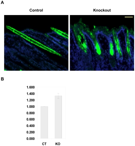
Dietary selenium is known to protect skin against UV-induced damage and cancer and its topical application improves skin surface parameters in humans, while selenium deficiency compromises protective antioxidant enzymes in skin. Furthermore, skin and hair abnormalities in humans and rodents may be caused by selenium deficiency, which are overcome by dietary selenium supplementation. Most important biological functions of selenium are attributed to selenoproteins, proteins containing selenium in the form of the amino acid, selenocysteine (Sec). Sec insertion into proteins depends on Sec tRNA; thus, knocking out the Sec tRNA gene (Trsp) ablates selenoprotein expression. We generated mice with targeted removal of selenoproteins in keratin 14 (K14) expressing cells and their differentiated descendents. The knockout progeny had a runt phenotype, developed skin abnormalities and experienced premature death. Lack of selenoproteins in epidermal cells led to the development of hyperplastic epidermis and aberrant hair follicle morphogenesis, accompanied by progressive alopecia after birth. Further analyses revealed that selenoproteins are essential antioxidants in skin and unveiled their role in keratinocyte growth and viability. This study links severe selenoprotein deficiency to abnormalities in skin and hair and provides genetic evidence for the role of these proteins in keratinocyte function and cutaneous development.
Overview
The claims and statistics of this paper haven’t been checked yet.
Run the check — free, no account needed. It reads the open-access text and checks the sources it cites.
Integrity signals
Registry
Cites
Statistics
Figures
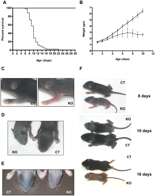
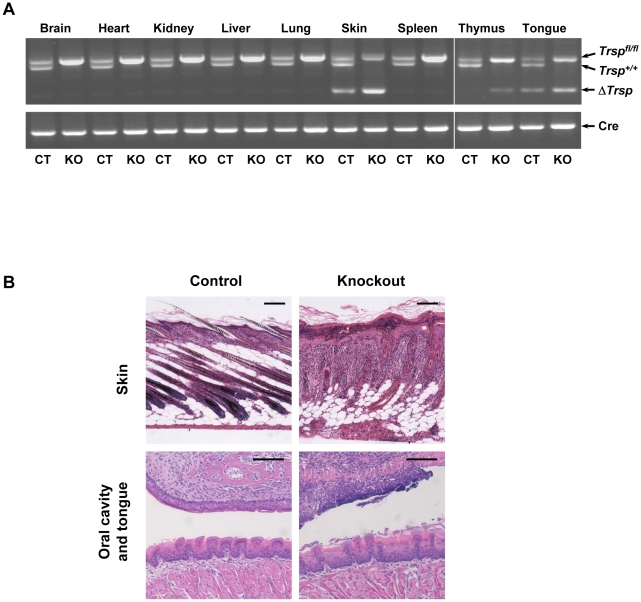
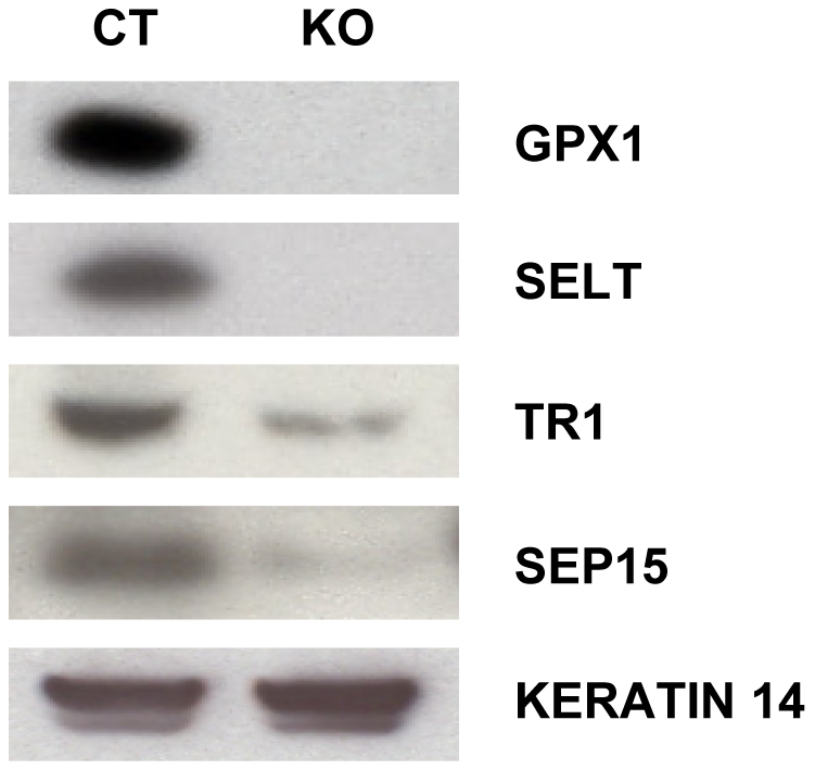
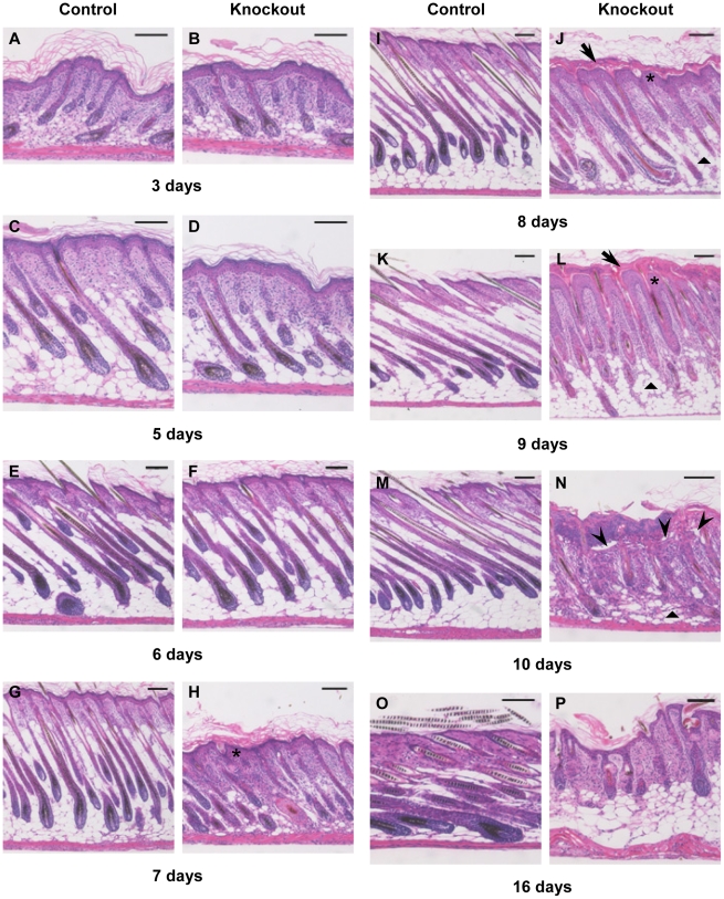
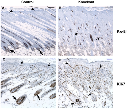
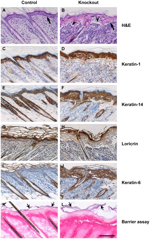
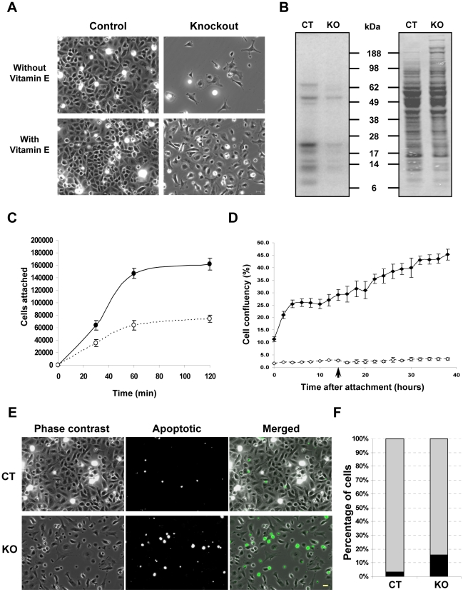
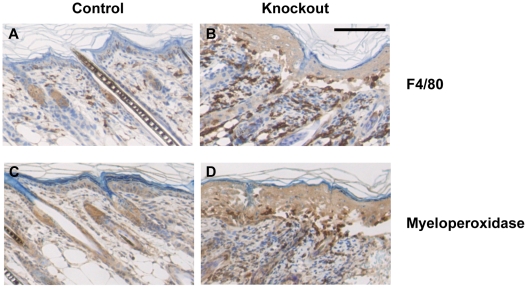
Verification
Claims the paper makes
The claims and statistics of this paper haven’t been checked yet.
Run the check — free, no account needed. It reads the open-access text and checks the sources it cites.
Claims other papers make about this one
Each is checked against this paper’s own text: does it say what the citing paper says it does?
In the citing paper
“Se-enriched proteins are also essential in keratinocyte development and function [67].”
From Diet and Skin Aging—From the Perspective of Food Nutrition (2020) · cites this paper as [67]
Self-check
Percentages, sums and reported differences re-computed from the paper’s own numbers.
The self-check needs the paper’s full text.
Citations
Cites
Characterization of Mammalian Selenoproteomes
Kryukov, G. V., Castellano, S., Novoselov, S. V., Lobanov, A. V., Zehtab, O., Guigó, R., & Gladyshev, V. N. (2003) · Science · doi:10.1126/science.1083516 · Cited by 2,385
Self-citationThe human keratins: biology and pathology
Moll, R., Divo, M., & Langbein, L. (2008) · Histochemistry and Cell Biology · doi:10.1007/s00418-008-0435-6 · Cited by 1,465
Jonkers, J., Meuwissen, R. L. J., van der Gulden, H. M. L., Peterse, H. L., van der Valk, M., & Berns, A. J. M. (2001) · Nature Genetics · doi:10.1038/ng747 · Cited by 1,044
Getting under the skin of epidermal morphogenesis
Fuchs, E., & Raghavan, S. (2002) · Nature Reviews Genetics · doi:10.1038/nrg758 · Cited by 753
How Selenium Has Altered Our Understanding of the Genetic Code
Hatfield, D. L., & Gladyshev, V. N. (2002) · Molecular and Cellular Biology · doi:10.1128/mcb.22.11.3565-3576.2002 · Cited by 681
Self-citationRole of integrins in regulating epidermal adhesion, growth and differentiation
Watt, F. M. (2002) · The EMBO Journal · doi:10.1093/emboj/cdf399 · Cited by 647
Enzymic and non-enzymic antioxidants in epidermis and dermis of human skin
Shindo, Y., Witt, E., Han, D., Epstein, W., & Packer, L. (1994) · Journal of Investigative Dermatology · doi:10.1111/1523-1747.ep12371744 · Cited by 546
Oxidative mechanisms in carcinogenesis
Guyton, K. Z., & Kensler, T. W. (1993) · British Medical Bulletin · doi:10.1093/oxfordjournals.bmb.a072628 · Cited by 465
Lichti, U. F., Anders, J., & Yuspa, S. H. (2008) · Nature Protocols · doi:10.1038/nprot.2008.50 · Cited by 444
Self-citationPatterned acquisition of skin barrier function during development
Hardman, M. J., Sisi, P., Banbury, D. N., & Byrne, C. (1998) · Development · doi:10.1242/dev.125.8.1541 · Cited by 385
Research works printed but not linked: 1
- HPM Gollnick, 1996, Systemic b carotene plus topical UV-sunscreen are an optimal protection against harmful effects of natural UV-sunlight: results of the Berlin-Eilath study., Eur J Dermatol, 6, 200
Cited by
Sorted by citations
Selenium Utilization by GPX4 Is Required to Prevent Hydroperoxide-Induced Ferroptosis
Ingold, I., Berndt, C., Schmitt, S., Doll, S. G., Poschmann, G., Buday, K., Roveri, A., Peng, X., Freitas, F. P., Seibt, T., et al. (2017) · Cell · doi:10.1016/j.cell.2017.11.048 · Cited by 1,701
Selenium and selenocysteine: roles in cancer, health, and development
Hatfield, D. L., Tsuji, P. A., Carlson, B. A., & Gladyshev, V. N. (2014) · Trends in Biochemical Sciences · doi:10.1016/j.tibs.2013.12.007 · Cited by 715
Self-citationParadoxical Roles of Antioxidant Enzymes: Basic Mechanisms and Health Implications
Lei, X. G., Zhu, J., Cheng, W.-H., Bao, Y., Ho, Y.-S., Reddi, A. R., Holmgren, A., & Arnér, E. S. J. (2015) · Physiological Reviews · doi:10.1152/physrev.00010.2014 · Cited by 455
Wang, G., Jin, W., Qasim, A. M., Gao, A., Peng, X. F., Li, W., Feng, H., & Chu, P. K.-H. (2017) · Biomaterials · doi:10.1016/j.biomaterials.2017.01.028 · Cited by 306
Diet and Skin Aging—From the Perspective of Food Nutrition
Cao, C., Xiao, Z., Wu, Y., & Ge, C. (2020) · Nutrients · doi:10.3390/nu12030870 · Cited by 231
Wang, G., Feng, H., Hu, L., Jin, W., Hao, Q., Gao, A., Peng, X. F., Wan, L., Wong, K., Wang, H., et al. (2018) · Nature Communications · doi:10.1038/s41467-018-04317-2 · Cited by 218
Selenium and GPX4, a vital symbiosis
Angeli, J. P. F., & Conrad, M. (2018) · Free Radical Biology and Medicine · doi:10.1016/j.freeradbiomed.2018.03.001 · Cited by 197
Diet and hair loss: effects of nutrient deficiency and supplement use
Guo, E. L., & Katta, R. (2017) · Dermatology Practical & Conceptual · doi:10.5826/dpc.0701a01 · Cited by 184
Integrative and Mechanistic Approach to the Hair Growth Cycle and Hair Loss
Natarelli, N., Gahoonia, N., & Sivamani, R. K. (2023) · Journal of Clinical Medicine · doi:10.3390/jcm12030893 · Cited by 174
Vollmer, D. L., West, V. A., & Lephart, E. D. (2018) · International Journal of Molecular Sciences · doi:10.3390/ijms19103059 · Cited by 132
McCann, J. C., & Ames, B. N. (2011) · The FASEB Journal · doi:10.1096/fj.11-180885 · Cited by 125
Selenoproteins in Tumorigenesis and Cancer Progression
Short, S. P., & Williams, C. S. (2017) · Advances in cancer research · doi:10.1016/bs.acr.2017.08.002 · Cited by 104
Understanding selenoprotein function and regulation through the use of rodent models
Kasaikina, M. V., Hatfield, D. L., & Gladyshev, V. N. (2012) · Biochimica et Biophysica Acta (BBA) - Molecular Cell Research · doi:10.1016/j.bbamcr.2012.02.018 · Cited by 98
Self-citationGoldberg, L. J., & Lenzy, Y. M. (2010) · Clinics in Dermatology · doi:10.1016/j.clindermatol.2010.03.038 · Cited by 96
Sengupta, A., Lichti, U. F., Carlson, B. A., Cataisson, C., Ryscavage, A. O., Mikulec, C. D., Conrad, M., Fischer, S. M., Hatfield, D. L., & Yuspa, S. H. (2013) · Journal of Investigative Dermatology · doi:10.1038/jid.2013.52 · Cited by 86
Self-citationLongo, N., Dimmock, D., Levy, H. L., Viau, K., Bausell, H., Bilder, D. A., Burton, B. K., Gross, C., Northrup, H., Rohr, F., et al. (2018) · Genetics in Medicine · doi:10.1038/s41436-018-0403-z · Cited by 89
Liu, C., Guo, H., DaSilva, N. A., Li, D., Zhang, K., Wan, Y., Gao, X., Chen, H., Seeram, N. P., & Ma, H. (2019) · Journal of Functional Foods · doi:10.1016/j.jff.2019.02.015 · Cited by 88
Selenoproteins and oxidative stress-induced inflammatory tumorigenesis in the gut
Barrett, C. W., Short, S. P., & Williams, C. S. (2016) · Cellular and Molecular Life Sciences · doi:10.1007/s00018-016-2339-2 · Cited by 73
Clinical and nutritional benefits of cysteine-enriched protein supplements
McPherson, R. A., & Hardy, G. (2011) · Current Opinion in Clinical Nutrition & Metabolic Care · doi:10.1097/mco.0b013e32834c1780 · Cited by 68
Thiry, C., Ruttens, A., Pussemier, L., & Schneider, Y. (2012) · British Journal Of Nutrition · doi:10.1017/s0007114512004412 · Cited by 66
Zhang, L., Zeng, H., & Cheng, W. (2018) · Free Radical Biology and Medicine · doi:10.1016/j.freeradbiomed.2018.05.067 · Cited by 67
Cox, A. G., Tsomides, A., Kim, A. J., Saunders, D. C., Hwang, K. L., Evason, K. J., Heidel, J. R., Brown, K., Yuan, M., Lien, E. C., et al. (2016) · Proceedings of the National Academy of Sciences · doi:10.1073/pnas.1600204113 · Cited by 63
Self-citationEspinosa-Leal, C. A., & García‐Lara, S. (2019) · Planta Medica · doi:10.1055/a-0857-6633 · Cited by 61
Interaction of mineral salts with the skin: A literature survey
Polefka, T. G., Bianchini, R. J., & Shapiro, S. (2012) · International Journal of Cosmetic Science · doi:10.1111/j.1468-2494.2012.00731.x · Cited by 56
Ren, F., Chen, X., Hesketh, J. E., Gan, F., & Huang, K. (2012) · PLoS ONE · doi:10.1371/journal.pone.0035375 · Cited by 55
Selenoprotein H Suppresses Cellular Senescence through Genome Maintenance and Redox Regulation
Wu, R. T. Y., Cao, L., Chen, B. P. C., & Cheng, W.-H. (2014) · Journal of Biological Chemistry · doi:10.1074/jbc.m114.611970 · Cited by 55
Mirastschijski, U., Martin, A., Jørgensen, L. N., Sampson, B., & Ågren, M. S. (2013) · Biological Trace Element Research · doi:10.1007/s12011-013-9658-z · Cited by 53
Tsuji, P. A., Santesmasses, D., Lee, B. J., Gladyshev, V. N., & Hatfield, D. L. (2021) · International Journal of Molecular Sciences · doi:10.3390/ijms23010005 · Cited by 54
Self-citationAn overview of the genetic aspects of hair loss and its connection with nutrition
Gökçe, N., Basgoz, N., Kenanoğlu, S., Akalın, H., Özkul, Y., Ergören, M. Ç., Beccari, T., Bertelli, M., & Dündar, M. (2022) · Journal of preventive medicine and hygiene · doi:10.15167/2421-4248/jpmh2022.63.2s3.2765 · Cited by 53
Katta, R., & Brown, D. N. (2015) · Journal of Skin Cancer · doi:10.1155/2015/893149 · Cited by 56
Predicted dietary intake of selenium by the general adult population in Belgium
Waegeneers, N., Thiry, C., Temmerman, L. D., & Ruttens, A. (2012) · Food Additives & Contaminants Part A · doi:10.1080/19440049.2012.746474 · Cited by 52
Savery, L. C., Evers, D. C., Wise, S. S., Falank, C. R., Wise, J. T. F., Gianios, C., Kerr, I., Payne, R. L., Thompson, W. D., Perkins, C. R., et al. (2013) · The Science of The Total Environment · doi:10.1016/j.scitotenv.2013.01.070 · Cited by 51
Ferroptosis: Mechanism and connections with cutaneous diseases
Liu, L., Lian, N., Shi, L., Hao, Z., & Chen, K. (2023) · Frontiers in Cell and Developmental Biology · doi:10.3389/fcell.2022.1079548 · Cited by 52
Selenium Preserves Keratinocyte Stemness and Delays Senescence by Maintaining Epidermal Adhesion
Jobeili, L., Rousselle, P., Béal, D., Blouin, E., Roussel, A.-M., Damour, O., & Rachidi, W. (2017) · Aging · doi:10.18632/aging.101322 · Cited by 44
Fu, S., Zhao, X., Yang, L. J., Qin, G. W., & Zhang, E. (2022) · Biomaterials Advances · doi:10.1016/j.msec.2022.112653 · Cited by 40
Jing, S., Liu, Y., Wang, B., Zhou, H., Zhang, H., Siwakoti, P., Qu, X., Ye, P., He, Y., Kumeria, T., et al. (2025) · Journal of Controlled Release · doi:10.1016/j.jconrel.2025.113597 · Cited by 44
Wu, R. T., Cao, L., Mattson, E., Witwer, K. W., Cao, J., Zeng, H., He, X., Combs, G. F., & Cheng, W. (2017) · Aging Cell · doi:10.1111/acel.12529 · Cited by 37
Favrot, C., Beal, D., Blouin, E., Leccia, M. T., Roussel, A. M., & Rachidi, W. (2018) · Oxidative Medicine and Cellular Longevity · doi:10.1155/2018/5895439 · Cited by 36
Pioneering Role of Marine Macroalgae in Cosmeceuticals
Kalasariya, H. S., Pereira, L., & Patel, N. B. (2022) · Phycology · doi:10.3390/phycology2010010 · Cited by 40
Chang, S. N., Khan, I., Dey, D. K., Cho, K.-H., Hwang, B. S., Bae, K. B., Kang, S. C., & Park, J. G. (2019) · Food and Chemical Toxicology · doi:10.1016/j.fct.2019.110699 · Cited by 33
Cáceres‐Saez, I., Goodall, R. N. P., Dellabianca, N. A., Cappozzo, H. L., & Guevara, S. R. (2015) · Chemosphere · doi:10.1016/j.chemosphere.2015.07.026 · Cited by 31
Human Genetic Disorders Resulting in Systemic Selenoprotein Deficiency
Schoenmakers, E., & Chatterjee, K. (2021) · International Journal of Molecular Sciences · doi:10.3390/ijms222312927 · Cited by 30
Wang, G., Feng, H., Jin, W., Gao, A., Peng, X. F., Wan, L., Wu, H., Li, Z., & Chu, P. K.-H. (2017) · Applied Surface Science · doi:10.1016/j.apsusc.2017.04.053 · Cited by 29
The Proteomic Profile of Keratinocytes and Lymphocytes in Psoriatic Patients
Gęgotek, A., Domingues, P. M., Wroński, A., Ambrożewicz, E., & Skrzydlewska, E. (2019) · PROTEOMICS - CLINICAL APPLICATIONS · doi:10.1002/prca.201800119 · Cited by 28
O′Connor, K., & Goldberg, L. J. (2021) · Clinics in Dermatology · doi:10.1016/j.clindermatol.2021.05.008 · Cited by 26
Insulin–transferrin–selenium as an alternative to foetal serum for epidermal equivalents
Mainzer, C., Barrichello, C., Debret, R., Remoué, N., Sigaudo‐Roussel, D., & Sommer, P. (2014) · International Journal of Cosmetic Science · doi:10.1111/ics.12141 · Cited by 24
Nuclear selenoproteins and genome maintenance
Zhang, X., Zhang, L., Zhu, J., & Cheng, W. (2015) · IUBMB Life · doi:10.1002/iub.1455 · Cited by 24
Falk, M., Bernhoft, A., Reinoso‐Maset, E., Salbu, B., Lebed, P. J., Framstad, T., Fuhrmann, H., & Oropeza-Moe, M. (2019) · Journal of Trace Elements in Medicine and Biology · doi:10.1016/j.jtemb.2019.126439 · Cited by 23
Hazane‐Puch, F., Champelovier, P., Arnaud, J., Garrel, C., Ballester, B., Faure, P., & Laporte, F. (2013) · Biological Trace Element Research · doi:10.1007/s12011-013-9709-5 · Cited by 21
Huang, Q., Yao, P., Eriani, G., & Wang, E. (2012) · Nucleic Acids Research · doi:10.1093/nar/gks783 · Cited by 21
Couto, N. A. S., Newton, J., Russo, C., Karunakaran, E., Achour, B., Al‐Majdoub, Z. M., Sidaway, J. E., Rostami‐Hodjegan, A., Clench, M. R., & Barber, J. (2020) · Drug Metabolism and Disposition · doi:10.1124/dmd.120.000168 · Cited by 19
In Vitro Evaluation of Bioavailability of Se from Daily Food Rations and Dietary Supplements
Bawiec, P., Sawicki, J., Łasińska-Pracuta, P., Czop, M., Sowa, I., Iłowiecka, K., & Koch, W. (2023) · Nutrients · doi:10.3390/nu15061511 · Cited by 18
Cell death as an architect of adult skin stem cell niches
Lecomte, K., Toniolo, A., & Hoste, E. (2024) · Cell Death and Differentiation · doi:10.1038/s41418-024-01297-3 · Cited by 16
Savery, L. C., Chen, T. L., Wise, J. T. F., Wise, S. S., Gianios, C., Buonagurio, J., Perkins, C. R., Falank, C. R., Zheng, T., Zhu, C., et al. (2015) · Comparative Biochemistry and Physiology Part C Toxicology & Pharmacology · doi:10.1016/j.cbpc.2015.09.012 · Cited by 15
Kreindl, C., Soto-Alarcón, S. A., Hidalgo, M., Riveros, A. L., Añazco, C. C., Pulgar, R., & Porras, O. H. (2024) · Antioxidants · doi:10.3390/antiox13020159 · Cited by 13
Nano and mesosized selenium and its synthesis using the ascorbic acid route
Štefanková, D., Skrbek, K., Pižl, M., & Bartůněk, V. (2023) · Journal of Non-Crystalline Solids · doi:10.1016/j.jnoncrysol.2023.122462 · Cited by 12
The role of trace elements for the function and health of the skin
Bjørklund, G., Shanaida, M., Hangan, T., Talgatkyzy, K. L., Kussainova, A. A., Semenova, Y. M., Gheorghe, E., Гонтова, Т. М., Voloshyn, V., Lysiuk, R., et al. (2025) · Journal of Trace Elements in Medicine and Biology · doi:10.1016/j.jtemb.2025.127674 · Cited by 14
Cáceres‐Saez, I., Haro, D., Blank, O., Aguayo‐Lobo, A., Dougnac, C., Arredondo, C., Cappozzo, H. L., & Guevara, S. R. (2021) · Environmental Science and Pollution Research · doi:10.1007/s11356-021-12890-1 · Cited by 11
Antonyak, H., Iskra, R., Panas, N., & Lysiuk, R. (2018) · Healthy ageing and longevity · doi:10.1007/978-3-030-03742-0_3 · Cited by 11
The Role of Dietary Antioxidants in Melanoma and Nonmelanoma Skin Cancer
Sable, K. A. (2023) · Cutis · doi:10.12788/cutis.0672 · Cited by 12
Knot Architecture for Biocompatible and Semiconducting 2D Electronic Fiber Transistors
Carey, T., Maughan, J., Doolan, L., Caffrey, E., Garcia, J. R., Liu, S., Kaur, H., Ilhan, C., Seyedin, S., & Coleman, J. N. (2024) · Small Methods · doi:10.1002/smtd.202301654 · Cited by 11
Minerals and Trace Elements: Key Protectors of Skin Health and Defenders against Skin Disorders
Bjørklund, G., Shanaida, M., Гонтова, Т. М., Gheorghe, E., Kassym, L., Kussainova, A., Voloshyn, V., Semenova, Y. M., Lysiuk, R., Voloshyn, O., et al. (2025) · Current Medicinal Chemistry · doi:10.2174/0109298673348175250214054101 · Cited by 10
Kosik-Bogacka, D. I., Osten‐Sacken, N., Łanocha-Arendarczyk, N., Kot, K., Pilarczyk, B., Tomza–Marciniak, A., Podlasińska, J., Chmielarz, M., Heddergott, M., Frantz, A. C., et al. (2019) · Ecotoxicology · doi:10.1007/s10646-019-02120-3 · Cited by 11
Wang, Z., Yang, S. F., Liu, X., Yang, T., Han, T., He, X., Jiang, Y., & Hao, Y. (2021) · Microchemical Journal · doi:10.1016/j.microc.2021.106681 · Cited by 9
Antioxidants in Skin Disorders
Kumar, S., Subba, D. P., Seema, Firdous, S. M., Odeku, O. A., Kumar, S., & Sindhu, R. K. (2024) · Antioxidants · doi:10.1002/9781394270576.ch16 · Cited by 11
Chen, Y., Liu, Y., Liu, Y., Li, S., Li, S., Li, S., Ying, L., Chen, T., & Chen, T. (2025) · Chinese Chemical Letters · doi:10.1016/j.cclet.2025.111298 · Cited by 8
Advantages of Functional Foods in Supporting and Maintaining Hair and Skin Health
Muraleedharan, V., Kamath, G. S., Sasikumar, G., & Panicker, S. P. (2023) · Springer eBooks · doi:10.1007/978-981-99-0534-8_12 · Cited by 9
The relevance of selenium to alopecias
Costa, V. D. T., Melo, D., & Matsunaga, A. (2018) · International Journal of Trichology · doi:10.4103/ijt.ijt_37_17 · Cited by 8
Mutations in Humans That Adversely Affect the Selenoprotein Synthesis Pathway
Schoenmakers, E., Schoenmakers, N. A., & Chatterjee, K. (2016) · Springer eBooks · doi:10.1007/978-3-319-41283-2_44 · Cited by 7
Microneedle technology integrated with diverse therapeutic modalities for hair regrowth in alopecia
Gong, M., Jiang, Z., Yang, Z., Chen, T., Hu, W., Zhou, C., Jian, J., Quan, G., Wu, C., Cai, J., et al. (2026) · Acta Pharmaceutica Sinica B · doi:10.1016/j.apsb.2026.03.006 · Cited by 6
Utilizing Robust Design to Optimize Composite Bioadhesive for Promoting Dermal Wound Repair
Pinnaratip, R., Zhang, Z., Smies, A., Forooshani, P. K., Tang, X., Rajachar, R. M., & Lee, B. P. (2023) · Polymers · doi:10.3390/polym15081905 · Cited by 5
Foord, C. S., Robb, K., & Nugegoda, D. (2024) · Marine Pollution Bulletin · doi:10.1016/j.marpolbul.2024.117130 · Cited by 4
Chan, Y., Robinson, N. J., Dourdeville, K., Haas, H. L., Nielsen, J. R., Paladino, F. V., Prescott, R., & Patel, S. H. (2025) · Chemosphere · doi:10.1016/j.chemosphere.2025.144190 · Cited by 4
Aging and Anti-Aging in Hair and Hair Loss
Yang, C., Chen, C., & Chen, W. (2013) · Elsevier eBooks · doi:10.1016/b978-0-12-397803-5.00019-8 · Cited by 5
The interaction of metals and the skin: The good, bad, and the ugly
Matsui, M. S., Carle, T. L., & Costa, M. (2022) · Elsevier eBooks · doi:10.1016/b978-0-12-823292-7.00011-5 · Cited by 4
Zhao, C., Duan, Y., Diao, X., He, L., & Zhang, W. (2024) · Animals · doi:10.3390/ani14030477 · Cited by 4
Carlson, B. A. (2016) · Springer eBooks · doi:10.1007/978-3-319-41283-2_46 · Cited by 4
Self-citationPicut, C. A., & Remick, A. K. (2019) · Springer eBooks · doi:10.1007/978-1-4939-9777-0_19 · Cited by 3
Protective Effects of Selenium in Patients with Beta-Thalassemia Major
aboutalebi, A., Jouyban, A., Chavoshi, H., Akbari, A. M., Shaseb, E., Sarbakhsh, P., & Ghaffary, S. (2020) · Pharmaceutical Sciences · doi:10.34172/ps.2019.53 · Cited by 3
Peran Selenium dan Vitamin E pada Ruminansia: ulasan singkat
Yanuartono, Y., Indarjulianto, S., & Paryuni, A. D. (2024) · Jurnal Ilmiah Ilmu-Ilmu Peternakan · doi:10.22437/jiiip.v27i1.31614 · Cited by 2
Selenocysteine tRNA[Ser]Sec: The Central Component of Selenoprotein Biosynthesis
Carlson, B. A., Tobe, R., Tsuji, P. A., Yoo, M., Feigenbaum, L., Tessarollo, L., Lee, B., Schweizer, U., Gladyshev, V. N., & Hatfield, D. L. (2015) · Oxidative stress and disease · doi:10.1201/b18810-7 · Cited by 2
Self-citationAn entropy-reducing data representation approach for bioinformatic data
McCulloch, A., Jáuregui, R., Maclean, P. H., Ashby, R., Moraga, R., Laugraud, A., Bräuning, R., Dodds, K. G., & McEwan, J. C. (2018) · Database · doi:10.1093/database/bay029 · Cited by 2
Селенодефицит и возрастзависимая патология (в фокусе дейодиназы)
Honcharova, O. A., & Illinа, I. M. (2016) · INTERNATIONAL JOURNAL OF ENDOCRINOLOGY (Ukraine) · doi:10.22141/2224-0721.4.68.2015.75020 · Cited by 2
Li, Q., Chan, H., Zhou, J., Zheng, W., Yang, B., & Liu, Z. (2025) · Journal of the European Academy of Dermatology and Venereology · doi:10.1111/jdv.20870 · Cited by 1
Chauhan, A., Vishwas, S., Singh, T., Kumar, M., Negi, P., Collet, T., Kour, A., & Awasthi, A. (2026) · International Journal of Pharmaceutics · doi:10.1016/j.ijpharm.2026.126815 · Cited by 2
Optimizing Skin Health through Nutrition: Evidence-Based Insights
Bakshi, N., Loomba, V., & Soin, A. S. (2026) · Recent Progress in Nutrition · doi:10.21926/rpn.2603017 · Cited by 1
Addition of Selenium to Carica papaya Linn Pulp Extract Enhances Dermal Wound Healing Activity
Nafiu, B. A., Akram, H., Bero, D. N., Mohamad, M., Malik, I., & Rahman, M. T. (2013) · Tropical Journal of Pharmaceutical Research · doi:10.4314/tjpr.v12i1.13 · Cited by 1
Curtis, E. E., & Gibran, N. S. (2019) · Springer eBooks · doi:10.1007/978-3-030-18940-2_32 · Cited by 1
Nutritional supplementation of an apple callus extract to target epidermal aging
Baumann, J., Wandrey, F., Nowak, K., & Grothe, T. (2024) · Journal of Food Nutrition and Diet Science · doi:10.55976/fnds.22024123718-28 · Cited by 1
Chernoff, G. (2021) · Journal of Clinical and Cosmetic Dermatology · doi:10.16966/2576-2826.162 · Cited by 1
Carlson, B. A., Yoo, M., Tsuji, P. A., Tobe, R., Naranjo‐Suarez, S., Chen, F., Feigenbaum, L., Tessarollo, L., Lee, B. J., Gladyshev, V. N., et al. (2011) · Springer eBooks · doi:10.1007/978-1-4614-1025-6_44 · Cited by 1
Self-citationLegumes and Preventive Dermatology
Porres, J. M., & Cheng, W. (2012) · Humana Press eBooks · doi:10.1007/978-1-62703-167-7_38
Untersuchungen zur Rolle der Glutathion-Peroxidase 4 als möglicher Redox-Sensor in Säugetierzellen
Grünler, N. (2015) · Site cant be reached · doi:10.5282/edoc.18272
Skin Cancer Chemoprevention: Current Status and Future Potential
Siddiqui, I. A., Adhami, V. M., & Mukhtar, H. (2011) · Springer eBooks · doi:10.1007/978-94-007-2630-7_13
Palafox-Vigil, G., Esquivel, M. A. G., & Martínez, S. G. (2015)
(2010) · Journal of Investigative Dermatology · doi:10.1038/jid.2010.321
Brown, D. A., & Gibran, N. S. (2012) · Springer eBooks · doi:10.1007/978-3-7091-0348-7_22
Anastassakis, K. (2022) · Springer eBooks · doi:10.1007/978-3-031-08057-9_44
Metadata
- Authors
- Sengupta, A., Lichti, U. F., Carlson, B. A., Ryscavage, A. O., Gladyshev, V. N., Yuspa, S. H., & Hatfield, D. L.
- Affiliations
- National Institutes of Health (US)
- Brigham and Women's Hospital (US)
- Corresponding author
- Yuspa, Stuart H.
- Hatfield, Dolph L.
- Venue
- PLoS ONE
- Publisher
- Public Library of Science (PLoS)
- Published
- 2010-08-18
- Volume
- 5
- Issue
- 8
- Pages
- e12249
- ISSN
- 1932-6203 (ISSN-L)
- PMC
- PMC2923614
- OpenAlex
- W2140893882
- Cited by
- 109
- References
- 41
- Citations by year
- 2026: 7 · 2025: 7 · 2024: 9 · 2023: 9 · 2022: 5 · 2021: 7 · 2020: 4 · 2019: 8 · 2018: 10 · 2017: 6 · 2016: 6 · 2015: 8 · 2014: 3 · 2013: 6 · 2012: 8
- FWCI
- 6.07
- Type
- journal article
- Language
- en
- Open access
- gold
- doi.org
- Licence
- Locations
- PLoS ONE (publishedVersion, public-domain)
- PubMed Central (publishedVersion, public-domain)
- PubMed (publishedVersion)
- Digital Access to Scholarship at Harvard (DASH) (Harvard University) (submittedVersion, cc-by)
- DOAJ (DOAJ: Directory of Open Access Journals) (submittedVersion, cc-by-sa)
- Figshare (submittedVersion, cc-by)
- Funders
- National Institutes of Health
- National Cancer Institute
- National Institute of General Medical Sciences (R01GM061603)
- Topics
- Health Sciences › Nursing › Nutrition and Dietetics › Selenium in Biological Systems
- Physical Sciences › Chemistry › Organic Chemistry › Organoselenium and organotellurium chemistry
- Health Sciences › Medicine › Dermatology › Hair Growth and Disorders
- Keywords
- selenoproteins, selenocysteine, keratinocytes, skin development, hair follicle morphogenesis, hair loss, selenium deficiency, keratin 14, epidermal hyperplasia, antioxidant enzymes, keratinocyte viability, knockout mice
- MeSH terms
- Epidermal Cells, Animals, Animals, Newborn, Antioxidants, Cell Adhesion, Cell Differentiation, Epidermis, Female, Gene Expression Regulation, Macrophages, Male, Organ Specificity, Recombination, Genetic, RNA, Messenger, RNA, Transfer, Amino Acid-Specific, Selenium, Skin, Lipid Peroxidation, Biomarkers, Keratinocytes, Gene Deletion, Reactive Oxygen Species, Hair Follicle, Cell Proliferation, Selenoproteins, Mice, Keratin-14, Gene Knockout Techniques
About these checks
Registry, retraction, Cites and statistics results are computed, not judged, with one exception: a printed reference that no index lists is read once by a model before it counts as a citation error. Claims are extracted by a model and kept only when their quote appears verbatim in the text; each cited source is then checked, with a model’s help, for whether it says what the claim relies on. Those verdicts can be wrong — read the claim’s quote and the cited source before relying on one.
Statistics and arithmetic are recomputed, not judged.
Confidence bands are shown only for fields calibrated on our annotated test set. None is calibrated yet, so no band is shown.
What counts as a claim ›How we measure confidence ›How we check this ›
Are you an author, or do you see a problem with this page? Request a review
