

Resting-State Functional Connectivity between Fronto-Parietal and Default Mode Networks in Obsessive-Compulsive Disorder
Open accessThe DOI matches its Crossref record; 76 of the 76 references it cites were found in a registry or an index.
Abstract
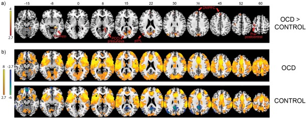
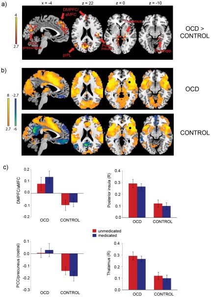
BACKGROUND: Obsessive-compulsive disorder (OCD) is characterized by an excessive focus on upsetting or disturbing thoughts, feelings, and images that are internally-generated. Internally-focused thought processes are subserved by the "default mode network" (DMN), which has been found to be hyperactive in OCD during cognitive tasks. In healthy individuals, disengagement from internally-focused thought processes may rely on interactions between DMN and a fronto-parietal network (FPN) associated with external attention and task execution. Altered connectivity between FPN and DMN may contribute to the dysfunctional behavior and brain activity found in OCD. METHODS: The current study examined interactions between FPN and DMN during rest in 30 patients with OCD (17 unmedicated) and 32 control subjects (17 unmedicated). Timecourses from seven fronto-parietal seeds were correlated across the whole brain and compared between groups. RESULTS: OCD patients exhibited altered connectivity between FPN seeds (primarily anterior insula) and several regions of DMN including posterior cingulate cortex, medial frontal cortex, posterior inferior parietal lobule, and parahippocampus. These differences were driven largely by a reduction of negative correlations among patients compared to controls. Patients also showed greater positive connectivity between FPN and regions outside DMN, including thalamus, lateral frontal cortex, and somatosensory/motor regions. CONCLUSIONS: OCD is associated with abnormal intrinsic functional connectivity between large-scale brain networks. Alteration of interactions between FPN and DMN at rest may contribute to aspects of the OCD phenotype, such as patients' inability to disengage from internally-generated scenarios and thoughts when performing everyday tasks requiring external attention.
Overview
The claims and statistics of this paper haven’t been checked yet.
Free: it reads the open-access text and checks the first 5 claims that cite a source we can check. Create a free account to check every claim.
Integrity signals
Registry
Cites
Statistics
Figures
Verification
Claims the paper makes
The claims and statistics of this paper haven’t been checked yet.
Free: it reads the open-access text and checks the first 5 claims that cite a source we can check. Create a free account to check every claim.
Claims other papers make about this one
Each is checked against this paper’s own text: does it say what the citing paper says it does?
The source backs part of the claim, not all of it — or backs it for a different population, condition or comparison.
In the citing paper
“Alterations in the DMN have been linked to increased self-focused rumination and intrusive thoughts in OCD patients (191).”
From Neural mechanisms underlying cognitive inflexibility in obsessive-compulsive disorder: a review (2026) · cites this paper as [191]
Experimental — accuracy not yet measuredThe source backs part of the claim, not all of it — or backs it for a different population, condition or comparison.
In the citing paper
“They are integral parts not only of the CSTC circuits but also the salience network, and the default mode network (DMN) – which have also been implicated in OCD (1, 191, 195).”
From Neural mechanisms underlying cognitive inflexibility in obsessive-compulsive disorder: a review (2026) · cites this paper as [191]
Experimental — accuracy not yet measuredThe source backs part of the claim, not all of it — or backs it for a different population, condition or comparison.
In the citing paper
“OCD patients frequently exhibit hyperactivity in the DMN, which correlates with increased rumination and obsessive thoughts (191).”
From Neural mechanisms underlying cognitive inflexibility in obsessive-compulsive disorder: a review (2026) · cites this paper as [191]
Experimental — accuracy not yet measuredThe cited source says what the claim attributes to it.
In the citing paper
“Hyperactivity in these regions might contribute to the excessive internal focus observed in OCD patients (191).”
From Neural mechanisms underlying cognitive inflexibility in obsessive-compulsive disorder: a review (2026) · cites this paper as [191]
In the citing paper
“Despite the extensively documented functional alterations of the CSTC loops in OCD, it is not clear how these FC abnormalities (in terms of increased/decreased FC within this network and in outward connections originating from these loops) are associated with specific OCD symptoms.”
From Functional connectivity changes in obsessive-compulsive disorder correspond to interference control and obsessions severity (2020) · cites this paper as [26]
3 more claims about this paper
Create a free account to see every claim other papers make about this one.
Self-check
Percentages, sums and reported differences re-computed from the paper’s own numbers.
The self-check needs the paper’s full text.
Citations
Cites
Hamilton, M. (1960) · Journal of Neurology, Neurosurgery & Psychiatry · doi:10.1136/jnnp.23.1.56 · Cited by 32,817
A default mode of brain function
Raichle, M. E., MacLeod, A. M., Snyder, A. Z., Powers, W. J., Gusnard, D. A., & Shulman, G. L. (2001) · Proceedings of the National Academy of Sciences · doi:10.1073/pnas.98.2.676 · Cited by 12,190
The assessment of anxiety states by rating
HAMILTON, M. (1959) · British Journal of Medical Psychology · doi:10.1111/j.2044-8341.1959.tb00467.x · Cited by 11,455
The Brain's Default Network: Anatomy, Function, and Relevance to Disease
Buckner, R. L., Andrews‐Hanna, J. R., & Schacter, D. L. (2008) · Annals of the New York Academy of Sciences · doi:10.1196/annals.1440.011 · Cited by 9,761
The human brain is intrinsically organized into dynamic, anticorrelated functional networks
Fox, M. D., Snyder, A. Z., Vincent, J. L., Corbetta, M., Van Essen, D. C., & Raichle, M. E. (2005) · Proceedings of the National Academy of Sciences · doi:10.1073/pnas.0504136102 · Cited by 8,553
Parallel organization of functionally segregated circuits linking basal ganglia and cortex
Alexander, G. E., DeLong, M. R., & Strick, P. L. (1986) · Annual Review of Neuroscience · doi:10.1146/annurev.ne.09.030186.002041 · Cited by 8,527 · cited 1×
“While dysfunction in orbital, medial frontal, and striatal areas composing fronto-striatal circuits [1] has long been found to be contribute to the pathogenesis of OCD [2–7], emerging evidence suggests there is broader cortical dysfunction in the disorder, including abnormality of dorsolateral prefrontal cortex, anter…”
BackgroundThe Yale-Brown Obsessive Compulsive Scale: I. Development, Use, and Reliability
Goodman, W. K. (1989) · Archives of General Psychiatry · doi:10.1001/archpsyc.1989.01810110048007 · Cited by 7,712 · cited 1×
“Obsessive-compulsive (OC) symptom severity (current and lifetime) was quantified using the Yale-Brown Obsessive-Compulsive Scale (Y-BOCS, [52].”
MethodDissociable intrinsic connectivity networks for salience processing and executive control
Seeley, W. W., Menon, V., Schatzberg, A. F., Keller, J., Glover, G. H., Kenna, H., Reiss, A. L., & Greicius, M. D. (2007) · The Journal of Neuroscience · doi:10.1523/jneurosci.5587-06.2007 · Cited by 7,426 · cited 3×
“Nevertheless, given recent evidence identifying dissociable cingulo-opercular and dorsolateral prefrontal and parietal systems within FPN [20,34,36], we feel that this approach is more informative than creating a global timecourse compiled from signals averaged across the entire network.”
BackgroundFunctional connectivity in the resting brain: a network analysis of the default mode hypothesis
Greicius, M. D., Krasnow, B., Reiss, A. L., & Menon, V. (2003) · Proceedings of the National Academy of Sciences · doi:10.1073/pnas.0135058100 · Cited by 6,358 · cited 3×
“FPN and DMN are also negatively correlated at rest, at least in healthy controls [18,35,46], suggesting that the unconstrained brain may be intrinsically organized to support competitive relationships between networks involved in external attention and internallyfocused thought processes.”
BackgroundSaliency, switching, attention and control: a network model of insula function
Menon, V., & Uddin, L. Q. (2010) · Brain Structure and Function · doi:10.1007/s00429-010-0262-0 · Cited by 5,592
Correspondence of the brain's functional architecture during activation and rest
Smith, S. M., Fox, P. T., Miller, K. L., Glahn, D. C., Fox, P. M., Mackay, C. E., Filippini, N., Watkins, K. E., Toro, R., Laird, A. R., et al. (2009) · Proceedings of the National Academy of Sciences · doi:10.1073/pnas.0905267106 · Cited by 5,150 · cited 1×
“Numerous rs-fcMRI studies have identified ‘‘intrinsic’’ large-scale brain networks that exhibit interactions at rest similar to those identified during task [18–21].”
BackgroundA component based noise correction method (CompCor) for BOLD and perfusion based fMRI
Behzadi, Y., Restom, K., Liau, J., & Liu, T. T. (2007) · NeuroImage · doi:10.1016/j.neuroimage.2007.04.042 · Cited by 4,502 · cited 1×
“This tool identifies principle components associated with segmented white matter (WM) and cerebrospinal fluid (CSF) images for each individual subject using the ‘‘CompCor’’ method [56].”
MethodSridharan, D., Levitin, D. J., & Menon, V. (2008) · Proceedings of the National Academy of Sciences · doi:10.1073/pnas.0800005105 · Cited by 2,892
Functional-anatomic fractionation of the brain’s default network
Andrews-Hanna, J. R., Reidler, J. S., Sepulcre, J., Poulin, R., & Buckner, R. L. (2010) · Neuron · doi:10.1016/j.neuron.2010.02.005 · Cited by 2,767
Distinct brain networks for adaptive and stable task control in humans
Dosenbach, N. U. F., Fair, D. A., Miezin, F. M., Cohen, A. L., Wenger, K. K., Dosenbach, R. A. T., Fox, M. D., Snyder, A. Z., Vincent, J. L., Raichle, M. E., et al. (2007) · Proceedings of the National Academy of Sciences · doi:10.1073/pnas.0704320104 · Cited by 2,632
Murphy, K., Birn, R. M., Handwerker, D. A., Jones, T. B., & Bandettini, P. A. (2009) · NeuroImage · doi:10.1016/j.neuroimage.2008.09.036 · Cited by 2,387
The Yale-Brown Obsessive-Compulsive Scale: Measures of internal consistency
Kim, S. W., Dysken, M. W., Pheley, A. M., & Hoover, K. M. (1994) · Psychiatry Research · doi:10.1016/0165-1781(94)90039-6 · Cited by 2,359
The influence of head motion on intrinsic functional connectivity MRI
Van Dijk, K. R. A., Sabuncu, M. R., & Buckner, R. L. (2012) · NeuroImage · doi:10.1016/j.neuroimage.2011.07.044 · Cited by 2,344
Spreng, R. N., Mar, R. A., & Kim, A. S. N. (2009) · Journal of Cognitive Neuroscience · doi:10.1162/jocn.2008.21029 · Cited by 2,143 · cited 2×
“Activation in these regions not only decreases during externally-directed attention, but also increases when attention is directed inward, such as when subjects think about their own personal qualities and take the perspective of others (theory of mind/mentalizing), remember personal episodes from their past, imagine…”
BackgroundCommon blood flow changes across visual tasks: II. Decreases in cerebral cortex
Shulman, G. L., Fiez, J. A., Corbetta, M., Buckner, R. L., Miezin, F. M., Raichle, M. E., & Petersen, S. E. (1997) · Journal of Cognitive Neuroscience · doi:10.1162/jocn.1997.9.5.648 · Cited by 1,890 · cited 2×
“Research on DMN has garnered much interest since the initial observation of reduced blood flow within several anatomicallywidespread brain regions during cognitive tasks versus passive viewing [38].”
BackgroundEvidence for a Frontoparietal Control System Revealed by Intrinsic Functional Connectivity
Vincent, J. L., Kahn, I., Snyder, A. Z., Raichle, M. E., & Buckner, R. L. (2008) · Journal of Neurophysiology · doi:10.1152/jn.90355.2008 · Cited by 1,869 · cited 1×
“Zhang et al [32] found altered resting-state connectivity within a ‘‘top-down control network’’ including posterior temporal cortex, lateral frontal and cingulate cortex, and precuneus, regions that partially overlap with the fairly well-delineated fronto-parietal network (FPN) composed of anterior insula, medial fron…”
BackgroundDefault-mode brain dysfunction in mental disorders: A systematic review
Broyd, S. J., Demanuele, C., Debener, S., Helps, S. K., James, C. J., & Sonuga-Barke, E. J. S. (2009) · Neuroscience & Biobehavioral Reviews · doi:10.1016/j.neubiorev.2008.09.002 · Cited by 1,675 · cited 1×
“To our knowledge, this is the first report of altered intrinsic connectivity between distributed regions of fronto-parietal and default mode networks in OCD. However, there are several limitations of the current study, many of which could be addressed by future research. Given that results were not corrected for multi…”
ResultThe neural bases of momentary lapses in attention
Weissman, D. H., Roberts, K. C., Visscher, K. M., & Woldorff, M. G. (2006) · Nature Neuroscience · doi:10.1038/nn1727 · Cited by 1,566 · cited 2×
“While the relationship between FPN and DMN during task depends upon the paradigm employed and the goals of the subject [41,42], reductions in DMN activity [43,44] and greater negative correlations between FPN and DMN [45] are associated with improved performance (and less ‘‘mind wandering’’) on tasks requiring externa…”
BackgroundMethodFransson, P. (2005) · Human Brain Mapping · doi:10.1002/hbm.20113 · Cited by 1,388
Paulus, M. P., & Stein, M. B. (2006) · Biological Psychiatry · doi:10.1016/j.biopsych.2006.03.042 · Cited by 1,325 · cited 1×
“Many anxiety disorders [72], including OCD [8,9,73,74], are characterized by increased activation of anterior insula; as such, the hyper-connectivity between insula and DMN revealed by the current study may be more related to an anxiety phenotype than other psychopathologies, although direct comparisons between disord…”
BackgroundCompetition between functional brain networks mediates behavioral variability
Kelly, A. M. C., Uddin, L. Q., Biswal, B. B., Castellanos, F. X., & Milham, M. P. (2008) · NeuroImage · doi:10.1016/j.neuroimage.2007.08.008 · Cited by 1,287 · cited 9×
“While the relationship between FPN and DMN during task depends upon the paradigm employed and the goals of the subject [41,42], reductions in DMN activity [43,44] and greater negative correlations between FPN and DMN [45] are associated with improved performance (and less ‘‘mind wandering’’) on tasks requiring externa…”
BackgroundMethodInfluentialSpreng, R. N., Stevens, W. D., Chamberlain, J. P., Gilmore, A. W., & Schacter, D. L. (2010) · NeuroImage · doi:10.1016/j.neuroimage.2010.06.016 · Cited by 1,256
Neural reuse: A fundamental organizational principle of the brain
Anderson, M. L. (2010) · Behavioral and Brain Sciences · doi:10.1017/s0140525x10000853 · Cited by 1,212
Menzies, L., Chamberlain, S. R., Laird, A. R., Thelen, S. M., Sahakian, B. J., & Bullmore, E. T. (2008) · Neuroscience & Biobehavioral Reviews · doi:10.1016/j.neubiorev.2007.09.005 · Cited by 1,190
Uddin, L. Q., Clare Kelly, A. M., Biswal, B. B., Xavier Castellanos, F., & Milham, M. P. (2009) · Human Brain Mapping · doi:10.1002/hbm.20531 · Cited by 1,135 · cited 1×
“Despite the consistent activation of these brain regions across several different tasks involving internally-directed cognition [61], there appears to be some measure of dissociation between different nodes within DMN, particularly between anterior medial frontal and posterior parietal/temporal regions [19,65,66].”
BackgroundFirst, M. B., Spitzer, R. L., Gibbon, M., & Williams, J. B. W. (1997) · American Psychiatric Press eBooks · Cited by 983
The Resting Brain: Unconstrained yet Reliable
Shehzad, Z., Kelly, A. M. C., Reiss, P. T., Gee, D. G., Gotimer, K., Uddin, L. Q., Lee, S. H., Margulies, D. S., Roy, A. K., Biswal, B. B., et al. (2009) · Cerebral Cortex · doi:10.1093/cercor/bhn256 · Cited by 908
Functional neuroimaging and the neuroanatomy of obsessive-compulsive disorder
Saxena, S., & Rauch, S. L. (2000) · Psychiatric Clinics of North America · doi:10.1016/s0193-953x(05)70181-7 · Cited by 840
Developing constructs for psychopathology research: Research domain criteria
Sanislow, C. A., Pine, D. S., Quinn, K. J., Kozak, M. J., Garvey, M. A., Heinssen, R. K., Wang, P. S.-E., & Cuthbert, B. N. (2010) · Journal of Abnormal Psychology · doi:10.1037/a0020909 · Cited by 793
Human insula activation reflects risk prediction errors as well as risk
Preuschoff, K., Quartz, S. R., & Bossaerts, P. (2008) · The Journal of Neuroscience · doi:10.1523/jneurosci.4286-07.2008 · Cited by 784
Learning sculpts the spontaneous activity of the resting human brain
Lewis, C. M., Baldassarre, A., Committeri, G., Romani, G. L., & Corbetta, M. (2009) · Proceedings of the National Academy of Sciences · doi:10.1073/pnas.0902455106 · Cited by 777 · cited 1×
“Despite being taskindependent, resting-state network connectivity is modulated by the preceding task [22,23] and is related to task-evoked neural activity and behavior [24,25], but with considerably more stability over time than task-related BOLD changes [26,27].”
BackgroundIntrinsic Fluctuations within Cortical Systems Account for Intertrial Variability in Human Behavior
Fox, M. D., Snyder, A. Z., Vincent, J. L., & Raichle, M. E. (2007) · Neuron · doi:10.1016/j.neuron.2007.08.023 · Cited by 758
Duvernoy, H. M. (1999) · Springer eBooks · doi:10.1007/978-3-7091-6792-2 · Cited by 756
The error-related negativity (ERN) and psychopathology: Toward an endophenotype
Olvet, D. M., & Hajcak, G. (2008) · Clinical Psychology Review · doi:10.1016/j.cpr.2008.07.003 · Cited by 578
Altered corticostriatal functional connectivity in obsessive-compulsive disorder
Harrison, B. J., Soriano-Mas, C., Pujol, J., Ortiz, H., López-Solà, M., Hernández-Ribas, R., Deus, J., Alonso, P., Yücel, M., Pantelis, C., et al. (2009) · Archives of General Psychiatry · doi:10.1001/archgenpsychiatry.2009.152 · Cited by 571
Laird, A. R., Eickhoff, S. B., Li, K., Robin, D. A., Glahn, D. C., & Fox, P. T. (2009) · The Journal of Neuroscience · doi:10.1523/jneurosci.4004-09.2009 · Cited by 558
Resting-state brain networks: literature review and clinical applications
Rosazza, C., & Minati, L. (2011) · Neurological Sciences · doi:10.1007/s10072-011-0636-y · Cited by 514
Association between Functional Connectivity Hubs and Brain Networks
Tomasi, D., & Volkow, N. D. (2011) · Cerebral Cortex · doi:10.1093/cercor/bhq268 · Cited by 439
Reduced orbitofrontal-striatal activity on a reversal learning task in obsessive-compulsive disorder
Remijnse, P. L., Nielen, M. M. A., van Balkom, A. J. L. M., Cath, D. C., van Oppen, P., Uylings, H. B. M., & Veltman, D. J. (2006) · Archives of General Psychiatry · doi:10.1001/archpsyc.63.11.1225 · Cited by 408
Associations and dissociations between default and self-reference networks in the human brain
Whitfield-Gabrieli, S., Moran, J. M., Nieto-Castañón, A., Triantafyllou, C., Saxe, R., & Gabrieli, J. D. E. (2011) · NeuroImage · doi:10.1016/j.neuroimage.2010.11.048 · Cited by 394
Smallwood, J., Brown, K., Baird, B., & Schooler, J. W. (2012) · Brain Research · doi:10.1016/j.brainres.2011.03.072 · Cited by 379 · cited 1×
“While the relationship between FPN and DMN during task depends upon the paradigm employed and the goals of the subject [41,42], reductions in DMN activity [43,44] and greater negative correlations between FPN and DMN [45] are associated with improved performance (and less ‘‘mind wandering’’) on tasks requiring externa…”
MethodError-related hyperactivity of the anterior cingulate cortex in obsessive-compulsive disorder
Fitzgerald, K. D., Welsh, R. C., Gehring, W. J., Abelson, J. L., Himle, J. A., Liberzon, I., & Taylor, S. F. (2005) · Biological Psychiatry · doi:10.1016/j.biopsych.2004.10.038 · Cited by 378
Self-citationConsistency and functional specialization in the default mode brain network
Harrison, B. J., Pujol, J., López-Solà, M., Hernández-Ribas, R., Deus, J., Ortiz, H., Soriano-Mas, C., Yücel, M., Pantelis, C., & Cardoner, N. (2008) · Proceedings of the National Academy of Sciences · doi:10.1073/pnas.0711791105 · Cited by 363 · cited 1×
“Despite the consistent activation of these brain regions across several different tasks involving internally-directed cognition [61], there appears to be some measure of dissociation between different nodes within DMN, particularly between anterior medial frontal and posterior parietal/temporal regions [19,65,66].”
BackgroundMennes, M., Kelly, C., Zuo, X.-N., Di Martino, A., Biswal, B. B., Castellanos, F. X., & Milham, M. P. (2010) · NeuroImage · doi:10.1016/j.neuroimage.2010.01.002 · Cited by 362
Ursu, S., Stenger, V. A., Shear, M. K., Jones, M. R., & Carter, C. S. (2003) · Psychological Science · doi:10.1111/1467-9280.24411 · Cited by 341 · cited 1×
“While dysfunction in orbital, medial frontal, and striatal areas composing fronto-striatal circuits [1] has long been found to be contribute to the pathogenesis of OCD [2–7], emerging evidence suggests there is broader cortical dysfunction in the disorder, including abnormality of dorsolateral prefrontal cortex, anter…”
BackgroundStawarczyk, D., Majerus, S., Maquet, P., & D'Argembeau, A. (2011) · PLoS ONE · doi:10.1371/journal.pone.0016997 · Cited by 305
Neural correlates of cognitive inflexibility during task-switching in obsessive-compulsive disorder
Gu, B.-M., Park, J.-Y., Kang, D.-H., Lee, S. J., Yoo, S. Y., Jo, H. J., Choi, C.-H., Lee, J.-M., & Kwon, J. S. (2007) · Brain · doi:10.1093/brain/awm277 · Cited by 284
Brain activation by disgust-inducing pictures in obsessive-compulsive disorder
Shapira, N. A., Liu, Y., He, A. G., Bradley, M. M., Lessig, M. C., James, G. A., Stein, D. J., Lang, P. J., & Goodman, W. K. (2003) · Biological Psychiatry · doi:10.1016/s0006-3223(03)00003-9 · Cited by 269
Rotge, J.-Y., Guehl, D., Dilharreguy, B., Cuny, E., Tignol, J., Bioulac, B., Allard, M., Burbaud, P., & Aouizerate, B. (2008) · Journal of Psychiatry and Neuroscience · doi:10.1139/jpn.0838 · Cited by 254
Modulation of Brain Resting-State Networks by Sad Mood Induction
Harrison, B. J., Pujol, J., Ortiz, H., Fornito, A., Pantelis, C., & Yücel, M. (2008) · PLoS ONE · doi:10.1371/journal.pone.0001794 · Cited by 206 · cited 1×
“Although this effect was found only at a lowered threshold, it is consistent with two prior reports [58,59], and is intriguing given the finding that negative affect reduces connectivity within DMN [69].”
ResultOverbeek, T., Schruers, K., Vermetten, E., & Griez, E. (2002) · The Journal of Clinical Psychiatry · doi:10.4088/jcp.v63n1204 · Cited by 196
Task-dependent organization of brain regions active during rest
Hasson, U., Nusbaum, H. C., & Small, S. L. (2009) · Proceedings of the National Academy of Sciences · doi:10.1073/pnas.0903253106 · Cited by 187
Roth, R. M., Saykin, A. J., Flashman, L. A., Pixley, H. S., West, J. D., & Mamourian, A. C. (2007) · Biological Psychiatry · doi:10.1016/j.biopsych.2006.12.007 · Cited by 181
Corticostriatal functional connectivity in non-medicated patients with obsessive-compulsive disorder
Sakai, Y., Narumoto, J., Nishida, S., Nakamae, T., Yamada, K., Nishimura, T., & Fukui, K. (2011) · European Psychiatry · doi:10.1016/j.eurpsy.2010.09.005 · Cited by 173
Rauch, S. L., Wedig, M. M., Wright, C. I., Martis, B., McMullin, K. G., Shin, L. M., Cannistraro, P. A., & Wilhelm, S. (2007) · Biological Psychiatry · doi:10.1016/j.biopsych.2005.12.012 · Cited by 167
Developmental alterations of frontal-striatal-thalamic connectivity in obsessive-compulsive disorder
Fitzgerald, K. D., Welsh, R. C., Stern, E. R., Angstadt, M., Hanna, G. L., Abelson, J. L., & Taylor, S. F. (2011) · Journal of the American Academy of Child & Adolescent Psychiatry · doi:10.1016/j.jaac.2011.06.011 · Cited by 165 · cited 1×
“However, as two prior studies have reported altered resting-state connectivity within DMN in OCD [29,59], we examined connectivity between these seeds and regions of DMN at an uncorrected threshold (p,.”
BackgroundSelf-citationSchienle, A., Schäfer, A., Stark, R., Walter, B., & Vaitl, D. (2005) · International Journal of Psychophysiology · doi:10.1016/j.ijpsycho.2004.12.013 · Cited by 158
Fitzgerald, K. D., Stern, E. R., Angstadt, M., Nicholson-Muth, K. C., Maynor, M. R., Welsh, R. C., Hanna, G. L., & Taylor, S. F. (2010) · Biological Psychiatry · doi:10.1016/j.biopsych.2010.08.018 · Cited by 152 · cited 3×
“Although not our main focus of analysis, we also sought to investigate within-DMN patterns of connectivity, due to two prior reports of reduced connectivity in OCD [58,59].”
BackgroundResultSelf-citationA functional magnetic resonance imaging study of inhibitory control in obsessive-compulsive disorder
Page, L. A., Rubia, K., Deeley, Q., Daly, E., Toal, F., Mataix-Cols, D., Giampietro, V., Schmitz, N., & Murphy, D. G. M. (2009) · Psychiatry Research: Neuroimaging · doi:10.1016/j.pscychresns.2009.05.002 · Cited by 147 · cited 1×
“While dysfunction in orbital, medial frontal, and striatal areas composing fronto-striatal circuits [1] has long been found to be contribute to the pathogenesis of OCD [2–7], emerging evidence suggests there is broader cortical dysfunction in the disorder, including abnormality of dorsolateral prefrontal cortex, anter…”
BackgroundStern, E. R., Welsh, R. C., Fitzgerald, K. D., Gehring, W. J., Lister, J. J., Himle, J. A., Abelson, J. L., & Taylor, S. F. (2011) · Biological Psychiatry · doi:10.1016/j.biopsych.2010.09.048 · Cited by 135 · cited 2×
“Many anxiety disorders [72], including OCD [8,9,73,74], are characterized by increased activation of anterior insula; as such, the hyper-connectivity between insula and DMN revealed by the current study may be more related to an anxiety phenotype than other psychopathologies, although direct comparisons between disord…”
BackgroundSelf-citationAbnormal small-world architecture of top-down control networks in obsessive-compulsive disorder
Zhang, T. (2011) · Journal of Psychiatry & Neuroscience · doi:10.1503/jpn.100006 · Cited by 134
Rauch, S. L., Whalen, P. J., Curran, T., Shin, L. M., Coffey, B. J., Savage, C. R., McInerney, S. C., Baer, L., & Jenike, M. A. (2001) · PubMed · Cited by 133
Jang, J. H., Kim, J., Jung, W. H., Choi, J., Jung, M. H., Lee, J., Choi, C., Kang, D., & Kwon, J. S. (2010) · Neuroscience Letters · doi:10.1016/j.neulet.2010.03.031 · Cited by 124
Grigg, O., & Grady, C. L. (2010) · PLoS ONE · doi:10.1371/journal.pone.0013311 · Cited by 115
Subjective uncertainty and limbic hyperactivation in obsessive‐compulsive disorder
Stern, E. R., Welsh, R. C., Gonzalez, R., Fitzgerald, K. D., Abelson, J. L., & Taylor, S. F. (2013) · Human Brain Mapping · doi:10.1002/hbm.22038 · Cited by 102
Self-citationYang, Y., Gu, H., Zhan, W., Xu, S., Silbersweig, D. A., & Stern, E. (2002) · Magnetic Resonance in Medicine · doi:10.1002/mrm.10196 · Cited by 77 · cited 1×
“sensitive to signal in ventral frontal regions [53].”
BackgroundSelf-citationJung, W. H., Kang, D.-H., Han, J. Y., Jang, J. H., Gu, B.-M., Choi, J.-S., Jung, M. H., Choi, C.-H., & Kwon, J. S. (2011) · Acta Psychiatrica Scandinavica · doi:10.1111/j.1600-0447.2010.01659.x · Cited by 68
Ursu, S., & Carter, C. S. (2009) · Neuropsychologia · doi:10.1016/j.neuropsychologia.2009.03.018 · Cited by 49
Fusion or Confusion in Obsessive-Compulsive Disorder
O'Connor, K., & Aardema, F. (2003) · Psychological Reports · doi:10.2466/pr0.2003.93.1.227 · Cited by 31
Investigating Functional Cooperation in the Human Brain Using Simple Graph-Theoretic Methods
Anderson, M. L., Brumbaugh, J., & Suben, A. (2010) · Springer optimization and its applications · doi:10.1007/978-0-387-88630-5_2 · Cited by 23
Glover (2000) · doi:10.1002/1522-2594(200007)44:1<162::aid-mrm23>3.0.co;2-e
Cited by
Controllability of structural brain networks
Gu, S., Pasqualetti, F., Cieslak, M., Telesford, Q. K., Yu, A. B., Kahn, A. E., Medaglia, J. D., Vettel, J. M., Miller, M. B., Grafton, S. T., et al. (2015) · Nature Communications · doi:10.1038/ncomms9414 · Cited by 912
“The medial frontal cortex is one of the key hubs of the fronto-parietal network 74,75 .”
InfluentialThe clinical basis of orthorexia nervosa: emerging perspectives
Koven, N., & Abry, A. (2015) · Neuropsychiatric Disease and Treatment · doi:10.2147/ndt.s61665 · Cited by 480
Development of human brain cortical network architecture during infancy
Gao, W., Alcauter, S., Smith, J. K., Gilmore, J. H., & Lin, W. (2015) · Brain Structure and Function · doi:10.1007/s00429-014-0710-3 · Cited by 356
“…and Gao In Press; Gao et al. 2013; Gao and Lin 2012; Spreng et al. 2010; Fornito et al. 2012) and abnormal brain functioning (Sripada et al. 2012; Stern et al. 2012), a systematic examination of all possible functional networks and their corresponding networklevel interactions during early…”
BackgroundBoedhoe, P. S. W., Schmaal, L., Abe, Y., Alonso, P., Ameis, S. H., Anticevic, A., Arnold, P. D., Batistuzzo, M. C., Benedetti, F., Beucke, J. C., et al. (2018) · American Journal of Psychiatry · doi:10.1176/appi.ajp.2017.17050485 · Cited by 271
Self-citationAnticevic, A., Hu, S., Zhang, S., Savic, A., Billingslea, E., Wasylink, S., Repovs, G., Cole, M. W., Bednarski, S., Krystal, J. H., et al. (2014) · Biological Psychiatry · doi:10.1016/j.biopsych.2013.10.021 · Cited by 268
Metadata
- Authors
- Stern, E. R., Fitzgerald, K. D., Welsh, R. C., Abelson, J. L., & Taylor, S. F.
- Affiliations
- Icahn School of Medicine at Mount Sinai (US)
- University of Michigan (US)
- Corresponding author
- Stern, Emily R.
- Venue
- PLoS ONE
- Publisher
- Public Library of Science (PLoS)
- Published
- 2012-05-03
- Volume
- 7
- Issue
- 5
- Pages
- e36356
- ISSN
- 1932-6203 (ISSN-L)
- PMC
- PMC3343054
- OpenAlex
- W1979588348
- Cited by
- 245
- References
- 76
- Citations by year
- 2026: 12 · 2025: 14 · 2024: 14 · 2023: 11 · 2022: 16 · 2021: 29 · 2020: 20 · 2019: 15 · 2018: 18 · 2017: 21 · 2016: 15 · 2015: 19 · 2014: 27 · 2013: 11 · 2012: 2
- FWCI
- 21.46
- Type
- journal article
- Language
- en
- Open access
- gold
- doi.org
- Locations
- PLoS ONE (publishedVersion, cc-by)
- Europe PMC (PubMed Central) (publishedVersion, cc-by)
- PubMed Central (publishedVersion, cc-by)
- PubMed (publishedVersion)
- DOAJ (DOAJ: Directory of Open Access Journals) (submittedVersion, cc-by-sa)
- Figshare (submittedVersion, cc-by)
- Funders
- National Alliance for Research on Schizophrenia and Depression
- National Institute of Mental Health (NIMH K23, R01MH071821, K23MH082176, F32MH082573)
- Topics
- Social Sciences › Psychology › Clinical Psychology › Obsessive-Compulsive Spectrum Disorders
- Life Sciences › Neuroscience › Cognitive Neuroscience › Functional Brain Connectivity Studies
- Life Sciences › Neuroscience › Cognitive Neuroscience › Neural and Behavioral Psychology Studies
- Keywords
- obsessive-compulsive disorder, resting-state functional connectivity, default mode network, frontoparietal network, large-scale brain networks, anterior insula, posterior cingulate cortex, medial frontal cortex, parahippocampal cortex, negative correlation, thalamus
- MeSH terms
- Adolescent, Adult, Brain Mapping, Frontal Lobe, Humans, Magnetic Resonance Imaging, Neural Pathways, Obsessive-Compulsive Disorder, Parietal Lobe, Young Adult
About these checks
Registry, retraction, Cites and statistics results are computed, not judged, with one exception: a printed reference that no index lists is read once by a model before it counts as a citation error. Claims are extracted by a model and kept only when their quote appears verbatim in the text; each cited source is then checked, with a model’s help, for whether it says what the claim relies on. Those verdicts can be wrong — read the claim’s quote and the cited source before relying on one.
Statistics and arithmetic are recomputed, not judged.
Confidence bands are shown only for fields calibrated on our annotated test set. None is calibrated yet, so no band is shown.
What counts as a claim ›How we measure confidence ›How we check this ›
Are you an author, or do you see a problem with this page? Request a review
Data sources
Bibliographic data: Semantic Scholar, OpenAlex, Crossref.
Contains information from the Semantic Scholar Academic Graph Datasets, made available under the ODC Attribution License (ODC-BY 1.0).
Semantic Scholar’s data is described in: Kinney, R., et al. (2023). The Semantic Scholar Open Data Platform. arXiv:2301.10140.
