

The DOI matches its Crossref record; 73 of the 73 references it cites were found in a registry or an index.
Characterizing Newly Repopulated Microglia in the Adult Mouse: Impacts on Animal Behavior, Cell Morphology, and Neuroinflammation
Open accessAbstract
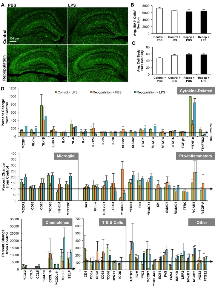
Microglia are the primary immune cell in the brain and are postulated to play important roles outside of immunity. Administration of the dual colony-stimulating factor 1 receptor (CSF1R)/c-Kit kinase inhibitor, PLX3397, to adult mice results in the elimination of ~99% of microglia, which remain eliminated for as long as treatment continues. Upon removal of the inhibitor, microglia rapidly repopulate the entire adult brain, stemming from a central nervous system (CNS) resident progenitor cell. Using this method of microglial elimination and repopulation, the role of microglia in both healthy and diseased states can be explored. Here, we examine the responsiveness of newly repopulated microglia to an inflammatory stimulus, as well as determine the impact of these cells on behavior, cognition, and neuroinflammation. Two month-old wild-type mice were placed on either control or PLX3397 diet for 21 d to eliminate microglia. PLX3397 diet was then removed in a subset of animals to allow microglia to repopulate and behavioral testing conducted beginning at 14 d repopulation. Finally, inflammatory profiling of the microglia-repopulated brain in response to lipopolysaccharide (LPS; 0.25 mg/kg) or phosphate buffered saline (PBS) was determined 21 d after inhibitor removal using quantitative real time polymerase chain reaction (RT-PCR), as well as detailed analyses of microglial morphologies. We find mice with repopulated microglia to perform similarly to controls by measures of behavior, cognition, and motor function. Compared to control/resident microglia, repopulated microglia had larger cell bodies and less complex branching in their processes, which resolved over time after inhibitor removal. Inflammatory profiling revealed that the mRNA gene expression of repopulated microglia was similar to normal resident microglia and that these new cells appear functional and responsive to LPS. Overall, these data demonstrate that newly repopulated microglia function similarly to the original resident microglia without any apparent adverse effects in healthy adult mice.
Overview
The claims and statistics of this paper haven’t been checked yet.
Run the check — free, no account needed. It reads the open-access text and checks the sources it cites.
Integrity signals
Registry
Cites
Statistics
Figures
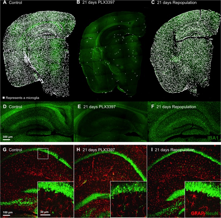
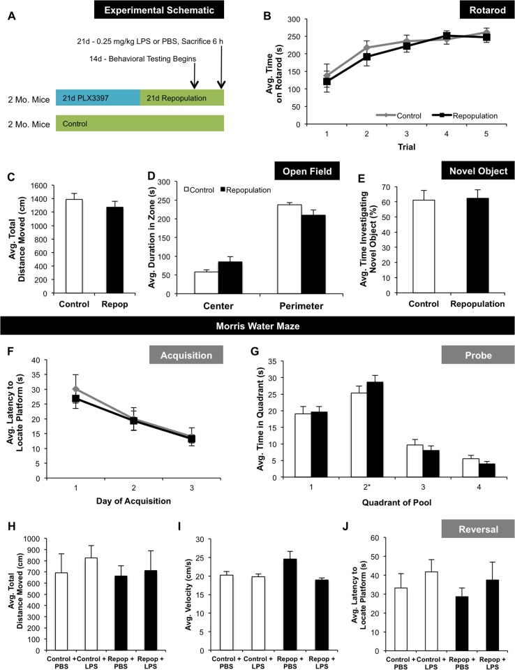
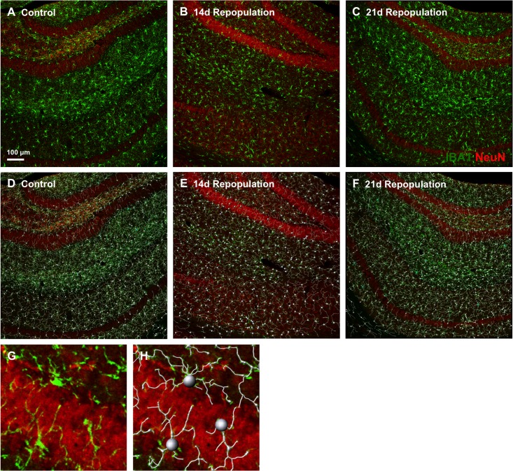
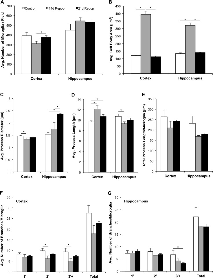
Verification
Claims the paper makes
The claims and statistics of this paper haven’t been checked yet.
Run the check — free, no account needed. It reads the open-access text and checks the sources it cites.
Claims other papers make about this one
Each is checked against this paper’s own text: does it say what the citing paper says it does?
In the citing paper
“EGFP+Iba1+ cells were once again abundant throughout analyzed BG nuclei, similar to repopulation dynamics reported in other brain regions (Elmore et al., 2015)”
From Local cues establish and maintain region-specific phenotypes of basal ganglia microglia (2017) · cites this paper as Elmore et al., 2015
Self-check
Percentages, sums and reported differences re-computed from the paper’s own numbers.
The self-check needs the paper’s full text.
Citations
Cites
Analysis of Relative Gene Expression Data Using Real-Time Quantitative PCR and the 2−ΔΔCT Method
Livak, K. J., & Schmittgen, T. D. (2001) · Methods · doi:10.1006/meth.2001.1262 · Cited by 180,860
Polazzi, E., Mengoni, I., Caprini, M., Peña‐Altamira, E., Kurtys, E., & Monti, B. (2012) · Neurosignals · doi:10.1159/000337115 · Cited by 9,023
Glutamate neurotoxicity and diseases of the nervous system
Choi, D. W. (1988) · Neuron · doi:10.1016/0896-6273(88)90162-6 · Cited by 8,188
From inflammation to sickness and depression: when the immune system subjugates the brain
Dantzer, R., O'Connor, J. C., Freund, G. G., Johnson, R. W., & Kelley, K. W. (2008) · Nature Reviews Neuroscience · doi:10.1038/nrn2297 · Cited by 7,395
Developments of a water-maze procedure for studying spatial learning in the rat
Morris, R. (1984) · Journal of Neuroscience Methods · doi:10.1016/0165-0270(84)90007-4 · Cited by 7,180
Fate Mapping Analysis Reveals That Adult Microglia Derive from Primitive Macrophages
Ginhoux, F., Greter, M., Leboeuf, M., Nandi, S., See, P., Gokhan, S., Mehler, M. F., Conway, S. J., Ng, L. G., Stanley, E. R., et al. (2010) · Science · doi:10.1126/science.1194637 · Cited by 5,175
Morris water maze: procedures for assessing spatial and related forms of learning and memory
Vorhees, C. V., & Williams, M. T. (2006) · Nature Protocols · doi:10.1038/nprot.2006.116 · Cited by 4,629
Synaptic Pruning by Microglia Is Necessary for Normal Brain Development
Paolicelli, R. C., Bolasco, G., Pagani, F., Maggi, L., Scianni, M., Panzanelli, P., Giustetto, M., Ferreira, T. A., Guiducci, E., Dumas, L., et al. (2011) · Science · doi:10.1126/science.1202529 · Cited by 4,244
Mutation of the mouse klotho gene leads to a syndrome resembling ageing
Kuro-o, M., Matsumura, Y., Aizawa, H., Kawaguchi, H., Suga, T., Utsugi, T., Ohyama, Y., Kurabayashi, M., Kaname, T., Kume, E., et al. (1997) · Nature · doi:10.1038/36285 · Cited by 3,837
Microglia: Active sensor and versatile effector cells in the normal and pathologic brain
Hanisch, U.-K., & Kettenmann, H. (2007) · Nature Neuroscience · doi:10.1038/nn1997 · Cited by 3,676
Cited by
Sorted by citations
Neurotoxic reactive astrocytes are induced by activated microglia
Liddelow, S. A., Guttenplan, K. A., Clarke, L. E., Bennett, F. C., Bohlen, C. J., Schirmer, L., Bennett, M. L., Münch, A. E., Chung, W.-S., Peterson, T. C., et al. (2017) · Nature · doi:10.1038/nature21029 · Cited by 7,971
Spangenberg, E. E., Lee, R. J., Najafi, A. R., Rice, R. A., Elmore, M. R. P., Blurton-Jones, M., West, B. L., & Green, K. N. (2016) · Brain · doi:10.1093/brain/aww016 · Cited by 695
Self-citationGlial Cells and Their Function in the Adult Brain: A Journey through the History of Their Ablation
Jäkel, S., & Dimou, L. (2017) · Frontiers in Cellular Neuroscience · doi:10.3389/fncel.2017.00024 · Cited by 571
Local cues establish and maintain region-specific phenotypes of basal ganglia microglia
De Biase, L. M., Schuebel, K. E., Fusfeld, Z. H., Jair, K., Hawes, I. A., Cimbro, R., Zhang, H.-Y., Liu, Q.-R., Shen, H., Xi, Z.-X., et al. (2017) · Neuron · doi:10.1016/j.neuron.2017.06.020 · Cited by 432
Elmore, M. R. P., Hohsfield, L. A., Kramár, E. A., Soreq, L., Lee, R. J., Pham, S. T., Najafi, A. R., Spangenberg, E. E., Wood, M. A., West, B. L., et al. (2018) · Aging Cell · doi:10.1111/acel.12832 · Cited by 380
Self-citationRojo, R., Raper, A., Ozdemir, D. D., Lefèvre, L., Grabert, K., Wollscheid-Lengeling, E., Bradford, B. M., Caruso, M., Gažová, I., Sánchez, A., et al. (2019) · Nature Communications · doi:10.1038/s41467-019-11053-8 · Cited by 390
Henry, R. J., Ritzel, R. M., Barrett, J. P., Doran, S. J., Jiao, Y., Leach, J. B., Szeto, G. L., Wu, J., Stoica, B. A., Faden, A. I., et al. (2020) · Journal of Neuroscience · doi:10.1523/jneurosci.2402-19.2020 · Cited by 344
Microglia Versus Myeloid Cell Nomenclature during Brain Inflammation
Greter, M., Lelios, I., & Croxford, A. L. (2015) · Frontiers in Immunology · doi:10.3389/fimmu.2015.00249 · Cited by 273
To Kill a Microglia: A Case for CSF1R Inhibitors
Green, K. N., Crapser, J. D., & Hohsfield, L. A. (2020) · Trends in Immunology · doi:10.1016/j.it.2020.07.001 · Cited by 265
Elimination of microglia improves cognitive function following cranial irradiation
Acharya, M. M., Green, K. N., Allen, B. D., Najafi, A. R., Syage, A., Minasyan, H., Le, M. T., Kawashita, T., Giedzinski, E., Parihar, V. K., et al. (2016) · Scientific Reports · doi:10.1038/srep31545 · Cited by 259
Microglia affect α-synuclein cell-to-cell transfer in a mouse model of Parkinson’s disease
George, S., Rey, N. L., Tyson, T., Esquibel, C., Meyerdirk, L., Schulz, E., Pierce, S., Burmeister, A. R., Madaj, Z., Steiner, J. A., et al. (2019) · Molecular Neurodegeneration · doi:10.1186/s13024-019-0335-3 · Cited by 229
Microglial repopulation resolves inflammation and promotes brain recovery after injury: RICE et al.
Rice, R. A., Pham, J., Lee, R. J., Najafi, A. R., West, B. L., & Green, K. N. (2017) · Glia · doi:10.1002/glia.23135 · Cited by 228
“Microglia remain eliminated for the duration of treatment, but withdrawal of CSF1R inhibition stimulates robust and rapid repopulation of the entire CNS with new cells that becomemicroglia (Elmore et al., 2015; Elmore et al., 2014), effectively replacing the entire microglial compartment.”
BackgroundSelf-citationMicroglia are required for protection against lethal coronavirus encephalitis in mice
Wheeler, D. L., Sariol, A., Meyerholz, D. K., & Perlman, S. (2018) · Journal of Clinical Investigation · doi:10.1172/jci97229 · Cited by 212
Transcriptional and epigenetic decoding of the microglial aging process
Li, X., Li, Y., Jin, Y., Zhang, Y., Wu, J., Xu, Z., Huang, Y., Cai, L., Gao, S., Liu, T., et al. (2023) · Nature Aging · doi:10.1038/s43587-023-00479-x · Cited by 234
Neuroprotective effects of chrysin: From chemistry to medicine
Nabavi, S. F., Braidy, N., Habtemariam, S., Orhan, İ., Daglia, M., Manayi, A., Gortzi, O., & Nabavi, S. M. (2015) · Neurochemistry International · doi:10.1016/j.neuint.2015.09.006 · Cited by 203
Sevenich, L. (2018) · Frontiers in Immunology · doi:10.3389/fimmu.2018.00697 · Cited by 206
Efficient Strategies for Microglia Replacement in the Central Nervous System
Xu, Z., Rao, Y., Huang, Y., Zhou, T. G., Feng, R., Xiong, S., Yuan, T., Qin, S., Lu, Y., Zhou, X., et al. (2020) · Cell Reports · doi:10.1016/j.celrep.2020.108041 · Cited by 189
Casali, B. T., MacPherson, K. P., Reed-Geaghan, E. G., & Landreth, G. E. (2020) · Neurobiology of Disease · doi:10.1016/j.nbd.2020.104956 · Cited by 186
Microglia Are Irrelevant for Neuronal Degeneration and Axon Regeneration after Acute Injury
Hilla, A. M., Diekmann, H., & Fischer, D. (2017) · Journal of Neuroscience · doi:10.1523/jneurosci.0584-17.2017 · Cited by 176
Crapser, J. D., Ochaba, J., Soni, N., Reidling, J. C., Thompson, L. M., & Green, K. N. (2020) · Brain · doi:10.1093/brain/awz363 · Cited by 157
Li, M., Li, Z., Ren, H., Jin, W., Wood, K., Liu, Q., Sheth, K. N., & Shi, F. (2016) · Journal of Cerebral Blood Flow & Metabolism · doi:10.1177/0271678x16666551 · Cited by 164
Maternal separation induces neuroinflammation and long-lasting emotional alterations in mice
Gracia-Rubio, I., Moscoso-Castro, M., Pozo, O. J., Marcos, J., Nadal, R., & Valverde, O. (2016) · Progress in Neuro-Psychopharmacology and Biological Psychiatry · doi:10.1016/j.pnpbp.2015.09.003 · Cited by 144
Zhang, Y., Zhao, L., Wang, X., Ma, W., Lazere, A., Qian, H., Zhang, J., Abu-Asab, M. S., Fariss, R. N., Roger, J. E., et al. (2018) · Science Advances · doi:10.1126/sciadv.aap8492 · Cited by 145
Activated microglia drive demyelination via CSF1R signaling
Marzan, D. E., Brügger‐Verdon, V., West, B. L., Liddelow, S., Samanta, J., & Salzer, J. L. (2021) · Glia · doi:10.1002/glia.23980 · Cited by 147
An updated assessment of microglia depletion: current concepts and future directions
Han, J., Harris, R. A., & Zhang, X.-M. (2017) · Molecular Brain · doi:10.1186/s13041-017-0307-x · Cited by 141
Lehmann, M. L., Weigel, T. K., Poffenberger, C. N., & Herkenham, M. (2019) · The Journal of Neuroscience · doi:10.1523/jneurosci.0184-19.2019 · Cited by 138
Protective Microglial Subset in Development, Aging, and Disease: Lessons From Transcriptomic Studies
Benmamar-Badel, A., Owens, T., & Wlodarczyk, A. (2020) · Frontiers in Immunology · doi:10.3389/fimmu.2020.00430 · Cited by 124
Beckmann, N., Giorgetti, E., Neuhaus, A., Zurbruegg, S., Accart, N., Smith, P., Perdoux, J., Perrot, L., Nash, M., Desrayaud, S., et al. (2018) · Acta Neuropathologica Communications · doi:10.1186/s40478-018-0510-8 · Cited by 131
Bordt, E. A., Ceasrine, A. M., & Bilbo, S. D. (2020) · Glia · doi:10.1002/glia.23753 · Cited by 125
Dynamic microglial modulation of spatial learning and social behavior
Torres, L., Danver, J., Ji, K., Miyauchi, J. T., Chen, D., Anderson, M. E., West, B. L., Robinson, J. K., & Tsirka, S. E. (2015) · Brain Behavior and Immunity · doi:10.1016/j.bbi.2015.09.001 · Cited by 133
Waltl, I., Käufer, C., Gerhauser, I., Chhatbar, C., Ghita, L., Kalinke, U., & Löscher, W. (2018) · Brain, Behavior, and Immunity · doi:10.1016/j.bbi.2018.09.006 · Cited by 124
Neal, M. L., Fleming, S. M., Budge, K. M., Boyle, A. M., Kim, C., Alam, G., Beier, E. E., Wu, L., & Richardson, J. R. (2020) · The FASEB Journal · doi:10.1096/fj.201900567rr · Cited by 123
Fard, M. T., & Stough, C. K. K. (2019) · Frontiers in Aging Neuroscience · doi:10.3389/fnagi.2019.00056 · Cited by 124
Unger, M. S., Schernthaner, P., Marschallinger, J., Mrowetz, H., & Aigner, L. (2018) · Journal of Neuroinflammation · doi:10.1186/s12974-018-1304-4 · Cited by 115
Gerber, Y. N., Saint-Martin, G., Bringuier, C. M., Bartolami, S., Goze‐Bac, C., Noristani, H. N., & Perrin, F. E. (2018) · Frontiers in Cellular Neuroscience · doi:10.3389/fncel.2018.00368 · Cited by 119
Han, J., Zhu, K., Zhang, X., & Harris, R. A. (2018) · Glia · doi:10.1002/glia.23529 · Cited by 116
A limited capacity for microglial repopulation in the adult brain
Najafi, A. R., Crapser, J., Jiang, S., Ng, W., Mortazavi, A., West, B. L., & Green, K. N. (2018) · Glia · doi:10.1002/glia.23477 · Cited by 99
Rosin, J. M., Vora, S. R., & Kurrasch, D. M. (2018) · Brain Behavior and Immunity · doi:10.1016/j.bbi.2018.07.023 · Cited by 113
Weber, M. D., McKim, D. B., Niraula, A., Witcher, K. G., Yin, W., Sobol, C. G., Wang, Y., Sawicki, C. M., Sheridan, J. F., & Godbout, J. P. (2018) · Biological Psychiatry · doi:10.1016/j.biopsych.2018.10.009 · Cited by 112
Complement Targets Newborn Retinal Ganglion Cells for Phagocytic Elimination by Microglia
Anderson, S., Zhang, J., Steele, M. R., Romero, C. O., Kautzman, A. G., Schafer, D. P., & Vetter, M. L. (2019) · Journal of Neuroscience · doi:10.1523/jneurosci.1854-18.2018 · Cited by 111
Microglia control glutamatergic synapses in the adult mouse hippocampus
Basilico, B., Ferrucci, L., Ratano, P., Golia, M. T., Grimaldi, A., Rosito, M., Ferretti, V., Reverte, I., Sanchini, C., Marrone, M. C., et al. (2021) · Glia · doi:10.1002/glia.24101 · Cited by 104
Liu, Y., Given, K. S., Dickson, E. L., Owens, G. P., Macklin, W. B., & Bennett, J. L. (2019) · Experimental Neurology · doi:10.1016/j.expneurol.2019.04.011 · Cited by 105
A systems-level framework for drug discovery identifies Csf1R as an anti-epileptic drug target
Srivastava, P. K., van Eyll, J. M., Godard, P., Mazzuferi, M., Delahaye‐Duriez, A., Steenwinckel, J. V., Gressèns, P., Danis, B., Vandenplas, C., Foerch, P., et al. (2018) · Nature Communications · doi:10.1038/s41467-018-06008-4 · Cited by 103
Microglia Are Critical in Host Defense against Prion Disease
Carroll, J. A., Race, B., Williams, K., Striebel, J., & Chesebro, B. (2018) · Journal of Virology · doi:10.1128/jvi.00549-18 · Cited by 94
Walker, D. G., Tang, T. M., & Lue, L. (2017) · Frontiers in Aging Neuroscience · doi:10.3389/fnagi.2017.00244 · Cited by 95
Post-Ischaemic Immunological Response in the Brain: Targeting Microglia in Ischaemic Stroke Therapy
Rawlinson, C., Jenkins, S. I., Thei, L., Dallas, M. L., & Chen, R. (2020) · Brain Sciences · doi:10.3390/brainsci10030159 · Cited by 99
Microglia: Driving critical periods and sexual differentiation of the brain
VanRyzin, J. W., Pickett, L. A., & McCarthy, M. M. (2017) · Developmental Neurobiology · doi:10.1002/dneu.22569 · Cited by 97
Han, X., Li, Q., Lan, X., El‐Mufti, L. H., Ren, H., & Wang, J. (2019) · Molecular Neurobiology · doi:10.1007/s12035-019-1502-9 · Cited by 92
Krukowski, K., Feng, X., Paladini, M. S., Chou, A., Sacramento, K., Grue, K., Riparip, L.-K., Jones, T., Campbell-Beachler, M., Nelson, G., et al. (2018) · Scientific Reports · doi:10.1038/s41598-018-26039-7 · Cited by 92
Coleman, L. G., Zou, J., & Crews, F. T. (2020) · Journal of Neuroinflammation · doi:10.1186/s12974-019-1678-y · Cited by 90
Senescent Microglia: The Key to the Ageing Brain?
Greenwood, E. K., & Brown, D. R. (2021) · International Journal of Molecular Sciences · doi:10.3390/ijms22094402 · Cited by 84
Neuroinflammation, Microglia, and Cell-Association during Prion Disease
Carroll, J. A., & Chesebro, B. W. (2019) · Viruses · doi:10.3390/v11010065 · Cited by 87
VanRyzin, J. W., Yu, S. J., Perez‐Pouchoulen, M., & McCarthy, M. M. (2016) · eNeuro · doi:10.1523/eneuro.0297-16.2016 · Cited by 83
Microglia as Therapeutic Target for Radiation-Induced Brain Injury
Liu, Q., Yan, H., Duan, M., Yang, Q., Ren, B., & Tang, F. R. (2022) · International Journal of Molecular Sciences · doi:10.3390/ijms23158286 · Cited by 84
Green, T. R. F., Murphy, S. M., & Rowe, R. K. (2022) · Scientific Reports · doi:10.1038/s41598-022-23091-2 · Cited by 87
Bray, C. E., Witcher, K. G., Adekunle-Adegbite, D., Ouviña, M., Witzel, M., Hans, E., Tapp, Z. M., Packer, J. M., Goodman, E. J., Zhao, F., et al. (2022) · Journal of Neuroscience · doi:10.1523/jneurosci.1910-21.2022 · Cited by 77
RNA-seq and network analysis reveal unique glial gene expression signatures during prion infection
Carroll, J. A., Race, B., Williams, K., Striebel, J., & Chesebro, B. (2020) · Molecular Brain · doi:10.1186/s13041-020-00610-8 · Cited by 66
Kerkhofs, D., van Hagen, B. T. J., Milanova, I. V., Schell, K. J., van Essen, H., Wijnands, E., Goossens, P., Blankesteijn, W. M., Unger, T., Prickaerts, J., et al. (2020) · Theranostics · doi:10.7150/thno.44394 · Cited by 75
Melchiorri, D., Merlo, S., Micallef, B., Borg, J.-J., & Dráfi, F. (2023) · Frontiers in Pharmacology · doi:10.3389/fphar.2023.1196413 · Cited by 73
Vogel Ciernia, A., Careaga, M., LaSalle, J. M., & Ashwood, P. (2018) · Glia · doi:10.1002/glia.23261 · Cited by 69
Microglia depletion fails to abrogate inflammation-induced sickness in mice and rats
Vichaya, E. G., Malik, S., Sominsky, L., Ford, B. G., Spencer, S. J., & Dantzer, R. (2020) · Journal of Neuroinflammation · doi:10.1186/s12974-020-01832-2 · Cited by 71
Immune cell modulation of oligodendrocyte lineage cells
Harrington, E. P., Bergles, D. E., & Calabresi, P. A. (2019) · Neuroscience Letters · doi:10.1016/j.neulet.2019.134601 · Cited by 70
Hohsfield, L. A., Najafi, A. R., Ghorbanian, Y., Soni, N., Hingco, E. E., Kim, S. J., Jue, A. D., Swarup, V., Inlay, M. A., & Green, K. N. (2020) · Journal of Neuroinflammation · doi:10.1186/s12974-020-01931-0 · Cited by 66
Mitigation of helium irradiation-induced brain injury by microglia depletion
Allen, B. D., Syage, A. R., Maroso, M., Baddour, A. A. D., Luong, V., Minasyan, H., Giedzinski, E., West, B. L., Soltész, I., Limoli, C. L., et al. (2020) · Journal of Neuroinflammation · doi:10.1186/s12974-020-01790-9 · Cited by 61
Circulating Pro-Inflammatory Exosomes Worsen Stroke Outcomes in Aging
Zhang, H., Lin, S., McElroy, C. L., Wang, B. Z., Jin, D., Uteshev, V. V., & Jin, K. (2021) · Circulation Research · doi:10.1161/circresaha.121.318897 · Cited by 60
Manickam, V., Gawande, D. Y., Stothert, A. R., Clayman, A. C., Batalkina, L., Warchol, M. E., Ohlemiller, K. K., & Kaur, T. (2023) · Journal of Neuroscience · doi:10.1523/jneurosci.1273-22.2023 · Cited by 65
Microglial Function and Regulation during Development, Homeostasis and Alzheimer’s Disease
Casali, B. T., & Reed-Geaghan, E. G. (2021) · Cells · doi:10.3390/cells10040957 · Cited by 70
Barca, C., Foray, C., Hermann, S., Herrlinger, U., Remory, I., Laoui, D., Schäfers, M., Grauer, O. M., Zinnhardt, B., & Jacobs, A. H. (2021) · Frontiers in Immunology · doi:10.3389/fimmu.2021.787307 · Cited by 64
Microglia maintain the normal structure and function of the hippocampal astrocyte network
Du, Y., Brennan, F. H., Popovich, P. G., & Zhou, M. (2022) · Glia · doi:10.1002/glia.24179 · Cited by 61
Poulen, G., Aloy, E., Bringuier, C. M., Mestre‐Francés, N., Artus, E. V. F., Cardoso, M., Perez, J., Goze‐Bac, C., Boukhaddaoui, H., Lonjon, N., et al. (2021) · Theranostics · doi:10.7150/thno.61833 · Cited by 64
Luca, S. N. D., Sominsky, L., Soch, A., Wang, H., Ziko, I., Rank, M. M., & Spencer, S. J. (2018) · Brain Behavior and Immunity · doi:10.1016/j.bbi.2018.12.008 · Cited by 61
Sabogal-Guáqueta, A. M., Marmolejo-Garza, A., de Pádua, V. P., Eggen, B. J. L., Boddeke, E., & Dolga, A. M. (2020) · Progress in Neurobiology · doi:10.1016/j.pneurobio.2020.101805 · Cited by 65
The ins and outs of microglial cells in brain health and disease
Pallarés-Moratalla, C., & Bergers, G. (2024) · Frontiers in Immunology · doi:10.3389/fimmu.2024.1305087 · Cited by 66
A story of birth and death: Insights into the formation and dynamics of the microglial population
Askew, K. E., & Gómez‐Nicola, D. (2017) · Brain Behavior and Immunity · doi:10.1016/j.bbi.2017.03.009 · Cited by 59
Tahmasebi, F., Pasbakhsh, P., Mortezaee, K., Madadi, S., Barati, S., & Kashani, I. R. (2019) · Journal of Cellular Biochemistry · doi:10.1002/jcb.28344 · Cited by 56
Hohsfield, L. A., Najafi, A. R., Ghorbanian, Y., Soni, N., Crapser, J. D., Velez, D. X. F., Jiang, S., Royer, S. E., Kim, S. J., Henningfield, C. M., et al. (2021) · eLife · doi:10.7554/elife.66738 · Cited by 50
Basilico, B., Ferrucci, L., Khan, A., Angelantonio, S. D., Ragozzino, D., & Reverte, I. (2022) · Frontiers in Cellular Neuroscience · doi:10.3389/fncel.2022.1022431 · Cited by 52
Role of Macrophages and Microglia in Zebrafish Regeneration
Var, S. R., & Byrd-Jacobs, C. A. (2020) · International Journal of Molecular Sciences · doi:10.3390/ijms21134768 · Cited by 52
Johnson, N. R., Yuan, P., Castillo, E., Lopez, T. P., Yue, W., Bond, A., Rivera, B. M., Sullivan, M. C., Hirouchi, M., Giles, K., et al. (2023) · Nature Communications · doi:10.1038/s41467-022-35753-w · Cited by 46
Mendes, M. S., Le, L., Atlas, J., Brehm, Z., Ladron-de-Guevara, A., Matei, E., Lamantia, C., McCall, M. N., & Majewska, A. K. (2021) · eLife · doi:10.7554/elife.61173 · Cited by 46
Ferro, A., Sheeler, C. A., Rosa, J. S., & Cvetanović, M. (2019) · Journal of Molecular Biology · doi:10.1016/j.jmb.2019.01.016 · Cited by 49
Tanzey, S. S., Shao, X., Stauff, J., Arteaga, J., Sherman, P. S., Scott, P. J. H., & Mossine, A. V. (2018) · Pharmaceuticals · doi:10.3390/ph11040136 · Cited by 44
Oh, S. J., Ahn, H., Jung, K.-H., Han, S. J., Nam, K. R., Kang, K. J., Park, J.-A., Lee, K. C., Lee, Y. J., & Choi, J. Y. (2020) · Molecular Imaging and Biology · doi:10.1007/s11307-020-01485-w · Cited by 50
Microglia in neuroimmunopharmacology and drug addiction
Li, H., Watkins, L. R., & Wang, X. (2024) · Molecular Psychiatry · doi:10.1038/s41380-024-02443-6 · Cited by 44
Effect of Inhibition of Colony-Stimulating Factor 1 Receptor on Choroidal Neovascularization in Mice
Schwarzer, P., Kokona, D., Ebneter, A., & Zinkernagel, M. S. (2019) · American Journal Of Pathology · doi:10.1016/j.ajpath.2019.10.011 · Cited by 40
Chen, Y., Hong, T., Chen, F., Sun, Y., Wang, Y., & Cui, L. (2021) · Frontiers in Aging Neuroscience · doi:10.3389/fnagi.2021.631827 · Cited by 40
The role of microglia in ischemic preconditioning
McDonough, A., & Weinstein, J. R. (2019) · Glia · doi:10.1002/glia.23695 · Cited by 39
Emerging roles for hypothalamic microglia as regulators of physiological homeostasis
Rosin, J. M., & Kurrasch, D. M. (2019) · Frontiers in Neuroendocrinology · doi:10.1016/j.yfrne.2019.100748 · Cited by 37
An overview of microglia ontogeny and maturation in the homeostatic and pathological brain
Mendes, M. S., & Majewska, A. K. (2021) · European Journal of Neuroscience · doi:10.1111/ejn.15225 · Cited by 35
Microglia and Their Promising Role in Ischemic Brain Injuries: An Update
Yu, L., Su, X., Li, S., Zhao, F., Mu, D., & Qu, Y. (2020) · Frontiers in Cellular Neuroscience · doi:10.3389/fncel.2020.00211 · Cited by 34
Regulation of Microglia Identity from an Epigenetic and Transcriptomic Point of View
Eggen, B. J. L., Boddeke, E. W. G. M., & Kooistra, S. M. (2019) · Neuroscience · doi:10.1016/j.neuroscience.2017.12.010 · Cited by 34
Yegla, B., Boles, J. S., Kumar, A., & Foster, T. C. (2021) · Glia · doi:10.1002/glia.23975 · Cited by 33
He, Y., Liu, T., He, Q., Ke, W., Li, X., Du, J., Deng, S., Shu, Z., Wu, J. S., Yang, B., et al. (2023) · eLife · doi:10.7554/elife.92252 · Cited by 33
Deplete and repeat: microglial CSF1R inhibition and traumatic brain injury
Boland, R., & Kokiko‐Cochran, O. N. (2024) · Frontiers in Cellular Neuroscience · doi:10.3389/fncel.2024.1352790 · Cited by 32
CuII(atsm) Attenuates Neuroinflammation
Choo, X. Y., Liddell, J. R., Huuskonen, M. T., Grubman, A., Moujalled, D., Roberts, J., Kysenius, K., Patten, L., Quek, H., Oikari, L. E., et al. (2018) · Frontiers in Neuroscience · doi:10.3389/fnins.2018.00668 · Cited by 31
Microglia activation is essential for BMP7-mediated retinal reactive gliosis
Dharmarajan, S., Fisk, D. L., Sorenson, C. M., Sheibani, N., & Belecky‐Adams, T. L. (2017) · Journal of Neuroinflammation · doi:10.1186/s12974-017-0855-0 · Cited by 31
Du, S., Ou, F., Drieu, A., Xu, E. Z., Cheng, Y., Storck, S. E., Mamuladze, T., Cao, J., Abduljawad, N., Bhattarai, B., et al. (2026) · Neuron · doi:10.1016/j.neuron.2026.01.032 · Cited by 32
Hohsfield, L. A., Tsourmas, K. I., Ghorbanian, Y., Syage, A. R., Kim, S. J., Cheng, Y., Furman, S., Inlay, M. A., Lane, T. E., & Green, K. N. (2022) · Glia · doi:10.1002/glia.24144 · Cited by 25
Microglial depletion and repopulation: a new era of regenerative medicine?
Coleman, L. G., Barnett, A., & Crews, F. T. (2020) · Neural Regeneration Research · doi:10.4103/1673-5374.300439 · Cited by 27
Picard, K., Corsi, G., Decoeur, F., Castro, M. A. D., Bordeleau, M., Persillet, M., Layé, S., Limatola, C., Tremblay, M., & Nadjar, A. (2022) · Brain Behavior and Immunity · doi:10.1016/j.bbi.2022.09.016 · Cited by 26
Metadata
- Authors
- Elmore, M. R. P., Lee, R. J., West, B. L., & Green, K. N.
- Affiliations
- University of California, Irvine (US)
- Plexxikon (United States) (US)
- Corresponding author
- Green, Kim N.
- Venue
- PLoS ONE
- Publisher
- Public Library of Science (PLoS)
- Published
- 2015-04-07
- Volume
- 10
- Issue
- 4
- Pages
- e0122912
- ISSN
- 1932-6203 (ISSN-L)
- PMC
- PMC4388515
- OpenAlex
- W2169700519
- Cited by
- 228
- References
- 73
- Citations by year
- 2026: 10 · 2025: 20 · 2024: 19 · 2023: 19 · 2022: 23 · 2021: 32 · 2020: 26 · 2019: 28 · 2018: 23 · 2017: 18 · 2016: 6 · 2015: 4
- FWCI
- 9.07
- Type
- journal article
- Language
- en
- Open access
- gold
- doi.org
- Licence
- Locations
- PLoS ONE (publishedVersion, cc-by)
- PubMed Central (publishedVersion, cc-by)
- PubMed (publishedVersion)
- DOAJ (DOAJ: Directory of Open Access Journals) (submittedVersion, cc-by-sa)
- eScholarship (California Digital Library) (submittedVersion)
- Funders
- Plexxikon
- National Institute on Aging (T32AG000096)
- National Institute of Neurological Disorders and Stroke (R01NS083801)
- Topics
- Life Sciences › Neuroscience › Neurology › Neuroinflammation and Neurodegeneration Mechanisms
- Life Sciences › Neuroscience › Developmental Neuroscience › Neurogenesis and neuroplasticity mechanisms
- Life Sciences › Neuroscience › Biological Psychiatry › Tryptophan and brain disorders
- Keywords
- microglia, microglial repopulation, PLX3397, CSF1R inhibition, lipopolysaccharide response, neuroinflammation, microglial morphology, cognitive function, motor function, brain immune cells
- MeSH terms
- Animals, Brain, Cells, Cultured, Central Nervous System Diseases, Cognition, Female, Inflammation, Lipopolysaccharides, Male, Mice, Inbred C57BL, Motor Activity, Nerve Regeneration, Microglia, Maze Learning, Rotarod Performance Test, Cell Shape
About these checks
Registry, retraction, Cites and statistics results are computed, not judged, with one exception: a printed reference that no index lists is read once by a model before it counts as a citation error. Claims are extracted by a model and kept only when their quote appears verbatim in the text; each cited source is then checked, with a model’s help, for whether it says what the claim relies on. Those verdicts can be wrong — read the claim’s quote and the cited source before relying on one.
Statistics and arithmetic are recomputed, not judged.
Confidence bands are shown only for fields calibrated on our annotated test set. None is calibrated yet, so no band is shown.
What counts as a claim ›How we measure confidence ›How we check this ›
Are you an author, or do you see a problem with this page? Request a review
