

The DOI matches its Crossref record; 36 of the 38 references it cites were found in a registry or an index.
Dendritic spines of CA 1 pyramidal cells in the rat hippocampus: serial electron microscopy with reference to their biophysical characteristics
Open accessAbstract
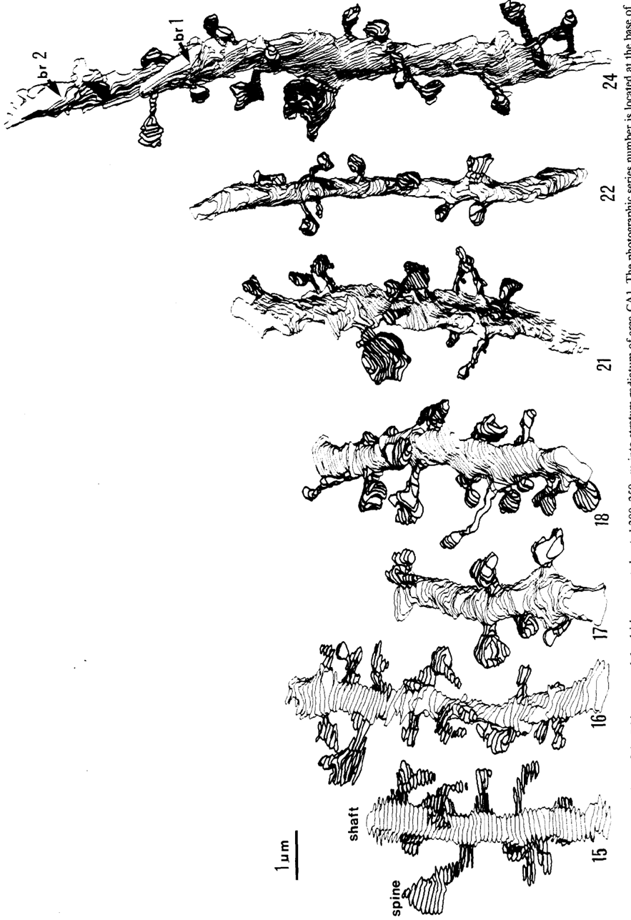
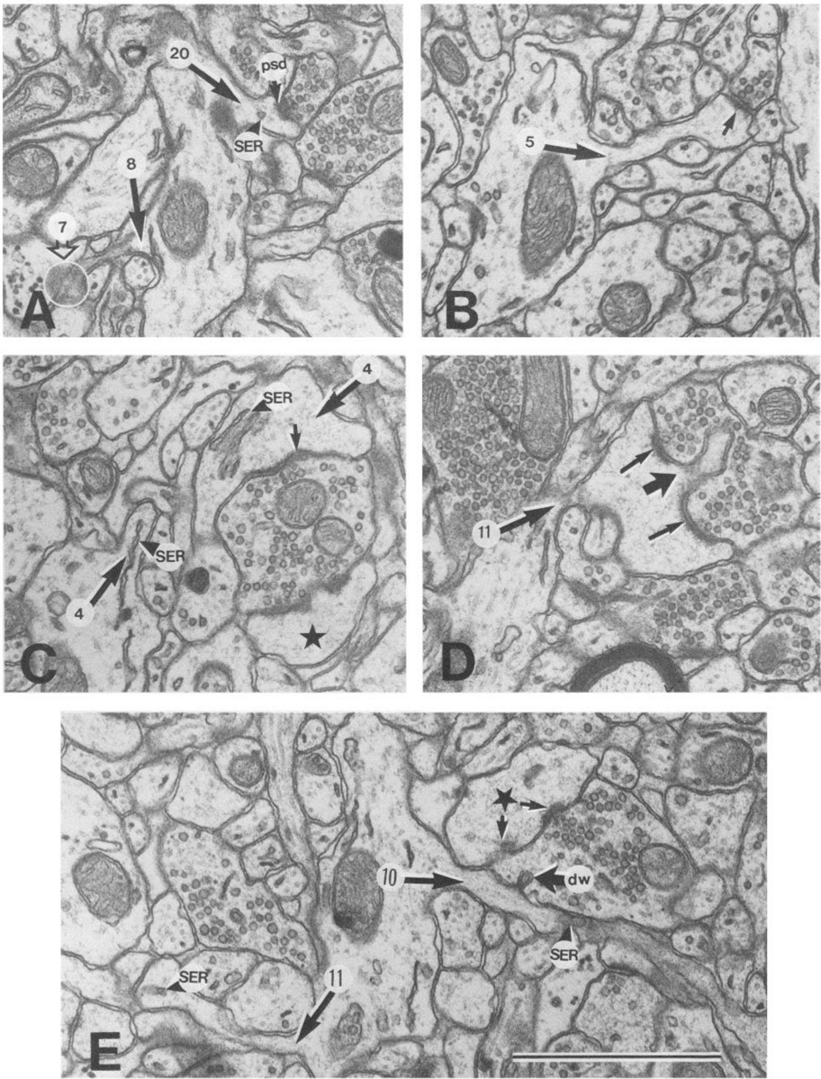
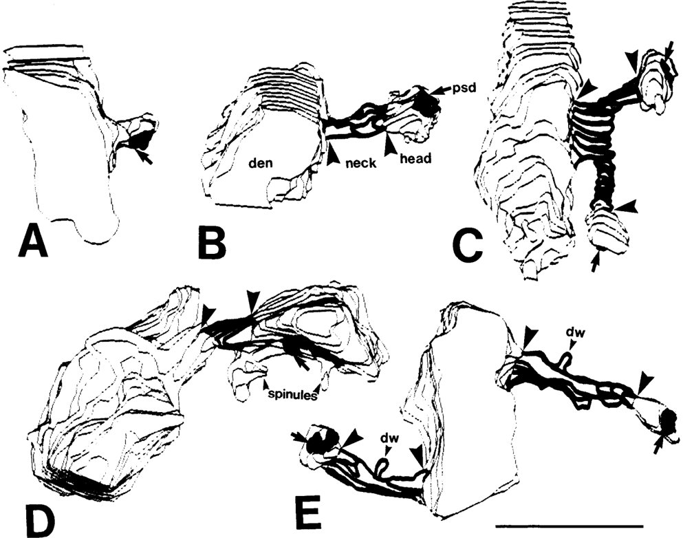
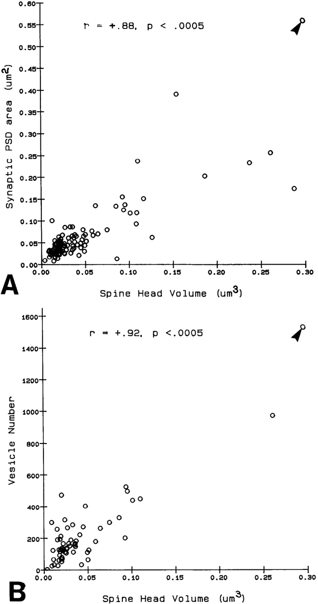
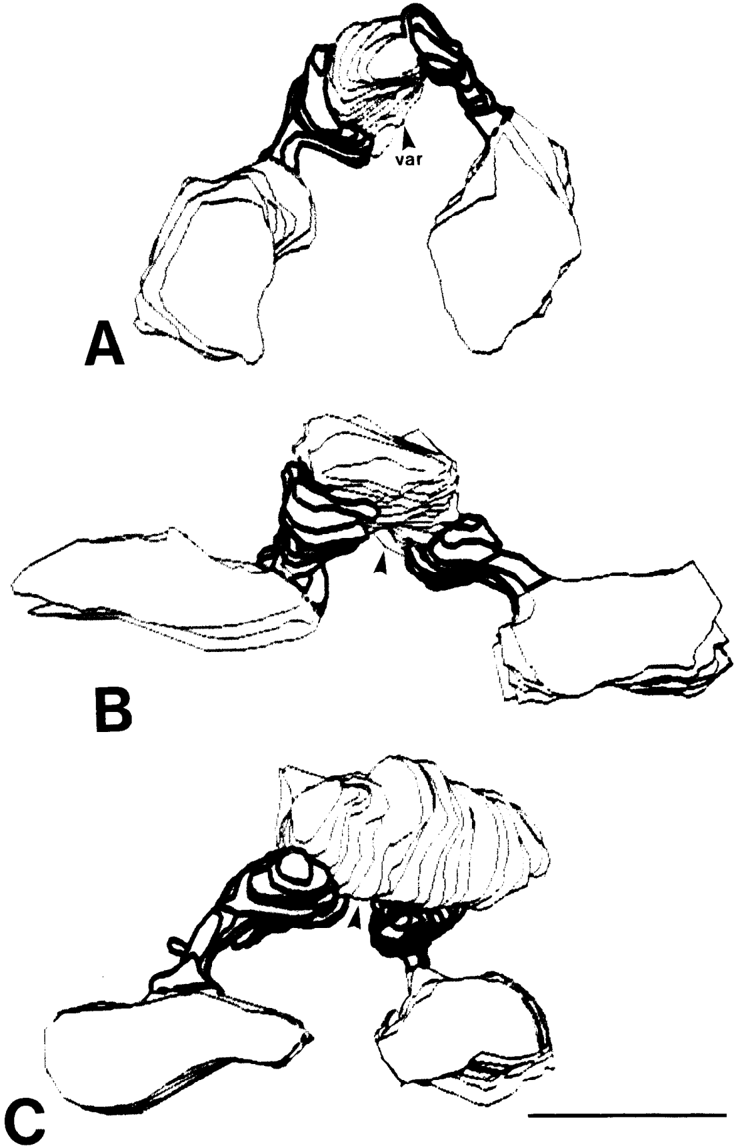
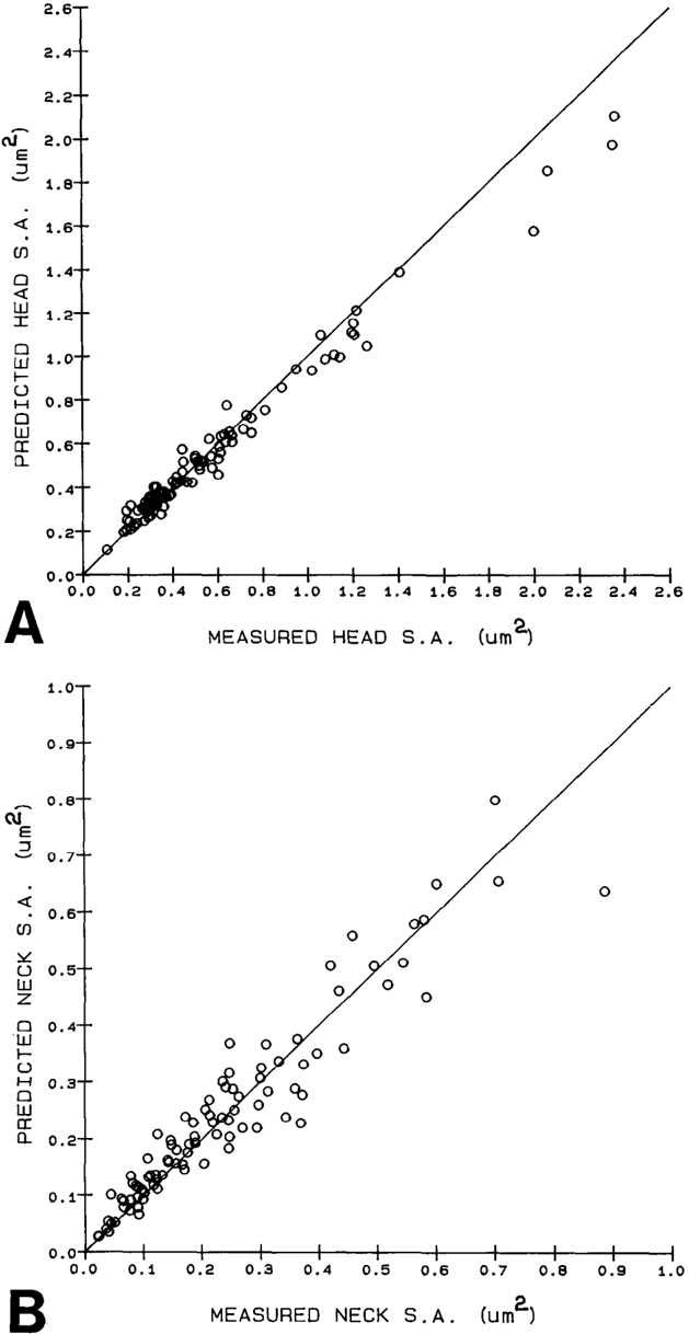
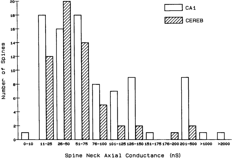
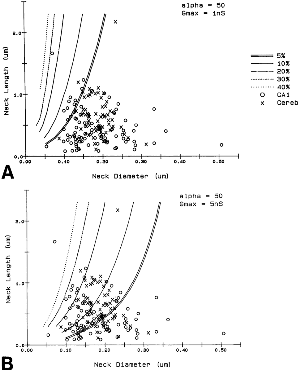
Serial electron microscopy and 3-D reconstructions of dendritic spines from hippocampal area CA 1 dendrites were obtained to evaluate 2 questions about relationships between spine geometry and synaptic efficacy. First, under what biophysical conditions are the spine necks likely to reduce the magnitude of charge transferred from the synapses on the spine heads to the recipient dendrite? Simulation software provided by Charles Wilson (1984) was used to determine that if synaptic conductance is 1 nS or less, only 1% of the hippocampal spine necks are sufficiently thin and long to reduce charge transfer by more than 10%. If synaptic conductance approaches 5 nS, however, 33% of the hippocampal spine necks are sufficiently thin and long to reduce charge transfer by more than 10%. Second, is spine geometry associated with other anatomical indicators of synaptic efficacy, including the area of the postsynaptic density and the number of vesicles in the presynaptic axon? Reconstructed spines were graphically edited into head and neck compartments, and their dimensions were measured, the areas of the postsynaptic densities (PSD) were measured, and all of the vesicles in the presynaptic axonal varicosities were counted. The dimensions of the spine head were well correlated with the area of PSD and the number of vesicles in the presynaptic axonal varicosity. Spine neck diameter and length were not correlated with PSD area, head volume, or the number of vesicles. These results suggest that the dimensions of the spine head, but not the spine neck, reflect differences in synaptic efficacy. We suggest that the constricted necks of hippocampal dendritic spines might reduce diffusion of activated molecules to neighboring synapses, thereby attributing specificity to activated or potentiated synapses.
Overview
The claims and statistics of this paper haven’t been checked yet.
Run the check — free, no account needed. It reads the open-access text and checks the sources it cites.
Integrity signals
Registry
Cites
Statistics
Figures
Verification
Claims the paper makes
The claims and statistics of this paper haven’t been checked yet.
Run the check — free, no account needed. It reads the open-access text and checks the sources it cites.
Claims other papers make about this one
Each is checked against this paper’s own text: does it say what the citing paper says it does?
The source backs part of the claim, not all of it — or backs it for a different population, condition or comparison.
In the citing paper
“This is probably one of the reasons why many ultrastructural studies that utilized high concentrations of potassium ferrocyanide/ferricyanide (1.5–3.0%) provided no data on SS (e.g., Harris and Stevens, 1989; Hayworth et al., 2014; Yakoubi et al., 2019a; Yin et al., 2020; Gour et al., 2021; Phelps et al., 2021; Peddie et al., 2022; Turner et al., 2022).”
From Unambiguous identification of asymmetric and symmetric synapses using volume electron microscopy (2024) · cites this paper as Harris and Stevens, 1989
Experimental — accuracy not yet measuredThe source backs part of the claim, not all of it — or backs it for a different population, condition or comparison.
In the citing paper
“Given that each spine receives only one synaptic input13, our data indicate the highest possible degree of freedom is available in synaptic connections in the dendrites, which has been assumed in the learning models of most neural network theories17.”
From Dendritic spine geometry is critical for AMPA receptor expression in hippocampal CA1 pyramidal neurons (2001) · cites this paper as [13]
Experimental — accuracy not yet measuredThe source backs part of the claim, not all of it — or backs it for a different population, condition or comparison.
In the citing paper
“This observation suggests that the expression of AMPA receptors is controlled independently at a level of the individual spine, consistent with the classical notion that a single spine may behave as an independent chemical compartment to regulate synaptic transmission13, 14.”
From Dendritic spine geometry is critical for AMPA receptor expression in hippocampal CA1 pyramidal neurons (2001) · cites this paper as [13]
Experimental — accuracy not yet measuredThe source backs part of the claim, not all of it — or backs it for a different population, condition or comparison.
In the citing paper
“Regions of glutamate sensitivity tended to be asymmetrically localized to one portion of a spine (Figs. 3h and 4c–j), consistent with the predominant localization of AMPA receptors within PSDs with a diameter of 0.1 to 0.5μm13, 19, 20.”
From Dendritic spine geometry is critical for AMPA receptor expression in hippocampal CA1 pyramidal neurons (2001) · cites this paper as [13]
Experimental — accuracy not yet measuredThe source backs part of the claim, not all of it — or backs it for a different population, condition or comparison.
In the citing paper
“Syn1a-CFP puncta area calculated as a measure of bouton area, which correlated with pHuse area, as expected42.”
From A trans-synaptic nanocolumn aligns neurotransmitter release to receptors (2016) · cites this paper as [42]
Experimental — accuracy not yet measured
The source backs part of the claim, not all of it — or backs it for a different population, condition or comparison.
In the citing paper
“In turn, the size of the postsynaptic density is correlated with the number of presynaptic vesicles, the number of postsynaptic receptors and the ready-releasable pool of transmitter (Harris and Stevens, 1989; Nusser et al., 1998; Schikorski and Stevens, 1999, 2001; Arellano et al., 2007a).”
From The influence of phospho-tau on dendritic spines of cortical pyramidal neurons in patients with Alzheimer’s disease (2013) · cites this paper as Harris and Stevens, 1989
Experimental — accuracy not yet measuredThe source backs part of the claim, not all of it — or backs it for a different population, condition or comparison.
In the citing paper
“Additionally, the volume of the dendritic spine head is related to the size of the postsynaptic density (Harris and Stevens, 1989; Schikorski and Stevens, 1997; Arellano et al., 2007b), the number of presynaptic vesicles, and the number of vesicles adjacent to the presynaptic cleft (Harris and Stevens, 1989).”
From The influence of phospho-tau on dendritic spines of cortical pyramidal neurons in patients with Alzheimer’s disease (2013) · cites this paper as Harris and Stevens, 1989
Experimental — accuracy not yet measuredThe source backs part of the claim, not all of it — or backs it for a different population, condition or comparison.
In the citing paper
“Furthermore, the volume of the head is directly proportional to the strength of synaptic currents (Harris and Stevens, 1989; Schikorski and Stevens, 1997; Arellano et al., 2007a).”
From The influence of phospho-tau on dendritic spines of cortical pyramidal neurons in patients with Alzheimer’s disease (2013) · cites this paper as Harris and Stevens, 1989
Experimental — accuracy not yet measuredThe cited source says what the claim attributes to it.
In the citing paper
“The relationship between spine geometry and expression of functional AMPA receptors demonstrated here provides substantial support to the hypothesis that neuronal fine structures have functional correlates in the brain13, 18, 21.”
From Dendritic spine geometry is critical for AMPA receptor expression in hippocampal CA1 pyramidal neurons (2001) · cites this paper as [13]
The cited source says what the claim attributes to it.
In the citing paper
“These data are consistent with the correlation between AMPA receptor immunoreactivity and the size of PSD19, 20, because the size of PSD is nearly proportional to the spine-head volume13.”
From Dendritic spine geometry is critical for AMPA receptor expression in hippocampal CA1 pyramidal neurons (2001) · cites this paper as [13]
The cited source says what the claim attributes to it.
In the citing paper
“whereas the spine head volume is correlated with the area of the postsynaptic density.”
From The influence of phospho-tau on dendritic spines of cortical pyramidal neurons in patients with Alzheimer’s disease (2013) · cites this paper as Harris and Stevens, 1989
Self-check
Percentages, sums and reported differences re-computed from the paper’s own numbers.
The self-check needs the paper’s full text.
Citations
Cites
The Synaptic Organization of the Brain
(2004) · Oxford University Press eBooks · doi:10.1093/acprof:oso/9780195159561.001.1 · Cited by 3,574
The Synaptic Organization of the Brain
Freemon, F. R. (1980) · JAMA · doi:10.1001/jama.1980.03300440052037 · Cited by 709
Chang, F., & Greenough, W. T. (1984) · Brain Research · doi:10.1016/0006-8993(84)91008-4 · Cited by 523
Lee, K. S., Schottler, F., Oliver, M. E., & LYNCH, G. S. (1980) · Journal of Neurophysiology · doi:10.1152/jn.1980.44.2.247 · Cited by 473
Harris, K., & Stevens, J. (1988) · The Journal of Neuroscience · doi:10.1523/jneurosci.08-12-04455.1988 · Cited by 318
Self-citationDesmond, N. L., & Levy, W. B. (1983) · Brain Research · doi:10.1016/0006-8993(83)91329-x · Cited by 276
Three-Dimensional analysis of dendritic spines
Špaček, J., & Hartmann, M. (1983) · Anatomy and Embryology · doi:10.1007/bf00298517 · Cited by 269
The anatomy of a memory: convergence of results across a diversity of tests
Greenough, W. T., & Bailey, C. H. (1988) · Trends in Neurosciences · doi:10.1016/0166-2236(88)90139-7 · Cited by 266
Crick, F. (1982) · Trends in Neurosciences · doi:10.1016/0166-2236(82)90020-0 · Cited by 254
Van Harreveld, A., & Fifkova, E. (1975) · Experimental Neurology · doi:10.1016/0014-4886(75)90055-2 · Cited by 239
Research works printed but not linked: 6
- Harris, K. M., F. E. Jensen, and B. Tsao (1988) Ultrastructure, de- velopment, and plasticity of synapses in area CA1 of the rat hippo- campus: Extending our vision with serial electron microscopy and three dimensional analyses. Neurol. Neurobiol. (in press).
- Andersen, P., T. Blackstad, G. Hulleberg, J. Line Vaaland, and M. Andersen, P., T. Blackstad, G. Hulleberg, M. Trommald, and J. L. Vaaland (1988b) Dimensions ofdendritic spines ofrat dentate gran- Trommald (1988a) Changes in spine morphology associated with ule cells during long-term potentiation (LTP). Proc. Physiol. Sot. P.C. 50. long-term potentiation in rat dentate granule cells. Proc. Physiol. Sot. s.
- Diamond, J., E. G. Gray, and G. M. Yasargil (1970) The function of dendritic spines: An hypothesis. In Excitatory Synaptic Mechanisms, P. Andersen and J. Jensen, eds., pp. 2 13-222, Universitets Forlaget, Oslo.
- Meek, G. A. (1976) Practical Electron Microscopyfor Biologists, Wiley, New York.
- Rall, W. (1970) Cable properties of dendrites and effects of synaptic location. In Excitatory Synaptic Mechanisms, P. Andersen and J. K. S. Jensen, eds., pp. 175-l 87, Universitets Forlaget, Oslo.
- Rall, W. (1978) Dendritic spines and synaptic potency. In Studies in NeurophysioloRy, A. K. McIntyre and K. Porter, eds., pp. 203-209, Cambridge U.-P., Cambridge.
Cited by
Sorted by citations
Items 801–900 of 999
Endocrine disrupting effects on morphological synaptic plasticity
Zsarnovszky, A., Alymbaeva, D., Jocsak, G., Szabo, C., Mária Schilling-Tóth, B., & Sandor Kiss, D. (2024) · Frontiers in Neuroendocrinology · doi:10.1016/j.yfrne.2024.101157 · Cited by 7
Cryo-electron tomographic investigation of native hippocampal glutamatergic synapsesPreprint
Matsui, A., Spangler, C. J., Elferich, J., Shiozaki, M., Jean, N., Zhao, X., Qin, M., Zhong, H., Yu, Z., & Gouaux, E. (2024) · eLife · doi:10.7554/elife.98458.3 · Cited by 40
Structural plasticity of dendritic spines
Zhang, S., Wang, J., & Wang, L. (2010) · Frontiers in Biology · doi:10.1007/s11515-010-0011-z · Cited by 7
Analysis of Actin Turnover and Spine Dynamics in Hippocampal Slice Cultures
Michaelsen‐Preusse, K., Kellner, Y., Körte, M., & Zagrebelsky, M. (2014) · Neuromethods · doi:10.1007/978-1-4939-0381-8_9 · Cited by 7
Liu, J., Gu, X., Zou, R.-X., Nan, W., Yang, S., Wang, H.-L., & Chen, X.-T. (2018) · Frontiers in Pharmacology · doi:10.3389/fphar.2018.01141 · Cited by 8
Early synaptic deficits in GSK-3β overexpressing mice
Banach, E., Jaworski, T., & Urban‐Ciećko, J. (2022) · Neuroscience Letters · doi:10.1016/j.neulet.2022.136744 · Cited by 7
Bell, M. K., Lee, C. T., & Rangamani, P. (2022) · The Journal of Physiology · doi:10.1113/jp283407 · Cited by 6
McCollum, L. A., Roche, J. K., & Roberts, R. C. (2011) · Synapse · doi:10.1002/syn.21502 · Cited by 7
Elliott, T. (2016) · Neural Computation · doi:10.1162/neco_a_00889 · Cited by 7
Sanchez, V., Bakhti‐Suroosh, A., Chen, A., Brunzell, D. H., Erişir, A., & Lynch, W. J. (2019) · European Journal of Neuroscience · doi:10.1111/ejn.14408 · Cited by 7
Buffering by Transporters Can Spare Geometric Hindrance in Controlling Glutamate Escape
Savtchenko, L. P., Zheng, K., & Rusakov, D. A. (2021) · Frontiers in Cellular Neuroscience · doi:10.3389/fncel.2021.707813 · Cited by 7
Cortical Presynaptic Boutons Progressively Engulf Spinules as They Mature
Campbell, C., Lindhartsen, S., Knyaz, A., Erişir, A., & Nahmani, M. (2020) · eNeuro · doi:10.1523/eneuro.0426-19.2020 · Cited by 7
Illuminating the location of brain glutamate receptors
Augustine, G. J. (2001) · Nature Neuroscience · doi:10.1038/nn1101-1051 · Cited by 7
Statistical Traces of Long-Term Memories Stored in Strengths and Patterns of Synaptic Connections
Stepanyants, A., & Escobar, G. M. (2011) · Journal of Neuroscience · doi:10.1523/jneurosci.0255-11.2011 · Cited by 7
Evidence of a Sensory Processing Unit in the Mammalian Macula
Chimento, T. C., & Ross, M. D. (1996) · Annals of the New York Academy of Sciences · doi:10.1111/j.1749-6632.1996.tb15702.x · Cited by 7
Chen, C.-Y., Chou, Y., & Hsueh, Y. (2020) · bioRxiv (Cold Spring Harbor Laboratory) · doi:10.1101/2020.12.03.409763 · Cited by 8
Nam, A. J., Kuwajima, M., Parker, P. H., Bowden, J. B., Abraham, W. C., & Harris, K. M. (2025) · Journal of Neuroscience · doi:10.1523/jneurosci.0943-25.2025 · Cited by 6
Self-citationSpine plasticity of dentate gyrus parvalbumin-positive interneurons is regulated by experience
Kaufhold, D., Casas, E. M. D. L., Ocaña-Fernández, M. D. Á., Cazala, A., Yuan, M., Kulik, Á., Cholvin, T., Bothe, S., Sauer, J., Eyre, M. D., et al. (2024) · Cell Reports · doi:10.1016/j.celrep.2024.113806 · Cited by 6
Neuronal ensembles in the amygdala allow social information to motivate later decisions
Kietzman, H. W., Trinoskey-Rice, G., Seo, E. H., Guo, J., & Gourley, S. L. (2024) · Journal of Neuroscience · doi:10.1523/jneurosci.1848-23.2024 · Cited by 5
Python-based geometry preparation and simulation visualization toolkits for STEPS
Chen, W., & Schutter, E. D. (2014) · Frontiers in Neuroinformatics · doi:10.3389/fninf.2014.00037 · Cited by 6
Musical Representation of Dendritic Spine Distribution: A New Exploratory Tool
Toharia, P., Morales, J., de Juan, O., Fernaud, I., Rodríguez, Á., & DeFelipe, J. (2014) · Neuroinformatics · doi:10.1007/s12021-013-9195-0 · Cited by 6
Li, S., Takumi, K., Iijima, N., & Ozawa, H. (2015) · Cell and Tissue Research · doi:10.1007/s00441-015-2335-0 · Cited by 6
Effect of Phosphorylated Tau on Cortical Pyramidal Neuron Morphology during Hibernation
Regalado-Reyes, M., Benavides‐Piccione, R., Fernaud-Espinosa, I., DeFelipe, J., & León‐Espinosa, G. (2020) · Cerebral Cortex Communications · doi:10.1093/texcom/tgaa018 · Cited by 6
Crimins, J. L., Puri, R., Calakos, K. C., Yuk, F., Janssen, W. G. M., Hara, Y., Rapp, P. R., & Morrison, J. H. (2018) · The Journal of Comparative Neurology · doi:10.1002/cne.24576 · Cited by 6
Ultrastructural Analysis of Spine Plasticity
Bourne, J. N., & Harris, K. M. (2009) · Elsevier eBooks · doi:10.1016/b978-008045046-9.01771-x · Cited by 6
Self-citationTian, C., Reyes, I., Johnson, A., & Masurkar, A. V. (2025) · Hippocampus · doi:10.1002/hipo.70025 · Cited by 6
Flexible neural connectivity under constraints on total connection strength
Ocker, G. K., & Buice, M. A. (2020) · PLoS Computational Biology · doi:10.1371/journal.pcbi.1008080 · Cited by 5
Plasticity of Dendritic Spines Underlies Fear Memory
Choi, J. E., & Kaang, B. (2023) · The Neuroscientist · doi:10.1177/10738584231185530 · Cited by 5
González‐Burgos, I., Velázquez-Zamora, D. A., & González-Tapia, D. (2024) · PubMed · doi:10.14670/hh-18-672 · Cited by 5
Vadakkan, K. I. (2015) · SpringerPlus · doi:10.1186/s40064-015-1568-4 · Cited by 6
The dendrites of granule cell layer neurons are the primary injury sites in the “Brain Diabetes” rat
Shingo, A. S., Mervis, R. F., Kanabayashi, T., Kito, S., & Murase, T. (2014) · Behavioural Brain Research · doi:10.1016/j.bbr.2014.11.041 · Cited by 5
Long-Term Potentiation: A Candidate Cellular Mechanism for Information Storage in the CNS ☆
Hawkins, K. E., Gavin, C. F., & Sweatt, J. D. (2017) · Elsevier eBooks · doi:10.1016/b978-0-12-809324-5.21103-6 · Cited by 7
Reid, C. A., Xu, S., & Williams, D. A. (2008) · Hippocampus · doi:10.1002/hipo.20419 · Cited by 5
Song, X.-M., Wang, Y., Zhu, Z., & Li, C.-Y. (2010) · PLoS ONE · doi:10.1371/journal.pone.0015025 · Cited by 5
The importance of stochastic signaling processes in the induction of long-term synaptic plasticity
Schutter, E. D. (2012) · Neural Networks · doi:10.1016/j.neunet.2012.11.015 · Cited by 5
Biophysics of computation in single hippocampal neurons
Brown, T. H., & Zador, A. M. (1993) · Cited by 5
Laser-scanning confocal microscopy and three-dimensional volume rendering of biological structures
Paddock, S. W., DeVries, P. J., Holy, J. M., & Schatten, G. P. (1990) · Proceedings of SPIE, the International Society for Optical Engineering/Proceedings of SPIE · doi:10.1117/12.17780 · Cited by 5
Super-Resolution Light-Sheet Fluorescence Microscopy by SOFIPreprint
Mizrachi, J., Narasimhan, A., Qi, X., Drewes, R., Wu, Z., Muñoz-Castañeda, R., Schaukowitch, K., Wernig, M., & Osten, P. (2020) · bioRxiv (Cold Spring Harbor Laboratory) · doi:10.1101/2020.08.17.254797 · Cited by 5
Patient mutations in DRP1 perturb synaptic maturation of cortical neuronsPreprint
Baum, T., Bodnya, C., Costanzo, J., & Gama, V. (2024) · bioRxiv (Cold Spring Harbor Laboratory) · doi:10.1101/2024.08.23.609462 · Cited by 5
Marafiga, J. R., & Calcagnotto, M. E. (2023) · Advances in neurobiology · doi:10.1007/978-3-031-36159-3_3 · Cited by 4
Primate gut microbiota induce evolutionarily salient changes in mouse neurodevelopment
DeCasien, A. R., Aronoff, J. E., Mallott, E. K., Kuthyar, S., Chitta, S., Layden, B. T., Sardaro, M. L. S., Gray, S. B., Williams, L. E., Liechty, E. R., et al. (2026) · Proceedings of the National Academy of Sciences · doi:10.1073/pnas.2426232122 · Cited by 4
Miyawaki, H., & Hirano, T. (2010) · Synapse · doi:10.1002/syn.20860 · Cited by 4
Costa, J. F., Dines, M., Agarwal, K., & Lamprecht, R. (2022) · Neuropsychopharmacology · doi:10.1038/s41386-022-01518-8 · Cited by 5
Ecker, A., Santander, D. E., Abdellah, M., Alonso, J. B., Bolaños‐Puchet, S., Chindemi, G., Mariyappan, D. P. G., Isbister, J. B., King, J. G., Kumbhar, P., et al. (2025) · eLife · doi:10.1101/2023.08.07.552264 · Cited by 4
Xu, Z., Foster, J. B., Lashley, R., Wang, X., Benson, E. K., Kidd, G. J., & Lin, C. G. (2023) · Frontiers in Cellular Neuroscience · doi:10.3389/fncel.2023.1229731 · Cited by 3
Smolen, P. (2015) · Neural Plasticity · doi:10.1155/2015/185410 · Cited by 5
Function Suggests Nano-Structure: Quantitative Structural Support for SNARE-Mediated Pore Formation
Hammel, I., & Meilijson, I. (2015) · Neurotoxicity Research · doi:10.1007/s12640-015-9559-3 · Cited by 4
Cytoskeletal Organization: Actin
Abouelezz, A. M., Minkeviciene, R., & Hotulainen, P. (2016) · Springer eBooks · doi:10.1007/978-4-431-56050-0_2 · Cited by 4
Cheng, L., Su, Y., Zhi, K., Xie, Y., Zhang, C., & Meng, X. (2022) · Frontiers in Cellular Neuroscience · doi:10.3389/fncel.2022.956029 · Cited by 4
NMDAR-Mediated Ca2+ Increase Shows Robust Information Transfer in Dendritic Spines
Tottori, T., Fujii, M., & Kuroda, S. (2019) · Biophysical Journal · doi:10.1016/j.bpj.2019.03.030 · Cited by 4
Imaging of calcium inside dendrites and spines: goals, issues and perspectives
Segal, M. (1995) · Journal of Neuroscience Methods · doi:10.1016/0165-0270(94)00190-r · Cited by 4
Ultrastructure of spines and associated terminals on brainstem neurons controlling auditory input
Brown, M. C., Lee, D. J., & Benson, T. E. (2013) · Brain Research · doi:10.1016/j.brainres.2013.04.020 · Cited by 4
Sojka, M., Davies, H. A., Rusakov, D. A., & Stewart, M. G. (1995) · Journal of Neuroscience Methods · doi:10.1016/0165-0270(95)00056-9 · Cited by 4
O’Boyle, M., Carnevale, N. T., Claiborne, B. J., & Brown, T. H. (1999) · Cited by 4
A novel method of estimating real [Ca2+]i dynamics from fluorescence signals.
Yamaguchi, I., & Ichikawa, K. (1998) · Neurosciences research · doi:10.1016/s0168-0102(97)00122-3 · Cited by 3
Sun, Y., Smirnov, M., Kamasawa, N., & Yasuda, R. (2019) · bioRxiv (Cold Spring Harbor Laboratory) · doi:10.1101/840629 · Cited by 4
Bourne, J. N., & Harris, K. M. (2007) · Wiley eBooks · doi:10.1002/9780470015902.a0000093.pub2 · Cited by 4
Self-citationRegional and LTP-Dependent Variation of Synaptic Information Storage Capacity in Rat HippocampusPreprint
Samavat, M., Bartol, T. M., Bromer, C., Bowden, J. B., Hubbard, D. D., Hanka, D. C., Kuwajima, M., Mendenhall, J. M., Parker, P. H., Abraham, W. C., et al. (2022) · bioRxiv (Cold Spring Harbor Laboratory) · doi:10.1101/2022.08.29.505464 · Cited by 3
Self-citationSamavat, M., Bartol, T. M., Bromer, C., Hubbard, D. D., Hanka, D. C., Kuwajima, M., Mendenhall, J. M., Parker, P. H., Bowden, J. B., Abraham, W. C., et al. (2024) · bioRxiv (Cold Spring Harbor Laboratory) · doi:10.1101/2024.01.12.574766 · Cited by 4
Self-citationZhang, C.-L., Moutoussamy, C., Tuffery, M., Varangot, A., Piskorowski, R. A., & Hanus, C. (2024) · bioRxiv (Cold Spring Harbor Laboratory) · doi:10.1101/2024.03.25.586577 · Cited by 5
Ecker, A., Santander, D. E., Abdellah, M., Alonso, J. B., Bolaños‐Puchet, S., Chindemi, G., Mariyappan, D. P. G., Isbister, J. B., King, J. G., Kumbhar, P., et al. (2024) · eLife · doi:10.7554/elife.101850.1 · Cited by 6
Early synaptic pathology is associated with small tau aggregates in Alzheimer’s disease
Fertan, E., Kedia, S., Nolan, G., Meisl, G., Cotton, M. W., Müller, K. H., Zhang, Z., Mureşan, L., Quaegebeur, A., Spillantini, M. G., et al. (2026) · Acta Neuropathologica · doi:10.1007/s00401-026-02977-9 · Cited by 3
Rößler, N., Smilović, D., Vukšić, M., Jedlička, P., & Deller, T. (2024) · The Journal of Comparative Neurology · doi:10.1002/cne.25645 · Cited by 3
Spadini, S., Racchetti, G., Adiletta, A., Lamanna, J., Moro, A. S., Ferro, M., Zimarino, V., & Malgaroli, A. (2021) · Journal of Neuroscience Methods · doi:10.1016/j.jneumeth.2021.109351 · Cited by 3
Kietzman, H. W., Shapiro, L. P., Trinoskey-Rice, G., & Gourley, S. L. (2022) · Developmental Cognitive Neuroscience · doi:10.1016/j.dcn.2022.101097 · Cited by 3
Ducote, A. L., Voglewede, R., & Mostany, R. (2024) · Journal of Neuroscience · doi:10.1523/jneurosci.1378-24.2024 · Cited by 5
B, T. J. S. A. (2016) · Research and perspectives in neurosciences · doi:10.1007/978-3-319-27777-6_1 · Cited by 3
Anderson, R. M., Glanz, R. M., Johnson, S. B., Miller, M. M., Romig‐Martin, S. A., & Radley, J. J. (2016) · The Journal of Comparative Neurology · doi:10.1002/cne.24019 · Cited by 4
Elliott, T. (2019) · Neural Computation · doi:10.1162/neco_a_01227 · Cited by 3
Long-Term Potentiation: A Candidate Cellular Mechanism for Information Storage in the CNS
Sweatt, J. D. (2008) · Elsevier eBooks · doi:10.1016/b978-012370509-9.00016-4 · Cited by 4
The Role of the Postsynaptic Density and the Spine Cytoskeleton in Synaptic Plasticity
Marcora, E. M., Carlisle, H. J., & Kennedy, M. B. (2008) · Elsevier eBooks · doi:10.1016/b978-012370509-9.00036-x · Cited by 4
Information storage capacity of synaptic connectivity patterns
Stepanyants, A., Hof, P. R., & Chklovskii, D. B. (2002) · Neurocomputing · doi:10.1016/s0925-2312(02)00454-x · Cited by 3
SpineS: An interactive time-series analysis software for dendritic spinesPreprint
Argunşah, A. Ö., Erdil, E., Ghani, M. U., Cortés, Y. R., Hobbiss, A. F., Karayannis, T., Çetin, M., Israely, I., & Ünay, D. (2020) · bioRxiv (Cold Spring Harbor Laboratory) · doi:10.1101/2020.09.12.294546 · Cited by 3
Dendritic spine neck plasticity controls synaptic expression of long-term potentiationPreprint
Gupta, R., & O’Donnell, C. (2023) · bioRxiv (Cold Spring Harbor Laboratory) · doi:10.1101/2023.01.27.525952 · Cited by 3
de Castilhos, J. (2005) · Americanae (AECID Library) · Cited by 3
Lack of support for surface diffusion of postsynaptic AMPARs in tuning synaptic transmission
Delgado, J. Y. (2021) · Biophysical Journal · doi:10.1016/j.bpj.2021.06.026 · Cited by 2
Heuser, J. E. (2022) · Frontiers in Cellular Neuroscience · doi:10.3389/fncel.2022.920360 · Cited by 2
Synaptic logistics: Competing over shared resources
Hafner, A.-S., & Triesch, J. (2023) · Molecular and Cellular Neuroscience · doi:10.1016/j.mcn.2023.103858 · Cited by 2
STED microscopy reveals dendrite-specificity of spines in turtle cortex
Knobloch, J. A., Laurent, G., & Lauterbach, M. A. (2023) · Progress in Neurobiology · doi:10.1016/j.pneurobio.2023.102541 · Cited by 2
The Golgi method. A historical through contemporary view.
González‐Burgos, I. (2025) · PubMed · doi:10.14670/hh-18-821 · Cited by 2
Murakoshi, H., & Shibata, A. C. E. (2015) · Springer eBooks · doi:10.1007/978-4-431-55516-2_12 · Cited by 2
Pytel, M., Mercik, K., & Mozrzymas, J. W. (2003) · PubMed · Cited by 2
Diversity in Synapse Structure and Composition
Hell, J. W., & Ehlers, M. (2008) · Springer eBooks · doi:10.1007/978-0-387-77232-5_1 · Cited by 2
Beňušková, Ľ. (2000) · Neuroscience Letters · doi:10.1016/s0304-3940(99)00971-4 · Cited by 2
CaMKII: A Master Functional and Structural Molecule in Synaptic Plasticity and Memory
Sanhueza, M., & Fernandez, G. (2015) · Springer eBooks · doi:10.1007/978-3-319-24364-1_3 · Cited by 2
The Role of Drebrin-Binding Stable Actin Filaments in Dendritic Spine Morphogenesis
Shirao, T., & Koganezawa, N. (2015) · Springer eBooks · doi:10.1007/978-1-4939-2904-7_15 · Cited by 2
Analyzing Synaptic Ultrastructure with Serial Section Electron Microscopy
Bourne, J. N. (2016) · Neuromethods · doi:10.1007/7657_2015_100 · Cited by 2
Three-Dimensional Electron Microscopy Imaging of Spines in Non-human Primates
Villalba, R. M., Paré, J., & Smith, Y. (2015) · Neuromethods · doi:10.1007/7657_2015_97 · Cited by 2
Gupta, R., Reneaux, M., & Karmeshu (2016) · PLoS ONE · doi:10.1371/journal.pone.0167505 · Cited by 2
Fernholz, B. D., Jordan, B. A., Neubert, T. A., & Ziff, E. B. (2006) · Frontiers in neuroscience · doi:10.1201/9780203486283.ch3 · Cited by 3
Müller, D., & Nikonenko, I. (2013) · Elsevier eBooks · doi:10.1016/b978-0-12-397267-5.00145-x · Cited by 4
Computational Neuroanatomy of the Rat Hippocampus
Ropireddy, D., Bachus, S. E., Scorcioni, R., & Ascoli, G. A. (2008) · Elsevier eBooks · doi:10.1016/b978-012373649-9.50009-0 · Cited by 2
STED nanoscopy of synaptic substructures in living mice
Masch, J.-M. (2018) · eDiss (Georg-August-Universität Göttingen) · doi:10.53846/goediss-6934 · Cited by 2
Ostroff, L. E., & Cain, C. K. (2022) · Learning & Memory · doi:10.1101/lm.053577.122 · Cited by 2
Photoemission electron microscopy for connectomicsPreprint
Boergens, K. M., Wildenberg, G. A., Li, R., Lambert, L., Moradi, A., Stam, G., Tromp, R. M., van der Molen, S. J., King, S. B., & Kasthuri, N. (2023) · bioRxiv (Cold Spring Harbor Laboratory) · doi:10.1101/2023.09.05.556423 · Cited by 2
Jones, G. C., Akter, Y., Shifflett, V., & Hruska, M. (2024) · bioRxiv (Cold Spring Harbor Laboratory) · doi:10.1101/2024.05.01.592008 · Cited by 2
Cryo-electron tomographic investigation of native hippocampal glutamatergic synapsesPreprint
Matsui, A., Spangler, C. J., Elferich, J., Shiozaki, M., Jean, N., Zhao, X., Qin, M., Zhong, H., Yu, Z., & Gouaux, E. (2024) · eLife · doi:10.7554/elife.98458.1 · Cited by 2
Connectomic analysis of astrocyte-synapse interactions in the cerebral cortexPreprint
Yener, Y., Motta, A., & Helmstaedter, M. N. (2025) · bioRxiv (Cold Spring Harbor Laboratory) · doi:10.1101/2025.02.20.639274 · Cited by 2
Homeostasis of dendritic spines in hippocampal CA1 cells
Mestrallet, C. A. (2011) · UCL Discovery (University College London) · Cited by 2
da Costa, N. M. (2016) · Springer eBooks · doi:10.1007/978-4-431-56050-0_6 · Cited by 1
Metadata
- Authors
- Harris, K., & Stevens, J.
- Affiliations
- Boston Children's Hospital (US)
- Venue
- The Journal of Neuroscience
- Publisher
- Society for Neuroscience
- Published
- 1989-08-01
- Volume
- 9
- Issue
- 8
- Pages
- 2982-2997
- ISSN
- 0270-6474 (ISSN-L)
- 1529-2401
- PMC
- PMC6569708
- OpenAlex
- W2117664079
- Cited by
- 1,073
- References
- 38
- Citations by year
- 2026: 17 · 2025: 26 · 2024: 35 · 2023: 33 · 2022: 31 · 2021: 50 · 2020: 39 · 2019: 43 · 2018: 47 · 2017: 33 · 2016: 40 · 2015: 48 · 2014: 44 · 2013: 50 · 2012: 42
- FWCI
- 5.92
- Type
- journal article
- Language
- en
- Open access
- hybrid
- doi.org
- Licence
- Locations
- Journal of Neuroscience (publishedVersion, cc-by-nc-sa)
- Europe PMC (PubMed Central) (publishedVersion)
- PubMed Central (publishedVersion)
- PubMed (publishedVersion)
- Funders
- Alfred P. Sloan Foundation
- National Institutes of Health (NS21184)
- Medical Research Council
- National Institute of Neurological Disorders and Stroke (NS21184, NS 20820)
- Eunice Kennedy Shriver National Institute of Child Health and Human Development (P30HD018655, P30HD-18655)
- Topics
- Life Sciences › Neuroscience › Cellular and Molecular Neuroscience › Neuroscience and Neuropharmacology Research
- Life Sciences › Neuroscience › Cognitive Neuroscience › Memory and Neural Mechanisms
- Life Sciences › Neuroscience › Cognitive Neuroscience › Neural dynamics and brain function
- Keywords
- dendritic spines, CA1 pyramidal neurons, hippocampus, synaptic efficacy, postsynaptic density, synaptic vesicles, charge transfer, synaptic conductance, dendritic spine morphology, synaptic specificity
- MeSH terms
- Animals, Axons, Biophysics, Dendrites, Electric Conductivity, Endoplasmic Reticulum, Hippocampus, Image Processing, Computer-Assisted, Male, Microscopy, Electron, Models, Neurological, Neurons, Purkinje Cells, Rats, Inbred Strains, Synapses, Rats, Biophysical Phenomena
About these checks
Registry, retraction, Cites and statistics results are computed, not judged, with one exception: a printed reference that no index lists is read once by a model before it counts as a citation error. Claims are extracted by a model and kept only when their quote appears verbatim in the text; each cited source is then checked, with a model’s help, for whether it says what the claim relies on. Those verdicts can be wrong — read the claim’s quote and the cited source before relying on one.
Statistics and arithmetic are recomputed, not judged.
Confidence bands are shown only for fields calibrated on our annotated test set. None is calibrated yet, so no band is shown.
What counts as a claim ›How we measure confidence ›How we check this ›
Are you an author, or do you see a problem with this page? Request a review
