

The DOI matches its Crossref record; 36 of the 38 references it cites were found in a registry or an index.
Dendritic spines of CA 1 pyramidal cells in the rat hippocampus: serial electron microscopy with reference to their biophysical characteristics
Open accessAbstract
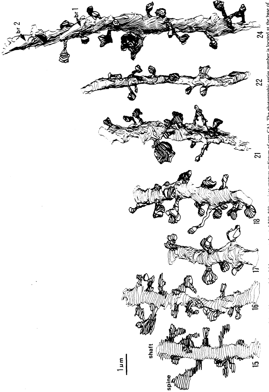
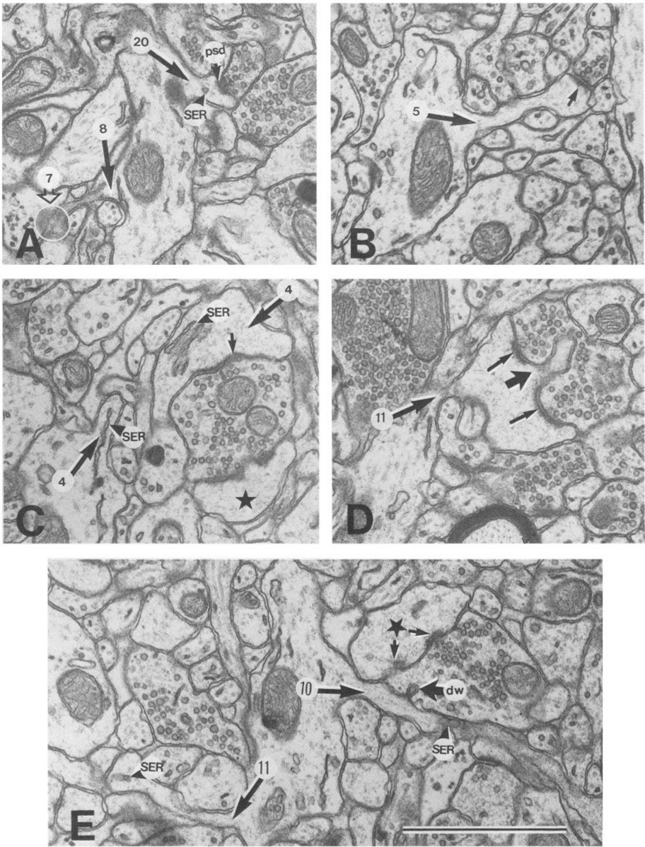
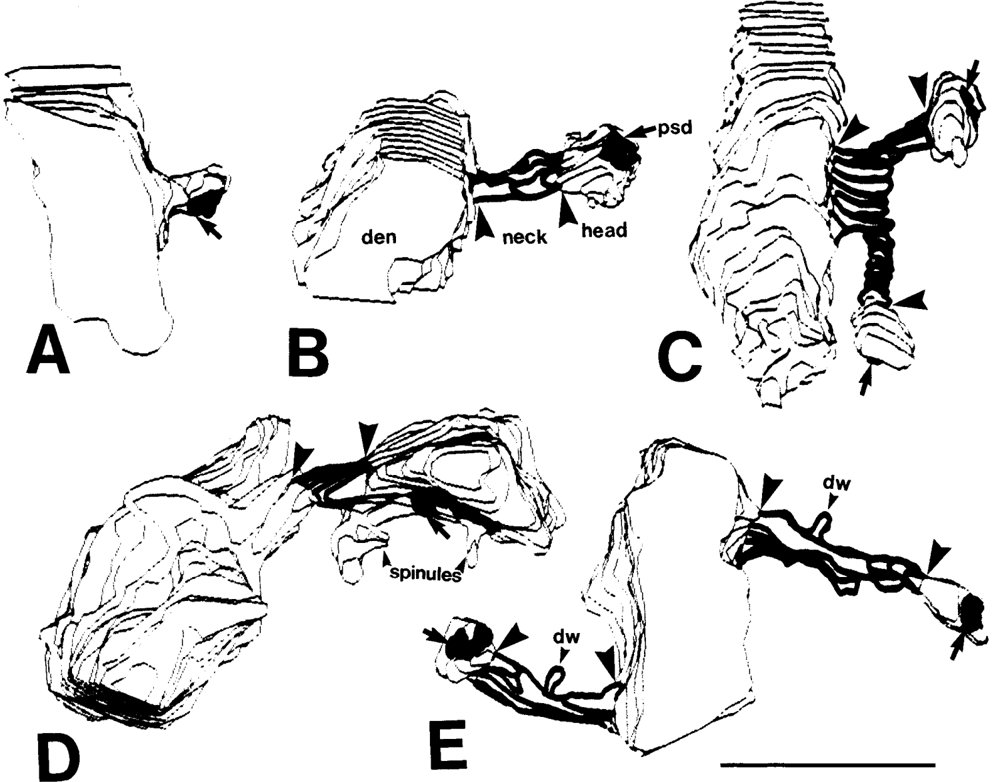
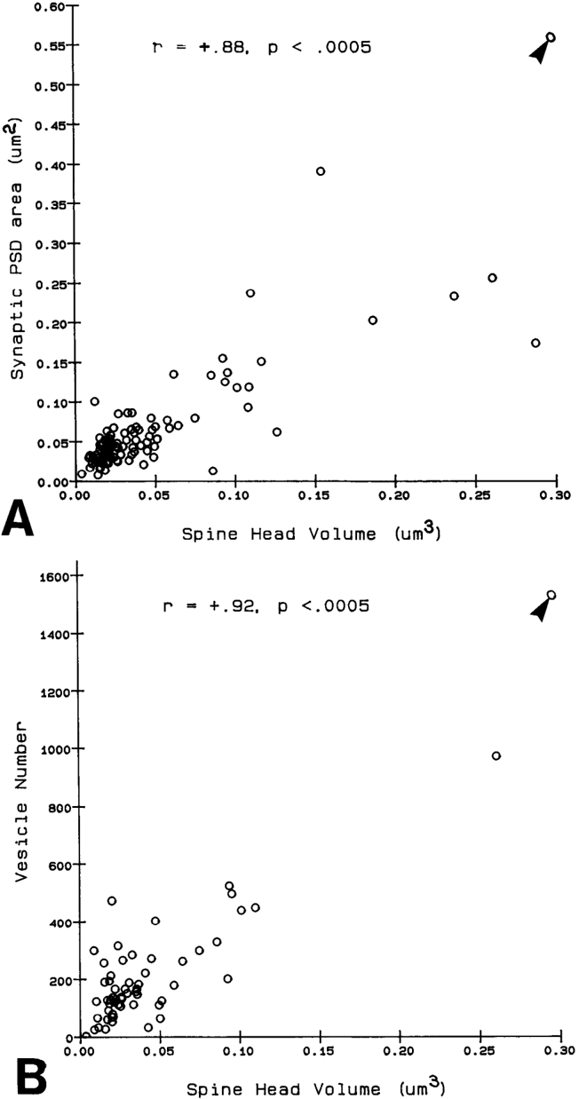
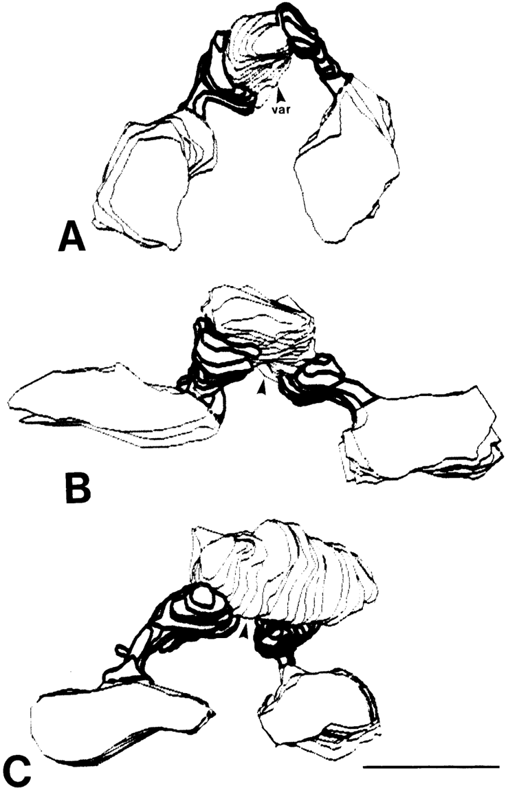
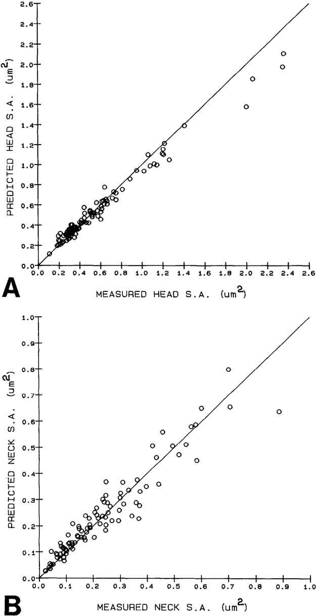
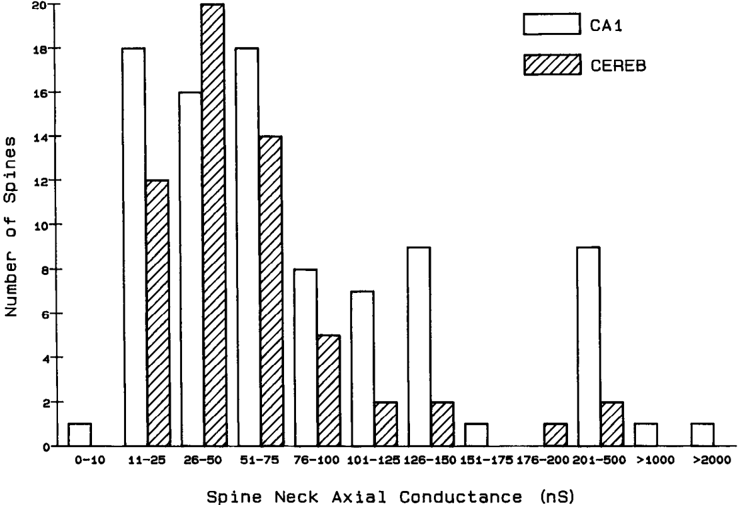
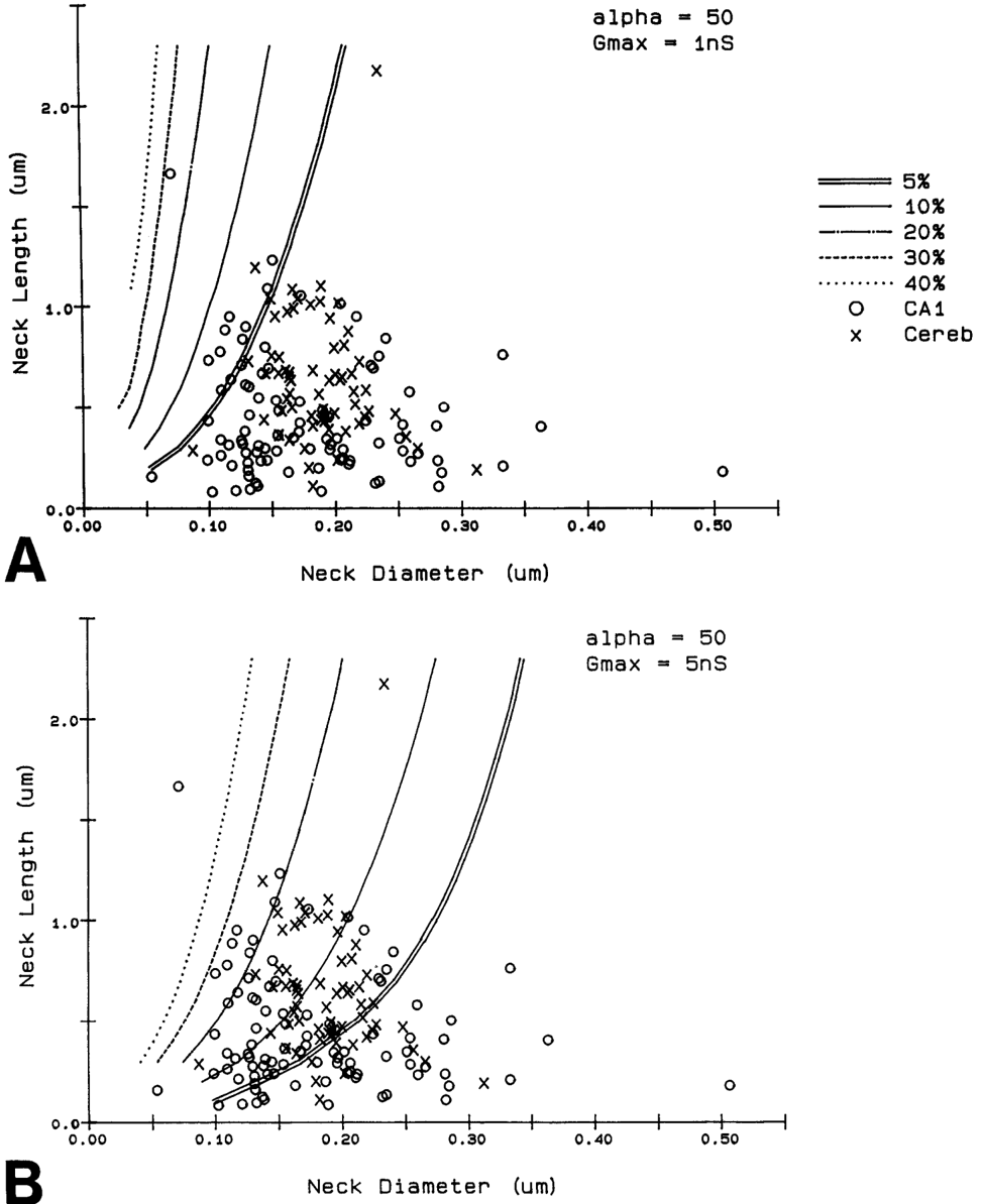
Serial electron microscopy and 3-D reconstructions of dendritic spines from hippocampal area CA 1 dendrites were obtained to evaluate 2 questions about relationships between spine geometry and synaptic efficacy. First, under what biophysical conditions are the spine necks likely to reduce the magnitude of charge transferred from the synapses on the spine heads to the recipient dendrite? Simulation software provided by Charles Wilson (1984) was used to determine that if synaptic conductance is 1 nS or less, only 1% of the hippocampal spine necks are sufficiently thin and long to reduce charge transfer by more than 10%. If synaptic conductance approaches 5 nS, however, 33% of the hippocampal spine necks are sufficiently thin and long to reduce charge transfer by more than 10%. Second, is spine geometry associated with other anatomical indicators of synaptic efficacy, including the area of the postsynaptic density and the number of vesicles in the presynaptic axon? Reconstructed spines were graphically edited into head and neck compartments, and their dimensions were measured, the areas of the postsynaptic densities (PSD) were measured, and all of the vesicles in the presynaptic axonal varicosities were counted. The dimensions of the spine head were well correlated with the area of PSD and the number of vesicles in the presynaptic axonal varicosity. Spine neck diameter and length were not correlated with PSD area, head volume, or the number of vesicles. These results suggest that the dimensions of the spine head, but not the spine neck, reflect differences in synaptic efficacy. We suggest that the constricted necks of hippocampal dendritic spines might reduce diffusion of activated molecules to neighboring synapses, thereby attributing specificity to activated or potentiated synapses.
Overview
The claims and statistics of this paper haven’t been checked yet.
Run the check — free, no account needed. It reads the open-access text and checks the sources it cites.
Integrity signals
Registry
Cites
Statistics
Figures
Verification
Claims the paper makes
The claims and statistics of this paper haven’t been checked yet.
Run the check — free, no account needed. It reads the open-access text and checks the sources it cites.
Claims other papers make about this one
Each is checked against this paper’s own text: does it say what the citing paper says it does?
The source backs part of the claim, not all of it — or backs it for a different population, condition or comparison.
In the citing paper
“This is probably one of the reasons why many ultrastructural studies that utilized high concentrations of potassium ferrocyanide/ferricyanide (1.5–3.0%) provided no data on SS (e.g., Harris and Stevens, 1989; Hayworth et al., 2014; Yakoubi et al., 2019a; Yin et al., 2020; Gour et al., 2021; Phelps et al., 2021; Peddie et al., 2022; Turner et al., 2022).”
From Unambiguous identification of asymmetric and symmetric synapses using volume electron microscopy (2024) · cites this paper as Harris and Stevens, 1989
Experimental — accuracy not yet measuredThe source backs part of the claim, not all of it — or backs it for a different population, condition or comparison.
In the citing paper
“Given that each spine receives only one synaptic input13, our data indicate the highest possible degree of freedom is available in synaptic connections in the dendrites, which has been assumed in the learning models of most neural network theories17.”
From Dendritic spine geometry is critical for AMPA receptor expression in hippocampal CA1 pyramidal neurons (2001) · cites this paper as [13]
Experimental — accuracy not yet measuredThe source backs part of the claim, not all of it — or backs it for a different population, condition or comparison.
In the citing paper
“This observation suggests that the expression of AMPA receptors is controlled independently at a level of the individual spine, consistent with the classical notion that a single spine may behave as an independent chemical compartment to regulate synaptic transmission13, 14.”
From Dendritic spine geometry is critical for AMPA receptor expression in hippocampal CA1 pyramidal neurons (2001) · cites this paper as [13]
Experimental — accuracy not yet measuredThe source backs part of the claim, not all of it — or backs it for a different population, condition or comparison.
In the citing paper
“Regions of glutamate sensitivity tended to be asymmetrically localized to one portion of a spine (Figs. 3h and 4c–j), consistent with the predominant localization of AMPA receptors within PSDs with a diameter of 0.1 to 0.5μm13, 19, 20.”
From Dendritic spine geometry is critical for AMPA receptor expression in hippocampal CA1 pyramidal neurons (2001) · cites this paper as [13]
Experimental — accuracy not yet measuredThe source backs part of the claim, not all of it — or backs it for a different population, condition or comparison.
In the citing paper
“Syn1a-CFP puncta area calculated as a measure of bouton area, which correlated with pHuse area, as expected42.”
From A trans-synaptic nanocolumn aligns neurotransmitter release to receptors (2016) · cites this paper as [42]
Experimental — accuracy not yet measured
The source backs part of the claim, not all of it — or backs it for a different population, condition or comparison.
In the citing paper
“In turn, the size of the postsynaptic density is correlated with the number of presynaptic vesicles, the number of postsynaptic receptors and the ready-releasable pool of transmitter (Harris and Stevens, 1989; Nusser et al., 1998; Schikorski and Stevens, 1999, 2001; Arellano et al., 2007a).”
From The influence of phospho-tau on dendritic spines of cortical pyramidal neurons in patients with Alzheimer’s disease (2013) · cites this paper as Harris and Stevens, 1989
Experimental — accuracy not yet measuredThe source backs part of the claim, not all of it — or backs it for a different population, condition or comparison.
In the citing paper
“Additionally, the volume of the dendritic spine head is related to the size of the postsynaptic density (Harris and Stevens, 1989; Schikorski and Stevens, 1997; Arellano et al., 2007b), the number of presynaptic vesicles, and the number of vesicles adjacent to the presynaptic cleft (Harris and Stevens, 1989).”
From The influence of phospho-tau on dendritic spines of cortical pyramidal neurons in patients with Alzheimer’s disease (2013) · cites this paper as Harris and Stevens, 1989
Experimental — accuracy not yet measuredThe source backs part of the claim, not all of it — or backs it for a different population, condition or comparison.
In the citing paper
“Furthermore, the volume of the head is directly proportional to the strength of synaptic currents (Harris and Stevens, 1989; Schikorski and Stevens, 1997; Arellano et al., 2007a).”
From The influence of phospho-tau on dendritic spines of cortical pyramidal neurons in patients with Alzheimer’s disease (2013) · cites this paper as Harris and Stevens, 1989
Experimental — accuracy not yet measuredThe cited source says what the claim attributes to it.
In the citing paper
“The relationship between spine geometry and expression of functional AMPA receptors demonstrated here provides substantial support to the hypothesis that neuronal fine structures have functional correlates in the brain13, 18, 21.”
From Dendritic spine geometry is critical for AMPA receptor expression in hippocampal CA1 pyramidal neurons (2001) · cites this paper as [13]
The cited source says what the claim attributes to it.
In the citing paper
“These data are consistent with the correlation between AMPA receptor immunoreactivity and the size of PSD19, 20, because the size of PSD is nearly proportional to the spine-head volume13.”
From Dendritic spine geometry is critical for AMPA receptor expression in hippocampal CA1 pyramidal neurons (2001) · cites this paper as [13]
The cited source says what the claim attributes to it.
In the citing paper
“whereas the spine head volume is correlated with the area of the postsynaptic density.”
From The influence of phospho-tau on dendritic spines of cortical pyramidal neurons in patients with Alzheimer’s disease (2013) · cites this paper as Harris and Stevens, 1989
Self-check
Percentages, sums and reported differences re-computed from the paper’s own numbers.
The self-check needs the paper’s full text.
Citations
Cites
The Synaptic Organization of the Brain
(2004) · Oxford University Press eBooks · doi:10.1093/acprof:oso/9780195159561.001.1 · Cited by 3,574
The Synaptic Organization of the Brain
Freemon, F. R. (1980) · JAMA · doi:10.1001/jama.1980.03300440052037 · Cited by 709
Chang, F., & Greenough, W. T. (1984) · Brain Research · doi:10.1016/0006-8993(84)91008-4 · Cited by 523
Lee, K. S., Schottler, F., Oliver, M. E., & LYNCH, G. S. (1980) · Journal of Neurophysiology · doi:10.1152/jn.1980.44.2.247 · Cited by 473
Harris, K., & Stevens, J. (1988) · The Journal of Neuroscience · doi:10.1523/jneurosci.08-12-04455.1988 · Cited by 318
Self-citationDesmond, N. L., & Levy, W. B. (1983) · Brain Research · doi:10.1016/0006-8993(83)91329-x · Cited by 276
Three-Dimensional analysis of dendritic spines
Špaček, J., & Hartmann, M. (1983) · Anatomy and Embryology · doi:10.1007/bf00298517 · Cited by 269
The anatomy of a memory: convergence of results across a diversity of tests
Greenough, W. T., & Bailey, C. H. (1988) · Trends in Neurosciences · doi:10.1016/0166-2236(88)90139-7 · Cited by 266
Crick, F. (1982) · Trends in Neurosciences · doi:10.1016/0166-2236(82)90020-0 · Cited by 254
Van Harreveld, A., & Fifkova, E. (1975) · Experimental Neurology · doi:10.1016/0014-4886(75)90055-2 · Cited by 239
Research works printed but not linked: 6
- Harris, K. M., F. E. Jensen, and B. Tsao (1988) Ultrastructure, de- velopment, and plasticity of synapses in area CA1 of the rat hippo- campus: Extending our vision with serial electron microscopy and three dimensional analyses. Neurol. Neurobiol. (in press).
- Andersen, P., T. Blackstad, G. Hulleberg, J. Line Vaaland, and M. Andersen, P., T. Blackstad, G. Hulleberg, M. Trommald, and J. L. Vaaland (1988b) Dimensions ofdendritic spines ofrat dentate gran- Trommald (1988a) Changes in spine morphology associated with ule cells during long-term potentiation (LTP). Proc. Physiol. Sot. P.C. 50. long-term potentiation in rat dentate granule cells. Proc. Physiol. Sot. s.
- Diamond, J., E. G. Gray, and G. M. Yasargil (1970) The function of dendritic spines: An hypothesis. In Excitatory Synaptic Mechanisms, P. Andersen and J. Jensen, eds., pp. 2 13-222, Universitets Forlaget, Oslo.
- Meek, G. A. (1976) Practical Electron Microscopyfor Biologists, Wiley, New York.
- Rall, W. (1970) Cable properties of dendrites and effects of synaptic location. In Excitatory Synaptic Mechanisms, P. Andersen and J. K. S. Jensen, eds., pp. 175-l 87, Universitets Forlaget, Oslo.
- Rall, W. (1978) Dendritic spines and synaptic potency. In Studies in NeurophysioloRy, A. K. McIntyre and K. Porter, eds., pp. 203-209, Cambridge U.-P., Cambridge.
Cited by
Sorted by citations
Items 201–300 of 999
Shimada, A., Mason, C. A., & Morrison, M. E. (1998) · Journal of Neuroscience · doi:10.1523/jneurosci.18-21-08559.1998 · Cited by 165
Ganeshina, O., Berry, R. W., Petralia, R. S., Nicholson, D. A., & Geinisman, Y. (2003) · The Journal of Comparative Neurology · doi:10.1002/cne.10950 · Cited by 165
Specialization of the Neocortical Pyramidal Cell during Primate Evolution
Elston, G. N. (2007) · Elsevier eBooks · doi:10.1016/b0-12-370878-8/00164-6 · Cited by 168
Postsynaptic signaling during plasticity of dendritic spines
Murakoshi, H., & Yasuda, R. (2012) · Trends in Neurosciences · doi:10.1016/j.tins.2011.12.002 · Cited by 158
Dependence of GABAergic Synaptic Areas on the Interneuron Type and Target Size
Kubota, Y., & Kawaguchi, Y. (2000) · Journal of Neuroscience · doi:10.1523/jneurosci.20-01-00375.2000 · Cited by 170
Extracellular sheets and tunnels modulate glutamate diffusion in hippocampal neuropil
Kinney, J. P., Špaček, J., Bartol, T. M., Bajaj, C., Harris, K. M., & B, T. J. S. A. (2012) · The Journal of Comparative Neurology · doi:10.1002/cne.23181 · Cited by 160
Self-citationBecker, N., Wierenga, C. J., Fonseca, R., Bonhoeffer, T., & Nägerl, U. V. (2008) · Neuron · doi:10.1016/j.neuron.2008.09.018 · Cited by 169
Spine neck plasticity controls postsynaptic calcium signals through electrical compartmentalization
Grunditz, Å., Holbro, N., Tian, L., Zuo, Y., & Oertner, T. G. (2008) · The Journal of Neuroscience · doi:10.1523/jneurosci.2702-08.2008 · Cited by 157
Quanbeck, C., Sherwood, N. M., Millar, R. P., & Terasawa, E. (1997) · The Journal of Comparative Neurology · doi:10.1002/(sici)1096-9861(19970414)380:3 · Cited by 120
An ultrastructural size principle
Pierce, J. P., & Lewin, G. R. (1994) · Neuroscience · doi:10.1016/0306-4522(94)90071-x · Cited by 157
Giessel, A. J., & Sabatini, B. L. (2010) · Neuron · doi:10.1016/j.neuron.2010.09.004 · Cited by 156
Ostroff, L. E., Cain, C. K., Bedont, J. L., Monfils, M., & LeDoux, J. E. (2010) · Proceedings of the National Academy of Sciences · doi:10.1073/pnas.0913384107 · Cited by 161
The next generation of approaches to investigate the link between synaptic plasticity and learning
Humeau, Y., & Choquet, D. (2019) · Nature Neuroscience · doi:10.1038/s41593-019-0480-6 · Cited by 158
Crystal structure and association behaviour of the GluR2 amino‐terminal domain
Jin, R., Singh, S. K., Gu, S., Furukawa, H., Sobolevsky, A. I., Zhou, J., Jin, Y., & Gouaux, E. (2009) · The EMBO Journal · doi:10.1038/emboj.2009.140 · Cited by 153
Dumitriu, D., Rodriguez, A., & Morrison, J. H. (2011) · Nature Protocols · doi:10.1038/nprot.2011.389 · Cited by 148
Membrane Potential Changes in Dendritic Spines during Action Potentials and Synaptic Input
Palmer, L. M., & Stuart, G. J. (2009) · Journal of Neuroscience · doi:10.1523/jneurosci.5847-08.2009 · Cited by 149
Amrock, L. G., Starner, M. L., Murphy, K. L., & Baxter, M. G. (2014) · Anesthesiology · doi:10.1097/aln.0000000000000477 · Cited by 152
Mechanisms underlying the inability to induce area CA1 LTP in the mouse after traumatic brain injury
Schwarzbach, E., Bonislawski, D. P., Xiong, G., & Cohen, A. S. (2006) · Hippocampus · doi:10.1002/hipo.20183 · Cited by 156
Bridgman, P. C. (1999) · The Journal of Cell Biology · doi:10.1083/jcb.146.5.1045 · Cited by 151
Multivesicular Release at Schaffer Collateral–CA1 Hippocampal Synapses
Christie, J. M., & Jahr, C. E. (2006) · Journal of Neuroscience · doi:10.1523/jneurosci.4307-05.2006 · Cited by 144
Long-term potentiation and contextual fear conditioning increase neuronal glutamate uptake
Levenson, J. M., Weeber, E. J., Selcher, J. C., Kategaya, L. S., Sweatt, J. D., & Eskin, A. (2002) · Nature Neuroscience · doi:10.1038/nn791 · Cited by 153
PSD-95 protects synapses from β-amyloid
Dore, K., Carrico, Z., Alfonso, S., Marino, M., Koymans, K., Kessels, H. W., & Malinow, R. (2021) · Cell Reports · doi:10.1016/j.celrep.2021.109194 · Cited by 149
Dynamic stochastic synapses as computational units.
Maass, W., & Zador, A. M. (1999) · Neural Computation · doi:10.1162/089976699300016494 · Cited by 149
Roo, M. D., Klauser, P., Méndez, P., Poglia, L., & Müller, D. (2007) · Cerebral Cortex · doi:10.1093/cercor/bhm041 · Cited by 156
Synaptic potentiation induces increased glial coverage of excitatory synapses in CA1 hippocampus
Lushnikova, I., Skibo, G. G., Müller, D., & Nikonenko, I. (2009) · Hippocampus · doi:10.1002/hipo.20551 · Cited by 146
BDNF signaling during the lifetime of dendritic spines
Zagrebelsky, M., Tacke, C., & Korte, M. (2020) · Cell and Tissue Research · doi:10.1007/s00441-020-03226-5 · Cited by 147
Comparison of Hippocampal Dendritic Spines in Culture and in Brain
Boyer, C., Schikorski, T., & Stevens, C. F. (1998) · Journal of Neuroscience · doi:10.1523/jneurosci.18-14-05294.1998 · Cited by 144
Density and morphology of dendritic spines in mouse neocortex.
Ballesteros-Yáñez, I., Benavides-Piccione, R., Elston, G. N., Yuste, R., & DeFelipe, J. (2006) · Neuroscience · doi:10.1016/j.neuroscience.2005.11.038 · Cited by 143
Reading the Book of Memory: Sparse Sampling versus Dense Mapping of Connectomes
Seung, H. S. (2009) · Neuron · doi:10.1016/j.neuron.2009.03.020 · Cited by 138
A Morphological Correlate of Synaptic Scaling in Visual Cortex
Wallace, W., & Bear, M. F. (2004) · Journal of Neuroscience · doi:10.1523/jneurosci.1110-04.2004 · Cited by 141
Long-term depression triggers the selective elimination of weakly integrated synapses
Wiegert, J. S., & Oertner, T. G. (2013) · Proceedings of the National Academy of Sciences · doi:10.1073/pnas.1315926110 · Cited by 139
Dendritic Spines as Tunable Regulators of Synaptic Signals
Tønnesen, J., & Nägerl, U. V. (2016) · Frontiers in Psychiatry · doi:10.3389/fpsyt.2016.00101 · Cited by 147
mRNA at the synapse: analysis of a synaptosomal preparation enriched in hippocampal dendritic spines
Chicurel, M., Terrian, D., & Potter, H. (1993) · Journal of Neuroscience · doi:10.1523/jneurosci.13-09-04054.1993 · Cited by 139
Emerging roles of mitochondria in synaptic transmission and neurodegeneration
Lee, A., Hirabayashi, Y., Kwon, S., Lewis, T. L., & Polleux, F. (2018) · Current Opinion in Physiology · doi:10.1016/j.cophys.2018.03.009 · Cited by 131
Measures for quantifying dendritic arborizations
Uylings, H. B. M., & van Pelt, J. (2002) · Network Computation in Neural Systems · doi:10.1088/0954-898x_13_3_309 · Cited by 138
Therapeutic implications of the mGluR theory of fragile X mental retardation
Bear, M. F. (2005) · Genes, Brain and Behavior · doi:10.1111/j.1601-183x.2005.00135.x · Cited by 137
Dendritic Pathology in Prion Disease Starts at the Synaptic Spine
Fuhrmann, M., Mitteregger, G., Kretzschmar, H. A., & Herms, J. W. (2007) · Journal of Neuroscience · doi:10.1523/jneurosci.5062-06.2007 · Cited by 146
Systematic regulation of spine sizes and densities in pyramidal neurons
Konur, S., Rabinowitz, D., Fenstermaker, V., & Yuste, R. (2003) · Journal of Neurobiology · doi:10.1002/neu.10229 · Cited by 137
Chapter 11 Spine dynamics and synapse remodeling during LTP and memory processes
Roo, M. D., Klauser, P., Garcia, P. M., Poglia, L., & Müller, D. (2008) · Progress in brain research · doi:10.1016/s0079-6123(07)00011-8 · Cited by 144
Merino-Serrais, P., Benavides-Piccione, R., Blazquez-Llorca, L., Kastanauskaite, A., Rábano, A., Avila, J., & DeFelipe, J. (2013) · Brain · doi:10.1093/brain/awt088 · Cited by 134
Geinisman, Y. (1993) · Hippocampus · doi:10.1002/hipo.450030404 · Cited by 134
Fossati, M., Pizzarelli, R., Schmidt, E. R., Kupferman, J. V., Stroebel, D., Polleux, F., & Charrier, C. (2016) · Neuron · doi:10.1016/j.neuron.2016.06.013 · Cited by 133
Structural plasticity with preserved topology in the postsynaptic protein network
Blanpied, T. A., Kerr, J. M., & Ehlers, M. (2008) · Proceedings of the National Academy of Sciences · doi:10.1073/pnas.0711669105 · Cited by 139
Excitatory amino acid involvement in dendritic spine formation, maintenance and remodelling
McKinney, R. A. (2009) · The Journal of Physiology · doi:10.1113/jphysiol.2009.178905 · Cited by 147
Serotonin/dopamine interaction in memory formation
González‐Burgos, I., & Feria‐Velasco, A. (2008) · Progress in brain research · doi:10.1016/s0079-6123(08)00928-x · Cited by 138
Graupner, M. (2010) · Frontiers in Computational Neuroscience · doi:10.3389/fncom.2010.00136 · Cited by 132
Axonal synapse sorting in medial entorhinal cortex
Schmidt, H., Gour, A., Straehle, J., Boergens, K. M., Brecht, M., & Helmstaedter, M. (2017) · Nature · doi:10.1038/nature24005 · Cited by 152
Proteostasis in complex dendrites
Hanus, C., & Schuman, E. M. (2013) · Nature reviews. Neuroscience · doi:10.1038/nrn3546 · Cited by 133
Increased axonal bouton dynamics in the aging mouse cortex
Grillo, F. W., Song, S., Ruivo, L. M. T.-G., Huang, L., Gao, G., Knott, G. W., Maco, B., Ferretti, V., Thompson, D., Little, G. E., et al. (2013) · Proceedings of the National Academy of Sciences · doi:10.1073/pnas.1218731110 · Cited by 130
Matikainen-Ankney, B. A., Kezunovic, N., Mesias, R. E., Tian, Y., Williams, F. M., Huntley, G. W., & Benson, D. L. (2016) · Journal of Neuroscience · doi:10.1523/jneurosci.3314-15.2016 · Cited by 139
Binary and analog variation of synapses between cortical pyramidal neurons
Dorkenwald, S., Turner, N. L., Macrina, T., Lee, K., Lu, R., Wu, J., Bodor, Á. L., Bleckert, A., Brittain, D., Kemnitz, N., et al. (2022) · eLife · doi:10.7554/elife.76120 · Cited by 127
SynGAP Regulates Steady-State and Activity-Dependent Phosphorylation of Cofilin
Carlisle, H. J., Manzerra, P., Marcora, E., & Kennedy, M. B. (2008) · The Journal of Neuroscience · doi:10.1523/jneurosci.4695-08.2008 · Cited by 128
Dendritic morphology of CA1 pyramidal neurones from the rat hippocampus: II. Spine distributions
Bannister, N. J., & Larkman, A. U. (1995) · The Journal of Comparative Neurology · doi:10.1002/cne.903600112 · Cited by 130
Cytoskeletal dynamics in dendritic spines: direct modulation by glutamate receptors?
van Rossum, D., & Hanisch, U. (1999) · Trends in Neurosciences · doi:10.1016/s0166-2236(99)01404-6 · Cited by 128
Lenz, M., Platschek, S., Priesemann, V., Becker, D., Willems, L., Ziemann, U., Deller, T., Müller-Dahlhaus, F., Jedlicka, P., & Vlachos, A. (2014) · Brain Structure and Function · doi:10.1007/s00429-014-0859-9 · Cited by 122
Propagation of dendritic spikes mediated by excitable spines: a continuum theory
Baer, S. M., & Rinzel, J. (1991) · Journal of Neurophysiology · doi:10.1152/jn.1991.65.4.874 · Cited by 124
Mechanism-based approaches to treating fragile X
Dölen, G., Carpenter, R. L., Ocain, T. D., & Bear, M. F. (2010) · Pharmacology & Therapeutics · doi:10.1016/j.pharmthera.2010.02.008 · Cited by 129
Cortical afferents of the perirhinal, postrhinal, and entorhinal cortices of the rat
Burwell, R. D., & Amaral, D. G. (1998) · The Journal of Comparative Neurology · doi:10.1002/(sici)1096-9861(19980824)398:2 · Cited by 734
Self-citationPeled, E. S., & Isacoff, E. Y. (2011) · Nature Neuroscience · doi:10.1038/nn.2767 · Cited by 123
Maiti, P., Manna, J., Ilavazhagan, G., Rossignol, J., & Dunbar, G. L. (2015) · Neuroscience & Biobehavioral Reviews · doi:10.1016/j.neubiorev.2015.09.020 · Cited by 128
Learning rules and persistence of dendritic spines
Kasai, H., Hayama, T., Ishikawa, M., Watanabe, S., Yagishita, S., & Noguchi, J. (2010) · European Journal of Neuroscience · doi:10.1111/j.1460-9568.2010.07344.x · Cited by 123
Radley, J. J., Anderson, R. M., Hamilton, B. A., Alcock, J. A., & Romig‐Martin, S. A. (2013) · Journal of Neuroscience · doi:10.1523/jneurosci.0287-13.2013 · Cited by 127
Borczyk, M., Śliwińska, M. A., Cały, A., Bernaś, T., & Radwańska, K. (2019) · Scientific Reports · doi:10.1038/s41598-018-38412-7 · Cited by 140
Spatial organization of cofilin in dendritic spines
Rácz, B., & Weinberg, R. J. (2006) · Neuroscience · doi:10.1016/j.neuroscience.2005.11.025 · Cited by 125
The role of postsynaptic calcium in the induction of long-term potentiation
Malenka, R. C. (1991) · Molecular Neurobiology · doi:10.1007/bf02935552 · Cited by 121
In Sickness and in Health: Perineuronal Nets and Synaptic Plasticity in Psychiatric Disorders
Pantazopoulos, H., & Berretta, S. (2015) · Neural Plasticity · doi:10.1155/2016/9847696 · Cited by 128
Robust nanoscopy of a synaptic protein in living mice by organic-fluorophore labeling
Masch, J.-M., Steffens, H., Fischer, J., Engelhardt, J., Hubrich, J., Keller‐Findeisen, J., D’Este, E., Urban, N. T., Grant, S. G. N., Sahl, S. J., et al. (2018) · Proceedings of the National Academy of Sciences · doi:10.1073/pnas.1807104115 · Cited by 119
Cranial Irradiation Alters Dendritic Spine Density and Morphology in the Hippocampus
Chakraborti, A., Allen, A. R., Allen, B. D., Rosi, S., & Fike, J. R. (2012) · PLoS ONE · doi:10.1371/journal.pone.0040844 · Cited by 126
Biphasic synaptic Ca influx arising from compartmentalized electrical signals in dendritic spines
Bloodgood, B. L., Giessel, A. J., & Sabatini, B. L. (2009) · PLoS Biology · doi:10.1371/journal.pbio.1000190 · Cited by 119
Homeostatic Plasticity of Subcellular Neuronal Structures: From Inputs to Outputs
Wefelmeyer, W., Puhl, C. J., & Burrone, J. (2016) · Trends in Neurosciences · doi:10.1016/j.tins.2016.08.004 · Cited by 122
Phosphorylation of dynamin I and synaptic-vesicle recycling
Robinson, P. J., Liu, J., Powell, K. A., Fykse, E. M., & Südhof, T. C. (1994) · Trends in Neurosciences · doi:10.1016/0166-2236(94)90179-1 · Cited by 118
Calcium dynamics of spines depend on their dendritic location
Holthoff, K., Tsay, D., & Yuste, R. (2002) · Neuron · doi:10.1016/s0896-6273(02)00576-7 · Cited by 117
Three‐dimensional relationships between perisynaptic astroglia and human hippocampal synapses
Witcher, M. R., Park, Y. D., Lee, M. R., Sharma, S., Harris, K. M., & Kirov, S. A. (2009) · Glia · doi:10.1002/glia.20946 · Cited by 121
Self-citationThe multifaceted role of mitochondria in autism spectrum disorder
Khaliulin, I., Hamoudi, W., & Amal, H. (2024) · Molecular Psychiatry · doi:10.1038/s41380-024-02725-z · Cited by 128
Spatiotemporal appearance of developing LHRH neurons in the rat brain
Daikoku, S., & KOIDE, I. (1998) · The Journal of Comparative Neurology · doi:10.1002/(sici)1096-9861(19980330)393:1 · Cited by 36
Self-citationFiumelli, H., Briner, A., Puskarjov, M., Blaesse, P., Belem, B. J., Dayer, A. G., Kaila, K., Martin, J., & Vutskits, L. (2012) · Cerebral Cortex · doi:10.1093/cercor/bhs027 · Cited by 126
Brito, V., Giralt, A., Enriquez-Barreto, L., Puigdellívol, M., Suelves, N., Zamora-Moratalla, A., Ballesteros, J. J., Martín, E. D., Dominguez-Iturza, N., Morales, M., et al. (2014) · Journal of Clinical Investigation · doi:10.1172/jci74809 · Cited by 114
Triggers and substrates of hippocampal synaptic plasticity
Massicotte, G., & Baudry, M. (1991) · Neuroscience & Biobehavioral Reviews · doi:10.1016/s0149-7634(05)80034-x · Cited by 114
Cauller, L., & Connors, B. W. (1992) · Elsevier eBooks · doi:10.1016/b978-0-12-484815-3.50014-1 · Cited by 113
Scheuß, V., Yasuda, R., Sobczyk, A., & Svoboda, K. (2006) · Journal of Neuroscience · doi:10.1523/jneurosci.1962-06.2006 · Cited by 111
Trommald, M., Hulleberg, G., & Andersen, P. N. (1996) · Learning & Memory · doi:10.1101/lm.3.2-3.218 · Cited by 113
Presynaptic mechanisms of motor fluctuations in Parkinson’s disease: a probabilistic model
de la Fuente‐Fernández, R., Schulzer, M., Mak, E., Calne, D. B., & Stoessl, A. J. (2004) · Brain · doi:10.1093/brain/awh102 · Cited by 112
Warren, M. S., Bradley, W. D., Gourley, S. L., Lin, Y., Simpson, M. A., Reichardt, L. F., Greer, C. A., Taylor, J. R., & Koleske, A. J. (2012) · Journal of Neuroscience · doi:10.1523/jneurosci.3942-11.2012 · Cited by 113
The Synaptic Connections between Cortical Areas V1 and V2 in Macaque Monkey
Anderson, J. C., & Martin, K. A. C. (2009) · Journal of Neuroscience · doi:10.1523/jneurosci.5757-08.2009 · Cited by 115
Advanced imaging and labelling methods to decipher brain cell organization and function
Choquet, D., Sainlos, M., & Sibarita, J. (2021) · Nature reviews. Neuroscience · doi:10.1038/s41583-021-00441-z · Cited by 117
The Role of Actin Cytoskeleton in Dendritic Spines in the Maintenance of Long-Term Memory
Basu, S., & Lamprecht, R. (2018) · Frontiers in Molecular Neuroscience · doi:10.3389/fnmol.2018.00143 · Cited by 117
The prevalence and specificity of local protein synthesis during neuronal synaptic plasticity
Sun, C., Nold, A., Fusco, C. M., Rangaraju, V., Tchumatchenko, T., Heilemann, M., & Schuman, E. M. (2021) · Science Advances · doi:10.1126/sciadv.abj0790 · Cited by 108
Shepherd, G. M., Raastad, M., & Andersen, P. N. (2002) · Proceedings of the National Academy of Sciences · doi:10.1073/pnas.052151299 · Cited by 108
Solutions for transients in arbitrarily branching cables: I. Voltage recording with a somatic shunt
Major, G., Evans, J. D., & Jack, J. J. (1993) · Biophysical Journal · doi:10.1016/s0006-3495(93)81037-3 · Cited by 107
Dendritic morphology of pyramidal neurones of the visual cortex of the rat. IV: Electrical geometry
Larkman, A. U., Major, G., Stratford, K. J., & Jack, J. (1992) · The Journal of Comparative Neurology · doi:10.1002/cne.903230202 · Cited by 106
Bopp, R., Holler-Rickauer, S., Martin, K. A. C., & Schuhknecht, G. F. P. (2017) · Journal of Neuroscience · doi:10.1523/jneurosci.2557-16.2017 · Cited by 106
Trommald, M., Jensen, V., & Andersen, P. N. (1995) · The Journal of Comparative Neurology · doi:10.1002/cne.903530208 · Cited by 106
Carlbom, I., Terzopoulos, D., & Harris, K. M. (1994) · IEEE Transactions on Medical Imaging · doi:10.1109/42.293928 · Cited by 106
Self-citationDendritic Morphology, Inward Rectification, and the Functional Properties of Neostriatal Neurons
Wilson, C. J. (1992) · Elsevier eBooks · doi:10.1016/b978-0-12-484815-3.50012-8 · Cited by 105
The Connection from Cortical Area V1 to V5: A Light and Electron Microscopic Study
Anderson, J. C., Binzegger, T., Martin, K. A. C., & Rockland, K. S. (1998) · Journal of Neuroscience · doi:10.1523/jneurosci.18-24-10525.1998 · Cited by 104
Stereotyped axonal arbors of retinal ganglion cell subsets in the mouse superior colliculus
Hong, Y. K., Kim, I., & Sanes, J. R. (2011) · The Journal of Comparative Neurology · doi:10.1002/cne.22595 · Cited by 107
Saturation of postsynaptic glutamate receptors after quantal release of transmitter
Tang, C., Margulis, M., Shi, Q., & Fielding, A. (1994) · Neuron · doi:10.1016/0896-6273(94)90423-5 · Cited by 106
On the electrical function of dendritic spines
Tsay, D., & Yuste, R. (2003) · Trends in Neurosciences · doi:10.1016/j.tins.2003.11.008 · Cited by 103
Coleman, J. E., Nahmani, M., Gavornik, J. P., Haslinger, R., Heynen, A. J., Erişir, A., & Bear, M. F. (2010) · Journal of Neuroscience · doi:10.1523/jneurosci.1248-10.2010 · Cited by 102
Anatomical and Electrophysiological Comparison of CA1 Pyramidal Neurons of the Rat and Mouse
Routh, B. N., Johnston, D., Harris, K. M., & Chitwood, R. A. (2009) · Journal of Neurophysiology · doi:10.1152/jn.00082.2009 · Cited by 107
Self-citation
Metadata
- Authors
- Harris, K., & Stevens, J.
- Affiliations
- Boston Children's Hospital (US)
- Venue
- The Journal of Neuroscience
- Publisher
- Society for Neuroscience
- Published
- 1989-08-01
- Volume
- 9
- Issue
- 8
- Pages
- 2982-2997
- ISSN
- 0270-6474 (ISSN-L)
- 1529-2401
- PMC
- PMC6569708
- OpenAlex
- W2117664079
- Cited by
- 1,073
- References
- 38
- Citations by year
- 2026: 17 · 2025: 26 · 2024: 35 · 2023: 33 · 2022: 31 · 2021: 50 · 2020: 39 · 2019: 43 · 2018: 47 · 2017: 33 · 2016: 40 · 2015: 48 · 2014: 44 · 2013: 50 · 2012: 42
- FWCI
- 5.92
- Type
- journal article
- Language
- en
- Open access
- hybrid
- doi.org
- Licence
- Locations
- Journal of Neuroscience (publishedVersion, cc-by-nc-sa)
- Europe PMC (PubMed Central) (publishedVersion)
- PubMed Central (publishedVersion)
- PubMed (publishedVersion)
- Funders
- Alfred P. Sloan Foundation
- National Institutes of Health (NS21184)
- Medical Research Council
- National Institute of Neurological Disorders and Stroke (NS21184, NS 20820)
- Eunice Kennedy Shriver National Institute of Child Health and Human Development (P30HD018655, P30HD-18655)
- Topics
- Life Sciences › Neuroscience › Cellular and Molecular Neuroscience › Neuroscience and Neuropharmacology Research
- Life Sciences › Neuroscience › Cognitive Neuroscience › Memory and Neural Mechanisms
- Life Sciences › Neuroscience › Cognitive Neuroscience › Neural dynamics and brain function
- Keywords
- dendritic spines, CA1 pyramidal neurons, hippocampus, synaptic efficacy, postsynaptic density, synaptic vesicles, charge transfer, synaptic conductance, dendritic spine morphology, synaptic specificity
- MeSH terms
- Animals, Axons, Biophysics, Dendrites, Electric Conductivity, Endoplasmic Reticulum, Hippocampus, Image Processing, Computer-Assisted, Male, Microscopy, Electron, Models, Neurological, Neurons, Purkinje Cells, Rats, Inbred Strains, Synapses, Rats, Biophysical Phenomena
About these checks
Registry, retraction, Cites and statistics results are computed, not judged, with one exception: a printed reference that no index lists is read once by a model before it counts as a citation error. Claims are extracted by a model and kept only when their quote appears verbatim in the text; each cited source is then checked, with a model’s help, for whether it says what the claim relies on. Those verdicts can be wrong — read the claim’s quote and the cited source before relying on one.
Statistics and arithmetic are recomputed, not judged.
Confidence bands are shown only for fields calibrated on our annotated test set. None is calibrated yet, so no band is shown.
What counts as a claim ›How we measure confidence ›How we check this ›
Are you an author, or do you see a problem with this page? Request a review
