

The DOI matches its Crossref record; 36 of the 38 references it cites were found in a registry or an index.
Dendritic spines of CA 1 pyramidal cells in the rat hippocampus: serial electron microscopy with reference to their biophysical characteristics
Open accessAbstract
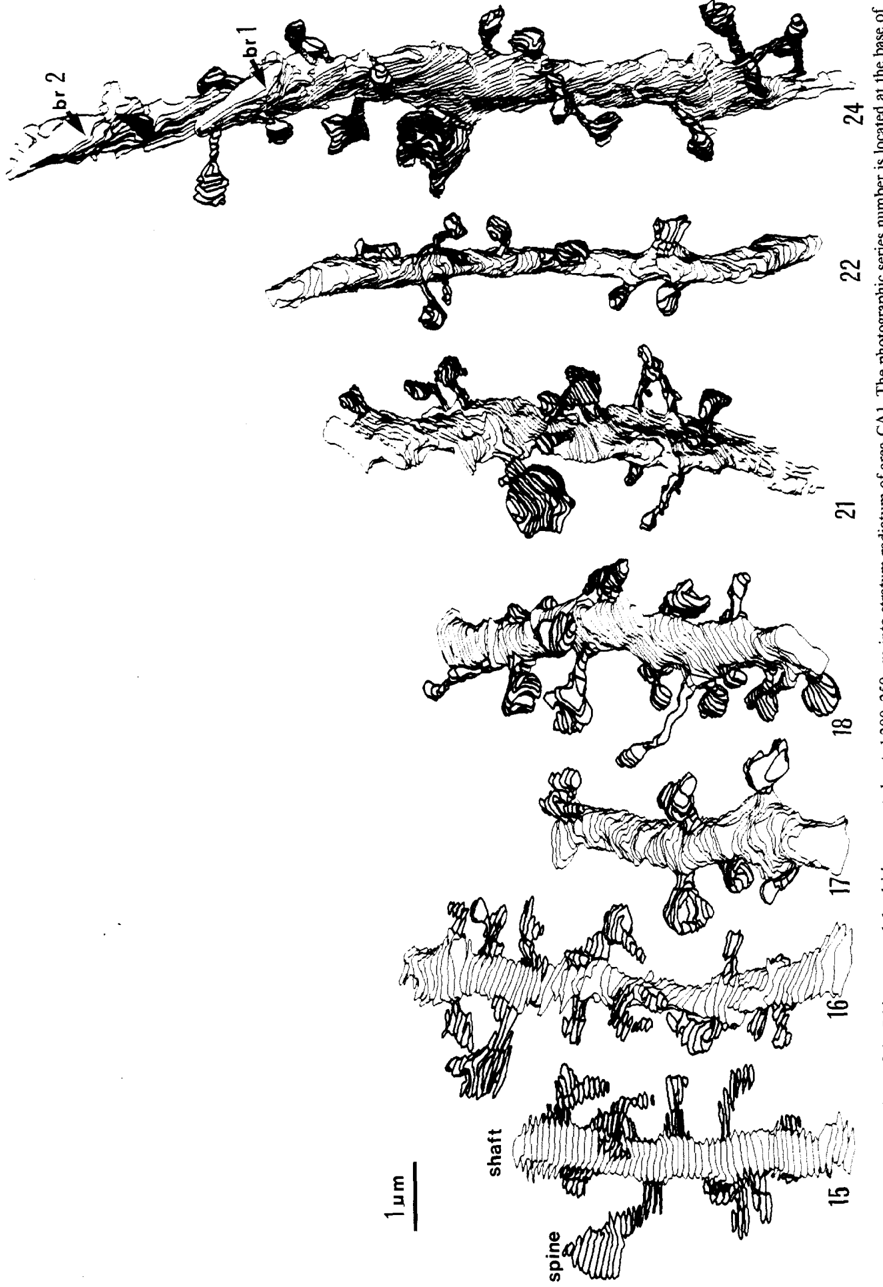
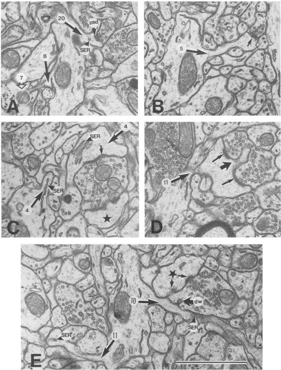
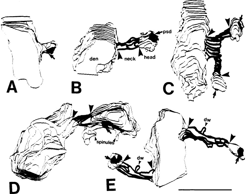
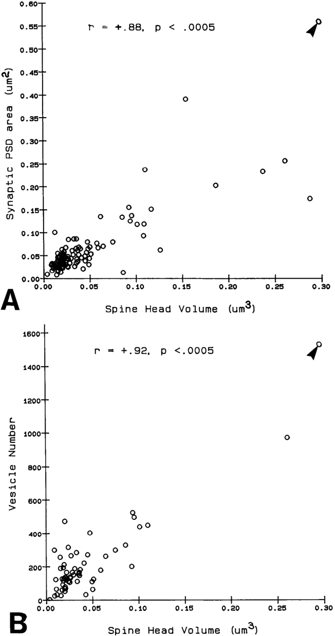
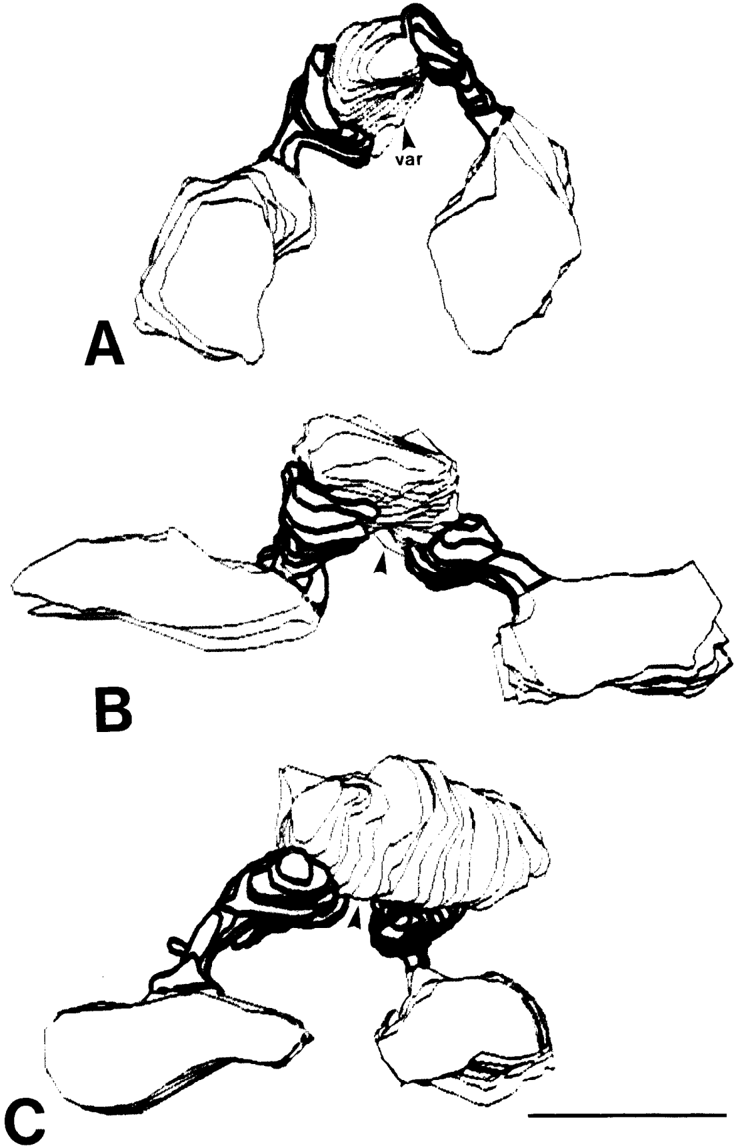
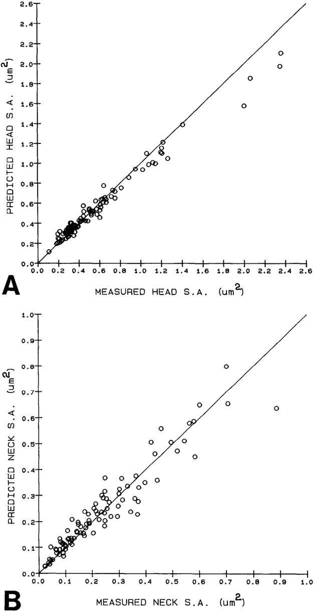
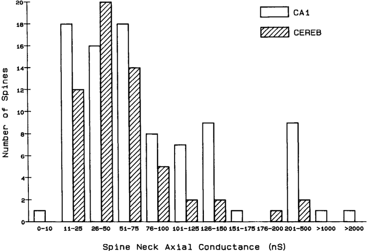
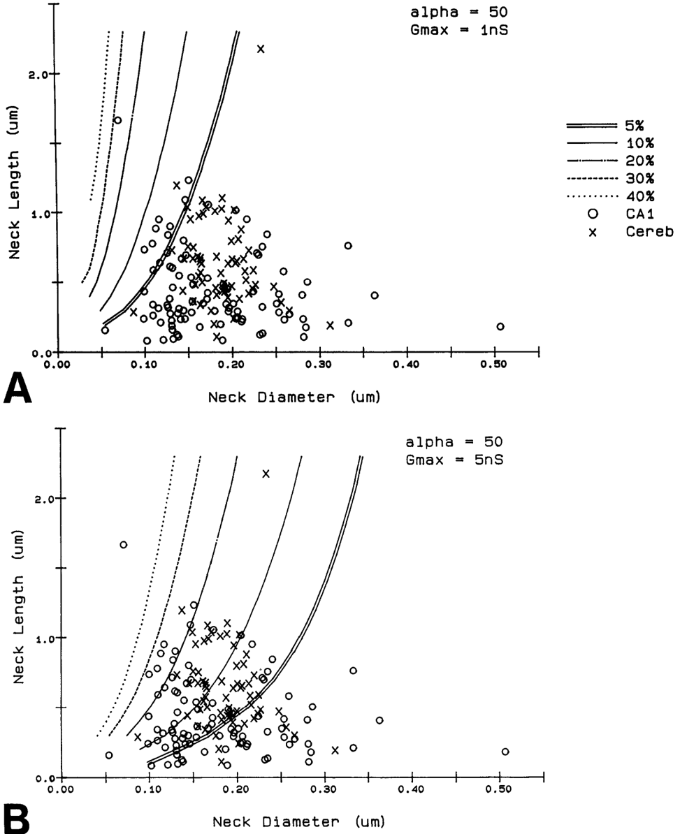
Serial electron microscopy and 3-D reconstructions of dendritic spines from hippocampal area CA 1 dendrites were obtained to evaluate 2 questions about relationships between spine geometry and synaptic efficacy. First, under what biophysical conditions are the spine necks likely to reduce the magnitude of charge transferred from the synapses on the spine heads to the recipient dendrite? Simulation software provided by Charles Wilson (1984) was used to determine that if synaptic conductance is 1 nS or less, only 1% of the hippocampal spine necks are sufficiently thin and long to reduce charge transfer by more than 10%. If synaptic conductance approaches 5 nS, however, 33% of the hippocampal spine necks are sufficiently thin and long to reduce charge transfer by more than 10%. Second, is spine geometry associated with other anatomical indicators of synaptic efficacy, including the area of the postsynaptic density and the number of vesicles in the presynaptic axon? Reconstructed spines were graphically edited into head and neck compartments, and their dimensions were measured, the areas of the postsynaptic densities (PSD) were measured, and all of the vesicles in the presynaptic axonal varicosities were counted. The dimensions of the spine head were well correlated with the area of PSD and the number of vesicles in the presynaptic axonal varicosity. Spine neck diameter and length were not correlated with PSD area, head volume, or the number of vesicles. These results suggest that the dimensions of the spine head, but not the spine neck, reflect differences in synaptic efficacy. We suggest that the constricted necks of hippocampal dendritic spines might reduce diffusion of activated molecules to neighboring synapses, thereby attributing specificity to activated or potentiated synapses.
Overview
The claims and statistics of this paper haven’t been checked yet.
Run the check — free, no account needed. It reads the open-access text and checks the sources it cites.
Integrity signals
Registry
Cites
Statistics
Figures
Verification
Claims the paper makes
The claims and statistics of this paper haven’t been checked yet.
Run the check — free, no account needed. It reads the open-access text and checks the sources it cites.
Claims other papers make about this one
Each is checked against this paper’s own text: does it say what the citing paper says it does?
The source backs part of the claim, not all of it — or backs it for a different population, condition or comparison.
In the citing paper
“This is probably one of the reasons why many ultrastructural studies that utilized high concentrations of potassium ferrocyanide/ferricyanide (1.5–3.0%) provided no data on SS (e.g., Harris and Stevens, 1989; Hayworth et al., 2014; Yakoubi et al., 2019a; Yin et al., 2020; Gour et al., 2021; Phelps et al., 2021; Peddie et al., 2022; Turner et al., 2022).”
From Unambiguous identification of asymmetric and symmetric synapses using volume electron microscopy (2024) · cites this paper as Harris and Stevens, 1989
Experimental — accuracy not yet measuredThe source backs part of the claim, not all of it — or backs it for a different population, condition or comparison.
In the citing paper
“Given that each spine receives only one synaptic input13, our data indicate the highest possible degree of freedom is available in synaptic connections in the dendrites, which has been assumed in the learning models of most neural network theories17.”
From Dendritic spine geometry is critical for AMPA receptor expression in hippocampal CA1 pyramidal neurons (2001) · cites this paper as [13]
Experimental — accuracy not yet measuredThe source backs part of the claim, not all of it — or backs it for a different population, condition or comparison.
In the citing paper
“This observation suggests that the expression of AMPA receptors is controlled independently at a level of the individual spine, consistent with the classical notion that a single spine may behave as an independent chemical compartment to regulate synaptic transmission13, 14.”
From Dendritic spine geometry is critical for AMPA receptor expression in hippocampal CA1 pyramidal neurons (2001) · cites this paper as [13]
Experimental — accuracy not yet measuredThe source backs part of the claim, not all of it — or backs it for a different population, condition or comparison.
In the citing paper
“Regions of glutamate sensitivity tended to be asymmetrically localized to one portion of a spine (Figs. 3h and 4c–j), consistent with the predominant localization of AMPA receptors within PSDs with a diameter of 0.1 to 0.5μm13, 19, 20.”
From Dendritic spine geometry is critical for AMPA receptor expression in hippocampal CA1 pyramidal neurons (2001) · cites this paper as [13]
Experimental — accuracy not yet measuredThe source backs part of the claim, not all of it — or backs it for a different population, condition or comparison.
In the citing paper
“Syn1a-CFP puncta area calculated as a measure of bouton area, which correlated with pHuse area, as expected42.”
From A trans-synaptic nanocolumn aligns neurotransmitter release to receptors (2016) · cites this paper as [42]
Experimental — accuracy not yet measured
The source backs part of the claim, not all of it — or backs it for a different population, condition or comparison.
In the citing paper
“In turn, the size of the postsynaptic density is correlated with the number of presynaptic vesicles, the number of postsynaptic receptors and the ready-releasable pool of transmitter (Harris and Stevens, 1989; Nusser et al., 1998; Schikorski and Stevens, 1999, 2001; Arellano et al., 2007a).”
From The influence of phospho-tau on dendritic spines of cortical pyramidal neurons in patients with Alzheimer’s disease (2013) · cites this paper as Harris and Stevens, 1989
Experimental — accuracy not yet measuredThe source backs part of the claim, not all of it — or backs it for a different population, condition or comparison.
In the citing paper
“Additionally, the volume of the dendritic spine head is related to the size of the postsynaptic density (Harris and Stevens, 1989; Schikorski and Stevens, 1997; Arellano et al., 2007b), the number of presynaptic vesicles, and the number of vesicles adjacent to the presynaptic cleft (Harris and Stevens, 1989).”
From The influence of phospho-tau on dendritic spines of cortical pyramidal neurons in patients with Alzheimer’s disease (2013) · cites this paper as Harris and Stevens, 1989
Experimental — accuracy not yet measuredThe source backs part of the claim, not all of it — or backs it for a different population, condition or comparison.
In the citing paper
“Furthermore, the volume of the head is directly proportional to the strength of synaptic currents (Harris and Stevens, 1989; Schikorski and Stevens, 1997; Arellano et al., 2007a).”
From The influence of phospho-tau on dendritic spines of cortical pyramidal neurons in patients with Alzheimer’s disease (2013) · cites this paper as Harris and Stevens, 1989
Experimental — accuracy not yet measuredThe cited source says what the claim attributes to it.
In the citing paper
“The relationship between spine geometry and expression of functional AMPA receptors demonstrated here provides substantial support to the hypothesis that neuronal fine structures have functional correlates in the brain13, 18, 21.”
From Dendritic spine geometry is critical for AMPA receptor expression in hippocampal CA1 pyramidal neurons (2001) · cites this paper as [13]
The cited source says what the claim attributes to it.
In the citing paper
“These data are consistent with the correlation between AMPA receptor immunoreactivity and the size of PSD19, 20, because the size of PSD is nearly proportional to the spine-head volume13.”
From Dendritic spine geometry is critical for AMPA receptor expression in hippocampal CA1 pyramidal neurons (2001) · cites this paper as [13]
The cited source says what the claim attributes to it.
In the citing paper
“whereas the spine head volume is correlated with the area of the postsynaptic density.”
From The influence of phospho-tau on dendritic spines of cortical pyramidal neurons in patients with Alzheimer’s disease (2013) · cites this paper as Harris and Stevens, 1989
Self-check
Percentages, sums and reported differences re-computed from the paper’s own numbers.
The self-check needs the paper’s full text.
Citations
Cites
The Synaptic Organization of the Brain
(2004) · Oxford University Press eBooks · doi:10.1093/acprof:oso/9780195159561.001.1 · Cited by 3,574
The Synaptic Organization of the Brain
Freemon, F. R. (1980) · JAMA · doi:10.1001/jama.1980.03300440052037 · Cited by 709
Chang, F., & Greenough, W. T. (1984) · Brain Research · doi:10.1016/0006-8993(84)91008-4 · Cited by 523
Lee, K. S., Schottler, F., Oliver, M. E., & LYNCH, G. S. (1980) · Journal of Neurophysiology · doi:10.1152/jn.1980.44.2.247 · Cited by 473
Harris, K., & Stevens, J. (1988) · The Journal of Neuroscience · doi:10.1523/jneurosci.08-12-04455.1988 · Cited by 318
Self-citationDesmond, N. L., & Levy, W. B. (1983) · Brain Research · doi:10.1016/0006-8993(83)91329-x · Cited by 276
Three-Dimensional analysis of dendritic spines
Špaček, J., & Hartmann, M. (1983) · Anatomy and Embryology · doi:10.1007/bf00298517 · Cited by 269
The anatomy of a memory: convergence of results across a diversity of tests
Greenough, W. T., & Bailey, C. H. (1988) · Trends in Neurosciences · doi:10.1016/0166-2236(88)90139-7 · Cited by 266
Crick, F. (1982) · Trends in Neurosciences · doi:10.1016/0166-2236(82)90020-0 · Cited by 254
Van Harreveld, A., & Fifkova, E. (1975) · Experimental Neurology · doi:10.1016/0014-4886(75)90055-2 · Cited by 239
Research works printed but not linked: 6
- Harris, K. M., F. E. Jensen, and B. Tsao (1988) Ultrastructure, de- velopment, and plasticity of synapses in area CA1 of the rat hippo- campus: Extending our vision with serial electron microscopy and three dimensional analyses. Neurol. Neurobiol. (in press).
- Andersen, P., T. Blackstad, G. Hulleberg, J. Line Vaaland, and M. Andersen, P., T. Blackstad, G. Hulleberg, M. Trommald, and J. L. Vaaland (1988b) Dimensions ofdendritic spines ofrat dentate gran- Trommald (1988a) Changes in spine morphology associated with ule cells during long-term potentiation (LTP). Proc. Physiol. Sot. P.C. 50. long-term potentiation in rat dentate granule cells. Proc. Physiol. Sot. s.
- Diamond, J., E. G. Gray, and G. M. Yasargil (1970) The function of dendritic spines: An hypothesis. In Excitatory Synaptic Mechanisms, P. Andersen and J. Jensen, eds., pp. 2 13-222, Universitets Forlaget, Oslo.
- Meek, G. A. (1976) Practical Electron Microscopyfor Biologists, Wiley, New York.
- Rall, W. (1970) Cable properties of dendrites and effects of synaptic location. In Excitatory Synaptic Mechanisms, P. Andersen and J. K. S. Jensen, eds., pp. 175-l 87, Universitets Forlaget, Oslo.
- Rall, W. (1978) Dendritic spines and synaptic potency. In Studies in NeurophysioloRy, A. K. McIntyre and K. Porter, eds., pp. 203-209, Cambridge U.-P., Cambridge.
Cited by
Sorted by citations
Items 401–500 of 999
Ultrastructural Correlates of Presynaptic Functional Heterogeneity in Hippocampal Synapses
Maus, L., Lee, C., Altas, B., Sertel, S. M., Weyand, K., Rizzoli, S. O., Rhee, J., Brose, N., Imig, C., & Cooper, B. H. (2020) · Cell Reports · doi:10.1016/j.celrep.2020.02.083 · Cited by 68
The mossy fiber bouton: the "common" or the "unique" synapse?
Rollenhagen, A. (2010) · Frontiers in Synaptic Neuroscience · doi:10.3389/fnsyn.2010.00002 · Cited by 66
Casting a net on dendritic spines: The extracellular matrix and its receptors
Dansie, L. E., & Ethell, I. M. (2011) · Developmental Neurobiology · doi:10.1002/dneu.20963 · Cited by 68
Heterogeneity of nervous system mitochondria: Location, location, location!
Dubinsky, J. M. (2009) · Experimental Neurology · doi:10.1016/j.expneurol.2009.05.020 · Cited by 67
Hara, Y., Punsoni, M., Yuk, F., Park, C. S., Janssen, W. G. M., Rapp, P. R., & Morrison, J. H. (2012) · Journal of Neuroscience · doi:10.1523/jneurosci.0605-12.2012 · Cited by 66
Chen, Y., Chen, C.-Y., Hu, H., & Hsueh, Y. (2012) · Molecular Biology of the Cell · doi:10.1091/mbc.e12-05-0365 · Cited by 66
Tiwari, S. S., Mizuno, K., Ghosh, A., Aziz, W., Troakes, C., Daoud, J., Golash, V., Noble, W., Hortobágyi, T., & Giese, K. P. (2016) · Brain · doi:10.1093/brain/aww205 · Cited by 65
Fife organizes synaptic vesicles and calcium channels for high-probability neurotransmitter release
Bruckner, J. J., Zhan, H., Gratz, S. J., Rao, M., Ukken, F. P., Zilberg, G., & O’Connor-Giles, K. M. (2016) · The Journal of Cell Biology · doi:10.1083/jcb.201601098 · Cited by 62
Sleep and dynamic stabilization of neural circuitry: A review and synthesis
Kavanau, J. L. (1994) · Behavioural Brain Research · doi:10.1016/0166-4328(94)90082-5 · Cited by 67
Mitochondrial plasticity and synaptic plasticity crosstalk; in health and Alzheimer's disease
Sayehmiri, F., Motamedi, F., Batool, Z., Naderi, N., Shaerzadeh, F., Zoghi, A., Rezaei, O., Khodagholi, F., & Pourbadie, H. G. (2024) · CNS Neuroscience & Therapeutics · doi:10.1111/cns.14897 · Cited by 68
Rescue of NMDAR-Dependent Synaptic Plasticity in Fmr1 Knock-Out Mice
Bostrom, C. A., Majaess, N. M., Morch, K., White, E. R., Eadie, B. D., & Christie, B. R. (2013) · Cerebral Cortex · doi:10.1093/cercor/bht237 · Cited by 67
Bourne, J. N., Chirillo, M. A., & Harris, K. M. (2013) · The Journal of Comparative Neurology · doi:10.1002/cne.23384 · Cited by 68
Self-citationBinary and analog variation of synapses between cortical pyramidal neurons
Dorkenwald, S., Turner, N. L., Macrina, T., Lee, K., Lu, R., Wu, J., Bodor, A. L., Bleckert, A. A., Brittain, D., Kemnitz, N., et al. (2019) · doi:10.1101/2019.12.29.890319 · Cited by 130
“6a), in accord with previous studies (Harris and Stevens, 1989; Arellano et al., 2007; Bartol et al., 2015).”
BackgroundMethodSpiga, S., Mulas, G., Piras, F., & Diana, M. (2014) · Frontiers in Neuroanatomy · doi:10.3389/fnana.2014.00110 · Cited by 63
Essential Role for Synaptopodin in Dendritic Spine Plasticity of the Developing Hippocampus
Zhang, X.-L., Poschel, B., Faul, C., Upreti, C., Stanton, P. K., & Mundel, P. H. (2013) · Journal of Neuroscience · doi:10.1523/jneurosci.2983-12.2013 · Cited by 62
Srinivas, K. V., Buss, E. W., Sun, Q., Santoro, B., Takahashi, H., Nicholson, D. A., & Siegelbaum, S. A. (2017) · Journal of Neuroscience · doi:10.1523/jneurosci.2219-16.2017 · Cited by 69
Androuin, A., Potier, B., Nägerl, U. V., Cattaert, D., Danglot, L., Thierry, M., Youssef, I., Triller, A., Duyckaerts, C. J., Hachimi, K. H. E., et al. (2018) · Acta Neuropathologica · doi:10.1007/s00401-018-1847-6 · Cited by 66
Long-term potentiation expands information content of hippocampal dentate gyrus synapses
Bromer, C., Bartol, T. M., Bowden, J. B., Hubbard, D. D., Hanka, D. C., Gonzalez, P. V., Kuwajima, M., Mendenhall, J. M., Parker, P. H., Abraham, W. C., et al. (2018) · Proceedings of the National Academy of Sciences · doi:10.1073/pnas.1716189115 · Cited by 60
Self-citationMiner, D. C., & Triesch, J. (2016) · PLoS Computational Biology · doi:10.1371/journal.pcbi.1004759 · Cited by 63
Anderson, R. M., Glanz, R. M., Johnson, S. B., Miller, M. M., Romig‐Martin, S. A., & Radley, J. J. (2016) · The Journal of Comparative Neurology · doi:10.1002/cne.24027 · Cited by 64
Kullmann, D. M., Min, M., Asztély, F., & Rusakov, D. A. (1999) · Philosophical Transactions of the Royal Society B Biological Sciences · doi:10.1098/rstb.1999.0392 · Cited by 61
The Role of Rac GTPase in Dendritic Spine Morphogenesis and Memory
Costa, J. F., Dines, M., & Lamprecht, R. (2020) · Frontiers in Synaptic Neuroscience · doi:10.3389/fnsyn.2020.00012 · Cited by 58
Terzic, B., Davatolhagh, M. F., Ho, Y., Tang, S., Liu, Y.-T., Xia, Z., Cui, Y., Fuccillo, M. V., & Zhou, Z. (2021) · Journal of Clinical Investigation · doi:10.1172/jci143655 · Cited by 63
Nicholson, D. A., & Geinisman, Y. (2008) · The Journal of Comparative Neurology · doi:10.1002/cne.21896 · Cited by 57
Signaling mechanisms mediating the regulation of synaptic plasticity and memory by estradiol.
Arevalo, M., Azcoitia, Í., González-Burgos, I., & Garcia-Segura, L. (2015) · Hormones and Behavior · doi:10.1016/j.yhbeh.2015.04.016 · Cited by 60
Structure and molecular organization of dendritic spines.
Tashiro, A., & Yuste, R. (2003) · PubMed · doi:10.14670/hh-18.617 · Cited by 60
Bello-Medina, P. C., Flores, G., Quirarte, G. L., McGaugh, J. L., & Prado-Alcalá, R. A. (2016) · Proceedings of the National Academy of Sciences · doi:10.1073/pnas.1613680113 · Cited by 61
Jones, D. G., & Harris, R. J. (1995) · Reviews in the Neurosciences · doi:10.1515/revneuro.1995.6.3.177 · Cited by 61
Schmidt, E. R. E., Kupferman, J. V., Stackmann, M., & Polleux, F. (2019) · Scientific Reports · doi:10.1038/s41598-019-54887-4 · Cited by 55
Super-resolution microscopy: a closer look at synaptic dysfunction in Alzheimer disease
Padmanabhan, P., Kneynsberg, A., & Götz, J. (2021) · Nature reviews. Neuroscience · doi:10.1038/s41583-021-00531-y · Cited by 59
Smolen, P. D., Baxter, D. A., & Byrne, J. H. (2012) · PLoS Computational Biology · doi:10.1371/journal.pcbi.1002620 · Cited by 62
Coss, R. G. (1991) · Ecological Psychology · doi:10.1207/s15326969eco0304_1 · Cited by 60
Spolidoro, M., Putignano, E., Munafò, C., Maffei, L., & Pizzorusso, T. (2012) · Cerebral Cortex · doi:10.1093/cercor/bhr158 · Cited by 58
Subcellular Location of PKA Controls Striatal Plasticity: Stochastic Simulations in Spiny Dendrites
Oliveira, R. F., Kim, M., & Blackwell, K. T. (2012) · PLoS Computational Biology · doi:10.1371/journal.pcbi.1002383 · Cited by 60
56Fe Irradiation Alters Spine Density and Dendritic Complexity in the Mouse Hippocampus
Allen, A. R., Raber, J., Chakraborti, A., Sharma, S., & Fike, J. R. (2015) · Radiation Research · doi:10.1667/rr14103.1 · Cited by 58
Efficient temporal processing with biologically realistic dynamic synapses
Natschlger, T., Maass, W., & Zador, A. M. (2001) · Network Computation in Neural Systems · doi:10.1088/0954-898x/12/1/305 · Cited by 67
Steroid Effects on Neuronal Activity: When is the Genome Involved?
McEwen, B. S., Coirini, H., & Schumacher, M. (2007) · Novartis Foundation symposium · doi:10.1002/9780470513989.ch2 · Cited by 58
Presubiculum layer III conveys retrosplenial input to the medial entorhinal cortex
Kononenko, N. L., & Witter, M. P. (2011) · Hippocampus · doi:10.1002/hipo.20949 · Cited by 56
Pardo, M., Cheng, Y., Sitbon, Y. H., Lowell, J. A., Grieco, S. F., Worthen, R. J., Desse, S. I., & Barreda-Diaz, A. (2018) · Neuroscience Research · doi:10.1016/j.neures.2018.10.012 · Cited by 61
Andersen, P., & Soleng, A. F. (1998) · Brain Research Reviews · doi:10.1016/s0165-0173(97)00042-8 · Cited by 57
Introduction to Confocal Microscopy
Wright, S. J., & Wright, D. J. (2002) · Methods in cell biology · doi:10.1016/s0091-679x(02)70002-2 · Cited by 56
Neurogranin controls the spatiotemporal pattern of postsynaptic Ca2+/CaM signaling.
Kubota, Y., Putkey, J., & Waxham, M. (2007) · Biophysical Journal · doi:10.1529/biophysj.107.106849 · Cited by 52
Nava, N., Treccani, G., Al‐Absi, A., Müller, H. K., Elfving, B., Popoli, M., Wegener, G., & Nyengaard, J. R. (2015) · Cerebral Cortex · doi:10.1093/cercor/bhv254 · Cited by 54
Lazarczyk, M. J., Kemmler, J. E., Eyford, B. A., Short, J. A., Varghese, M., Sowa, A., Dickstein, D. R., Yuk, F. J., Puri, R., Biron, K. E., et al. (2016) · Scientific Reports · doi:10.1038/srep26199 · Cited by 55
PTEN knockdown alters dendritic spine/protrusion morphology, not density
Haws, M. E., Jaramillo, T. C., Espinosa, F., Widman, A. J., Stuber, G. D., Sparta, D. R., Tye, K. M., Russo, S. J., Parada, L. F., Stavarache, M. A., et al. (2013) · The Journal of Comparative Neurology · doi:10.1002/cne.23488 · Cited by 56
How multiple-synapse boutons could preserve input specificity during an interneuronal spread of LTP
Harris, K. M. (1995) · Trends in Neurosciences · doi:10.1016/0166-2236(95)93930-v · Cited by 54
Self-citationDendritic integration in pyramidal neurons during network activity and disease
Palmer, L. M. (2013) · Brain Research Bulletin · doi:10.1016/j.brainresbull.2013.09.010 · Cited by 56
Shinohara, Y. (2009) · Frontiers in Neuroanatomy · doi:10.3389/neuro.05.010.2009 · Cited by 53
Massie, A., Cnops, L., Smolders, I. J., McCullumsmith, R. E., Kooijman, R., Kwak, S., Arckens, L., & Michotte, Y. M. (2008) · The Journal of Comparative Neurology · doi:10.1002/cne.21823 · Cited by 53
Brunkhorst, R., Kanaan, N., Koch, A. H., Ferreirós, N., Mirceska, A., Zeiner, P. S., Mittelbronn, M. G. A., Derouiche, A., Steinmetz, H., Foerch, C., et al. (2013) · PLoS ONE · doi:10.1371/journal.pone.0070124 · Cited by 53
Biophysical model of the role of actin remodeling on dendritic spine morphology
Miermans, C. A., Kusters, R., Hoogenraad, C. C., & Storm, C. (2017) · PLoS ONE · doi:10.1371/journal.pone.0170113 · Cited by 60
Synaptic Distinction of Laminar-specific Prefrontal-temporal Pathways in Primates
Germuska, M. A., Saha, S. G., Fiala, J. C., & Barbas, H. (2005) · Cerebral Cortex · doi:10.1093/cercor/bhj030 · Cited by 52
Neurofibromin Is the Major Ras Inactivator in Dendritic Spines
Oliveira, A. F., & Yasuda, R. (2014) · Journal of Neuroscience · doi:10.1523/jneurosci.3096-13.2014 · Cited by 52
Kerrisk, M. E., Greer, C. A., & Koleske, A. J. (2013) · Journal of Neuroscience · doi:10.1523/jneurosci.0528-13.2013 · Cited by 53
Radley, J. J., Anderson, R. M., Cosme, C. V., Glanz, R. M., Miller, M. C., Romig‐Martin, S. A., & LaLumiere, R. T. (2015) · Journal of Neuroscience · doi:10.1523/jneurosci.4961-14.2015 · Cited by 56
Redundancy in synaptic connections enables neurons to learn optimally
Hiratani, N., & Fukai, T. (2018) · Proceedings of the National Academy of Sciences · doi:10.1073/pnas.1803274115 · Cited by 58
Takasaki, K., & Sabatini, B. L. (2014) · Frontiers in Neuroanatomy · doi:10.3389/fnana.2014.00029 · Cited by 51
Bączyńska, E., Pels, K. K., Basu, S., Włodarczyk, J., & Ruszczycki, B. (2021) · International Journal of Molecular Sciences · doi:10.3390/ijms22084053 · Cited by 49
Heck, N., Bétuing, S., Vanhoutte, P., & Caboche, J. (2011) · Brain Structure and Function · doi:10.1007/s00429-011-0340-y · Cited by 58
Sauerbeck, A. D., Gangolli, M., Reitz, S. J., Salyards, M. H., Kim, S., Hemingway, C., Gratuze, M., Makkapati, T., Kerschensteiner, M., Holtzman, D. M., et al. (2020) · Neuron · doi:10.1016/j.neuron.2020.04.012 · Cited by 53
Cognitive aging is associated with redistribution of synaptic weights in the hippocampus
Buss, E. W., Corbett, N. J., Roberts, J. G., Ybarra, N., Musial, T. F., Simkin, D., Molina-Campos, E., Oh, K.-J., Nielsen, L. L., Ayala, G. D., et al. (2021) · Proceedings of the National Academy of Sciences · doi:10.1073/pnas.1921481118 · Cited by 52
Sun, Y., Smirnov, M., Kamasawa, N., & Yasuda, R. (2021) · The Journal of Neuroscience · doi:10.1523/jneurosci.1964-20.2021 · Cited by 51
Ultrastructure of a Somatic Spine Mat for Nicotinic Signaling in Neurons
Shoop, R. D., Esquenazi, E., Yamada, N., Ellisman, M. H., & Berg, D. K. (2002) · Journal of Neuroscience · doi:10.1523/jneurosci.22-03-00748.2002 · Cited by 52
Harris, K. M., Špaček, J., Bell, M. E., Parker, P. H., Lindsey, L. F., Baden, A. D., Vogelstein, J. T., & Burns, R. C. (2015) · Scientific Data · doi:10.1038/sdata.2015.46 · Cited by 51
Self-citationLayer‐specific alterations to CA1 dendritic spines in a mouse model of Alzheimer's disease
Merino‐Serrais, P., Knafo, S., Alonso‐Nanclares, L., Fernaud-Espinosa, I., & DeFelipe, J. (2010) · Hippocampus · doi:10.1002/hipo.20861 · Cited by 51
Moderate AMPA receptor clustering on the nanoscale can efficiently potentiate synaptic current
Savtchenko, L. P., & Rusakov, D. A. (2013) · Philosophical Transactions of the Royal Society B Biological Sciences · doi:10.1098/rstb.2013.0167 · Cited by 51
Hosseiny, S., Piétri, M., Petit-Paitel, A., Zarif, H., Heurteaux, C., Chabry, J., & Guyon, A. (2014) · Brain Structure and Function · doi:10.1007/s00429-014-0865-y · Cited by 50
CREB regulates spine density of lateral amygdala neurons: implications for memory allocation
Sargin, D., Mercaldo, V., Yiu, A. P., Higgs, G. V., Han, J., Frankland, P. W., & Josselyn, S. A. (2013) · Frontiers in Behavioral Neuroscience · doi:10.3389/fnbeh.2013.00209 · Cited by 50
Shi, S., Cheng, T., Jan, L. Y., & Jan, Y. N. (2004) · Proceedings of the National Academy of Sciences · doi:10.1073/pnas.0405371101 · Cited by 50
Lin, H., Huganir, R. L., & Liao, D. (2004) · Biochemical and Biophysical Research Communications · doi:10.1016/j.bbrc.2004.02.086 · Cited by 53
Cohen, R. S., & Pfaff, D. W. (1992) · Progress in Neurobiology · doi:10.1016/0301-0082(92)90045-g · Cited by 50
Kim, M., Park, A. J., Havekes, R., Chay, A., Guercio, L. A., Oliveira, R. F., Abel, T. G., & Blackwell, K. T. (2011) · PLoS Computational Biology · doi:10.1371/journal.pcbi.1002084 · Cited by 55
Synapse formation in adult barrel cortex following naturalistic environmental enrichment
Landers, M. S., Knott, G. W., Lipp, H., Полетаева, И. И., & Welker, E. (2011) · Neuroscience · doi:10.1016/j.neuroscience.2011.10.040 · Cited by 49
Organization of mitochondria in olfactory bulb granule cell dendritic spines
Cameron, H. A., Kaliszewski, C. K., & Greer, C. A. (1991) · Synapse · doi:10.1002/syn.890080205 · Cited by 49
Coexistence of Multiple Types of Synaptic Plasticity in Individual Hippocampal CA1 Pyramidal Neurons
Edelmann, E., Cepeda-Prado, E., & Leßmann, V. (2017) · Frontiers in Synaptic Neuroscience · doi:10.3389/fnsyn.2017.00007 · Cited by 48
Effects of Environment on Enhancing Functional Plasticity following Cerebral Ischemia
Briones, T. L., Therrien, B. A., & Metzger, B. L. (2000) · Biological Research For Nursing · doi:10.1177/109980040000100406 · Cited by 49
Effects of sleep and waking on the synaptic ultrastructure
Cirelli, C., & Tononi, G. (2020) · Philosophical Transactions of the Royal Society B Biological Sciences · doi:10.1098/rstb.2019.0235 · Cited by 47
Neuronal Ca2+: getting it up and keeping it up
Miller, R. J. (1992) · Trends in Neurosciences · doi:10.1016/0166-2236(92)90045-a · Cited by 48
Takahashi, N., Sawada, W., Noguchi, J., Watanabe, S., Ucar, H., Hayashi-Takagi, A., Yagishita, S., Ohno, M., Tokumaru, H., & Kasai, H. (2015) · Nature Communications · doi:10.1038/ncomms9531 · Cited by 46
Travaglia, A., Bisaz, R., Cruz, E., & Alberini, C. M. (2016) · Neurobiology of Learning and Memory · doi:10.1016/j.nlm.2016.08.005 · Cited by 50
Chapter 7 Compartmentalizing the Neuronal Plasma Membrane
Lasiecka, Z. M., Yap, C. C., Vakulenko, M., & Winckler, B. (2008) · International review of cytology · doi:10.1016/s1937-6448(08)01607-9 · Cited by 52
Steffens, H., Mott, A. C., Li, S., Wegner, W., Švehla, P., Kan, V. W. Y., Wolf, F., Liebscher, S., & Willig, K. I. (2021) · Science Advances · doi:10.1126/sciadv.abf2806 · Cited by 45
Actin Cytoskeleton Role in the Maintenance of Neuronal Morphology and Long-Term Memory
Lamprecht, R. (2021) · Cells · doi:10.3390/cells10071795 · Cited by 48
Jiang, G., Xie, G., Li, X., & Xiong, J. (2025) · Biomolecules · doi:10.3390/biom15060831 · Cited by 48
Role of Rho GTPases in the Morphogenesis and Motility of Dendritic Spines
Tashiro, A., & Yuste, R. (2008) · Methods in enzymology on CD-ROM/Methods in enzymology · doi:10.1016/s0076-6879(07)00421-1 · Cited by 47
Specialized Pathways from the Primate Amygdala to Posterior Orbitofrontal Cortex
Timbie, C. M., & Barbas, H. (2014) · Journal of Neuroscience · doi:10.1523/jneurosci.5014-13.2014 · Cited by 46
He, K., Lee, A., Song, L., Kanold, P. O., & Lee, H. (2011) · Journal of Neurophysiology · doi:10.1152/jn.00913.2010 · Cited by 43
Developmental Patterns of Sleep Slow Wave Activity and Synaptic Density in Adolescent Mice
de Vivo, L., Faraguna, U., Nelson, A. B., Pfister‐Genskow, M., Klapperich, M. E., Tononi, G., & Cirelli, C. (2014) · SLEEP · doi:10.5665/sleep.3570 · Cited by 45
Li, M.-Y., Miao, W., Wu, Q.-Z., He, S., Yan, G., Yang, Y., Liu, J., Taketo, M. M., & Yu, X. (2017) · Neuron · doi:10.1016/j.neuron.2017.05.022 · Cited by 46
Structural plasticity of dendritic secretory compartments during LTP-induced synaptogenesis
Kulik, Y. D., Watson, D. J., Cao, G., Kuwajima, M., & Harris, K. M. (2019) · eLife · doi:10.7554/elife.46356 · Cited by 46
Self-citationRegulation of synaptic signalling by postsynaptic, non‐glutamate receptor ion channels
Bloodgood, B. L., & Sabatini, B. L. (2007) · The Journal of Physiology · doi:10.1113/jphysiol.2007.148353 · Cited by 45
Structural dynamics of dendritic spines: Molecular composition, geometry and functional regulation
Ebrahimi, S., & Okabe, S. (2014) · Biochimica et Biophysica Acta (BBA) - Biomembranes · doi:10.1016/j.bbamem.2014.06.002 · Cited by 51
Andreasen, M., & Lambert, J. D. C. (1998) · The Journal of Physiology · doi:10.1111/j.1469-7793.1998.441bt.x · Cited by 45
Frerking, M., Borges, S., & Wilson, M. (1997) · Journal of Neurophysiology · doi:10.1152/jn.1997.78.3.1293 · Cited by 50
Kuwajima, M., Špaček, J., & Harris, K. M. (2012) · Neuroscience · doi:10.1016/j.neuroscience.2012.04.061 · Cited by 45
Self-citationMonocular deprivation induces dendritic spine elimination in the developing mouse visual cortex
Zhou, Y., Lai, B., & Gan, W. (2017) · Scientific Reports · doi:10.1038/s41598-017-05337-6 · Cited by 45
Loss of thin spines and small synapses contributes to defective hippocampal function in aged mice
Xu, B., Sun, A., He, Y., Qian, F., Xi, S., Long, D., & Chen, Y. (2018) · Neurobiology of Aging · doi:10.1016/j.neurobiolaging.2018.07.010 · Cited by 40
Ultrastructural comparison of dendritic spine morphology preserved with cryo and chemical fixation
Tamada, H., Blanc, J., Korogod, N., Petersen, C. C. H., & Knott, G. W. (2020) · eLife · doi:10.7554/elife.56384 · Cited by 44
Roles of palmitoylation in structural long-term synaptic plasticity
Ji, B., & Skup, M. (2021) · Molecular Brain · doi:10.1186/s13041-020-00717-y · Cited by 44
Branching of active dendritic spines as a mechanism for controlling synaptic efficacy
Rusakov, D. A., Stewart, M. G., & Коrogod, S. М. (1996) · Neuroscience · doi:10.1016/0306-4522(96)00253-9 · Cited by 44
Metadata
- Authors
- Harris, K., & Stevens, J.
- Affiliations
- Boston Children's Hospital (US)
- Venue
- The Journal of Neuroscience
- Publisher
- Society for Neuroscience
- Published
- 1989-08-01
- Volume
- 9
- Issue
- 8
- Pages
- 2982-2997
- ISSN
- 0270-6474 (ISSN-L)
- 1529-2401
- PMC
- PMC6569708
- OpenAlex
- W2117664079
- Cited by
- 1,073
- References
- 38
- Citations by year
- 2026: 17 · 2025: 26 · 2024: 35 · 2023: 33 · 2022: 31 · 2021: 50 · 2020: 39 · 2019: 43 · 2018: 47 · 2017: 33 · 2016: 40 · 2015: 48 · 2014: 44 · 2013: 50 · 2012: 42
- FWCI
- 5.92
- Type
- journal article
- Language
- en
- Open access
- hybrid
- doi.org
- Licence
- Locations
- Journal of Neuroscience (publishedVersion, cc-by-nc-sa)
- Europe PMC (PubMed Central) (publishedVersion)
- PubMed Central (publishedVersion)
- PubMed (publishedVersion)
- Funders
- Alfred P. Sloan Foundation
- National Institutes of Health (NS21184)
- Medical Research Council
- National Institute of Neurological Disorders and Stroke (NS21184, NS 20820)
- Eunice Kennedy Shriver National Institute of Child Health and Human Development (P30HD018655, P30HD-18655)
- Topics
- Life Sciences › Neuroscience › Cellular and Molecular Neuroscience › Neuroscience and Neuropharmacology Research
- Life Sciences › Neuroscience › Cognitive Neuroscience › Memory and Neural Mechanisms
- Life Sciences › Neuroscience › Cognitive Neuroscience › Neural dynamics and brain function
- Keywords
- dendritic spines, CA1 pyramidal neurons, hippocampus, synaptic efficacy, postsynaptic density, synaptic vesicles, charge transfer, synaptic conductance, dendritic spine morphology, synaptic specificity
- MeSH terms
- Animals, Axons, Biophysics, Dendrites, Electric Conductivity, Endoplasmic Reticulum, Hippocampus, Image Processing, Computer-Assisted, Male, Microscopy, Electron, Models, Neurological, Neurons, Purkinje Cells, Rats, Inbred Strains, Synapses, Rats, Biophysical Phenomena
About these checks
Registry, retraction, Cites and statistics results are computed, not judged, with one exception: a printed reference that no index lists is read once by a model before it counts as a citation error. Claims are extracted by a model and kept only when their quote appears verbatim in the text; each cited source is then checked, with a model’s help, for whether it says what the claim relies on. Those verdicts can be wrong — read the claim’s quote and the cited source before relying on one.
Statistics and arithmetic are recomputed, not judged.
Confidence bands are shown only for fields calibrated on our annotated test set. None is calibrated yet, so no band is shown.
What counts as a claim ›How we measure confidence ›How we check this ›
Are you an author, or do you see a problem with this page? Request a review
