

The DOI matches its Crossref record; 36 of the 38 references it cites were found in a registry or an index.
Dendritic spines of CA 1 pyramidal cells in the rat hippocampus: serial electron microscopy with reference to their biophysical characteristics
Open accessAbstract
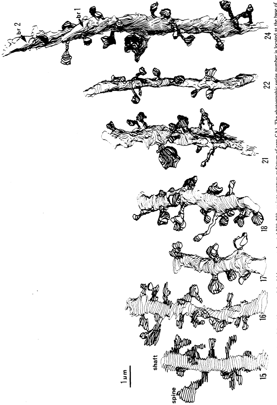
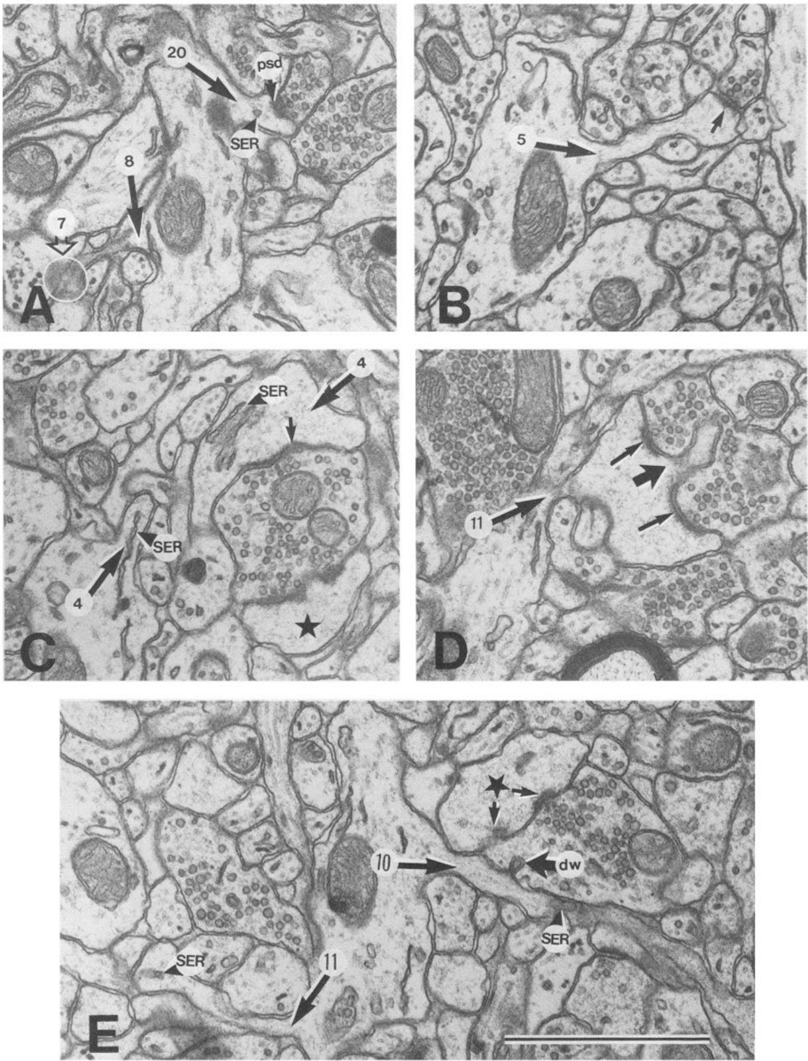
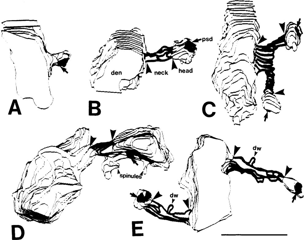
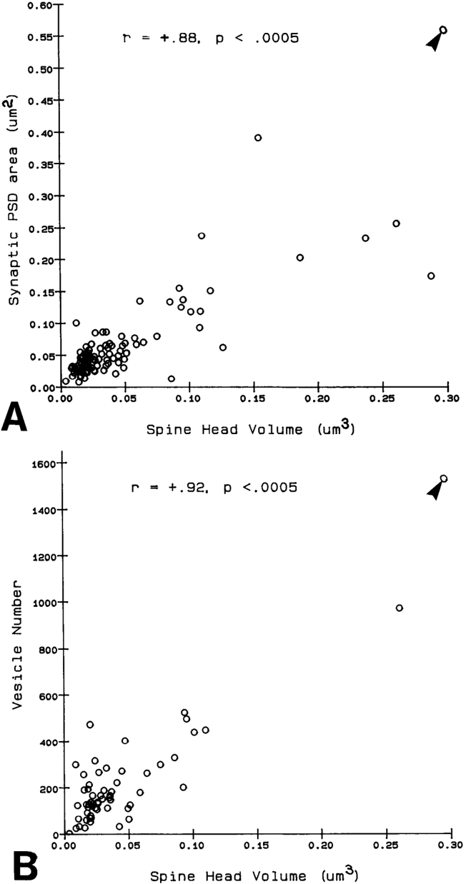
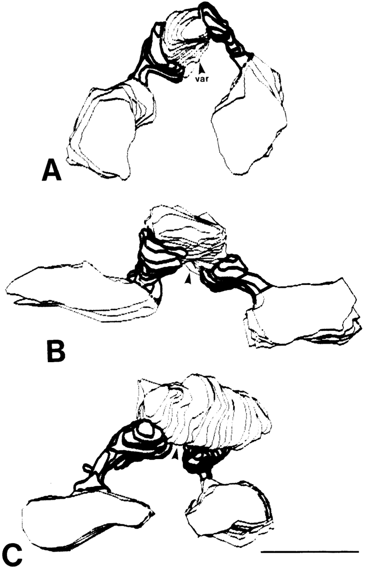
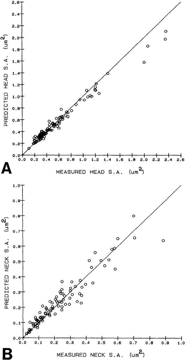
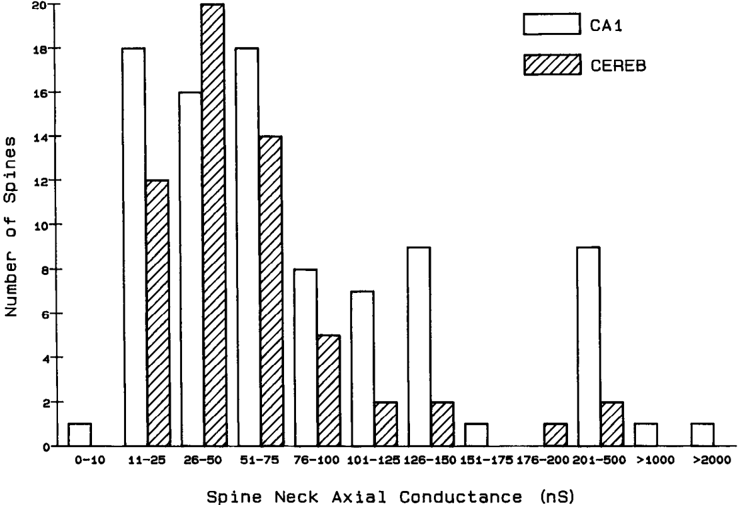
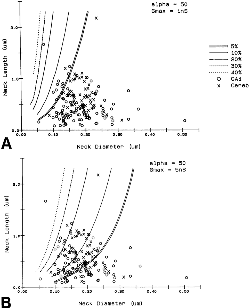
Serial electron microscopy and 3-D reconstructions of dendritic spines from hippocampal area CA 1 dendrites were obtained to evaluate 2 questions about relationships between spine geometry and synaptic efficacy. First, under what biophysical conditions are the spine necks likely to reduce the magnitude of charge transferred from the synapses on the spine heads to the recipient dendrite? Simulation software provided by Charles Wilson (1984) was used to determine that if synaptic conductance is 1 nS or less, only 1% of the hippocampal spine necks are sufficiently thin and long to reduce charge transfer by more than 10%. If synaptic conductance approaches 5 nS, however, 33% of the hippocampal spine necks are sufficiently thin and long to reduce charge transfer by more than 10%. Second, is spine geometry associated with other anatomical indicators of synaptic efficacy, including the area of the postsynaptic density and the number of vesicles in the presynaptic axon? Reconstructed spines were graphically edited into head and neck compartments, and their dimensions were measured, the areas of the postsynaptic densities (PSD) were measured, and all of the vesicles in the presynaptic axonal varicosities were counted. The dimensions of the spine head were well correlated with the area of PSD and the number of vesicles in the presynaptic axonal varicosity. Spine neck diameter and length were not correlated with PSD area, head volume, or the number of vesicles. These results suggest that the dimensions of the spine head, but not the spine neck, reflect differences in synaptic efficacy. We suggest that the constricted necks of hippocampal dendritic spines might reduce diffusion of activated molecules to neighboring synapses, thereby attributing specificity to activated or potentiated synapses.
Overview
The claims and statistics of this paper haven’t been checked yet.
Run the check — free, no account needed. It reads the open-access text and checks the sources it cites.
Integrity signals
Registry
Cites
Statistics
Figures
Verification
Claims the paper makes
The claims and statistics of this paper haven’t been checked yet.
Run the check — free, no account needed. It reads the open-access text and checks the sources it cites.
Claims other papers make about this one
Each is checked against this paper’s own text: does it say what the citing paper says it does?
The source backs part of the claim, not all of it — or backs it for a different population, condition or comparison.
In the citing paper
“This is probably one of the reasons why many ultrastructural studies that utilized high concentrations of potassium ferrocyanide/ferricyanide (1.5–3.0%) provided no data on SS (e.g., Harris and Stevens, 1989; Hayworth et al., 2014; Yakoubi et al., 2019a; Yin et al., 2020; Gour et al., 2021; Phelps et al., 2021; Peddie et al., 2022; Turner et al., 2022).”
From Unambiguous identification of asymmetric and symmetric synapses using volume electron microscopy (2024) · cites this paper as Harris and Stevens, 1989
Experimental — accuracy not yet measuredThe source backs part of the claim, not all of it — or backs it for a different population, condition or comparison.
In the citing paper
“Given that each spine receives only one synaptic input13, our data indicate the highest possible degree of freedom is available in synaptic connections in the dendrites, which has been assumed in the learning models of most neural network theories17.”
From Dendritic spine geometry is critical for AMPA receptor expression in hippocampal CA1 pyramidal neurons (2001) · cites this paper as [13]
Experimental — accuracy not yet measuredThe source backs part of the claim, not all of it — or backs it for a different population, condition or comparison.
In the citing paper
“This observation suggests that the expression of AMPA receptors is controlled independently at a level of the individual spine, consistent with the classical notion that a single spine may behave as an independent chemical compartment to regulate synaptic transmission13, 14.”
From Dendritic spine geometry is critical for AMPA receptor expression in hippocampal CA1 pyramidal neurons (2001) · cites this paper as [13]
Experimental — accuracy not yet measuredThe source backs part of the claim, not all of it — or backs it for a different population, condition or comparison.
In the citing paper
“Regions of glutamate sensitivity tended to be asymmetrically localized to one portion of a spine (Figs. 3h and 4c–j), consistent with the predominant localization of AMPA receptors within PSDs with a diameter of 0.1 to 0.5μm13, 19, 20.”
From Dendritic spine geometry is critical for AMPA receptor expression in hippocampal CA1 pyramidal neurons (2001) · cites this paper as [13]
Experimental — accuracy not yet measuredThe source backs part of the claim, not all of it — or backs it for a different population, condition or comparison.
In the citing paper
“Syn1a-CFP puncta area calculated as a measure of bouton area, which correlated with pHuse area, as expected42.”
From A trans-synaptic nanocolumn aligns neurotransmitter release to receptors (2016) · cites this paper as [42]
Experimental — accuracy not yet measured
The source backs part of the claim, not all of it — or backs it for a different population, condition or comparison.
In the citing paper
“In turn, the size of the postsynaptic density is correlated with the number of presynaptic vesicles, the number of postsynaptic receptors and the ready-releasable pool of transmitter (Harris and Stevens, 1989; Nusser et al., 1998; Schikorski and Stevens, 1999, 2001; Arellano et al., 2007a).”
From The influence of phospho-tau on dendritic spines of cortical pyramidal neurons in patients with Alzheimer’s disease (2013) · cites this paper as Harris and Stevens, 1989
Experimental — accuracy not yet measuredThe source backs part of the claim, not all of it — or backs it for a different population, condition or comparison.
In the citing paper
“Additionally, the volume of the dendritic spine head is related to the size of the postsynaptic density (Harris and Stevens, 1989; Schikorski and Stevens, 1997; Arellano et al., 2007b), the number of presynaptic vesicles, and the number of vesicles adjacent to the presynaptic cleft (Harris and Stevens, 1989).”
From The influence of phospho-tau on dendritic spines of cortical pyramidal neurons in patients with Alzheimer’s disease (2013) · cites this paper as Harris and Stevens, 1989
Experimental — accuracy not yet measuredThe source backs part of the claim, not all of it — or backs it for a different population, condition or comparison.
In the citing paper
“Furthermore, the volume of the head is directly proportional to the strength of synaptic currents (Harris and Stevens, 1989; Schikorski and Stevens, 1997; Arellano et al., 2007a).”
From The influence of phospho-tau on dendritic spines of cortical pyramidal neurons in patients with Alzheimer’s disease (2013) · cites this paper as Harris and Stevens, 1989
Experimental — accuracy not yet measuredThe cited source says what the claim attributes to it.
In the citing paper
“The relationship between spine geometry and expression of functional AMPA receptors demonstrated here provides substantial support to the hypothesis that neuronal fine structures have functional correlates in the brain13, 18, 21.”
From Dendritic spine geometry is critical for AMPA receptor expression in hippocampal CA1 pyramidal neurons (2001) · cites this paper as [13]
The cited source says what the claim attributes to it.
In the citing paper
“These data are consistent with the correlation between AMPA receptor immunoreactivity and the size of PSD19, 20, because the size of PSD is nearly proportional to the spine-head volume13.”
From Dendritic spine geometry is critical for AMPA receptor expression in hippocampal CA1 pyramidal neurons (2001) · cites this paper as [13]
The cited source says what the claim attributes to it.
In the citing paper
“whereas the spine head volume is correlated with the area of the postsynaptic density.”
From The influence of phospho-tau on dendritic spines of cortical pyramidal neurons in patients with Alzheimer’s disease (2013) · cites this paper as Harris and Stevens, 1989
Self-check
Percentages, sums and reported differences re-computed from the paper’s own numbers.
The self-check needs the paper’s full text.
Citations
Cites
The Synaptic Organization of the Brain
(2004) · Oxford University Press eBooks · doi:10.1093/acprof:oso/9780195159561.001.1 · Cited by 3,574
The Synaptic Organization of the Brain
Freemon, F. R. (1980) · JAMA · doi:10.1001/jama.1980.03300440052037 · Cited by 709
Chang, F., & Greenough, W. T. (1984) · Brain Research · doi:10.1016/0006-8993(84)91008-4 · Cited by 523
Lee, K. S., Schottler, F., Oliver, M. E., & LYNCH, G. S. (1980) · Journal of Neurophysiology · doi:10.1152/jn.1980.44.2.247 · Cited by 473
Harris, K., & Stevens, J. (1988) · The Journal of Neuroscience · doi:10.1523/jneurosci.08-12-04455.1988 · Cited by 318
Self-citationDesmond, N. L., & Levy, W. B. (1983) · Brain Research · doi:10.1016/0006-8993(83)91329-x · Cited by 276
Three-Dimensional analysis of dendritic spines
Špaček, J., & Hartmann, M. (1983) · Anatomy and Embryology · doi:10.1007/bf00298517 · Cited by 269
The anatomy of a memory: convergence of results across a diversity of tests
Greenough, W. T., & Bailey, C. H. (1988) · Trends in Neurosciences · doi:10.1016/0166-2236(88)90139-7 · Cited by 266
Crick, F. (1982) · Trends in Neurosciences · doi:10.1016/0166-2236(82)90020-0 · Cited by 254
Van Harreveld, A., & Fifkova, E. (1975) · Experimental Neurology · doi:10.1016/0014-4886(75)90055-2 · Cited by 239
Research works printed but not linked: 6
- Harris, K. M., F. E. Jensen, and B. Tsao (1988) Ultrastructure, de- velopment, and plasticity of synapses in area CA1 of the rat hippo- campus: Extending our vision with serial electron microscopy and three dimensional analyses. Neurol. Neurobiol. (in press).
- Andersen, P., T. Blackstad, G. Hulleberg, J. Line Vaaland, and M. Andersen, P., T. Blackstad, G. Hulleberg, M. Trommald, and J. L. Vaaland (1988b) Dimensions ofdendritic spines ofrat dentate gran- Trommald (1988a) Changes in spine morphology associated with ule cells during long-term potentiation (LTP). Proc. Physiol. Sot. P.C. 50. long-term potentiation in rat dentate granule cells. Proc. Physiol. Sot. s.
- Diamond, J., E. G. Gray, and G. M. Yasargil (1970) The function of dendritic spines: An hypothesis. In Excitatory Synaptic Mechanisms, P. Andersen and J. Jensen, eds., pp. 2 13-222, Universitets Forlaget, Oslo.
- Meek, G. A. (1976) Practical Electron Microscopyfor Biologists, Wiley, New York.
- Rall, W. (1970) Cable properties of dendrites and effects of synaptic location. In Excitatory Synaptic Mechanisms, P. Andersen and J. K. S. Jensen, eds., pp. 175-l 87, Universitets Forlaget, Oslo.
- Rall, W. (1978) Dendritic spines and synaptic potency. In Studies in NeurophysioloRy, A. K. McIntyre and K. Porter, eds., pp. 203-209, Cambridge U.-P., Cambridge.
Cited by
Sorted by citations
Items 101–200 of 999
Chen, X., Levy, J. M., Hou, A., Winters, C., Azzam, R., Sousa, A. A., Leapman, R. D., Nicoll, R. A., & Reese, T. S. (2015) · Proceedings of the National Academy of Sciences · doi:10.1073/pnas.1517045112 · Cited by 310
Visualization of changes in presynaptic function during long-term synaptic plasticity
Zakharenko, S. S., Zablow, L., & Siegelbaum, S. A. (2001) · Nature Neuroscience · doi:10.1038/89498 · Cited by 305
NMDA Receptor Content of Synapses in Stratum Radiatum of the Hippocampal CA1 Area
Racca, C., Stephenson, F. A., Streit, P., Roberts, J. D. B., & Somogyi, P. (2000) · The Journal of Neuroscience · doi:10.1523/jneurosci.20-07-02512.2000 · Cited by 304
Super-Resolution Imaging of the Extracellular Space in Living Brain Tissue
Tønnesen, J., Inavalli, V. V. G. K., & Nägerl, U. V. (2018) · Cell · doi:10.1016/j.cell.2018.02.007 · Cited by 303
Dendritic spines: from structure to in vivo function
Rochefort, N. L., & Konnerth, A. (2012) · The EMBO Reports · doi:10.1038/embor.2012.102 · Cited by 299
Dendritic Spine Loss and Synaptic Alterations in Alzheimer’s Disease
Knobloch, M., & Mansuy, I. M. (2008) · Molecular Neurobiology · doi:10.1007/s12035-008-8018-z · Cited by 296
High-contrast en bloc staining of neuronal tissue for field emission scanning electron microscopy
Tapia, J. C., Kasthuri, N., Hayworth, K. J., Schalek, R., Lichtman, J. W., Smith, S. J., & Buchanan, J. (2012) · Nature Protocols · doi:10.1038/nprot.2011.439 · Cited by 296
Harris, K. M., & Sultan, P. G. (1995) · Neuropharmacology · doi:10.1016/0028-3908(95)00142-s · Cited by 294
Self-citationDetailed passive cable models of whole-cell recorded CA3 pyramidal neurons in rat hippocampal slices
Major, G., Larkman, A. U., Jónás, P., Sakmann, B. J., & Jack, J. (1994) · Journal of Neuroscience · doi:10.1523/jneurosci.14-08-04613.1994 · Cited by 290
Organization of the core structure of the postsynaptic density
Chen, X., Winters, C. A., Azzam, R. N., Li, X., Galbraith, J. A., Leapman, R. D., & Reese, T. S. (2008) · Proceedings of the National Academy of Sciences · doi:10.1073/pnas.0800897105 · Cited by 299
NMDA Receptor Subunit-Dependent [Ca2+] Signaling in Individual Hippocampal Dendritic Spines
Sobczyk, A., Scheuß, V., & Svoboda, K. (2005) · Journal of Neuroscience · doi:10.1523/jneurosci.1221-05.2005 · Cited by 294
Slices Have More Synapses than Perfusion-Fixed Hippocampus from both Young and Mature Rats
Kirov, S. A., Sorra, K. E., & Harris, K. M. (1999) · Journal of Neuroscience · doi:10.1523/jneurosci.19-08-02876.1999 · Cited by 279
Self-citationIntegration of biochemical signalling in spines
Kennedy, M. B., Beale, H. C., Carlisle, H. J., & Washburn, L. R. (2005) · Nature Reviews Neuroscience · doi:10.1038/nrn1685 · Cited by 286
Zagrebelsky, M., Holz, A., Dechant, G., Barde, Y.-A., Bonhoeffer, T., & Korte, M. (2005) · The Journal of Neuroscience · doi:10.1523/jneurosci.2492-05.2005 · Cited by 286
Plasticity of perisynaptic astroglia during synaptogenesis in the mature rat hippocampus
Witcher, M. R., Kirov, S. A., & Harris, K. M. (2006) · Glia · doi:10.1002/glia.20415 · Cited by 306
Self-citationVesicle pool mobilization during action potential firing at hippocampal synapses
Ryan, T. A., & Smith, S. J. (1995) · Neuron · doi:10.1016/0896-6273(95)90336-4 · Cited by 282
Quantal analysis and synaptic anatomy--integrating two views of hippocampal plasticity.
Lisman, J., & Harris, K. (1993) · Trends in Neurosciences · doi:10.1016/0166-2236(93)90122-3 · Cited by 271
Self-citationHayashi, M. L., Choi, S.-Y., Rao, B. S. S., Jung, H.-Y., Lee, H.-K., Zhang, D., Chattarji, S., Kirkwood, A., & Tonegawa, S. (2004) · Neuron · doi:10.1016/j.neuron.2004.05.003 · Cited by 280
Role for metabotropic glutamate receptor 5 (mGluR5) in the pathogenesis of fragile X syndrome
Dölen, G., & Bear, M. F. (2008) · The Journal of Physiology · doi:10.1113/jphysiol.2008.150722 · Cited by 278
Ultrastructural Analysis of Hippocampal Neuropil from the Connectomics Perspective
Mishchenko, Y., Hu, T., Špaček, J., Mendenhall, J. M., Harris, K. M., & Chklovskii, D. B. (2010) · Neuron · doi:10.1016/j.neuron.2010.08.014 · Cited by 276
Self-citationDendritic morphology of pyramidal neurones of the visual cortex of the rat: III. Spine distributions
Larkman, A. U. (1991) · The Journal of Comparative Neurology · doi:10.1002/cne.903060209 · Cited by 274
Rapid functional maturation of nascent dendritic spines
Zito, K., Scheuss, V., Knott, G., Hill, T., & Svoboda, K. (2009) · Neuron · doi:10.1016/j.neuron.2008.10.054 · Cited by 272
High-accuracy neurite reconstruction for high-throughput neuroanatomy
Helmstaedter, M. N., Briggman, K. L., & Denk, W. T. (2011) · Nature Neuroscience · doi:10.1038/nn.2868 · Cited by 269
Monosomes actively translate synaptic mRNAs in neuronal processes
Biever, A., Glock, C., Tushev, G., Ciirdaeva, E., Dalmay, T., Langer, J. D., & Schuman, E. M. (2020) · Science · doi:10.1126/science.aay4991 · Cited by 283
NMDA Receptor-Mediated Subthreshold Ca2+Signals in Spines of Hippocampal Neurons
Kovalchuk, Y., Eilers, J., Lisman, J. E., & Konnerth, A. (2000) · Journal of Neuroscience · doi:10.1523/jneurosci.20-05-01791.2000 · Cited by 270
Spine synapse remodeling in the pathophysiology and treatment of depression
Duman, C. H., & Duman, R. S. (2015) · Neuroscience Letters · doi:10.1016/j.neulet.2015.01.022 · Cited by 257
Cooney, J. R., Hurlburt, J. L., Selig, D. K., Harris, K. M., & Fiala, J. C. (2002) · The Journal of Neuroscience · doi:10.1523/jneurosci.22-06-02215.2002 · Cited by 269
Self-citationHestrin, S. (1992) · Neuron · doi:10.1016/0896-6273(92)90250-h · Cited by 234
Development and Regulation of Dendritic Spine Synapses
Calabrese, B., Wilson, M. S., & Halpain, S. (2006) · Physiology · doi:10.1152/physiol.00042.2005 · Cited by 287
Independent regulation of calcium revealed by imaging dendritic spines
Guthrie, P. B., Segal, M., & Kater, S. B. (1991) · Nature · doi:10.1038/354076a0 · Cited by 262
Steiner, P., Higley, M. J., Xu, W., Czervionke, B. L., Malenka, R. C., & Sabatini, B. L. (2008) · Neuron · doi:10.1016/j.neuron.2008.10.014 · Cited by 259
Loewenstein, Y., Kuras, A., & Rumpel, S. (2011) · Journal of Neuroscience · doi:10.1523/jneurosci.6130-10.2011 · Cited by 259
Estrogen Alters Spine Number and Morphology in Prefrontal Cortex of Aged Female Rhesus Monkeys
Hao, J., Rapp, P. R., Leffler, A. E., Leffler, S. R., Janssen, W. G. M., Lou, W. Y. W., McKay, H. M., Roberts, J. A., Wearne, S. L., Hof, P. R., et al. (2006) · Journal of Neuroscience · doi:10.1523/jneurosci.3440-05.2006 · Cited by 264
Dendritic spines: Revisiting the physiological role
Chidambaram, S. B., Rathipriya, A. G., Bolla, S. R., Bhat, A., Ray, B., Mahalakshmi, A. M., Manivasagam, T., Thenmozhi, A. J., Essa, M. M., Guillemin, G. J., et al. (2019) · Progress in Neuro-Psychopharmacology and Biological Psychiatry · doi:10.1016/j.pnpbp.2019.01.005 · Cited by 266
Sorra, K., & Harris, K. M. (1993) · Journal of Neuroscience · doi:10.1523/jneurosci.13-09-03736.1993 · Cited by 249
Self-citationStructure and function of a neocortical synapse
Holler, S., Köstinger, G., Martin, K. A. C., Schuhknecht, G. F. P., & Stratford, K. J. (2021) · Nature · doi:10.1038/s41586-020-03134-2 · Cited by 249
Tashiro, A., & Yuste, R. (2004) · Molecular and Cellular Neuroscience · doi:10.1016/j.mcn.2004.04.001 · Cited by 265
Lin, Y., & Koleske, A. J. (2010) · Annual Review of Neuroscience · doi:10.1146/annurev-neuro-060909-153204 · Cited by 255
Majewska, A., Brown, E., Ross, J., & Yuste, R. (2000) · The Journal of Neuroscience · doi:10.1523/jneurosci.20-05-01722.2000 · Cited by 248
Keck, T., Scheuß, V., Jacobsen, R. I., Wierenga, C. J., Eysel, U. T., Bonhoeffer, T., & Hübener, M. (2011) · Neuron · doi:10.1016/j.neuron.2011.06.034 · Cited by 252
Structural determinants of transmission at large hippocampal mossy fiber synapses
Rollenhagen, A., Sätzler, K., Rodríguez, E. P., Jonas, P., Frotscher, M., & Lübke, J. H. R. (2007) · The Journal of Neuroscience · doi:10.1523/jneurosci.1946-07.2007 · Cited by 246
LAR receptor protein tyrosine phosphatases in the development and maintenance of excitatory synapses
Dunah, A. W., Hueske, E., Wyszynski, M. W., Hoogenraad, C. C., Jaworski, J., Pak, D. T. S., Simonetta, A., Liu, G., & Sheng, M. (2005) · Nature Neuroscience · doi:10.1038/nn1416 · Cited by 244
Remodeling of Synaptic Membranes after Induction of Long-Term Potentiation
Toni, N., Buchs, P.-A., Nikonenko, I., Povilaitite, P., Parisi, L., & Muller, D. (2001) · The Journal of Neuroscience · doi:10.1523/jneurosci.21-16-06245.2001 · Cited by 244
Briner, A., Roo, M. D., Dayer, A. G., Müller, D., Habre, W., & Vutskits, L. (2010) · Anesthesiology · doi:10.1097/aln.0b013e3181cd7942 · Cited by 249
A Unified Model of the Presynaptic and Postsynaptic Changes During LTP at CA1 Synapses
Lisman, J., & Raghavachari, S. (2006) · Science's STKE · doi:10.1126/stke.3562006re11 · Cited by 241
Biophysical model of a Hebbian synapse.
Zador, A. M., Koch, C., & Brown, T. H. (1990) · Proceedings of the National Academy of Sciences · doi:10.1073/pnas.87.17.6718 · Cited by 241
Umbriaco, D., Watkins, K. C., Descarries, L., Cozzari, C., & Hartman, B. K. (1994) · The Journal of Comparative Neurology · doi:10.1002/cne.903480304 · Cited by 237
Briner, A., Nikonenko, I., Roo, M. D., Dayer, A. G., Müller, D., & Vutskits, L. (2011) · Anesthesiology · doi:10.1097/aln.0b013e318221fbbd · Cited by 237
The timing of synaptic vesicle endocytosis.
Ryan, T. A., Smith, S. J., & REUTER, H. (1996) · Proceedings of the National Academy of Sciences · doi:10.1073/pnas.93.11.5567 · Cited by 236
Synaptic nanomodules underlie the organization and plasticity of spine synapses
Hruska, M., Henderson, N., Le Marchand, S. J., Jafri, H., & Dalva, M. B. (2018) · Nature Neuroscience · doi:10.1038/s41593-018-0138-9 · Cited by 233
Yankova, M. P., Hart, S. A., & Woolley, C. S. (2001) · Proceedings of the National Academy of Sciences · doi:10.1073/pnas.051624598 · Cited by 232
Calcium concentration dynamics produced by synaptic activation of CA1 hippocampal pyramidal cells
Regehr, W. G., & Tank, D. W. (1992) · Journal of Neuroscience · doi:10.1523/jneurosci.12-11-04202.1992 · Cited by 230
Synaptic Connectivity and Neuronal Morphology
Chklovskii, D. B. (2004) · Neuron · doi:10.1016/s0896-6273(04)00498-2 · Cited by 263
Molecular anatomy of the postsynaptic density.
Okabe, S. (2007) · Molecular and Cellular Neuroscience · doi:10.1016/j.mcn.2007.01.006 · Cited by 225
The Number of Glutamate Receptors Opened by Synaptic Stimulation in Single Hippocampal Spines
Nimchinsky, E. A., Yasuda, R., Oertner, T. G., & Svoboda, K. (2004) · Journal of Neuroscience · doi:10.1523/jneurosci.5066-03.2004 · Cited by 219
Ionotropic glutamate receptors
Asztely, F., & Gustafsson, B. (1996) · Molecular Neurobiology · doi:10.1007/bf02740744 · Cited by 90
Evidence for reduced experience-dependent dendritic spine plasticity in the aging prefrontal cortex
Bloss, E. B., Janssen, W. G., Ohm, D. T., Yuk, F. J., Wadsworth, S., Saardi, K. M., McEwen, B. S., & Morrison, J. H. (2011) · The Journal of Neuroscience · doi:10.1523/jneurosci.0839-11.2011 · Cited by 219
Direct measurement of quantal changes underlying long-term potentiation in CA1 hippocampus.
Liao, D., Jones, A., & Malinow, R. (1992) · Neuron · doi:10.1016/0896-6273(92)90068-o · Cited by 193
Modulatory Role of Drebrin on the Cytoskeleton within Dendritic Spines in the Rat Cerebral Cortex
Hayashi, K., Ishikawa, R., Ye, L., He, X., Takata, K., Kohama, K., & Shirao, T. (1996) · Journal of Neuroscience · doi:10.1523/jneurosci.16-22-07161.1996 · Cited by 217
The Relationship between PSD-95 Clustering and Spine Stability In Vivo
Cane, M., Maco, B., Knott, G., & Holtmaat, A. (2014) · The Journal of Neuroscience · doi:10.1523/jneurosci.3353-13.2014 · Cited by 216
Factors mediating powerful voltage attenuation along CA1 pyramidal neuron dendrites
Golding, N. L., Mickus, T. J., Katz, Y., Kath, W. L., & Spruston, N. (2005) · The Journal of Physiology · doi:10.1113/jphysiol.2005.086793 · Cited by 216
Woolley, C. S., Wenzel, H. J., & Schwartzkroin, P. A. (1996) · The Journal of Comparative Neurology · doi:10.1002/(sici)1096-9861(19960909)373:1 · Cited by 213
Cortical area and species differences in dendritic spine morphology
Benavides-Piccione, R., Ballesteros-Yáñez, I., DeFelipe, J., & Yuste, R. (2002) · Journal of Neurocytology · doi:10.1023/a:1024134312173 · Cited by 207
Regulation of Spine Calcium Dynamics by Rapid Spine Motility
Majewska, A. K., Tashiro, A., & Yuste, R. (2000) · Journal of Neuroscience · doi:10.1523/jneurosci.20-22-08262.2000 · Cited by 208
Ventral hippocampal projections to the medial prefrontal cortex regulate social memory
Phillips, M. L., Robinson, H. A., & Pozzo-Miller, L. (2019) · eLife · doi:10.7554/elife.44182 · Cited by 205
Quantitative fine-structural analysis of olfactory cortical synapses
Schikorski, T., & Stevens, C. F. (1999) · Proceedings of the National Academy of Sciences · doi:10.1073/pnas.96.7.4107 · Cited by 204
Extending Unbiased Stereology of Brain Ultrastructure to Three-dimensional Volumes
Fiala, J. C., & Harris, K. M. (2001) · Journal of the American Medical Informatics Association · doi:10.1136/jamia.2001.0080001 · Cited by 203
Self-citationAstrocyte lipid metabolism is critical for synapse development and function in vivo
van Deijk, A. F., Camargo, N., Timmerman, J., Heistek, T., Brouwers, J. F., Mogavero, F., Mansvelder, H. D., Smit, A. B., & Verheijen, M. H. G. (2017) · Glia · doi:10.1002/glia.23120 · Cited by 201
Biochemical Computation for Spine Structural Plasticity
Nishiyama, J., & Yasuda, R. (2015) · Neuron · doi:10.1016/j.neuron.2015.05.043 · Cited by 199
Activation of AMPA, Kainate, and Metabotropic Receptors at Hippocampal Mossy Fiber Synapses
Min, M., Rusakov, D. A., & Kullmann, D. M. (1998) · Neuron · doi:10.1016/s0896-6273(00)80566-8 · Cited by 199
Properties of Quantal Transmission at CA1 Synapses
Raghavachari, S., & Lisman, J. E. (2004) · Journal of Neurophysiology · doi:10.1152/jn.00258.2004 · Cited by 197
Okamoto, K., Bosch, M., & Hayashi, Y. (2009) · Physiology · doi:10.1152/physiol.00029.2009 · Cited by 213
Early Language Development And Its Neural Correlates
Bates, E. A., Thal, D. J., Finlay, B. L., & Clancy, B. (1992) · Cited by 195
Dudman, J. T., Tsay, D., & Siegelbaum, S. A. (2007) · Neuron · doi:10.1016/j.neuron.2007.10.020 · Cited by 192
Chronic 2P-STED imaging reveals high turnover of dendritic spines in the hippocampus in vivo
Pfeiffer, T., Poll, S., Bancelin, S., Angibaud, J., Inavalli, V. K., Keppler, K., Mittag, M., Fuhrmann, M., & Nägerl, U. V. (2018) · eLife · doi:10.7554/elife.34700 · Cited by 192
Dendritic pathology in mental retardation: from molecular genetics to neurobiology
Dierssen, M., & Ramakers, G. J. A. (2006) · Genes Brain & Behavior · doi:10.1111/j.1601-183x.2006.00224.x · Cited by 191
Hosokawa, T., Rusakov, D., Bliss, T., & Fine, A. (1995) · Journal of Neuroscience · doi:10.1523/jneurosci.15-08-05560.1995 · Cited by 187
Calcium Signaling in Dendritic SpinesPreprint
Higley, M. J., & Sabatini, B. L. (2012) · Cold Spring Harbor Perspectives in Biology · doi:10.1101/cshperspect.a005686 · Cited by 187
Li, Y., & Tsien, R. W. (2012) · Nature Neuroscience · doi:10.1038/nn.3126 · Cited by 183
Some assembly required: the development of neuronal synapses
Li, Z., & Sheng, M. (2003) · Nature Reviews Molecular Cell Biology · doi:10.1038/nrm1242 · Cited by 195
Spine dynamics in the brain, mental disorders and artificial neural networks
Kasai, H., Ziv, N. E., Okazaki, H., Yagishita, S., & Toyoizumi, T. (2021) · Nature reviews. Neuroscience · doi:10.1038/s41583-021-00467-3 · Cited by 184
Stability in Synapse Number and Size at 2 Hr after Long-Term Potentiation in Hippocampal Area CA1
Sorra, K. E., & Harris, K. M. (1998) · Journal of Neuroscience · doi:10.1523/jneurosci.18-02-00658.1998 · Cited by 181
Self-citationStructural changes at dendritic spine synapses during long-term potentiation
Harris, K. M., Fiala, J. C., & Ostroff, L. (2003) · Philosophical Transactions of the Royal Society of London. Series B: Biological Sciences · doi:10.1098/rstb.2002.1254 · Cited by 180
Self-citationWilke, S. A., Antonios, J. K., Bushong, E. A., Badkoobehi, A., Malek, E., Hwang, M., Terada, M., Ellisman, M. H., & Ghosh, A. (2013) · The Journal of Neuroscience · doi:10.1523/jneurosci.1600-12.2013 · Cited by 179
Lai, K., & Ip, N. Y. (2013) · Biochimica et Biophysica Acta (BBA) - Molecular Basis of Disease · doi:10.1016/j.bbadis.2013.08.012 · Cited by 182
Structural Contributions to Short-Term Synaptic Plasticity
Xu‐Friedman, M. A., & Regehr, W. G. (2004) · Physiological Reviews · doi:10.1152/physrev.00016.2003 · Cited by 178
PSD-95 promotes synaptogenesis and multiinnervated spine formation through nitric oxide signaling
Nikonenko, I., Boda, B., Steen, S., Knott, G. W., Welker, E., & Müller, D. (2008) · The Journal of Cell Biology · doi:10.1083/jcb.200805132 · Cited by 176
Hao, J., Rapp, P. R., Janssen, W. G. M., Lou, W. Y. W., Lasley, B. L., Hof, P. R., & Morrison, J. H. (2007) · Proceedings of the National Academy of Sciences · doi:10.1073/pnas.0704757104 · Cited by 178
A Single Packet of Transmitter Does Not Saturate Postsynaptic Glutamate Receptors
Ishikawa, T., Sahara, Y., & Takahashi, T. (2002) · Neuron · doi:10.1016/s0896-6273(02)00692-x · Cited by 176
Quantitative ultrastructure of Ia boutons in the ventral horn: scaling and positional relationships
Pierce, J., & Mendell, L. M. (1993) · Journal of Neuroscience · doi:10.1523/jneurosci.13-11-04748.1993 · Cited by 174
Synapse Distribution Suggests a Two-Stage Model of Dendritic Integration in CA1 Pyramidal Neurons
Katz, Y., Menon, V., Nicholson, D. A., Geinisman, Y., Kath, W. L., & Spruston, N. (2009) · Neuron · doi:10.1016/j.neuron.2009.06.023 · Cited by 168
Benavides‐Piccione, R., Fernaud-Espinosa, I., Robles, V., Yuste, R., & DeFelipe, J. (2012) · Cerebral Cortex · doi:10.1093/cercor/bhs154 · Cited by 180
Optical detection of a quantal presynaptic membrane turnover
Ryan, T. A., REUTER, H., & Smith, S. J. (1997) · Nature · doi:10.1038/41335 · Cited by 173
Jones, K. A., & Baughman, R. (1991) · Neuron · doi:10.1016/0896-6273(91)90372-7 · Cited by 154
How to build a central synapse: clues from cell culture
Craig, A. M., Graf, E. R., & Linhoff, M. W. (2005) · Trends in Neurosciences · doi:10.1016/j.tins.2005.11.002 · Cited by 170
Long-Term Relationships between Synaptic Tenacity, Synaptic Remodeling, and Network Activity
Minerbi, A., Kahana, R., Goldfeld, L., Kaufman, M., Marom, S., & Ziv, N. E. (2009) · PLoS Biology · doi:10.1371/journal.pbio.1000136 · Cited by 170
Actin and the agile spine: How and why do dendritic spines dance?
Halpain, S. (2000) · Trends in Neurosciences · doi:10.1016/s0166-2236(00)01576-9 · Cited by 169
The loading of neurotransmitters into synaptic vesicles
Gasnier, B. (2000) · Biochimie · doi:10.1016/s0300-9084(00)00221-2 · Cited by 171
Kalirin-7 Is Required for Synaptic Structure and Function
Ma, X., Kiraly, D. D., Gaier, E. D., Wang, Y., Kim, E., Levine, E. S., Eipper, B. A., & Mains, R. E. (2008) · Journal of Neuroscience · doi:10.1523/jneurosci.4269-08.2008 · Cited by 169
Oh, W. C., Hill, T. C., & Zito, K. (2012) · Proceedings of the National Academy of Sciences · doi:10.1073/pnas.1214705110 · Cited by 175
Metadata
- Authors
- Harris, K., & Stevens, J.
- Affiliations
- Boston Children's Hospital (US)
- Venue
- The Journal of Neuroscience
- Publisher
- Society for Neuroscience
- Published
- 1989-08-01
- Volume
- 9
- Issue
- 8
- Pages
- 2982-2997
- ISSN
- 0270-6474 (ISSN-L)
- 1529-2401
- PMC
- PMC6569708
- OpenAlex
- W2117664079
- Cited by
- 1,073
- References
- 38
- Citations by year
- 2026: 17 · 2025: 26 · 2024: 35 · 2023: 33 · 2022: 31 · 2021: 50 · 2020: 39 · 2019: 43 · 2018: 47 · 2017: 33 · 2016: 40 · 2015: 48 · 2014: 44 · 2013: 50 · 2012: 42
- FWCI
- 5.92
- Type
- journal article
- Language
- en
- Open access
- hybrid
- doi.org
- Licence
- Locations
- Journal of Neuroscience (publishedVersion, cc-by-nc-sa)
- Europe PMC (PubMed Central) (publishedVersion)
- PubMed Central (publishedVersion)
- PubMed (publishedVersion)
- Funders
- Alfred P. Sloan Foundation
- National Institutes of Health (NS21184)
- Medical Research Council
- National Institute of Neurological Disorders and Stroke (NS21184, NS 20820)
- Eunice Kennedy Shriver National Institute of Child Health and Human Development (P30HD018655, P30HD-18655)
- Topics
- Life Sciences › Neuroscience › Cellular and Molecular Neuroscience › Neuroscience and Neuropharmacology Research
- Life Sciences › Neuroscience › Cognitive Neuroscience › Memory and Neural Mechanisms
- Life Sciences › Neuroscience › Cognitive Neuroscience › Neural dynamics and brain function
- Keywords
- dendritic spines, CA1 pyramidal neurons, hippocampus, synaptic efficacy, postsynaptic density, synaptic vesicles, charge transfer, synaptic conductance, dendritic spine morphology, synaptic specificity
- MeSH terms
- Animals, Axons, Biophysics, Dendrites, Electric Conductivity, Endoplasmic Reticulum, Hippocampus, Image Processing, Computer-Assisted, Male, Microscopy, Electron, Models, Neurological, Neurons, Purkinje Cells, Rats, Inbred Strains, Synapses, Rats, Biophysical Phenomena
About these checks
Registry, retraction, Cites and statistics results are computed, not judged, with one exception: a printed reference that no index lists is read once by a model before it counts as a citation error. Claims are extracted by a model and kept only when their quote appears verbatim in the text; each cited source is then checked, with a model’s help, for whether it says what the claim relies on. Those verdicts can be wrong — read the claim’s quote and the cited source before relying on one.
Statistics and arithmetic are recomputed, not judged.
Confidence bands are shown only for fields calibrated on our annotated test set. None is calibrated yet, so no band is shown.
What counts as a claim ›How we measure confidence ›How we check this ›
Are you an author, or do you see a problem with this page? Request a review
