

The DOI matches its Crossref record; 36 of the 38 references it cites were found in a registry or an index.
Dendritic spines of CA 1 pyramidal cells in the rat hippocampus: serial electron microscopy with reference to their biophysical characteristics
Open accessAbstract
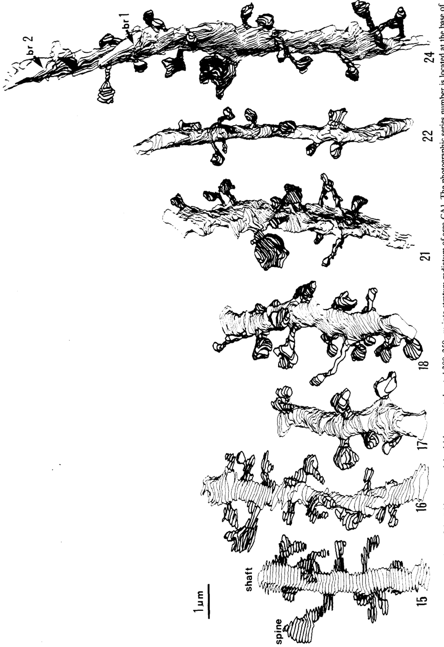
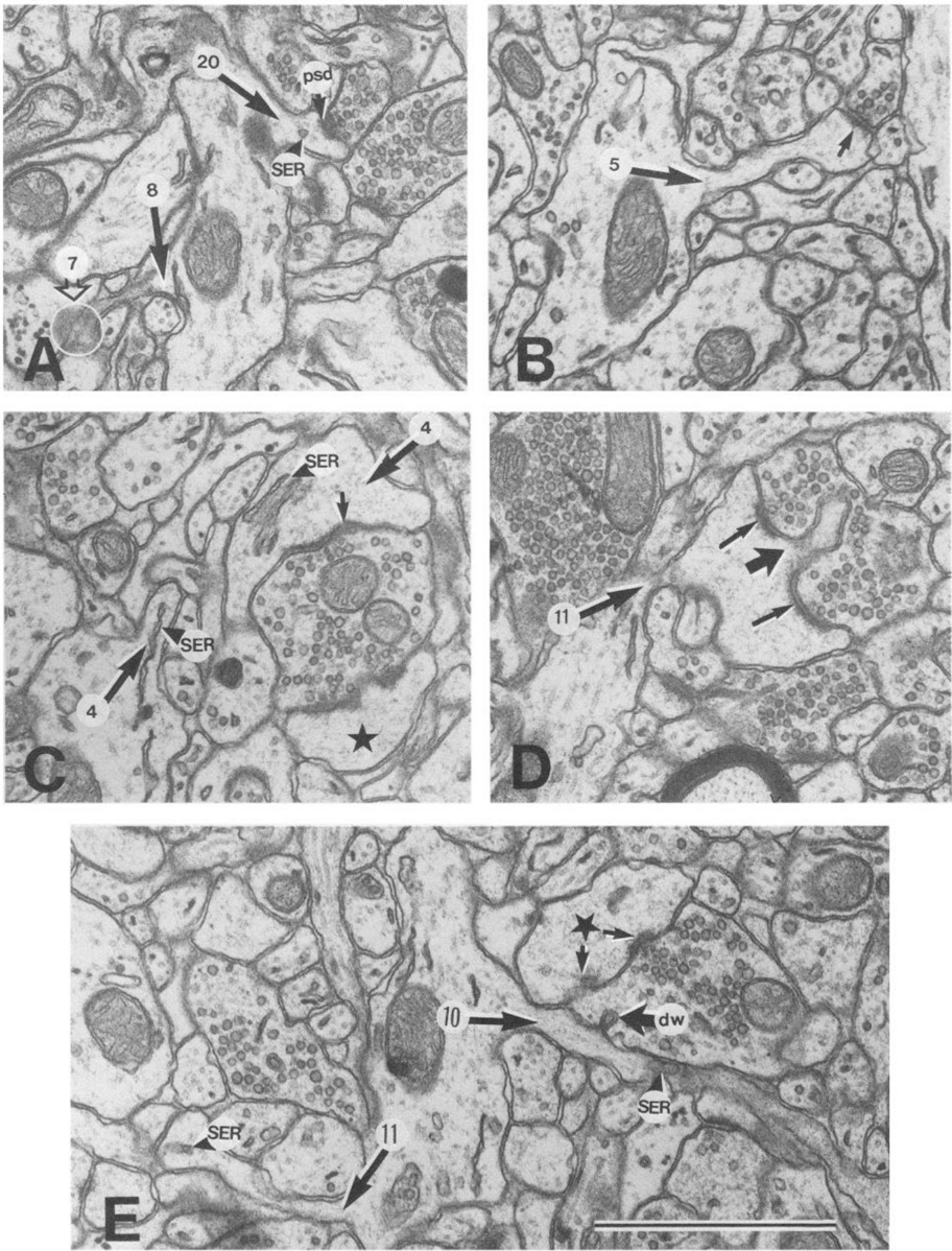
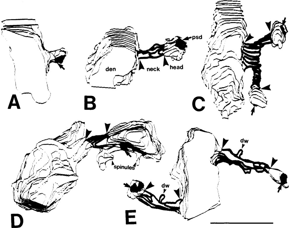
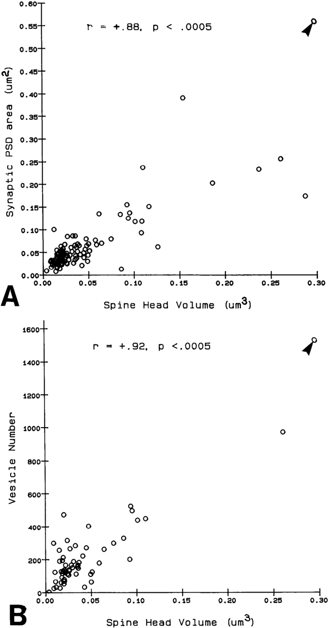
Serial electron microscopy and 3-D reconstructions of dendritic spines from hippocampal area CA 1 dendrites were obtained to evaluate 2 questions about relationships between spine geometry and synaptic efficacy. First, under what biophysical conditions are the spine necks likely to reduce the magnitude of charge transferred from the synapses on the spine heads to the recipient dendrite? Simulation software provided by Charles Wilson (1984) was used to determine that if synaptic conductance is 1 nS or less, only 1% of the hippocampal spine necks are sufficiently thin and long to reduce charge transfer by more than 10%. If synaptic conductance approaches 5 nS, however, 33% of the hippocampal spine necks are sufficiently thin and long to reduce charge transfer by more than 10%. Second, is spine geometry associated with other anatomical indicators of synaptic efficacy, including the area of the postsynaptic density and the number of vesicles in the presynaptic axon? Reconstructed spines were graphically edited into head and neck compartments, and their dimensions were measured, the areas of the postsynaptic densities (PSD) were measured, and all of the vesicles in the presynaptic axonal varicosities were counted. The dimensions of the spine head were well correlated with the area of PSD and the number of vesicles in the presynaptic axonal varicosity. Spine neck diameter and length were not correlated with PSD area, head volume, or the number of vesicles. These results suggest that the dimensions of the spine head, but not the spine neck, reflect differences in synaptic efficacy. We suggest that the constricted necks of hippocampal dendritic spines might reduce diffusion of activated molecules to neighboring synapses, thereby attributing specificity to activated or potentiated synapses.
Overview
The claims and statistics of this paper haven’t been checked yet.
Run the check — free, no account needed. It reads the open-access text and checks the sources it cites.
Integrity signals
Registry
Cites
Statistics
Figures
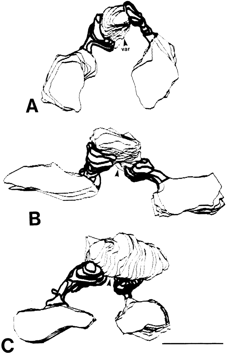
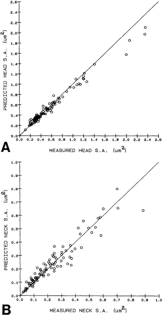
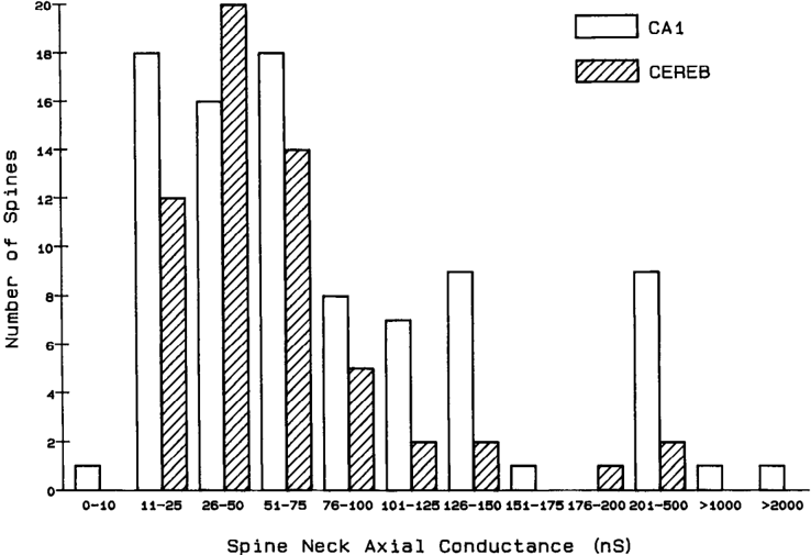
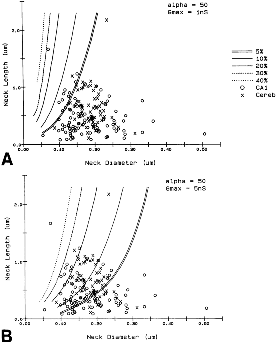
Verification
Claims the paper makes
The claims and statistics of this paper haven’t been checked yet.
Run the check — free, no account needed. It reads the open-access text and checks the sources it cites.
Claims other papers make about this one
Each is checked against this paper’s own text: does it say what the citing paper says it does?
The source backs part of the claim, not all of it — or backs it for a different population, condition or comparison.
In the citing paper
“This is probably one of the reasons why many ultrastructural studies that utilized high concentrations of potassium ferrocyanide/ferricyanide (1.5–3.0%) provided no data on SS (e.g., Harris and Stevens, 1989; Hayworth et al., 2014; Yakoubi et al., 2019a; Yin et al., 2020; Gour et al., 2021; Phelps et al., 2021; Peddie et al., 2022; Turner et al., 2022).”
From Unambiguous identification of asymmetric and symmetric synapses using volume electron microscopy (2024) · cites this paper as Harris and Stevens, 1989
Experimental — accuracy not yet measuredThe source backs part of the claim, not all of it — or backs it for a different population, condition or comparison.
In the citing paper
“Given that each spine receives only one synaptic input13, our data indicate the highest possible degree of freedom is available in synaptic connections in the dendrites, which has been assumed in the learning models of most neural network theories17.”
From Dendritic spine geometry is critical for AMPA receptor expression in hippocampal CA1 pyramidal neurons (2001) · cites this paper as [13]
Experimental — accuracy not yet measuredThe source backs part of the claim, not all of it — or backs it for a different population, condition or comparison.
In the citing paper
“This observation suggests that the expression of AMPA receptors is controlled independently at a level of the individual spine, consistent with the classical notion that a single spine may behave as an independent chemical compartment to regulate synaptic transmission13, 14.”
From Dendritic spine geometry is critical for AMPA receptor expression in hippocampal CA1 pyramidal neurons (2001) · cites this paper as [13]
Experimental — accuracy not yet measuredThe source backs part of the claim, not all of it — or backs it for a different population, condition or comparison.
In the citing paper
“Regions of glutamate sensitivity tended to be asymmetrically localized to one portion of a spine (Figs. 3h and 4c–j), consistent with the predominant localization of AMPA receptors within PSDs with a diameter of 0.1 to 0.5μm13, 19, 20.”
From Dendritic spine geometry is critical for AMPA receptor expression in hippocampal CA1 pyramidal neurons (2001) · cites this paper as [13]
Experimental — accuracy not yet measuredThe source backs part of the claim, not all of it — or backs it for a different population, condition or comparison.
In the citing paper
“Syn1a-CFP puncta area calculated as a measure of bouton area, which correlated with pHuse area, as expected42.”
From A trans-synaptic nanocolumn aligns neurotransmitter release to receptors (2016) · cites this paper as [42]
Experimental — accuracy not yet measured
The source backs part of the claim, not all of it — or backs it for a different population, condition or comparison.
In the citing paper
“In turn, the size of the postsynaptic density is correlated with the number of presynaptic vesicles, the number of postsynaptic receptors and the ready-releasable pool of transmitter (Harris and Stevens, 1989; Nusser et al., 1998; Schikorski and Stevens, 1999, 2001; Arellano et al., 2007a).”
From The influence of phospho-tau on dendritic spines of cortical pyramidal neurons in patients with Alzheimer’s disease (2013) · cites this paper as Harris and Stevens, 1989
Experimental — accuracy not yet measuredThe source backs part of the claim, not all of it — or backs it for a different population, condition or comparison.
In the citing paper
“Additionally, the volume of the dendritic spine head is related to the size of the postsynaptic density (Harris and Stevens, 1989; Schikorski and Stevens, 1997; Arellano et al., 2007b), the number of presynaptic vesicles, and the number of vesicles adjacent to the presynaptic cleft (Harris and Stevens, 1989).”
From The influence of phospho-tau on dendritic spines of cortical pyramidal neurons in patients with Alzheimer’s disease (2013) · cites this paper as Harris and Stevens, 1989
Experimental — accuracy not yet measuredThe source backs part of the claim, not all of it — or backs it for a different population, condition or comparison.
In the citing paper
“Furthermore, the volume of the head is directly proportional to the strength of synaptic currents (Harris and Stevens, 1989; Schikorski and Stevens, 1997; Arellano et al., 2007a).”
From The influence of phospho-tau on dendritic spines of cortical pyramidal neurons in patients with Alzheimer’s disease (2013) · cites this paper as Harris and Stevens, 1989
Experimental — accuracy not yet measuredThe cited source says what the claim attributes to it.
In the citing paper
“The relationship between spine geometry and expression of functional AMPA receptors demonstrated here provides substantial support to the hypothesis that neuronal fine structures have functional correlates in the brain13, 18, 21.”
From Dendritic spine geometry is critical for AMPA receptor expression in hippocampal CA1 pyramidal neurons (2001) · cites this paper as [13]
The cited source says what the claim attributes to it.
In the citing paper
“These data are consistent with the correlation between AMPA receptor immunoreactivity and the size of PSD19, 20, because the size of PSD is nearly proportional to the spine-head volume13.”
From Dendritic spine geometry is critical for AMPA receptor expression in hippocampal CA1 pyramidal neurons (2001) · cites this paper as [13]
The cited source says what the claim attributes to it.
In the citing paper
“whereas the spine head volume is correlated with the area of the postsynaptic density.”
From The influence of phospho-tau on dendritic spines of cortical pyramidal neurons in patients with Alzheimer’s disease (2013) · cites this paper as Harris and Stevens, 1989
Self-check
Percentages, sums and reported differences re-computed from the paper’s own numbers.
The self-check needs the paper’s full text.
Citations
Cites
The Synaptic Organization of the Brain
(2004) · Oxford University Press eBooks · doi:10.1093/acprof:oso/9780195159561.001.1 · Cited by 3,574
The Synaptic Organization of the Brain
Freemon, F. R. (1980) · JAMA · doi:10.1001/jama.1980.03300440052037 · Cited by 709
Chang, F., & Greenough, W. T. (1984) · Brain Research · doi:10.1016/0006-8993(84)91008-4 · Cited by 523
Lee, K. S., Schottler, F., Oliver, M. E., & LYNCH, G. S. (1980) · Journal of Neurophysiology · doi:10.1152/jn.1980.44.2.247 · Cited by 473
Harris, K., & Stevens, J. (1988) · The Journal of Neuroscience · doi:10.1523/jneurosci.08-12-04455.1988 · Cited by 318
Self-citationDesmond, N. L., & Levy, W. B. (1983) · Brain Research · doi:10.1016/0006-8993(83)91329-x · Cited by 276
Three-Dimensional analysis of dendritic spines
Špaček, J., & Hartmann, M. (1983) · Anatomy and Embryology · doi:10.1007/bf00298517 · Cited by 269
The anatomy of a memory: convergence of results across a diversity of tests
Greenough, W. T., & Bailey, C. H. (1988) · Trends in Neurosciences · doi:10.1016/0166-2236(88)90139-7 · Cited by 266
Crick, F. (1982) · Trends in Neurosciences · doi:10.1016/0166-2236(82)90020-0 · Cited by 254
Van Harreveld, A., & Fifkova, E. (1975) · Experimental Neurology · doi:10.1016/0014-4886(75)90055-2 · Cited by 239
Research works printed but not linked: 6
- Harris, K. M., F. E. Jensen, and B. Tsao (1988) Ultrastructure, de- velopment, and plasticity of synapses in area CA1 of the rat hippo- campus: Extending our vision with serial electron microscopy and three dimensional analyses. Neurol. Neurobiol. (in press).
- Andersen, P., T. Blackstad, G. Hulleberg, J. Line Vaaland, and M. Andersen, P., T. Blackstad, G. Hulleberg, M. Trommald, and J. L. Vaaland (1988b) Dimensions ofdendritic spines ofrat dentate gran- Trommald (1988a) Changes in spine morphology associated with ule cells during long-term potentiation (LTP). Proc. Physiol. Sot. P.C. 50. long-term potentiation in rat dentate granule cells. Proc. Physiol. Sot. s.
- Diamond, J., E. G. Gray, and G. M. Yasargil (1970) The function of dendritic spines: An hypothesis. In Excitatory Synaptic Mechanisms, P. Andersen and J. Jensen, eds., pp. 2 13-222, Universitets Forlaget, Oslo.
- Meek, G. A. (1976) Practical Electron Microscopyfor Biologists, Wiley, New York.
- Rall, W. (1970) Cable properties of dendrites and effects of synaptic location. In Excitatory Synaptic Mechanisms, P. Andersen and J. K. S. Jensen, eds., pp. 175-l 87, Universitets Forlaget, Oslo.
- Rall, W. (1978) Dendritic spines and synaptic potency. In Studies in NeurophysioloRy, A. K. McIntyre and K. Porter, eds., pp. 203-209, Cambridge U.-P., Cambridge.
Cited by
Sorted by citations
Items 301–400 of 999
Chindemi, G., Abdellah, M., Amsalem, O., Benavides-Piccione, R., Delattre, V., Doron, M., Ecker, A., Jaquier, A. T., King, J., Kumbhar, P., et al. (2022) · Nature Communications · doi:10.1038/s41467-022-30214-w · Cited by 102
Dobi, A., Seabold, G. K., Christensen, C. H., Bock, R., & Alvarez, V. A. (2011) · Journal of Neuroscience · doi:10.1523/jneurosci.5375-10.2011 · Cited by 114
Computational reconstitution of spine calcium transients from individual proteins
Bartol, T. M., Keller, D. X., Kinney, J. P., Bajaj, C., Harris, K. M., B, T. J. S. A., & Kennedy, M. B. (2015) · Frontiers in Synaptic Neuroscience · doi:10.3389/fnsyn.2015.00017 · Cited by 98
Self-citationAlterations in Purkinje cell spines of calbindin D‐28 k and parvalbumin knock‐out mice
Vecellio, M., Schwaller, B., Meyer, M., Hunziker, W. H., & Celio, M. R. (2000) · European Journal of Neuroscience · doi:10.1046/j.1460-9568.2000.00986.x · Cited by 101
Structure and function of dendritic spines within the hippocampus
Halbach, O. V. B. U. (2009) · Annals of Anatomy - Anatomischer Anzeiger · doi:10.1016/j.aanat.2009.08.006 · Cited by 112
Cotman, C. W., Hailer, N. P., Pfister, K. K., Soltész, I., & Schachner, M. (1998) · Progress in Neurobiology · doi:10.1016/s0301-0082(98)00025-2 · Cited by 100
Factors critical for the plasticity of dendritic spines and memory storage
Matsuzaki, M. (2006) · Neuroscience Research · doi:10.1016/j.neures.2006.09.017 · Cited by 102
Michelsen, K. A., van den Hove, D. L., Schmitz, C., Segers, O., Prickaerts, J., & Steinbusch, H. W. (2007) · BMC Neuroscience · doi:10.1186/1471-2202-8-107 · Cited by 100
Temporally-specific retrograde amnesia in two cases of discrete bilateral hippocampal pathology
Kapur, N., & Brooks, D. J. (1999) · Hippocampus · doi:10.1002/(sici)1098-1063(1999)9:3 · Cited by 142
Saturation of postsynaptic receptors at central synapses?
Frerking, M. E., & Wilson, M. (1996) · Current Opinion in Neurobiology · doi:10.1016/s0959-4388(96)80125-5 · Cited by 100
Dendritic Spine Elimination: Molecular Mechanisms and Implications
Stein, I. S., & Zito, K. (2018) · The Neuroscientist · doi:10.1177/1073858418769644 · Cited by 110
Unveiling the Extracellular Space of the Brain: From Super-resolved Microstructure toIn VivoFunction
Hrabětová, S., Cognet, L., Rusakov, D. A., & Nägerl, U. V. (2018) · Journal of Neuroscience · doi:10.1523/jneurosci.1664-18.2018 · Cited by 104
Facilitation of L-Type Ca2+Channels in Dendritic Spines by Activation of β2Adrenergic Receptors
Hoogland, T. M., & Saggau, P. (2004) · Journal of Neuroscience · doi:10.1523/jneurosci.1677-04.2004 · Cited by 103
Dendritic Spines: Convergence of Theory and Experiment
Christof Koch, A., Zador, A. M., & Brown, T. H. (1992) · Science · doi:10.1126/science.1589781 · Cited by 99
Giant miniature EPSCs at the hippocampal mossy fiber to CA3 pyramidal cell synapse are monoquantal.
Henze, D., Mcmahon, D., Harris, K., & Barrionuevo, G. (2002) · Journal of Neurophysiology · doi:10.1152/jn.00394.2001 · Cited by 105
Self-citationRocha, M., & Sur, M. (1995) · Proceedings of the National Academy of Sciences · doi:10.1073/pnas.92.17.8026 · Cited by 98
Extracellular matrix control of dendritic spine and synapse structure and plasticity in adulthood
Levy, A. D., Omar, M. H., & Koleske, A. J. (2014) · Frontiers in Neuroanatomy · doi:10.3389/fnana.2014.00116 · Cited by 102
Wang, A. C. J., Hara, Y., Janssen, W. G. M., Rapp, P. R., & Morrison, J. H. (2010) · Journal of Neuroscience · doi:10.1523/jneurosci.3192-10.2010 · Cited by 101
Benke, T. A., Jones, O., Collingridge, G. L., & Angelides, K. J. (1993) · Proceedings of the National Academy of Sciences · doi:10.1073/pnas.90.16.7819 · Cited by 97
Padmashri, R., Reiner, B. C., Suresh, A., Spartz, E., & Dunaevsky, A. (2013) · The Journal of Neuroscience · doi:10.1523/jneurosci.2514-13.2013 · Cited by 96
Dendritic spine dysgenesis in Rett syndrome
Xu, X., Miller, E. C., & Pozzo‐Miller, L. D. (2014) · Frontiers in Neuroanatomy · doi:10.3389/fnana.2014.00097 · Cited by 98
Degenerating Synaptic Boutons in Prion Disease
Šišková, Z., Page, A. M., O'Connor, V. M., & Perry, V. H. (2009) · American Journal Of Pathology · doi:10.2353/ajpath.2009.090372 · Cited by 100
Tang, S., Wang, I.-T. J., Yue, C., Takano, H., Terzic, B., Pance, K., Lee, J. Y., Cui, Y., Coulter, D. A., & Zhou, Z. (2017) · Journal of Neuroscience · doi:10.1523/jneurosci.0539-17.2017 · Cited by 101
Spano, G. M., Banningh, S. W., Marshall, W., de Vivo, L., Bellesi, M., Loschky, S. S., Tononi, G., & Cirelli, C. (2019) · Journal of Neuroscience · doi:10.1523/jneurosci.0380-19.2019 · Cited by 99
Microtubule Dynamics in Neuronal Development, Plasticity, and Neurodegeneration
Penazzi, L., Bakota, L., & Brandt, R. (2015) · International review of cytology · doi:10.1016/bs.ircmb.2015.09.004 · Cited by 97
Pi, H. J., Otmakhov, N., Gaamouch, F. E., Lemelin, D., Koninck, P. D., & Lisman, J. E. (2010) · Proceedings of the National Academy of Sciences · doi:10.1073/pnas.1009268107 · Cited by 97
Plasticity of dendritic spines: Molecular function and dysfunction in neurodevelopmental disorders
Nishiyama, J. (2019) · Psychiatry and Clinical Neurosciences · doi:10.1111/pcn.12899 · Cited by 96
Subsynaptic positioning of AMPARs by LRRTM2 controls synaptic strength
Ramsey, A. M., Tang, A., LeGates, T. A., Gou, X., Carbone, B. E., Thompson, S. M., Biederer, T., & Blanpied, T. A. (2021) · Science Advances · doi:10.1126/sciadv.abf3126 · Cited by 90
Yiu, A. P., Rashid, A. J., & Josselyn, S. A. (2011) · Neuropsychopharmacology · doi:10.1038/npp.2011.107 · Cited by 99
Mainen, Z. F., Carnevale, N. T., Zador, A. M., Claiborne, B. J., & Brown, T. H. (1996) · Journal of Neurophysiology · doi:10.1152/jn.1996.76.3.1904 · Cited by 93
Dendritic spines for neuroprotection: a hypothesis
Segal, M. (1995) · Trends in Neurosciences · doi:10.1016/0166-2236(95)92765-i · Cited by 93
Sociability Deficits and Altered Amygdala Circuits in Mice Lacking Pcdh10, an Autism Associated Gene
Schoch, H., Kreibich, A. S., Ferri, S. L., White, R. S., Bohorquez, D., Banerjee, A., Port, R. G., Dow, H. C., Cordero, L., Pallathra, A. A., et al. (2017) · Biological Psychiatry · doi:10.1016/j.biopsych.2016.06.008 · Cited by 92
When is an inhibitory synapse effective?
Ning Qian, A., & B, T. J. S. A. (1990) · Proceedings of the National Academy of Sciences · doi:10.1073/pnas.87.20.8145 · Cited by 92
Palisade pattern of mormyrid Purkinje cells: A correlated light and electron microscopic study
Meek, J., & Nieuwenhuys, R. (1991) · The Journal of Comparative Neurology · doi:10.1002/cne.903060111 · Cited by 92
The morphology of excitatory central synapses: from structure to function.
Rollenhagen, A., & Lübke, J. H. R. (2006) · Cell and Tissue Research · doi:10.1007/s00441-006-0288-z · Cited by 99
Biophysics of Biochemical Signaling in Dendritic Spines: Implications in Synaptic Plasticity
Yasuda, R. (2017) · Biophysical Journal · doi:10.1016/j.bpj.2017.07.029 · Cited by 85
Kerr, J. M., & Blanpied, T. A. (2012) · Journal of Neuroscience · doi:10.1523/jneurosci.2927-11.2012 · Cited by 97
Chen, Y.-K., & Hsueh, Y.-P. (2012) · The Journal of Neuroscience · doi:10.1523/jneurosci.4405-11.2012 · Cited by 90
Montero‐Crespo, M., Domínguez-Álvaro, M., Alonso‐Nanclares, L., DeFelipe, J., & Blázquez‐Llorca, L. (2020) · Brain · doi:10.1093/brain/awaa406 · Cited by 89
Juárez, I., Gratton, A., & Flores, G. (2008) · Journal of Comparative Neurology · doi:10.1002/cne.21651 · Cited by 89
Synaptic Strength of Individual Spines Correlates with Bound Ca 2+ –Calmodulin-Dependent Kinase II
Asrican, B., Lisman, J. E., & Otmakhov, N. (2007) · Journal of Neuroscience · doi:10.1523/jneurosci.3587-07.2007 · Cited by 85
Fluorescent ratiometric pH indicator SypHer2: Applications in neuroscience and regenerative biology
Matlashov, M. E., Bogdanova, Y. A., Ermakova, G. V., Mishina, N. M., Ermakova, Y. G., Nikitin, E. S., Balaban, P. M., Okabe, S., Lukyanov, S., Enikolopov, G., et al. (2015) · Biochimica et Biophysica Acta (BBA) - General Subjects · doi:10.1016/j.bbagen.2015.08.002 · Cited by 87
Dynamin, endocytosis and intracellular signalling (Review)
McClure, S. J., & Robinson, P. J. (1996) · Molecular Membrane Biology · doi:10.3109/09687689609160598 · Cited by 87
Chapter 1 Introduction to Confocal Microscopy and Three-Dimensional Reconstruction
Wright, S. J., Centonze, V. E., Stricker, S. A., DeVries, P. J., Paddock, S. W., & Schatten, G. P. (1993) · Methods in cell biology · doi:10.1016/s0091-679x(08)60998-x · Cited by 90
Homeostatic plasticity during alcohol exposure promotes enlargement of dendritic spines
Carpenter‐Hyland, E. P., & Chandler, L. J. (2006) · European Journal of Neuroscience · doi:10.1111/j.1460-9568.2006.05247.x · Cited by 87
Loss of PSD-95 Enrichment Is Not a Prerequisite for Spine Retraction
Woods, G., Oh, W. C., Boudewyn, L. C., Mikula, S. K., & Zito, K. (2011) · Journal of Neuroscience · doi:10.1523/jneurosci.6662-10.2011 · Cited by 88
The integrative properties of spiny distal dendrites
Jaslove, S. W. (1992) · Neuroscience · doi:10.1016/0306-4522(92)90161-t · Cited by 86
Belichenko, P. V., & Dahlström, A. B. (1995) · Journal of Neuroscience Methods · doi:10.1016/0165-0270(94)00125-z · Cited by 84
Matrix metalloproteinase-9 involvement in the structural plasticity of dendritic spines
Stawarski, M., Stefaniuk, M., & Włodarczyk, J. (2014) · Frontiers in Neuroanatomy · doi:10.3389/fnana.2014.00068 · Cited by 87
Automatic Dendritic Spine Quantification from Confocal Data with Neurolucida 360
Dickstein, D. L., Dickstein, D. R., Janssen, W. G. M., Hof, P. R., Glaser, J. R., Rodriguez, A., O'Connor, N., Angstman, P. J., & Tappan, S. J. (2016) · Current Protocols in Neuroscience · doi:10.1002/cpns.16 · Cited by 78
Actin Matrix of Dendritic Spines, Synaptic Plasticity, and Long-Term Potentiation
Fifkova´, E., & Morales, M. (1992) · International review of cytology · doi:10.1016/s0074-7696(08)61414-x · Cited by 84
Synaptic Consolidation Normalizes AMPAR Quantal Size following MAGUK Loss
Levy, J. M., Chen, X., Reese, T. S., & Nicoll, R. A. (2015) · Neuron · doi:10.1016/j.neuron.2015.07.015 · Cited by 83
Two-Photon Microscopy for Chemical Neuroscience
Ellis‐Davies, G. C. R. (2011) · ACS Chemical Neuroscience · doi:10.1021/cn100111a · Cited by 85
Parajuli, L. K., Nakajima, C., Kulik, Á., Matsui, K., Schneider, T., Shigemoto, R., & Fukazawa, Y. (2012) · Journal of Neuroscience · doi:10.1523/jneurosci.1142-12.2012 · Cited by 81
Swanger, S. A., Yao, X., Groß, C., & Bassell, G. J. (2011) · Molecular Brain · doi:10.1186/1756-6606-4-38 · Cited by 81
Linearized models of calcium dynamics: formal equivalence to the cable equation
Zador, A. M., & Christof Koch, A. (1994) · Journal of Neuroscience · doi:10.1523/jneurosci.14-08-04705.1994 · Cited by 82
Smeets, W. J. A. J., Alonso, J. R., & Gonz�lez, A. (1997) · The Journal of Comparative Neurology · doi:10.1002/(sici)1096-9861(19970106)377:1 · Cited by 71
Gold, J. I., & Bear, M. F. (1994) · Proceedings of the National Academy of Sciences · doi:10.1073/pnas.91.9.3941 · Cited by 84
Serial reconstructions of granule cell spines in the mammalian olfactory bulb
Woolf, T. B., Shepherd, G. M., & Greer, C. A. (1991) · Synapse · doi:10.1002/syn.890070303 · Cited by 82
Wheal, H. V., Chen, Y., Mitchell, J., Schachner, M., Maerz, W., Wieland, H., van Rossum, D., & Kirsch, J. (1998) · Progress in Neurobiology · doi:10.1016/s0301-0082(98)00026-4 · Cited by 93
Woolf, T. B., Shepherd, G. M., & Greer, C. (1991) · Journal of Neuroscience · doi:10.1523/jneurosci.11-06-01837.1991 · Cited by 81
Microtubule-based transport is essential to distribute RNA and nascent protein in skeletal muscle
Denes, L. T., Kelley, C. P., & Wang, E. T. (2021) · Nature Communications · doi:10.1038/s41467-021-26383-9 · Cited by 85
Dendritic spine plasticity—Current understanding from in vivo studies
Knott, G. W., & Holtmaat, A. J. D. G. (2008) · Brain Research Reviews · doi:10.1016/j.brainresrev.2008.01.002 · Cited by 86
Valosin-containing protein and neurofibromin interact to regulate dendritic spine density
Wang, H.-F., Shih, Y.-T., Chen, C.-Y., Chao, H.-W., Lee, M.-J., & Hsueh, Y.-P. (2011) · Journal of Clinical Investigation · doi:10.1172/jci45677 · Cited by 80
Dendritic Spines Prevent Synaptic Voltage Clamp
Beaulieu-Laroche, L., & Harnett, M. T. (2018) · Neuron · doi:10.1016/j.neuron.2017.11.016 · Cited by 79
Computational geometry analysis of dendritic spines by structured illumination microscopy
Kashiwagi, Y., Higashi, T., Obashi, K., Sato, Y., Komiyama, N. H., Grant, S. G. N., & Okabe, S. (2019) · Nature Communications · doi:10.1038/s41467-019-09337-0 · Cited by 82
Emerging roles for MEF2 transcription factors in memory
Rashid, A. J., Cole, C. J., & Josselyn, S. A. (2013) · Genes Brain & Behavior · doi:10.1111/gbb.12058 · Cited by 79
Rusakov, D. A., Harrison, E., & Stewart, M. G. (1998) · Neuropharmacology · doi:10.1016/s0028-3908(98)00023-9 · Cited by 79
Dendritic Spine Dynamics Regulate the Long-Term Stability of Synaptic Plasticity
O’Donnell, C., Nolan, M. F., & van Rossum, M. C. W. (2011) · Journal of Neuroscience · doi:10.1523/jneurosci.2520-11.2011 · Cited by 78
Synaptic correlates of memory and menopause in the hippocampal dentate gyrus in rhesus monkeys
Hara, Y., Park, C. S., Janssen, W. G. M., Roberts, M. T., Morrison, J. H., & Rapp, P. R. (2010) · Neurobiology of Aging · doi:10.1016/j.neurobiolaging.2010.09.014 · Cited by 78
Organization of the Arp2/3 Complex in Hippocampal Spines
Rácz, B., & Weinberg, R. J. (2008) · Journal of Neuroscience · doi:10.1523/jneurosci.0756-08.2008 · Cited by 84
The role of drebrin in dendritic spines
Koganezawa, N., Hanamura, K., Sekino, Y., & Shirao, T. (2017) · Molecular and Cellular Neuroscience · doi:10.1016/j.mcn.2017.01.004 · Cited by 80
Vassilev, P. M., Mitchel, J., Vassilev, M., Kanazirska, M., & Brown, E. M. (1997) · Biophysical Journal · doi:10.1016/s0006-3495(97)78853-2 · Cited by 78
Two‐photon imaging of dendritic spine development in the mouse cortex
Pan, F., & Gan, W. (2008) · Developmental Neurobiology · doi:10.1002/dneu.20630 · Cited by 79
A genetically specified connectomics approach applied to long-range feeding regulatory circuits
Atasoy, D., Betley, J. N., Li, W., Su, H. H., Sertel, S. M., Scheffer, L. K., Simpson, J. H., Fetter, R. D., & Sternson, S. M. (2014) · Nature Neuroscience · doi:10.1038/nn.3854 · Cited by 76
Lebeau, G., Maher-Laporte, M., Topolnik, L., Laurent, C. E., Sossin, W., DesGroseillers, L., & Lacaille, J. (2008) · Molecular and Cellular Biology · doi:10.1128/mcb.01844-07 · Cited by 86
Regehr, W. G., & Tank, D. W. (1994) · Current Opinion in Neurobiology · doi:10.1016/0959-4388(94)90099-x · Cited by 74
Gong, Y., & Lippa, C. F. (2010) · American Journal of Alzheimer's Disease & Other Dementias® · doi:10.1177/1533317510382893 · Cited by 76
Differential distribution of KCC2 along the axo‐somato‐dendritic axis of hippocampal principal cells
Báldi, R., Varga, C., & Tamás, G. (2010) · European Journal of Neuroscience · doi:10.1111/j.1460-9568.2010.07361.x · Cited by 76
Molecular Mechanisms of Psychostimulant-induced Structural Plasticity
Dietz, D., Dietz, K. C., Nestler, E. J., & Russo, S. J. (2009) · Pharmacopsychiatry · doi:10.1055/s-0029-1202847 · Cited by 78
SYNGAP1 mutations: Clinical, genetic, and pathophysiological features
Agarwal, M., Johnston, M. V., & Stafstrom, C. E. (2019) · International Journal of Developmental Neuroscience · doi:10.1016/j.ijdevneu.2019.08.003 · Cited by 79
Mechanical actions of dendritic-spine enlargement on presynaptic exocytosis
Ucar, H., Watanabe, S., Noguchi, J., Morimoto, Y., Iino, Y., Yagishita, S., Takahashi, N. K., & Kasai, H. (2021) · Nature · doi:10.1038/s41586-021-04125-7 · Cited by 75
Lesiak, A. J., Pelz, C., Ando, H., Zhu, M., Davare, M. A., Lambert, T. J., Hansen, K. F., Obrietan, K. H., Appleyard, S. M., Impey, S., et al. (2013) · PLoS ONE · doi:10.1371/journal.pone.0064658 · Cited by 77
α-Actinin Anchors PSD-95 at Postsynaptic Sites
Matt, L., Kim, K., Hergarden, A. C., Patriarchi, T., Malik, Z. A., Park, D. K., Chowdhury, D., Buonarati, O. R., Henderson, P. B., Gökçek-Saraç, Ç., et al. (2018) · Neuron · doi:10.1016/j.neuron.2018.01.036 · Cited by 77
Knafo, S., Venero, C., Merino‐Serrais, P., Fernaud-Espinosa, I., González‐Soriano, J., Ferrer, I., Santpere, G., & DeFelipe, J. (2009) · The Journal of Pathology · doi:10.1002/path.2565 · Cited by 80
Electrical Advantages of Dendritic Spines
Gulledge, A. T., Carnevale, N. T., & Stuart, G. J. (2012) · PLoS ONE · doi:10.1371/journal.pone.0036007 · Cited by 80
Villalba, R. M., & Smith, Y. (2010) · The Journal of Comparative Neurology · doi:10.1002/cne.22563 · Cited by 78
Jaffe, D. B., Fisher, S. A., & Brown, T. H. (1994) · Journal of Neurobiology · doi:10.1002/neu.480250303 · Cited by 73
Bell, M. E., Bourne, J. N., Chirillo, M. A., Mendenhall, J. M., Kuwajima, M., & Harris, K. M. (2014) · Journal of Comparative Neurology · doi:10.1002/cne.23646 · Cited by 72
Self-citationA role for synaptopodin and the spine apparatus in hippocampal synaptic plasticity
Deller, T., Orth, C. B., Turco, D. D., Vlachos, A., Burbach, G. J., Drakew, A., Chabanis, S., Körte, M., Schwegler, H., Haas, C. A., et al. (2006) · Annals of Anatomy - Anatomischer Anzeiger · doi:10.1016/j.aanat.2006.06.013 · Cited by 71
Local Efficacy of Glutamate Uptake Decreases with Synapse Size
Herde, M. K., Bohmbach, K., Domingos, C., Vana, N., Komorowska‐Müller, J. A., Passlick, S., Schwarz, I., Jackson, C. J., Dietrich, D., Schwarz, M. K., et al. (2020) · Cell Reports · doi:10.1016/j.celrep.2020.108182 · Cited by 71
SNAP-25 regulates spine formation through postsynaptic binding to p140Cap
Tomasoni, R., Repetto, D., Morini, R., Elia, C., Gardoni, F., Di Luca, M., Turco, E., Defilippi, P., & Matteoli, M. (2013) · Nature Communications · doi:10.1038/ncomms3136 · Cited by 71
Santucci, D. M., & Raghavachari, S. (2008) · PLoS Computational Biology · doi:10.1371/journal.pcbi.1000208 · Cited by 73
Conti, R., & Lisman, J. (2003) · Proceedings of the National Academy of Sciences of the United States of America · doi:10.1073/pnas.0630290100 · Cited by 77
Early calcium dysregulation in Alzheimer’s disease: setting the stage for synaptic dysfunction
Chakroborty, S., & Stutzmann, G. E. (2011) · Science China Life Sciences · doi:10.1007/s11427-011-4205-7 · Cited by 71
Murthy, V. N., Sejnowski, T. J., & Stevens, C. F. (2000) · Proceedings of the National Academy of Sciences · doi:10.1073/pnas.97.2.901 · Cited by 70
Patriarchi, T., Buonarati, O. R., & Hell, J. W. (2018) · The EMBO Journal · doi:10.15252/embj.201899771 · Cited by 68
Pansynaptic Enlargement at Adult Cortical Connections Strengthened by Experience
Cheetham, C. E. J., Barnes, S. J., Albieri, G., Knott, G. W., & Finnerty, G. T. (2012) · Cerebral Cortex · doi:10.1093/cercor/bhs334 · Cited by 70
Morimura, N., Yasuda, H., Yamaguchi, K., Katayama, K.-I., Hatayama, M., Tomioka, N. H., Odagawa, M., Kamiya, A., Iwayama, Y., Maekawa, M., et al. (2017) · Nature Communications · doi:10.1038/ncomms15800 · Cited by 73
Calì, C., Wawrzyniak, M., Becker, C., Maco, B., Cantoni, M., Jorstad, A., Nigro, B., Grillo, F. W., Paola, V. D., Fua, P., et al. (2018) · PLoS ONE · doi:10.1371/journal.pone.0198131 · Cited by 68
Metadata
- Authors
- Harris, K., & Stevens, J.
- Affiliations
- Boston Children's Hospital (US)
- Venue
- The Journal of Neuroscience
- Publisher
- Society for Neuroscience
- Published
- 1989-08-01
- Volume
- 9
- Issue
- 8
- Pages
- 2982-2997
- ISSN
- 0270-6474 (ISSN-L)
- 1529-2401
- PMC
- PMC6569708
- OpenAlex
- W2117664079
- Cited by
- 1,073
- References
- 38
- Citations by year
- 2026: 17 · 2025: 26 · 2024: 35 · 2023: 33 · 2022: 31 · 2021: 50 · 2020: 39 · 2019: 43 · 2018: 47 · 2017: 33 · 2016: 40 · 2015: 48 · 2014: 44 · 2013: 50 · 2012: 42
- FWCI
- 5.92
- Type
- journal article
- Language
- en
- Open access
- hybrid
- doi.org
- Licence
- Locations
- Journal of Neuroscience (publishedVersion, cc-by-nc-sa)
- Europe PMC (PubMed Central) (publishedVersion)
- PubMed Central (publishedVersion)
- PubMed (publishedVersion)
- Funders
- Alfred P. Sloan Foundation
- National Institutes of Health (NS21184)
- Medical Research Council
- National Institute of Neurological Disorders and Stroke (NS21184, NS 20820)
- Eunice Kennedy Shriver National Institute of Child Health and Human Development (P30HD018655, P30HD-18655)
- Topics
- Life Sciences › Neuroscience › Cellular and Molecular Neuroscience › Neuroscience and Neuropharmacology Research
- Life Sciences › Neuroscience › Cognitive Neuroscience › Memory and Neural Mechanisms
- Life Sciences › Neuroscience › Cognitive Neuroscience › Neural dynamics and brain function
- Keywords
- dendritic spines, CA1 pyramidal neurons, hippocampus, synaptic efficacy, postsynaptic density, synaptic vesicles, charge transfer, synaptic conductance, dendritic spine morphology, synaptic specificity
- MeSH terms
- Animals, Axons, Biophysics, Dendrites, Electric Conductivity, Endoplasmic Reticulum, Hippocampus, Image Processing, Computer-Assisted, Male, Microscopy, Electron, Models, Neurological, Neurons, Purkinje Cells, Rats, Inbred Strains, Synapses, Rats, Biophysical Phenomena
About these checks
Registry, retraction, Cites and statistics results are computed, not judged, with one exception: a printed reference that no index lists is read once by a model before it counts as a citation error. Claims are extracted by a model and kept only when their quote appears verbatim in the text; each cited source is then checked, with a model’s help, for whether it says what the claim relies on. Those verdicts can be wrong — read the claim’s quote and the cited source before relying on one.
Statistics and arithmetic are recomputed, not judged.
Confidence bands are shown only for fields calibrated on our annotated test set. None is calibrated yet, so no band is shown.
What counts as a claim ›How we measure confidence ›How we check this ›
Are you an author, or do you see a problem with this page? Request a review
