

The DOI matches its Crossref record; 36 of the 38 references it cites were found in a registry or an index.
Dendritic spines of CA 1 pyramidal cells in the rat hippocampus: serial electron microscopy with reference to their biophysical characteristics
Open accessAbstract
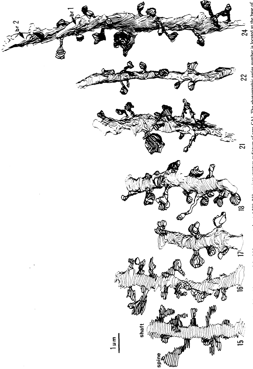
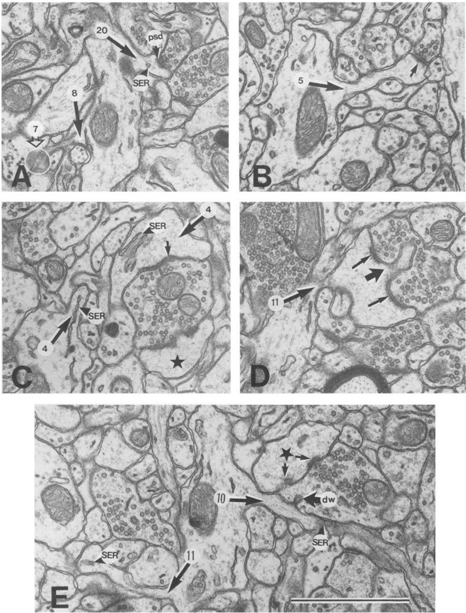
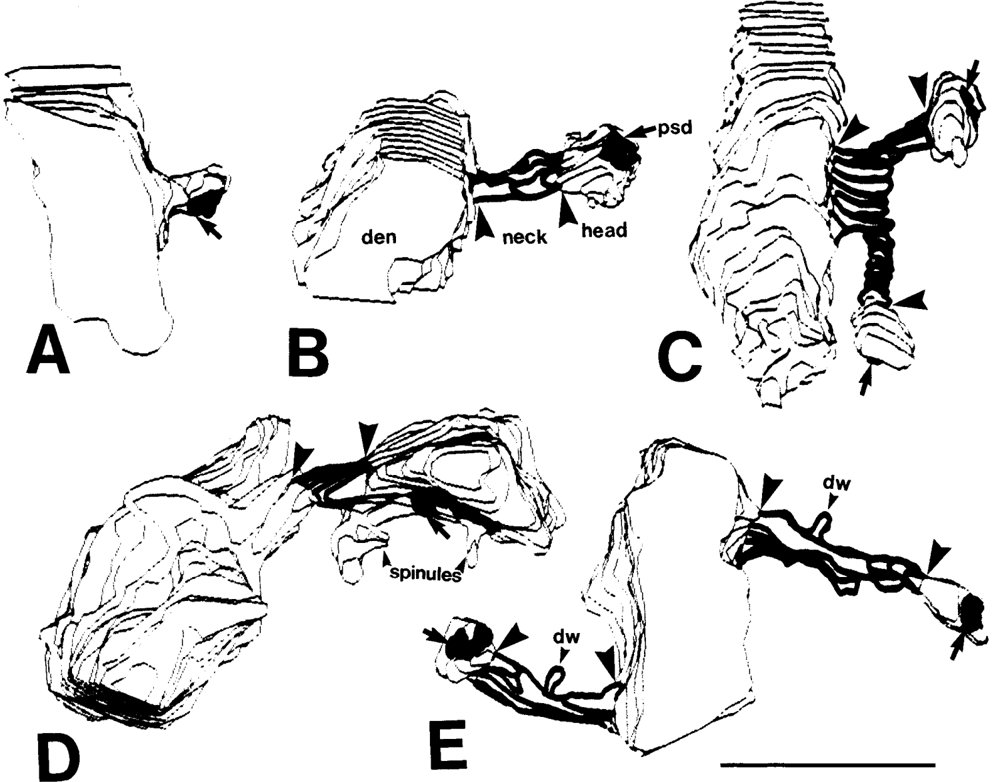
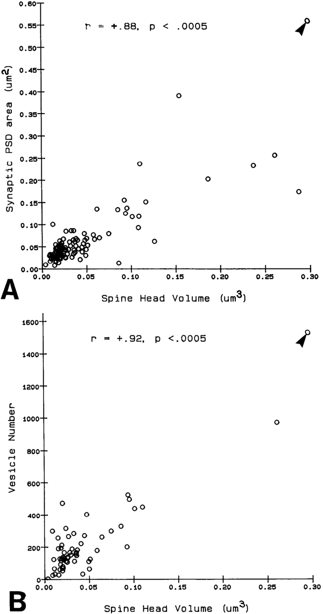
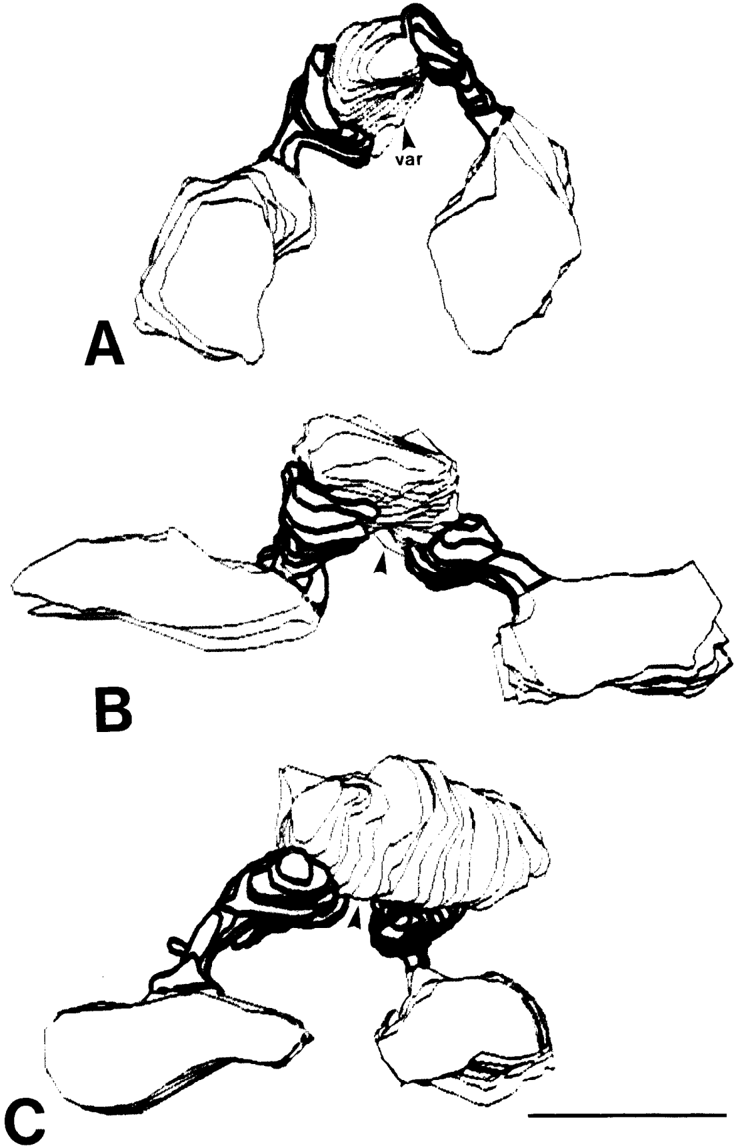
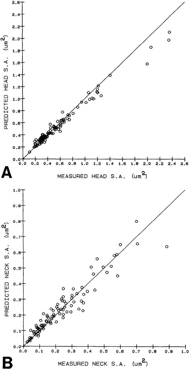
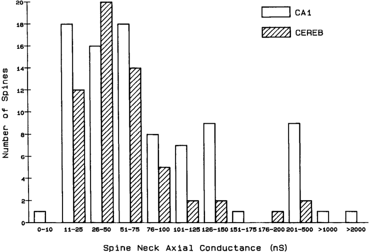
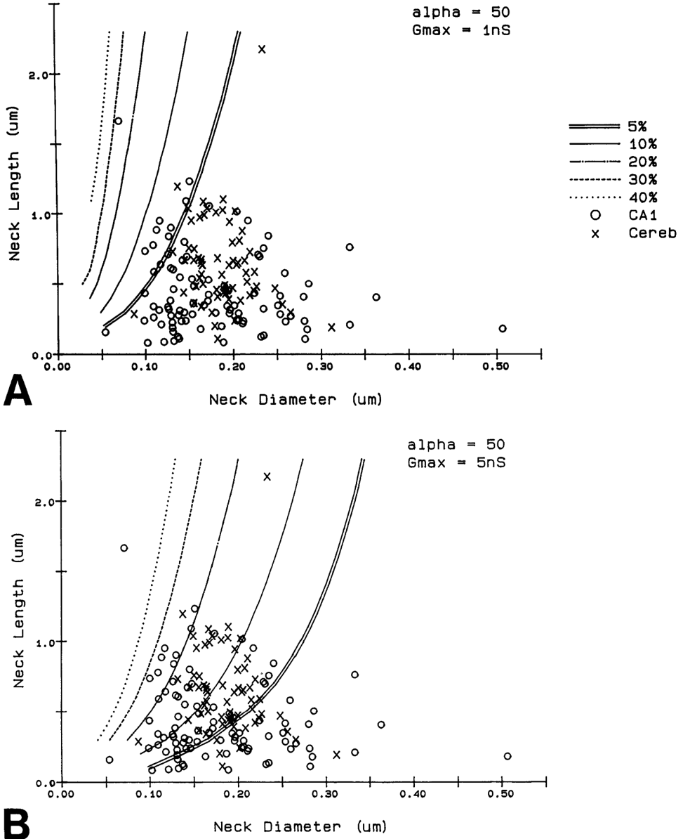
Serial electron microscopy and 3-D reconstructions of dendritic spines from hippocampal area CA 1 dendrites were obtained to evaluate 2 questions about relationships between spine geometry and synaptic efficacy. First, under what biophysical conditions are the spine necks likely to reduce the magnitude of charge transferred from the synapses on the spine heads to the recipient dendrite? Simulation software provided by Charles Wilson (1984) was used to determine that if synaptic conductance is 1 nS or less, only 1% of the hippocampal spine necks are sufficiently thin and long to reduce charge transfer by more than 10%. If synaptic conductance approaches 5 nS, however, 33% of the hippocampal spine necks are sufficiently thin and long to reduce charge transfer by more than 10%. Second, is spine geometry associated with other anatomical indicators of synaptic efficacy, including the area of the postsynaptic density and the number of vesicles in the presynaptic axon? Reconstructed spines were graphically edited into head and neck compartments, and their dimensions were measured, the areas of the postsynaptic densities (PSD) were measured, and all of the vesicles in the presynaptic axonal varicosities were counted. The dimensions of the spine head were well correlated with the area of PSD and the number of vesicles in the presynaptic axonal varicosity. Spine neck diameter and length were not correlated with PSD area, head volume, or the number of vesicles. These results suggest that the dimensions of the spine head, but not the spine neck, reflect differences in synaptic efficacy. We suggest that the constricted necks of hippocampal dendritic spines might reduce diffusion of activated molecules to neighboring synapses, thereby attributing specificity to activated or potentiated synapses.
Overview
The claims and statistics of this paper haven’t been checked yet.
Run the check — free, no account needed. It reads the open-access text and checks the sources it cites.
Integrity signals
Registry
Cites
Statistics
Figures
Verification
Claims the paper makes
The claims and statistics of this paper haven’t been checked yet.
Run the check — free, no account needed. It reads the open-access text and checks the sources it cites.
Claims other papers make about this one
Each is checked against this paper’s own text: does it say what the citing paper says it does?
The source backs part of the claim, not all of it — or backs it for a different population, condition or comparison.
In the citing paper
“This is probably one of the reasons why many ultrastructural studies that utilized high concentrations of potassium ferrocyanide/ferricyanide (1.5–3.0%) provided no data on SS (e.g., Harris and Stevens, 1989; Hayworth et al., 2014; Yakoubi et al., 2019a; Yin et al., 2020; Gour et al., 2021; Phelps et al., 2021; Peddie et al., 2022; Turner et al., 2022).”
From Unambiguous identification of asymmetric and symmetric synapses using volume electron microscopy (2024) · cites this paper as Harris and Stevens, 1989
Experimental — accuracy not yet measuredThe source backs part of the claim, not all of it — or backs it for a different population, condition or comparison.
In the citing paper
“Given that each spine receives only one synaptic input13, our data indicate the highest possible degree of freedom is available in synaptic connections in the dendrites, which has been assumed in the learning models of most neural network theories17.”
From Dendritic spine geometry is critical for AMPA receptor expression in hippocampal CA1 pyramidal neurons (2001) · cites this paper as [13]
Experimental — accuracy not yet measuredThe source backs part of the claim, not all of it — or backs it for a different population, condition or comparison.
In the citing paper
“This observation suggests that the expression of AMPA receptors is controlled independently at a level of the individual spine, consistent with the classical notion that a single spine may behave as an independent chemical compartment to regulate synaptic transmission13, 14.”
From Dendritic spine geometry is critical for AMPA receptor expression in hippocampal CA1 pyramidal neurons (2001) · cites this paper as [13]
Experimental — accuracy not yet measuredThe source backs part of the claim, not all of it — or backs it for a different population, condition or comparison.
In the citing paper
“Regions of glutamate sensitivity tended to be asymmetrically localized to one portion of a spine (Figs. 3h and 4c–j), consistent with the predominant localization of AMPA receptors within PSDs with a diameter of 0.1 to 0.5μm13, 19, 20.”
From Dendritic spine geometry is critical for AMPA receptor expression in hippocampal CA1 pyramidal neurons (2001) · cites this paper as [13]
Experimental — accuracy not yet measuredThe source backs part of the claim, not all of it — or backs it for a different population, condition or comparison.
In the citing paper
“Syn1a-CFP puncta area calculated as a measure of bouton area, which correlated with pHuse area, as expected42.”
From A trans-synaptic nanocolumn aligns neurotransmitter release to receptors (2016) · cites this paper as [42]
Experimental — accuracy not yet measured
The source backs part of the claim, not all of it — or backs it for a different population, condition or comparison.
In the citing paper
“In turn, the size of the postsynaptic density is correlated with the number of presynaptic vesicles, the number of postsynaptic receptors and the ready-releasable pool of transmitter (Harris and Stevens, 1989; Nusser et al., 1998; Schikorski and Stevens, 1999, 2001; Arellano et al., 2007a).”
From The influence of phospho-tau on dendritic spines of cortical pyramidal neurons in patients with Alzheimer’s disease (2013) · cites this paper as Harris and Stevens, 1989
Experimental — accuracy not yet measuredThe source backs part of the claim, not all of it — or backs it for a different population, condition or comparison.
In the citing paper
“Additionally, the volume of the dendritic spine head is related to the size of the postsynaptic density (Harris and Stevens, 1989; Schikorski and Stevens, 1997; Arellano et al., 2007b), the number of presynaptic vesicles, and the number of vesicles adjacent to the presynaptic cleft (Harris and Stevens, 1989).”
From The influence of phospho-tau on dendritic spines of cortical pyramidal neurons in patients with Alzheimer’s disease (2013) · cites this paper as Harris and Stevens, 1989
Experimental — accuracy not yet measuredThe source backs part of the claim, not all of it — or backs it for a different population, condition or comparison.
In the citing paper
“Furthermore, the volume of the head is directly proportional to the strength of synaptic currents (Harris and Stevens, 1989; Schikorski and Stevens, 1997; Arellano et al., 2007a).”
From The influence of phospho-tau on dendritic spines of cortical pyramidal neurons in patients with Alzheimer’s disease (2013) · cites this paper as Harris and Stevens, 1989
Experimental — accuracy not yet measuredThe cited source says what the claim attributes to it.
In the citing paper
“The relationship between spine geometry and expression of functional AMPA receptors demonstrated here provides substantial support to the hypothesis that neuronal fine structures have functional correlates in the brain13, 18, 21.”
From Dendritic spine geometry is critical for AMPA receptor expression in hippocampal CA1 pyramidal neurons (2001) · cites this paper as [13]
The cited source says what the claim attributes to it.
In the citing paper
“These data are consistent with the correlation between AMPA receptor immunoreactivity and the size of PSD19, 20, because the size of PSD is nearly proportional to the spine-head volume13.”
From Dendritic spine geometry is critical for AMPA receptor expression in hippocampal CA1 pyramidal neurons (2001) · cites this paper as [13]
The cited source says what the claim attributes to it.
In the citing paper
“whereas the spine head volume is correlated with the area of the postsynaptic density.”
From The influence of phospho-tau on dendritic spines of cortical pyramidal neurons in patients with Alzheimer’s disease (2013) · cites this paper as Harris and Stevens, 1989
Self-check
Percentages, sums and reported differences re-computed from the paper’s own numbers.
The self-check needs the paper’s full text.
Citations
Cites
The Synaptic Organization of the Brain
(2004) · Oxford University Press eBooks · doi:10.1093/acprof:oso/9780195159561.001.1 · Cited by 3,574
The Synaptic Organization of the Brain
Freemon, F. R. (1980) · JAMA · doi:10.1001/jama.1980.03300440052037 · Cited by 709
Chang, F., & Greenough, W. T. (1984) · Brain Research · doi:10.1016/0006-8993(84)91008-4 · Cited by 523
Lee, K. S., Schottler, F., Oliver, M. E., & LYNCH, G. S. (1980) · Journal of Neurophysiology · doi:10.1152/jn.1980.44.2.247 · Cited by 473
Harris, K., & Stevens, J. (1988) · The Journal of Neuroscience · doi:10.1523/jneurosci.08-12-04455.1988 · Cited by 318
Self-citationDesmond, N. L., & Levy, W. B. (1983) · Brain Research · doi:10.1016/0006-8993(83)91329-x · Cited by 276
Three-Dimensional analysis of dendritic spines
Špaček, J., & Hartmann, M. (1983) · Anatomy and Embryology · doi:10.1007/bf00298517 · Cited by 269
The anatomy of a memory: convergence of results across a diversity of tests
Greenough, W. T., & Bailey, C. H. (1988) · Trends in Neurosciences · doi:10.1016/0166-2236(88)90139-7 · Cited by 266
Crick, F. (1982) · Trends in Neurosciences · doi:10.1016/0166-2236(82)90020-0 · Cited by 254
Van Harreveld, A., & Fifkova, E. (1975) · Experimental Neurology · doi:10.1016/0014-4886(75)90055-2 · Cited by 239
Research works printed but not linked: 6
- Harris, K. M., F. E. Jensen, and B. Tsao (1988) Ultrastructure, de- velopment, and plasticity of synapses in area CA1 of the rat hippo- campus: Extending our vision with serial electron microscopy and three dimensional analyses. Neurol. Neurobiol. (in press).
- Andersen, P., T. Blackstad, G. Hulleberg, J. Line Vaaland, and M. Andersen, P., T. Blackstad, G. Hulleberg, M. Trommald, and J. L. Vaaland (1988b) Dimensions ofdendritic spines ofrat dentate gran- Trommald (1988a) Changes in spine morphology associated with ule cells during long-term potentiation (LTP). Proc. Physiol. Sot. P.C. 50. long-term potentiation in rat dentate granule cells. Proc. Physiol. Sot. s.
- Diamond, J., E. G. Gray, and G. M. Yasargil (1970) The function of dendritic spines: An hypothesis. In Excitatory Synaptic Mechanisms, P. Andersen and J. Jensen, eds., pp. 2 13-222, Universitets Forlaget, Oslo.
- Meek, G. A. (1976) Practical Electron Microscopyfor Biologists, Wiley, New York.
- Rall, W. (1970) Cable properties of dendrites and effects of synaptic location. In Excitatory Synaptic Mechanisms, P. Andersen and J. K. S. Jensen, eds., pp. 175-l 87, Universitets Forlaget, Oslo.
- Rall, W. (1978) Dendritic spines and synaptic potency. In Studies in NeurophysioloRy, A. K. McIntyre and K. Porter, eds., pp. 203-209, Cambridge U.-P., Cambridge.
Cited by
Sorted by citations
The synaptic vesicle cycle: a cascade of protein–protein interactions
Südhof, T. C. (1995) · Nature · doi:10.1038/375645a0 · Cited by 1,991
Experience-dependent structural synaptic plasticity in the mammalian brain
Holtmaat, A., & Svoboda, K. (2009) · Nature Reviews Neuroscience · doi:10.1038/nrn2699 · Cited by 1,934
Long-term in vivo imaging of experience-dependent synaptic plasticity in adult cortex
Trachtenberg, J. T., Chen, B. E., Knott, G. W., Feng, G., Sanes, J. R., Welker, E., & Svoboda, K. (2002) · Nature · doi:10.1038/nature01273 · Cited by 1,927
Pyramidal neurons: dendritic structure and synaptic integration
Spruston, N. (2008) · Nature Reviews Neuroscience · doi:10.1038/nrn2286 · Cited by 1,688
Shankar, G. M., Bloodgood, B. L., Townsend, M., Walsh, D. M., Selkoe, D. J., & Sabatini, B. L. (2007) · The Journal of Neuroscience · doi:10.1523/jneurosci.4970-06.2007 · Cited by 1,592
The mGluR theory of fragile X mental retardation
Bear, M. F., Huber, K. M., & Warren, S. T. (2004) · Trends in Neurosciences · doi:10.1016/j.tins.2004.04.009 · Cited by 1,580
Matsuzaki, M., Ellis-Davies, G. C. R., Nemoto, T., Miyashita, Y., Iino, M., & Kasai, H. (2001) · Nature Neuroscience · doi:10.1038/nn736 · Cited by 1,563
Distinct morphological stages of dentate granule neuron maturation in the adult mouse hippocampus
Zhao, C., Teng, E. M., Summers, R. G., Ming, G.-L., & Gage, F. H. (2006) · The Journal of Neuroscience · doi:10.1523/jneurosci.3648-05.2006 · Cited by 1,257
Structure and function of dendritic spines
Nimchinsky, E. A., Sabatini, B. L., & Svoboda, K. (2002) · Annual Review of Physiology · doi:10.1146/annurev.physiol.64.081501.160008 · Cited by 1,208
Long-term dendritic spine stability in the adult cortex
Grutzendler, J., Kasthuri, N., & Gan, W.-B. (2002) · Nature · doi:10.1038/nature01276 · Cited by 1,198
Rapid Dendritic Morphogenesis in CA1 Hippocampal Dendrites Induced by Synaptic Activity
Maletic-Savatic, M., Malinow, R., & Svoboda, K. (1999) · Science · doi:10.1126/science.283.5409.1923 · Cited by 1,009
Naturally occurring fluctuation in dendritic spine density on adult hippocampal pyramidal neurons
Woolley, C., Gould, E., Frankfurt, M., & McEwen, B. (1990) · Journal of Neuroscience · doi:10.1523/jneurosci.10-12-04035.1990 · Cited by 1,107
Shrinkage of dendritic spines associated with long-term depression of hippocampal synapses
Zhou, Q., Homma, K. J., & Poo, M.-M. (2004) · Neuron · doi:10.1016/j.neuron.2004.11.011 · Cited by 1,025
Structural plasticity and memory
Lamprecht, R., & LeDoux, J. (2004) · Nature Reviews Neuroscience · doi:10.1038/nrn1301 · Cited by 1,010
Harris, K. M., & Kater, S. B. (1994) · Annual Review of Neuroscience · doi:10.1146/annurev.ne.17.030194.002013 · Cited by 1,020
Self-citationQuantitative ultrastructural analysis of hippocampal excitatory synapses
Schikorski, T., & Stevens, C. F. (1997) · The Journal of Neuroscience · doi:10.1523/jneurosci.17-15-05858.1997 · Cited by 997
Harris, K. M., Jensen, F. E., & Tsao, B. (1992) · Journal of Neuroscience · doi:10.1523/jneurosci.12-07-02685.1992 · Cited by 1,036
Self-citationWoolley, C. S., & McEwen, B. (1992) · Journal of Neuroscience · doi:10.1523/jneurosci.12-07-02549.1992 · Cited by 998
The log-dynamic brain: how skewed distributions affect network operations
Buzsáki, G., & Mizuseki, K. (2014) · Nature reviews. Neuroscience · doi:10.1038/nrn3687 · Cited by 1,019
Balancing Structure and Function at Hippocampal Dendritic Spines
Bourne, J. N., & Harris, K. M. (2008) · Annual Review of Neuroscience · doi:10.1146/annurev.neuro.31.060407.125646 · Cited by 916
Self-citationThree-Dimensional Relationships between Hippocampal Synapses and Astrocytes
Ventura, R. E., & Harris, K. M. (1999) · Journal of Neuroscience · doi:10.1523/jneurosci.19-16-06897.1999 · Cited by 907
Self-citationOkamoto, K.-I., Nagai, T., Miyawaki, A., & Hayashi, Y. (2004) · Nature Neuroscience · doi:10.1038/nn1311 · Cited by 854
Structure-stability-function relationships of dendritic spines
Kasai, H., Matsuzaki, M., Noguchi, J., Yasumatsu, N., & Nakahara, H. (2003) · Trends in Neurosciences · doi:10.1016/s0166-2236(03)00162-0 · Cited by 847
Koffie, R. M., Meyer-Luehmann, M., Hashimoto, T., Adams, K. W., Mielke, M. L., Garcia-Alloza, M., Micheva, K. D., Smith, S. J., Kim, M. L., Lee, V. M., et al. (2009) · Proceedings of the National Academy of Sciences · doi:10.1073/pnas.0811698106 · Cited by 850
Actin in action: the interplay between the actin cytoskeleton and synaptic efficacy.
Cingolani, L. A., & Goda, Y. (2008) · Nature Reviews Neuroscience · doi:10.1038/nrn2373 · Cited by 831
Repeated Stress Induces Dendritic Spine Loss in the Rat Medial Prefrontal Cortex
Radley, J. J., Rocher, A. B., Miller, M., Janssen, W. G. M., Liston, C., Hof, P. R., McEwen, B. S., & Morrison, J. H. (2006) · Cerebral Cortex · doi:10.1093/cercor/bhi104 · Cited by 820
Dendritic Spine Pathology: Cause or Consequence of Neurological Disorders?
Fiala, J. C., Spacek, J., & Harris, K. M. (2002) · Brain Research Reviews · doi:10.1016/s0165-0173(02)00158-3 · Cited by 811
Self-citationDendritic Excitability and Synaptic Plasticity
Sjöström, P. J., Rancz, E. A., Roth, A., & Häusser, M. (2008) · Physiological Reviews · doi:10.1152/physrev.00016.2007 · Cited by 740
Jonas, P., Major, G., & Sakmann, B. (1993) · The Journal of Physiology · doi:10.1113/jphysiol.1993.sp019965 · Cited by 720
Filipello, F., Morini, R., Corradini, I., Zerbi, V., Canzi, A., Michalski, B., Erreni, M., Markicevic, M., Starvaggi-Cucuzza, C., Otero, K., et al. (2018) · Immunity · doi:10.1016/j.immuni.2018.04.016 · Cited by 695
Development of long-term dendritic spine stability in diverse regions of cerebral cortex
Zuo, Y., Lin, A., Chang, P., & Gan, W.-B. (2005) · Neuron · doi:10.1016/j.neuron.2005.04.001 · Cited by 684
A trans-synaptic nanocolumn aligns neurotransmitter release to receptors
Tang, A.-H., Chen, H., Li, T. P., Metzbower, S. R., MacGillavry, H. D., & Blanpied, T. A. (2016) · Nature · doi:10.1038/nature19058 · Cited by 677
Structural and Molecular Remodeling of Dendritic Spine Substructures during Long-Term Potentiation
Bosch, M., Castro, J., Saneyoshi, T., Matsuno, H., Sur, M., & Hayashi, Y. (2014) · Neuron · doi:10.1016/j.neuron.2014.03.021 · Cited by 637
Inactivity produces increases in neurotransmitter release and synapse size
Murthy, V. N., Schikorski, T., Stevens, C. F., & Zhu, Y. (2001) · Neuron · doi:10.1016/s0896-6273(01)00500-1 · Cited by 612
Experience-dependent and cell-type-specific spine growth in the neocortex
Holtmaat, A., Wilbrecht, L., Knott, G. W., Welker, E., & Svoboda, K. (2006) · Nature · doi:10.1038/nature04783 · Cited by 631
Nonuniform probability of glutamate release at a hippocampal synapse.
Rosenmund, C., Clements, J., & Westbrook, G. (1993) · Science · doi:10.1126/science.7901909 · Cited by 616
Spacek, J., & Harris, K. M. (1997) · The Journal of Neuroscience · doi:10.1523/jneurosci.17-01-00190.1997 · Cited by 599
Self-citationTransmitter timecourse in the synaptic cleft: its role in central synaptic function
Clements, J. D. (1996) · Trends in Neurosciences · doi:10.1016/s0166-2236(96)10024-2 · Cited by 598
On the Origin of the Extracellular Action Potential Waveform: A Modeling Study
Gold, C., Henze, D. A., Christof Koch, A., & Buzsáki, G. (2006) · Journal of Neurophysiology · doi:10.1152/jn.00979.2005 · Cited by 587
Synapse formation on neurons born in the adult hippocampus
Toni, N., Teng, E. M., Bushong, E. A., Aimone, J. B., Zhao, C., Consiglio, A., van Praag, H., Martone, M. E., Ellisman, M. H., & Gage, F. H. (2007) · Nature Neuroscience · doi:10.1038/nn1908 · Cited by 579
Ultrastructural evidence for synaptic scaling across the wake/sleep cycle
de Vivo, L., Bellesi, M., Marshall, W., Bushong, E. A., Ellisman, M. H., Tononi, G., & Cirelli, C. (2017) · Science · doi:10.1126/science.aah5982 · Cited by 574
Spine growth precedes synapse formation in the adult neocortex in vivo
Knott, G. W., Holtmaat, A., Wilbrecht, L., Welker, E., & Svoboda, K. (2006) · Nature Neuroscience · doi:10.1038/nn1747 · Cited by 563
REM sleep selectively prunes and maintains new synapses in development and learning
Li, W., Ma, L., Yang, G., & Gan, W.-B. (2017) · Nature Neuroscience · doi:10.1038/nn.4479 · Cited by 543
Anatomical and Physiological Plasticity of Dendritic Spines
Alvarez, V. A., & Sabatini, B. L. (2007) · Annual Review of Neuroscience · doi:10.1146/annurev.neuro.30.051606.094222 · Cited by 629
Ultrastructure of dendritic spines: correlation between synaptic and spine morphologies
Arellano, J. I. (2007) · Frontiers in Neuroscience · doi:10.3389/neuro.01.1.1.010.2007 · Cited by 529
Miniature synaptic events maintain dendritic spines via AMPA receptor activation
McKinney, R. A., Capogna, M., Dürr, R., Gähwiler, B. H., & Thompson, A. S. M. (1999) · Nature Neuroscience · doi:10.1038/4548 · Cited by 523
The kinetics of synaptic vesicle recycling measured at single presynaptic boutons
Ryan, T. A., REUTER, H., Wendland, B., Schweizer, F. E., Tsien, R. W., & Smith, S. J. (1993) · Neuron · doi:10.1016/0896-6273(93)90081-2 · Cited by 517
Spectrum of tumor angiogenesis in the bone marrow of children with acute lymphoblastic leukemia.
Pérez‐Atayde, A. R., Sallan, S. E., Tedrow, U. B., Connors, S. L., Allred, E. N., & Folkman, J. (1997) · Europe PMC (PubMed Central) · Cited by 516
Anatomy and function of an excitatory network in the visual cortex
Lee, W.-C. A., Bonin, V., Reed, M., Graham, B. J., Hood, G., Glattfelder, K. J., & Reid, R. C. (2016) · Nature · doi:10.1038/nature17192 · Cited by 496
Branch-specific dendritic Ca2+ spikes cause persistent synaptic plasticity
Cichon, J., & Gan, W. (2015) · Nature · doi:10.1038/nature14251 · Cited by 527
Shepherd, G. M. G., & Harris, K. M. (1998) · The Journal of Neuroscience · doi:10.1523/jneurosci.18-20-08300.1998 · Cited by 502
Self-citationThe Subspine Organization of Actin Fibers Regulates the Structure and Plasticity of Dendritic Spines
Honkura, N., Matsuzaki, M., Noguchi, J., Ellis-Davies, G. C. R., & Kasai, H. (2008) · Neuron · doi:10.1016/j.neuron.2008.01.013 · Cited by 490
MECHANISMS OF VERTEBRATE SYNAPTOGENESIS
Waites, C. L., Craig, A. M., & Garner, C. C. (2005) · Annual Review of Neuroscience · doi:10.1146/annurev.neuro.27.070203.144336 · Cited by 483
Dendritic spines as individual neuronal compartments for synaptic Ca2+ responses
Müller, W. S., & Connor, J. A. (1991) · Nature · doi:10.1038/354073a0 · Cited by 479
Direct measurement of coupling between dendritic spines and shafts
Svoboda, K., Tank, D. W., & Denk, W. (1996) · Science · doi:10.1126/science.272.5262.716 · Cited by 472
Dendritic Spines: The Locus of Structural and Functional Plasticity
Sala, C., & Segal, M. (2014) · Physiological Reviews · doi:10.1152/physrev.00012.2013 · Cited by 491
Spine loss and other dendritic abnormalities in epilepsy
Swann, J. W., Al‐Noori, S., Jiang, M., & Lee, C. L. (2000) · Hippocampus · doi:10.1002/1098-1063(2000)10:5 · Cited by 268
Self-citationInhibition of SRGAP2 function by its human-specific paralogs induces neoteny during spine maturation
Charrier, C., Joshi, K., Coutinho-Budd, J., Kim, J.-E., Lambert, N., de Marchena, J., Jin, W.-L., Vanderhaeghen, P., Ghosh, A., Sassa, T., et al. (2012) · Cell · doi:10.1016/j.cell.2012.03.034 · Cited by 454
The actin cytoskeleton: integrating form and function at the synapse
Dillon, C., & Goda, Y. (2005) · Annual Review of Neuroscience · doi:10.1146/annurev.neuro.28.061604.135757 · Cited by 447
Spine neck plasticity regulates compartmentalization of synapses
Tønnesen, J., Katona, G., Rózsa, B., & Nägerl, U. V. (2014) · Nature Neuroscience · doi:10.1038/nn.3682 · Cited by 444
Nanoscale Scaffolding Domains within the Postsynaptic Density Concentrate Synaptic AMPA Receptors
MacGillavry, H. D., Song, Y., Raghavachari, S., & Blanpied, T. A. (2013) · Neuron · doi:10.1016/j.neuron.2013.03.009 · Cited by 442
Dumitriu, D., Hao, J., Hara, Y., Kaufmann, J., Janssen, W. G. M., Lou, W., Rapp, P. R., & Morrison, J. H. (2010) · The Journal of Neuroscience · doi:10.1523/jneurosci.6410-09.2010 · Cited by 439
Zeng, M., Chen, X., Guan, D., Xu, J., Wu, H., Tong, P., & Zhang, M. (2018) · Cell · doi:10.1016/j.cell.2018.06.047 · Cited by 438
Local externalization of phosphatidylserine mediates developmental synaptic pruning by microglia
Scott‐Hewitt, N., Perrucci, F., Morini, R., Erreni, M., Mahoney, M., Witkowska, A., Carey, A. E., Faggiani, E., Schuetz, L. T., Mason, S., et al. (2020) · The EMBO Journal · doi:10.15252/embj.2020105380 · Cited by 436
A model of spike initiation in neocortical pyramidal neurons
Mainen, Z. F., Joerges, J., Huguenard, J. R., & B, T. J. S. A. (1995) · Neuron · doi:10.1016/0896-6273(95)90020-9 · Cited by 431
Chicurel, M. E., & Harris, K. M. (1992) · Journal of Comparative Neurology · doi:10.1002/cne.903250204 · Cited by 417
Self-citationStructural plasticity of dendritic spines
Bosch, M., & Hayashi, Y. (2012) · Current Opinion in Neurobiology · doi:10.1016/j.conb.2011.09.002 · Cited by 415
Ultrastructural analysis of adult mouse neocortex comparing aldehyde perfusion with cryo fixation
Korogod, N., Petersen, C. C., & Knott, G. W. (2015) · eLife · doi:10.7554/elife.05793 · Cited by 408
Homer: a link between neural activity and glutamate receptor function
Xiao, B., Tu, J. C., & Worley, P. F. (2000) · Current Opinion in Neurobiology · doi:10.1016/s0959-4388(00)00087-8 · Cited by 406
Release probability of hippocampal glutamatergic terminals scales with the size of the active zone
Holderith, N., Lorincz, A., Katona, G., Rózsa, B., Kulik, A., Watanabe, M., & Nusser, Z. (2012) · Nature Neuroscience · doi:10.1038/nn.3137 · Cited by 405
Spine-Neck Geometry Determines NMDA Receptor-Dependent Ca2+ Signaling in Dendrites
Noguchi, J., Matsuzaki, M., Ellis-Davies, G. C. R., & Kasai, H. (2005) · Neuron · doi:10.1016/j.neuron.2005.03.015 · Cited by 419
Impermanence of dendritic spines in live adult CA1 hippocampus
Attardo, A., Fitzgerald, J. E., & Schnitzer, M. J. (2015) · Nature · doi:10.1038/nature14467 · Cited by 399
Structural Components of Synaptic Plasticity and Memory ConsolidationPreprint
Bailey, C. H., Kandel, E. R., & Harris, K. M. (2015) · Cold Spring Harbor Perspectives in Biology · doi:10.1101/cshperspect.a021758 · Cited by 419
Self-citationBalance and Stability of Synaptic Structures during Synaptic Plasticity
Meyer, D., Bonhoeffer, T., & Scheuss, V. (2014) · Neuron · doi:10.1016/j.neuron.2014.02.031 · Cited by 395
Bhatt, D. H., Zhang, S., & Gan, W.-B. (2009) · Annual Review of Physiology · doi:10.1146/annurev.physiol.010908.163140 · Cited by 380
Repeated stress alters dendritic spine morphology in the rat medial prefrontal cortex
Radley, J. J., Rocher, A. B., Rodriguez, A., Ehlenberger, D. B., Dammann, M., McEwen, B. S., Morrison, J. H., Wearne, S. L., & Hof, P. R. (2007) · The Journal of Comparative Neurology · doi:10.1002/cne.21588 · Cited by 393
From form to function: calcium compartmentalization in dendritic spines
Yuste, R., Majewska, A. K., & Holthoff, K. (2000) · Nature Neuroscience · doi:10.1038/76609 · Cited by 378
Koch, C., & Zador, A. M. (1993) · Journal of Neuroscience · doi:10.1523/jneurosci.13-02-00413.1993 · Cited by 372
Glutamate receptor dynamics in dendritic microdomains.
Newpher, T. M., & Ehlers, M. D. (2008) · Neuron · doi:10.1016/j.neuron.2008.04.030 · Cited by 356
Miller, O. H., Yang, L., Wang, C.-C., Hargroder, E. A., Zhang, Y., Delpire, E., & Hall, B. J. (2014) · eLife · doi:10.7554/elife.03581 · Cited by 358
Sleep and Synaptic Homeostasis: Structural Evidence in Drosophila
Bushey, D., Tononi, G., & Cirelli, C. (2011) · Science · doi:10.1126/science.1202839 · Cited by 357
Recombinant Probes for Visualizing Endogenous Synaptic Proteins in Living Neurons
Gross, G. G., Junge, J. A., Mora, R. J., Kwon, H., Olson, C. A., Takahashi, T. T., Liman, E. R., Ellis‐Davies, G. C. R., McGee, A. W., Sabatini, B. L., et al. (2013) · Neuron · doi:10.1016/j.neuron.2013.04.017 · Cited by 346
Hung, A. Y., Futai, K., Sala, C., Valtschanoff, J. G., Ryu, J., Woodworth, M. A., Kidd, F. L., Sung, C. C., Miyakawa, T., Bear, M. F., et al. (2008) · The Journal of Neuroscience · doi:10.1523/jneurosci.3032-07.2008 · Cited by 350
Spine architecture and synaptic plasticity
Carlisle, H. J., & Kennedy, M. B. (2005) · Trends in Neurosciences · doi:10.1016/j.tins.2005.01.008 · Cited by 357
Boeckers, T. M., Bockmann, J., Kreutz, M. R., & Gundelfinger, E. D. (2002) · Journal of Neurochemistry · doi:10.1046/j.1471-4159.2002.00931.x · Cited by 349
Molecular mechanisms of dendritic spine development and remodeling
Ethell, I. M., & Pasquale, E. B. (2005) · Progress in Neurobiology · doi:10.1016/j.pneurobio.2005.02.003 · Cited by 367
Differential Control of Vesicle Priming and Short-Term Plasticity by Munc13 Isoforms
Rosenmund, C., Sigler, A., Augustin, I., Reim, K., Brose, N., & Rhee, J. (2002) · Neuron · doi:10.1016/s0896-6273(02)00568-8 · Cited by 346
Rapid Redistribution of Synaptic PSD-95 in the Neocortex In Vivo
Gray, N. W., Weimer, R. M., Bureau, I., & Svoboda, K. (2006) · PLoS Biology · doi:10.1371/journal.pbio.0040370 · Cited by 350
Murase, S., Mosser, E. A., & Schuman, E. M. (2002) · Neuron · doi:10.1016/s0896-6273(02)00764-x · Cited by 337
Block of glutamate transporters potentiates postsynaptic excitation.
Tong, G., & Jahr, C. (1994) · Neuron · doi:10.1016/0896-6273(94)90057-4 · Cited by 361
Bonhoeffer, T., & Yuste, R. (2002) · Neuron · doi:10.1016/s0896-6273(02)00906-6 · Cited by 337
Volume electron microscopy for neuronal circuit reconstruction
Briggman, K. L., & Bock, D. D. (2011) · Current Opinion in Neurobiology · doi:10.1016/j.conb.2011.10.022 · Cited by 352
Presynaptic Regulation of Quantal Size by the Vesicular Glutamate Transporter VGLUT1
Wilson, N. R., Kang, J., Hueske, E. V., Leung, T., Varoqui, H., Murnick, J. G., Erickson, J. D., & Liu, G. (2005) · The Journal of Neuroscience · doi:10.1523/jneurosci.3003-04.2005 · Cited by 335
Dense connectomic reconstruction in layer 4 of the somatosensory cortex
Motta, A., Berning, M., Boergens, K. M., Staffler, B., Beining, M., Loomba, S., Hennig, P., Wissler, H., & Helmstaedter, M. (2019) · Science · doi:10.1126/science.aay3134 · Cited by 333
Multivesicular release from excitatory synapses of cultured hippocampal neurons
Tong, G., & Jahr, C. E. (1994) · Neuron · doi:10.1016/0896-6273(94)90151-1 · Cited by 330
Facilitation at single synapses probed with optical quantal analysis
Oertner, T. G., Sabatini, B. L., Nimchinsky, E. A., & Svoboda, K. (2002) · Nature Neuroscience · doi:10.1038/nn867 · Cited by 320
Principles of Long-Term Dynamics of Dendritic Spines
Yasumatsu, N., Matsuzaki, M., Miyazaki, T., Noguchi, J., & Kasai, H. (2008) · Journal of Neuroscience · doi:10.1523/jneurosci.0603-08.2008 · Cited by 321
Nanoconnectomic upper bound on the variability of synaptic plasticity
Bartol, T. M., Bromer, C., Kinney, J. P., Chirillo, M. A., Bourne, J. N., Harris, K. M., & B, T. J. S. A. (2015) · eLife · doi:10.7554/elife.10778 · Cited by 341
Self-citationSynaptic calcium transients in single spines indicate that NMDA receptors are not saturated
Mainen, Z. F., Malinow, R., & Svoboda, K. (1999) · Nature · doi:10.1038/20187 · Cited by 315
Geometry and structural plasticity of synaptic connectivity
Stepanyants, A., Hof, P. R., & Chklovskii, D. B. (2002) · Neuron · doi:10.1016/s0896-6273(02)00652-9 · Cited by 312
Metadata
- Authors
- Harris, K., & Stevens, J.
- Affiliations
- Boston Children's Hospital (US)
- Venue
- The Journal of Neuroscience
- Publisher
- Society for Neuroscience
- Published
- 1989-08-01
- Volume
- 9
- Issue
- 8
- Pages
- 2982-2997
- ISSN
- 0270-6474 (ISSN-L)
- 1529-2401
- PMC
- PMC6569708
- OpenAlex
- W2117664079
- Cited by
- 1,073
- References
- 38
- Citations by year
- 2026: 17 · 2025: 26 · 2024: 35 · 2023: 33 · 2022: 31 · 2021: 50 · 2020: 39 · 2019: 43 · 2018: 47 · 2017: 33 · 2016: 40 · 2015: 48 · 2014: 44 · 2013: 50 · 2012: 42
- FWCI
- 5.92
- Type
- journal article
- Language
- en
- Open access
- hybrid
- doi.org
- Licence
- Locations
- Journal of Neuroscience (publishedVersion, cc-by-nc-sa)
- Europe PMC (PubMed Central) (publishedVersion)
- PubMed Central (publishedVersion)
- PubMed (publishedVersion)
- Funders
- Alfred P. Sloan Foundation
- National Institutes of Health (NS21184)
- Medical Research Council
- National Institute of Neurological Disorders and Stroke (NS21184, NS 20820)
- Eunice Kennedy Shriver National Institute of Child Health and Human Development (P30HD018655, P30HD-18655)
- Topics
- Life Sciences › Neuroscience › Cellular and Molecular Neuroscience › Neuroscience and Neuropharmacology Research
- Life Sciences › Neuroscience › Cognitive Neuroscience › Memory and Neural Mechanisms
- Life Sciences › Neuroscience › Cognitive Neuroscience › Neural dynamics and brain function
- Keywords
- dendritic spines, CA1 pyramidal neurons, hippocampus, synaptic efficacy, postsynaptic density, synaptic vesicles, charge transfer, synaptic conductance, dendritic spine morphology, synaptic specificity
- MeSH terms
- Animals, Axons, Biophysics, Dendrites, Electric Conductivity, Endoplasmic Reticulum, Hippocampus, Image Processing, Computer-Assisted, Male, Microscopy, Electron, Models, Neurological, Neurons, Purkinje Cells, Rats, Inbred Strains, Synapses, Rats, Biophysical Phenomena
About these checks
Registry, retraction, Cites and statistics results are computed, not judged, with one exception: a printed reference that no index lists is read once by a model before it counts as a citation error. Claims are extracted by a model and kept only when their quote appears verbatim in the text; each cited source is then checked, with a model’s help, for whether it says what the claim relies on. Those verdicts can be wrong — read the claim’s quote and the cited source before relying on one.
Statistics and arithmetic are recomputed, not judged.
Confidence bands are shown only for fields calibrated on our annotated test set. None is calibrated yet, so no band is shown.
What counts as a claim ›How we measure confidence ›How we check this ›
Are you an author, or do you see a problem with this page? Request a review
