

The DOI matches its Crossref record; 36 of the 38 references it cites were found in a registry or an index.
Dendritic spines of CA 1 pyramidal cells in the rat hippocampus: serial electron microscopy with reference to their biophysical characteristics
Open accessAbstract
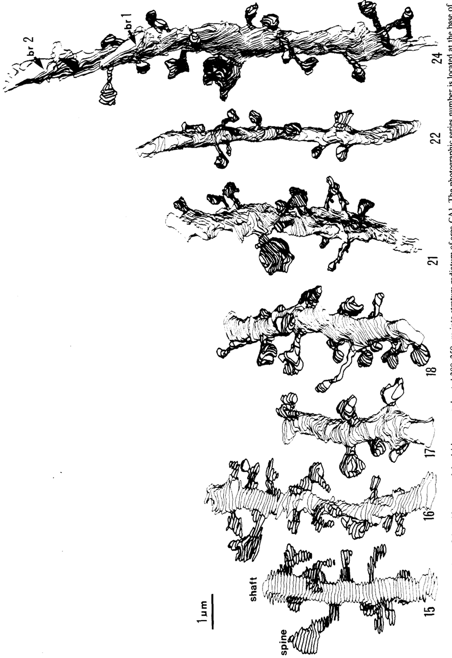
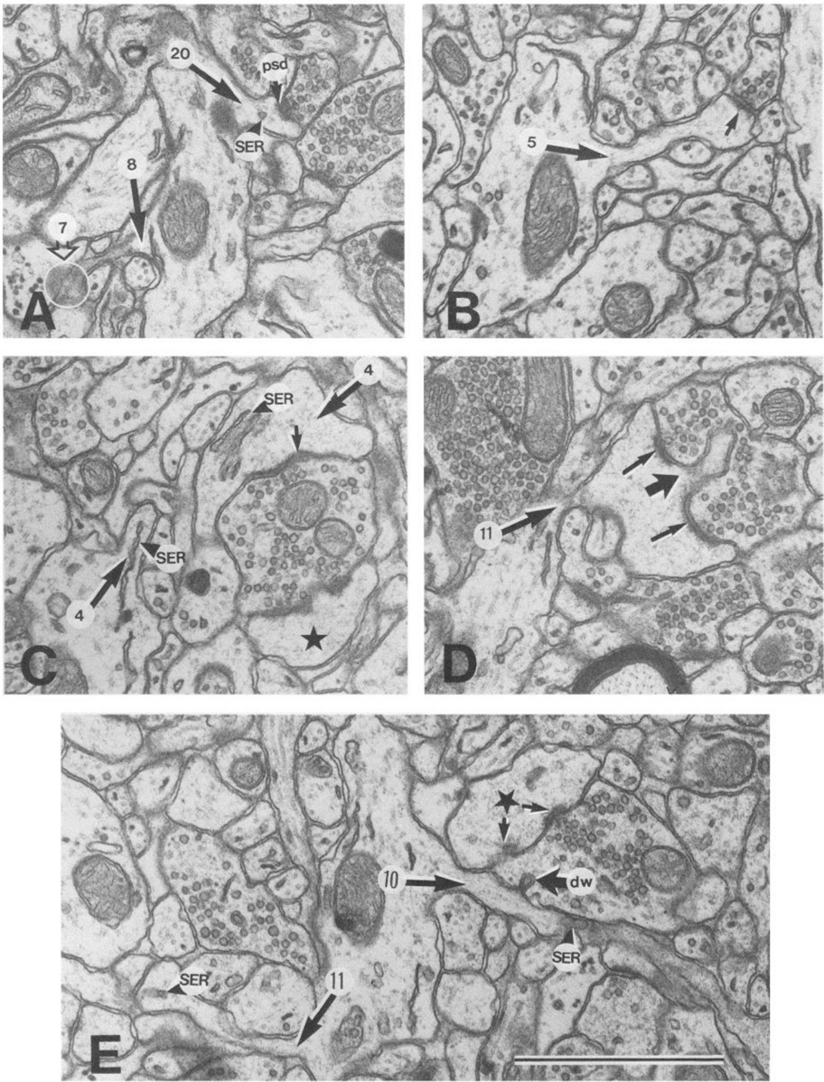
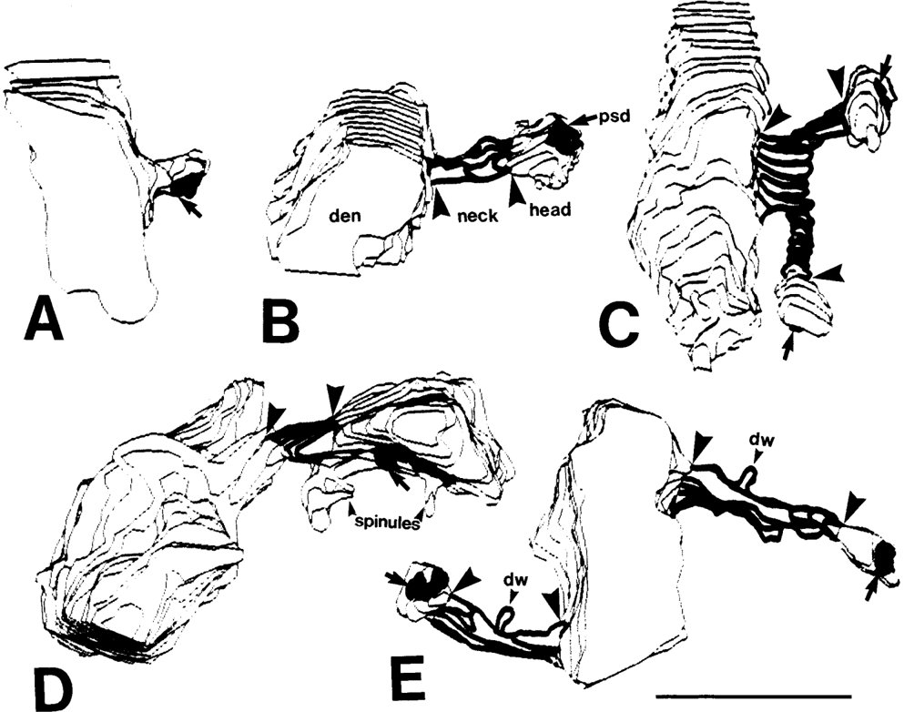
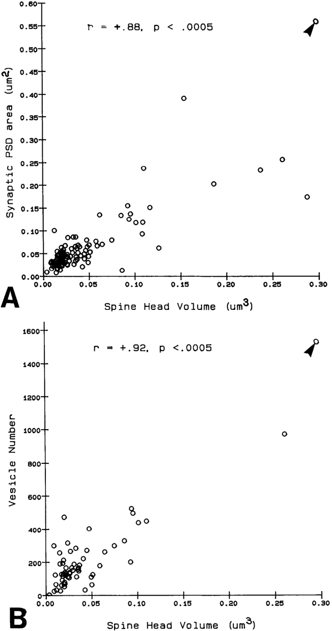
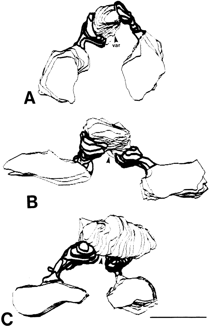
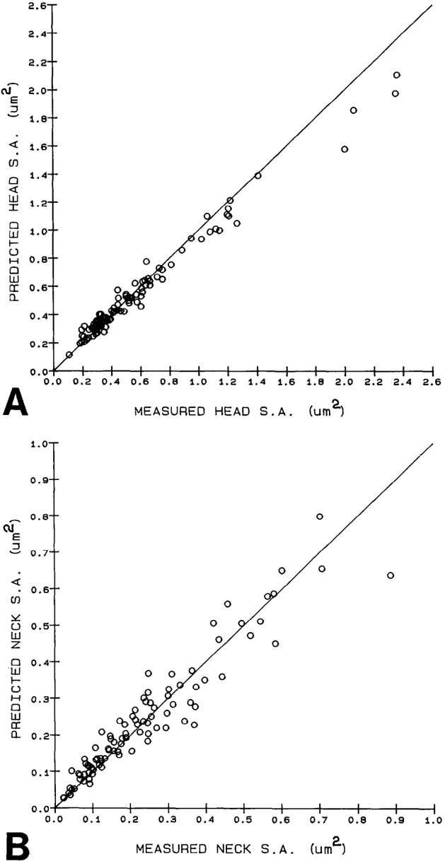
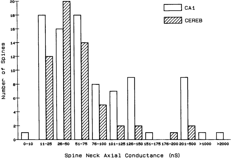
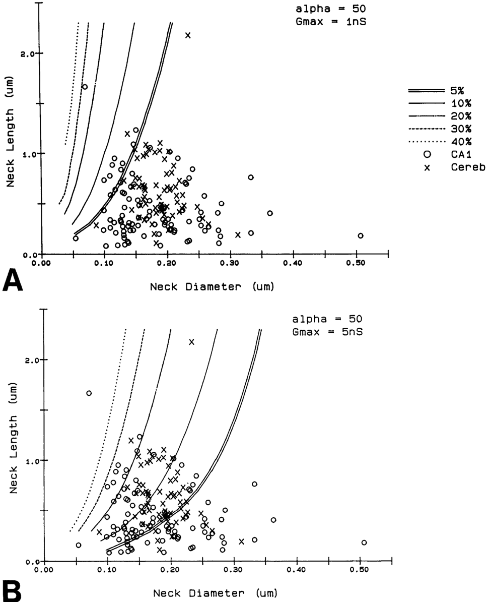
Serial electron microscopy and 3-D reconstructions of dendritic spines from hippocampal area CA 1 dendrites were obtained to evaluate 2 questions about relationships between spine geometry and synaptic efficacy. First, under what biophysical conditions are the spine necks likely to reduce the magnitude of charge transferred from the synapses on the spine heads to the recipient dendrite? Simulation software provided by Charles Wilson (1984) was used to determine that if synaptic conductance is 1 nS or less, only 1% of the hippocampal spine necks are sufficiently thin and long to reduce charge transfer by more than 10%. If synaptic conductance approaches 5 nS, however, 33% of the hippocampal spine necks are sufficiently thin and long to reduce charge transfer by more than 10%. Second, is spine geometry associated with other anatomical indicators of synaptic efficacy, including the area of the postsynaptic density and the number of vesicles in the presynaptic axon? Reconstructed spines were graphically edited into head and neck compartments, and their dimensions were measured, the areas of the postsynaptic densities (PSD) were measured, and all of the vesicles in the presynaptic axonal varicosities were counted. The dimensions of the spine head were well correlated with the area of PSD and the number of vesicles in the presynaptic axonal varicosity. Spine neck diameter and length were not correlated with PSD area, head volume, or the number of vesicles. These results suggest that the dimensions of the spine head, but not the spine neck, reflect differences in synaptic efficacy. We suggest that the constricted necks of hippocampal dendritic spines might reduce diffusion of activated molecules to neighboring synapses, thereby attributing specificity to activated or potentiated synapses.
Overview
The claims and statistics of this paper haven’t been checked yet.
Run the check — free, no account needed. It reads the open-access text and checks the sources it cites.
Integrity signals
Registry
Cites
Statistics
Figures
Verification
Claims the paper makes
The claims and statistics of this paper haven’t been checked yet.
Run the check — free, no account needed. It reads the open-access text and checks the sources it cites.
Claims other papers make about this one
Each is checked against this paper’s own text: does it say what the citing paper says it does?
The source backs part of the claim, not all of it — or backs it for a different population, condition or comparison.
In the citing paper
“This is probably one of the reasons why many ultrastructural studies that utilized high concentrations of potassium ferrocyanide/ferricyanide (1.5–3.0%) provided no data on SS (e.g., Harris and Stevens, 1989; Hayworth et al., 2014; Yakoubi et al., 2019a; Yin et al., 2020; Gour et al., 2021; Phelps et al., 2021; Peddie et al., 2022; Turner et al., 2022).”
From Unambiguous identification of asymmetric and symmetric synapses using volume electron microscopy (2024) · cites this paper as Harris and Stevens, 1989
Experimental — accuracy not yet measuredThe source backs part of the claim, not all of it — or backs it for a different population, condition or comparison.
In the citing paper
“Given that each spine receives only one synaptic input13, our data indicate the highest possible degree of freedom is available in synaptic connections in the dendrites, which has been assumed in the learning models of most neural network theories17.”
From Dendritic spine geometry is critical for AMPA receptor expression in hippocampal CA1 pyramidal neurons (2001) · cites this paper as [13]
Experimental — accuracy not yet measuredThe source backs part of the claim, not all of it — or backs it for a different population, condition or comparison.
In the citing paper
“This observation suggests that the expression of AMPA receptors is controlled independently at a level of the individual spine, consistent with the classical notion that a single spine may behave as an independent chemical compartment to regulate synaptic transmission13, 14.”
From Dendritic spine geometry is critical for AMPA receptor expression in hippocampal CA1 pyramidal neurons (2001) · cites this paper as [13]
Experimental — accuracy not yet measuredThe source backs part of the claim, not all of it — or backs it for a different population, condition or comparison.
In the citing paper
“Regions of glutamate sensitivity tended to be asymmetrically localized to one portion of a spine (Figs. 3h and 4c–j), consistent with the predominant localization of AMPA receptors within PSDs with a diameter of 0.1 to 0.5μm13, 19, 20.”
From Dendritic spine geometry is critical for AMPA receptor expression in hippocampal CA1 pyramidal neurons (2001) · cites this paper as [13]
Experimental — accuracy not yet measuredThe source backs part of the claim, not all of it — or backs it for a different population, condition or comparison.
In the citing paper
“Syn1a-CFP puncta area calculated as a measure of bouton area, which correlated with pHuse area, as expected42.”
From A trans-synaptic nanocolumn aligns neurotransmitter release to receptors (2016) · cites this paper as [42]
Experimental — accuracy not yet measured
The source backs part of the claim, not all of it — or backs it for a different population, condition or comparison.
In the citing paper
“In turn, the size of the postsynaptic density is correlated with the number of presynaptic vesicles, the number of postsynaptic receptors and the ready-releasable pool of transmitter (Harris and Stevens, 1989; Nusser et al., 1998; Schikorski and Stevens, 1999, 2001; Arellano et al., 2007a).”
From The influence of phospho-tau on dendritic spines of cortical pyramidal neurons in patients with Alzheimer’s disease (2013) · cites this paper as Harris and Stevens, 1989
Experimental — accuracy not yet measuredThe source backs part of the claim, not all of it — or backs it for a different population, condition or comparison.
In the citing paper
“Additionally, the volume of the dendritic spine head is related to the size of the postsynaptic density (Harris and Stevens, 1989; Schikorski and Stevens, 1997; Arellano et al., 2007b), the number of presynaptic vesicles, and the number of vesicles adjacent to the presynaptic cleft (Harris and Stevens, 1989).”
From The influence of phospho-tau on dendritic spines of cortical pyramidal neurons in patients with Alzheimer’s disease (2013) · cites this paper as Harris and Stevens, 1989
Experimental — accuracy not yet measuredThe source backs part of the claim, not all of it — or backs it for a different population, condition or comparison.
In the citing paper
“Furthermore, the volume of the head is directly proportional to the strength of synaptic currents (Harris and Stevens, 1989; Schikorski and Stevens, 1997; Arellano et al., 2007a).”
From The influence of phospho-tau on dendritic spines of cortical pyramidal neurons in patients with Alzheimer’s disease (2013) · cites this paper as Harris and Stevens, 1989
Experimental — accuracy not yet measuredThe cited source says what the claim attributes to it.
In the citing paper
“The relationship between spine geometry and expression of functional AMPA receptors demonstrated here provides substantial support to the hypothesis that neuronal fine structures have functional correlates in the brain13, 18, 21.”
From Dendritic spine geometry is critical for AMPA receptor expression in hippocampal CA1 pyramidal neurons (2001) · cites this paper as [13]
The cited source says what the claim attributes to it.
In the citing paper
“These data are consistent with the correlation between AMPA receptor immunoreactivity and the size of PSD19, 20, because the size of PSD is nearly proportional to the spine-head volume13.”
From Dendritic spine geometry is critical for AMPA receptor expression in hippocampal CA1 pyramidal neurons (2001) · cites this paper as [13]
The cited source says what the claim attributes to it.
In the citing paper
“whereas the spine head volume is correlated with the area of the postsynaptic density.”
From The influence of phospho-tau on dendritic spines of cortical pyramidal neurons in patients with Alzheimer’s disease (2013) · cites this paper as Harris and Stevens, 1989
Self-check
Percentages, sums and reported differences re-computed from the paper’s own numbers.
The self-check needs the paper’s full text.
Citations
Cites
The Synaptic Organization of the Brain
(2004) · Oxford University Press eBooks · doi:10.1093/acprof:oso/9780195159561.001.1 · Cited by 3,574
The Synaptic Organization of the Brain
Freemon, F. R. (1980) · JAMA · doi:10.1001/jama.1980.03300440052037 · Cited by 709
Chang, F., & Greenough, W. T. (1984) · Brain Research · doi:10.1016/0006-8993(84)91008-4 · Cited by 523
Lee, K. S., Schottler, F., Oliver, M. E., & LYNCH, G. S. (1980) · Journal of Neurophysiology · doi:10.1152/jn.1980.44.2.247 · Cited by 473
Harris, K., & Stevens, J. (1988) · The Journal of Neuroscience · doi:10.1523/jneurosci.08-12-04455.1988 · Cited by 318
Self-citationDesmond, N. L., & Levy, W. B. (1983) · Brain Research · doi:10.1016/0006-8993(83)91329-x · Cited by 276
Three-Dimensional analysis of dendritic spines
Špaček, J., & Hartmann, M. (1983) · Anatomy and Embryology · doi:10.1007/bf00298517 · Cited by 269
The anatomy of a memory: convergence of results across a diversity of tests
Greenough, W. T., & Bailey, C. H. (1988) · Trends in Neurosciences · doi:10.1016/0166-2236(88)90139-7 · Cited by 266
Crick, F. (1982) · Trends in Neurosciences · doi:10.1016/0166-2236(82)90020-0 · Cited by 254
Van Harreveld, A., & Fifkova, E. (1975) · Experimental Neurology · doi:10.1016/0014-4886(75)90055-2 · Cited by 239
Research works printed but not linked: 6
- Harris, K. M., F. E. Jensen, and B. Tsao (1988) Ultrastructure, de- velopment, and plasticity of synapses in area CA1 of the rat hippo- campus: Extending our vision with serial electron microscopy and three dimensional analyses. Neurol. Neurobiol. (in press).
- Andersen, P., T. Blackstad, G. Hulleberg, J. Line Vaaland, and M. Andersen, P., T. Blackstad, G. Hulleberg, M. Trommald, and J. L. Vaaland (1988b) Dimensions ofdendritic spines ofrat dentate gran- Trommald (1988a) Changes in spine morphology associated with ule cells during long-term potentiation (LTP). Proc. Physiol. Sot. P.C. 50. long-term potentiation in rat dentate granule cells. Proc. Physiol. Sot. s.
- Diamond, J., E. G. Gray, and G. M. Yasargil (1970) The function of dendritic spines: An hypothesis. In Excitatory Synaptic Mechanisms, P. Andersen and J. Jensen, eds., pp. 2 13-222, Universitets Forlaget, Oslo.
- Meek, G. A. (1976) Practical Electron Microscopyfor Biologists, Wiley, New York.
- Rall, W. (1970) Cable properties of dendrites and effects of synaptic location. In Excitatory Synaptic Mechanisms, P. Andersen and J. K. S. Jensen, eds., pp. 175-l 87, Universitets Forlaget, Oslo.
- Rall, W. (1978) Dendritic spines and synaptic potency. In Studies in NeurophysioloRy, A. K. McIntyre and K. Porter, eds., pp. 203-209, Cambridge U.-P., Cambridge.
Cited by
Sorted by citations
Items 501–600 of 999
Three-dimensional volume imaging with electron microscopy toward connectome
Ohno, N., Katoh, M., Saitoh, Y., Saitoh, S., & Ohno, S. (2014) · Microscopy · doi:10.1093/jmicro/dfu112 · Cited by 43
Amplifying mitochondrial function rescues adult neurogenesis in a mouse model of Alzheimer's disease
Richetin, K., Moulis, M., Millet, A. M. C., Arrázola, M. S., Andraini, T., Hua, J., Davezac, N., Roybon, L., Belenguer, P., Miquel, M., et al. (2017) · Neurobiology of Disease · doi:10.1016/j.nbd.2017.03.002 · Cited by 44
Quantitative 3-D morphometric analysis of individual dendritic spines
Basu, S., Saha, P. K., Roszkowska, M., Magnowska, M., Bączyńska, E., Das, N., Plewczyński, D., & Włodarczyk, J. (2018) · Scientific Reports · doi:10.1038/s41598-018-21753-8 · Cited by 43
Experience-Dependent, Rapid Structural Changes in Hippocampal Pyramidal Cell Spines
Kitanishi, T., Ikegaya, Y., Matsuki, N., & Yamada, M. K. (2009) · Cerebral Cortex · doi:10.1093/cercor/bhp012 · Cited by 42
Smolen, P. D., Baxter, D. A., & Byrne, J. H. (2007) · American Journal of Physiology-Cell Physiology · doi:10.1152/ajpcell.00447.2007 · Cited by 43
Flanigan, T. J., Anderson, J. E., Elayan, I., Allen, A. R., & Ferguson, S. A. (2017) · Toxicological Sciences · doi:10.1093/toxsci/kfx267 · Cited by 42
Function of Dendritic Spines on Hippocampal Inhibitory Neurons
Scheuß, V., & Bonhoeffer, T. (2013) · Cerebral Cortex · doi:10.1093/cercor/bht171 · Cited by 46
The impacts of geometry and binding on CaMKII diffusion and retention in dendritic spines
Byrne, M. J., Waxham, M. N., & Kubota, Y. (2011) · Journal of Computational Neuroscience · doi:10.1007/s10827-010-0293-9 · Cited by 41
Long‐term stability of axonal boutons in the mouse barrel cortex
Qiao, Q., Ma, L., Li, W., Tsai, J., Yang, G., & Gan, W. (2015) · Developmental Neurobiology · doi:10.1002/dneu.22311 · Cited by 42
Neuronal activity drives matching of pre- and postsynaptic function during synapse maturation
Kay, L., Humphreys, L., Eickholt, B. J., & Burrone, J. (2011) · Nature Neuroscience · doi:10.1038/nn.2826 · Cited by 41
Synapse adhesion: a dynamic equilibrium conferring stability and flexibility
Benson, D. L., & Huntley, G. W. (2011) · Current Opinion in Neurobiology · doi:10.1016/j.conb.2011.09.011 · Cited by 43
Lepeta, K., Purzycka, K. J., Pachulska‐Wieczorek, K., Mitjans, M., Begemann, M., Vafadari, B., Bijata, K., Adamiak, R. W., Ehrenreich, H., Dziembowska, M., et al. (2017) · EMBO Molecular Medicine · doi:10.15252/emmm.201707723 · Cited by 39
NEURD offers automated proofreading and feature extraction for connectomics
Celii, B., Papadopoulos, S., Ding, Z., Fahey, P. G., Wang, E. Y., Papadopoulos, C., Kunin, A. B., Patel, S. S., Bae, J. A., Bodor, Á. L., et al. (2025) · Nature · doi:10.1038/s41586-025-08660-5 · Cited by 41
The organization of AMPA receptor subunits at the postsynaptic membrane
Jacob, A., & Weinberg, R. J. (2014) · Hippocampus · doi:10.1002/hipo.22404 · Cited by 43
García-López, P., García‐Marín, V., & Freire, M. (2010) · Neural Plasticity · doi:10.1155/2010/769207 · Cited by 47
Bourne, J. N., Kirov, S. A., Sorra, K. E., & Harris, K. M. (2006) · Neuropharmacology · doi:10.1016/j.neuropharm.2006.06.020 · Cited by 40
Self-citationStructural role of zinc ions bound to postsynaptic densities
Jan, H., Chen, I., Tsai, Y., & Chang, Y. (2002) · Journal of Neurochemistry · doi:10.1046/j.1471-4159.2002.01093.x · Cited by 40
AMPA receptor trafficking pathways and links to dendritic spine morphogenesis
Hanley, J. G. (2008) · Cell Adhesion & Migration · doi:10.4161/cam.2.4.6510 · Cited by 42
Nakamura, Y., Tamura, H., Horinouchi, K., & Shiosaka, S. (2006) · Journal of Cell Science · doi:10.1242/jcs.02862 · Cited by 40
18 Quantal analysis of the synaptic excitation of Ca1 hippocampal pyramidal cells
Jack, J., Larkman, A. U., Major, G., & Stratford, K. J. (1994) · Advances in second messenger and phosphoprotein research · doi:10.1016/s1040-7952(06)80021-2 · Cited by 39
Yoshino, Y., Shimazawa, M., Nakamura, S., Inoue, S., Yoshida, H., Shimoda, M., Okada, Y., & Hara, H. (2018) · Biochemical and Biophysical Research Communications · doi:10.1016/j.bbrc.2018.07.138 · Cited by 39
Dvorkin, R., & Ziv, N. E. (2016) · PLoS Biology · doi:10.1371/journal.pbio.1002572 · Cited by 38
Boivin, J. R., & Nedivi, E. (2018) · Current Opinion in Neurobiology · doi:10.1016/j.conb.2018.01.013 · Cited by 41
Rollenhagen, A., Ohana, O., Sätzler, K., Hilgetag, C. C., Kuhl, D., & Lübke, J. H. R. (2018) · Frontiers in Synaptic Neuroscience · doi:10.3389/fnsyn.2018.00024 · Cited by 45
High levels of 27-hydroxycholesterol results in synaptic plasticity alterations in the hippocampus
Loera-Valencia, R., Vazquez-Juarez, E., Muñoz, A., Gerenu, G., Gómez-Galán, M., Lindskog, M., DeFelipe, J., Cedazo-Minguez, A., & Merino-Serrais, P. (2021) · Scientific Reports · doi:10.1038/s41598-021-83008-3 · Cited by 37
Intra-synapse-type and inter-synapse-type relationships between synaptic size and AMPAR expression
Fukazawa, Y., & Shigemoto, R. (2012) · Current Opinion in Neurobiology · doi:10.1016/j.conb.2012.01.006 · Cited by 38
Ropireddy, D., & Ascoli, G. A. (2011) · Frontiers in Neuroinformatics · doi:10.3389/fninf.2011.00005 · Cited by 37
Clarkson, C., Smeal, R. M., Hasenoehrl, M. G., White, J. A., Rubio, M. E., & Wilcox, K. S. (2020) · Experimental Neurology · doi:10.1016/j.expneurol.2020.113196 · Cited by 38
Interrogating structural plasticity among synaptic engrams
Choi, D. I., & Kaang, B. (2022) · Current Opinion in Neurobiology · doi:10.1016/j.conb.2022.102552 · Cited by 40
Measuring CaMKII concentration in dendritic spines
Otmakhov, N., & Lisman, J. E. (2011) · Journal of Neuroscience Methods · doi:10.1016/j.jneumeth.2011.09.022 · Cited by 38
Microdomains in Forebrain Spines: an Ultrastructural Perspective
Rácz, B., & Weinberg, R. J. (2012) · Molecular Neurobiology · doi:10.1007/s12035-012-8345-y · Cited by 41
Hippocampal neurons with stable excitatory connectivity become part of neuronal representations
Castello-Waldow, T. P., Weston, G., Ulivi, A. F., Chenani, A., Loewenstein, Y., Chen, A., & Attardo, A. (2020) · PLoS Biology · doi:10.1371/journal.pbio.3000928 · Cited by 33
Location and Function of the Slow Afterhyperpolarization Channels in the Basolateral Amygdala
Power, J., Bocklisch, C., Curby, P., & Sah, P. (2011) · Journal of Neuroscience · doi:10.1523/jneurosci.1045-10.2011 · Cited by 37
Cajal and brain plasticity: Insights relevant to emerging concepts of mind
Azmitia, E. C. (2007) · Brain Research Reviews · doi:10.1016/j.brainresrev.2007.01.010 · Cited by 42
Cabezas, C., & Buño, W. (2006) · Journal of Neurophysiology · doi:10.1152/jn.00739.2005 · Cited by 38
Ultrastructural Correlates of Quantal Synaptic Function at Single CNS Synapses
Mackenzie, P. J., Kenner, G. S., Prange, O., Shayan, H., Umemiya, M., & Murphy, T. H. (1999) · Journal of Neuroscience · doi:10.1523/jneurosci.19-12-j0003.1999 · Cited by 37
Ostroff, L. E., Botsford, B. W., Gindina, S., Cowansage, K. K., LeDoux, J. E., Klann, E., & Hoeffer, C. A. (2017) · Journal of Neuroscience · doi:10.1523/jneurosci.3301-16.2017 · Cited by 37
McLaurin, K. A., Cook, A. K., Li, H., League, A. F., Mactutus, C. F., & Booze, R. M. (2018) · Frontiers in Behavioral Neuroscience · doi:10.3389/fnbeh.2018.00285 · Cited by 36
C. elegans neurons have functional dendritic spines
Cuentas-Condori, A., Mulcahy, B., He, S., Palumbos, S. D., Zhen, M., & Miller, D. M. (2019) · eLife · doi:10.7554/elife.47918 · Cited by 38
Mechanical regulation of synapse formation and plasticity
Minegishi, T., Kastian, R. F., & Inagaki, N. (2022) · Seminars in Cell and Developmental Biology · doi:10.1016/j.semcdb.2022.05.017 · Cited by 39
Esparza, M. A., Bollati, F. A., García‐Keller, C., Virgolini, M. B., Lopez, L. M., Brusco, A., Shen, H., Kalivas, P. W., & Cancela, L. M. (2012) · European Journal of Neuroscience · doi:10.1111/j.1460-9568.2012.08239.x · Cited by 36
Nestor, M. W., Cai, X., Stone, M. R., Bloch, R. J., & Thompson, S. M. (2011) · PLoS ONE · doi:10.1371/journal.pone.0016197 · Cited by 37
Rose, G., & Call, S. E. (1993) · Journal of Neuroscience · doi:10.1523/jneurosci.13-03-01178.1993 · Cited by 36
Enríquez-Barreto, L., Cuesto, G., Domıńguez-Iturza, N., Gavilán, E., Ruano, D., Sandi, C., Fernández‐Ruiz, A., Martín-Vázquez, G., Herreras, Ó., & Morales, M. (2014) · Frontiers in Molecular Neuroscience · doi:10.3389/fnmol.2013.00054 · Cited by 33
Poppe, L., Rué, L., Timmers, M., Lenaerts, A., Storm, A., Callaerts‐Vegh, Z., Courtand, G., de Boer, A., Smolders, S., Damme, P. V., et al. (2019) · Alzheimer s Research & Therapy · doi:10.1186/s13195-019-0554-4 · Cited by 38
Tecuatl, C., Wheeler, D. W., Sutton, N. M., & Ascoli, G. A. (2020) · Journal of Neuroscience · doi:10.1523/jneurosci.1193-20.2020 · Cited by 36
Quantification of dendritic spine populations using image analysis and a tilting disector
Rusakov, D. A., & Stewart, M. G. (1995) · Journal of Neuroscience Methods · doi:10.1016/0165-0270(94)00215-3 · Cited by 35
Dhanrajan, T. M., Lynch, M. A., Kelly, A. M., Popov, V. I., Rusakov, D. A., & Stewart, M. G. (2004) · Hippocampus · doi:10.1002/hipo.10172 · Cited by 34
Romanski, L. M., Giguere, M., Bates, J. F., & Goldman-Rakic, P. S. (1997) · The Journal of Comparative Neurology · doi:10.1002/(sici)1096-9861(19970317)379:3 · Cited by 323
Neher, E. (2023) · The Journal of General Physiology · doi:10.1085/jgp.202313454 · Cited by 34
Liebscher, S., Page, R., Kaefer, K., Winkler, E., Quinn, K., Goldbach, E. G., Brigham, E. F., Quincy, D. A., Basi, G. S., Schenk, D., et al. (2013) · Molecular Psychiatry · doi:10.1038/mp.2013.122 · Cited by 33
Postsynaptic integrative properties of dorsal CA1 pyramidal neuron subpopulations
Masurkar, A. V., Tian, C., Warren, R., Reyes, I., Lowes, D. C., Brann, D. H., & Siegelbaum, S. A. (2020) · Journal of Neurophysiology · doi:10.1152/jn.00397.2019 · Cited by 34
Real-time nanoscale organization of amyloid precursor protein
Kedia, S., Ramakrishna, P., Netrakanti, P. R., Jose, M., Sibarita, J., Nadkarni, S., & Nair, D. K. (2020) · Nanoscale · doi:10.1039/d0nr00052c · Cited by 33
Briner, A., Roo, M. D., Dayer, A. G., Müller, D., Kiss, J. Z., & Vutskits, L. (2009) · The Journal of Comparative Neurology · doi:10.1002/cne.22297 · Cited by 36
Rose, G. J., & Call, S. (1992) · Proceedings of the National Academy of Sciences · doi:10.1073/pnas.89.20.9662 · Cited by 34
3D morphology-based clustering and simulation of human pyramidal cell dendritic spines
Luengo-Sánchez, S., Fernaud-Espinosa, I., Bielza, C., Benavides‐Piccione, R., Larrañaga, P., & DeFelipe, J. (2018) · PLoS Computational Biology · doi:10.1371/journal.pcbi.1006221 · Cited by 32
Bancelin, S., Mercier, L., Murana, E., & Nägerl, U. V. (2021) · Neurophotonics · doi:10.1117/1.nph.8.3.035001 · Cited by 31
Anderson, R. M., Johnson, S. B., Lingg, R. T., Hinz, D. C., Romig‐Martin, S. A., & Radley, J. J. (2019) · Cerebral Cortex · doi:10.1093/cercor/bhz092 · Cited by 38
Liu, P.-W., Hosokawa, T., & Hayashi, Y. (2021) · Current Opinion in Neurobiology · doi:10.1016/j.conb.2021.02.004 · Cited by 32
Structural Plasticity of Circuits in Cortical Neuropil
Escobar, G. M., Fares, T., & Stepanyants, A. (2008) · Journal of Neuroscience · doi:10.1523/jneurosci.2046-08.2008 · Cited by 33
Hook, M. J. V., Parmelee, C., Chen, M., Cork, K. M., Curto, C., & Thoreson, W. B. (2014) · The Journal of General Physiology · doi:10.1085/jgp.201411229 · Cited by 32
Cellular and subcellular localization of alpha-1 adrenoceptors in the rat visual cortex
Nakadate, K., Imamura, K., & Watanabe, Y. (2006) · Neuroscience · doi:10.1016/j.neuroscience.2006.05.031 · Cited by 32
Estrogen modulates the sexually dimorphic synaptic connectivity of the ventromedial nucleus
Sá, S. I., & Madeira, M. D. (2005) · The Journal of Comparative Neurology · doi:10.1002/cne.20451 · Cited by 33
Molecular Regulation of Dendritic Spine Shape and Function
Sala, C. (2002) · Neurosignals · doi:10.1159/000065433 · Cited by 32
MacGillavry, H. D., & Hoogenraad, C. C. (2015) · Experimental Cell Research · doi:10.1016/j.yexcr.2015.02.024 · Cited by 35
Gordon‐Weeks, P. R. (2016) · Brain Research Bulletin · doi:10.1016/j.brainresbull.2016.06.015 · Cited by 31
Dickstein, D. L., Talty, R., Bresnahan, E. R., Varghese, M., Perry, B. C., Janssen, W. G. M., Sowa, A., Giedzinski, E., Apodaca, L. A., Baulch, J. E., et al. (2018) · The Journal of Comparative Neurology · doi:10.1002/cne.24530 · Cited by 33
Comparative Study of ROCK1 and ROCK2 in Hippocampal Spine Formation and Synaptic Function
Yan, J., Pan, Y., Zheng, X., Zhu, C., Zhang, Y., Shi, G., Yao, L., Chen, Y., & Xu, N. (2019) · Neuroscience Bulletin · doi:10.1007/s12264-019-00351-2 · Cited by 31
Evidence for sleep-dependent synaptic renormalization in mouse pups
de Vivo, L., Nagai, H., Wispelaere, N. D., Spano, G. M., Marshall, W., Bellesi, M., Nemec, K. M., Schiereck, S. S., Nagai, M., Tononi, G., et al. (2019) · SLEEP · doi:10.1093/sleep/zsz184 · Cited by 30
Wu, H., Chen, X., Shen, Z., Li, H., Liang, S., Lu, Y., & Zhang, M. (2023) · Molecular Cell · doi:10.1016/j.molcel.2023.11.011 · Cited by 33
Structural Plasticity Can Produce Metaplasticity
Kalantzis, G., & Shouval, H. Z. (2009) · PLoS ONE · doi:10.1371/journal.pone.0008062 · Cited by 31
Confocal fluorescence microscopy and three‐dimensional reconstruction
Wright, S. J., & Schatten, G. P. (1991) · Journal of Electron Microscopy Technique · doi:10.1002/jemt.1060180103 · Cited by 31
Verzi, D. W., Rheuben, M. B., & Baer, S. M. (2005) · Journal of Neurophysiology · doi:10.1152/jn.00373.2004 · Cited by 31
Astrocyte glypican 5 regulates synapse maturation and stabilization
Bosworth, A. P., Contreras, M., Sancho, L., Salas, I. H., Paumier, A., Novak, S. W., Manor, U., & Allen, N. J. (2025) · Cell Reports · doi:10.1016/j.celrep.2025.115374 · Cited by 32
Brain is modulated by neuronal plasticity during postnatal development
Kourosh‐Arami, M., Hosseini, N., & Komaki, A. (2021) · The Journal of Physiological Sciences · doi:10.1186/s12576-021-00819-9 · Cited by 36
Popov, V. I., & Stewart, M. G. (2009) · Synapse · doi:10.1002/syn.20613 · Cited by 30
Llamosas, N., Pérez-Caballero, L., Berrocoso, E., Bruzos-Cidón, C., Ugedo, L., & Torrecilla, M. (2018) · Progress in Neuro-Psychopharmacology and Biological Psychiatry · doi:10.1016/j.pnpbp.2018.07.022 · Cited by 32
Computational Models of Hippocampal Neurons
Claiborne, B. J., Zador, A. M., Mainen, Z. F., & Brown, T. H. (1992) · Elsevier eBooks · doi:10.1016/b978-0-12-484815-3.50009-8 · Cited by 30
Lauterborn, J. C., Jafari, M., Babayan, A. H., & Gall, C. M. (2013) · Cerebral Cortex · doi:10.1093/cercor/bht249 · Cited by 31
Li, T. P., & Blanpied, T. A. (2016) · Frontiers in Synaptic Neuroscience · doi:10.3389/fnsyn.2016.00019 · Cited by 30
Geometry and the Organizational Principle of Spine Synapses along a Dendrite
Parajuli, L. K., Urakubo, H., Takahashi-Nakazato, A., Ogelman, R., Iwasaki, H., Koike, M., Kwon, H., Ishii, S., Oh, W. C., Fukazawa, Y., et al. (2020) · eNeuro · doi:10.1523/eneuro.0248-20.2020 · Cited by 27
García-López, P., García‐Marín, V., & Freire, M. (2006) · Journal of Neuroscience · doi:10.1523/jneurosci.3543-06.2006 · Cited by 32
Physiological roles of spine motility: development, plasticity and disorders
McKinney, R. A. (2005) · Biochemical Society Transactions · doi:10.1042/bst20051299 · Cited by 29
Geinisman, Y., de Toledo‐Morrell, L., & Morrell, F. (1990) · Brain Research · doi:10.1016/0006-8993(90)91107-r · Cited by 29
Specificity and randomness: structure–function relationships in neural circuits
Lee, W.-C. A., & Reid, R. C. (2011) · Current Opinion in Neurobiology · doi:10.1016/j.conb.2011.07.004 · Cited by 29
Bertling, E., & Hotulainen, P. (2017) · Molecular and Cellular Neuroscience · doi:10.1016/j.mcn.2017.05.002 · Cited by 28
An interactive time series image analysis software for dendritic spines
Argunşah, A. Ö., Erdil, E., Ghani, M. U., Ramiro‐Cortés, Y., Hobbiss, A. F., Karayannis, T., Çetin, M., Israely, I., & Ünay, D. (2022) · Scientific Reports · doi:10.1038/s41598-022-16137-y · Cited by 22
Plasticity of postsynaptic nanostructure
Droogers, W. J., & MacGillavry, H. D. (2023) · Molecular and Cellular Neuroscience · doi:10.1016/j.mcn.2023.103819 · Cited by 28
Shirai, K., Mizui, T., Suzuki, Y., Okamoto, M., Hanamura, K., Yoshida, Y., Hino, M., Noda, S.-E., Al-Jahdari, W. S., Chakravarti, A., et al. (2013) · Radiation Research · doi:10.1667/rr3098.1 · Cited by 28
Ca2+ sensor proteins in dendritic spines: a race for Ca2+
Raghuram, V., Sharma, Y., & Kreutz, M. R. (2012) · Frontiers in Molecular Neuroscience · doi:10.3389/fnmol.2012.00061 · Cited by 27
Calcium influx and postsynaptic proteins coordinate the dendritic filopodium‐spine transition
Hu, H., & Hsueh, Y. (2014) · Developmental Neurobiology · doi:10.1002/dneu.22181 · Cited by 28
Focal Local Field Potential Signature of the Single-Axon Monosynaptic Thalamocortical Connection
Hagen, E., Fossum, J. C., Pettersen, K. H., Alonso, J., Swadlow, H. A., & Einevoll, G. T. (2017) · Journal of Neuroscience · doi:10.1523/jneurosci.2715-16.2017 · Cited by 29
Kopec, C. D., & Malinow, R. (2006) · Science · doi:10.1126/science.1137595 · Cited by 28
Chapter 9 Changing views of Cajal's neuron: the case of the dendritic spine
Segal, M. (2002) · Progress in brain research · doi:10.1016/s0079-6123(02)36011-4 · Cited by 28
Mishchenko, Y. (2010) · Nature Precedings · doi:10.1371/journal.pone.0008853 · Cited by 28
Signaling to the microtubule cytoskeleton: An unconventional role for C a MKII
McVicker, D. P., Millette, M. M., & Dent, E. W. (2014) · Developmental Neurobiology · doi:10.1002/dneu.22227 · Cited by 27
Soltani, A., Lebrun, S., Carpentier, G., Zunino, G., Chantepie, S., Maïza, A., Bozzi, Y., Desnos, C., Darchen, F., & Stettler, O. (2017) · PLoS ONE · doi:10.1371/journal.pone.0181350 · Cited by 27
Imaging dendritic spines: molecular organization and signaling for plasticity
Suratkal, S. S., Yen, Y., & Nishiyama, J. (2020) · Current Opinion in Neurobiology · doi:10.1016/j.conb.2020.08.006 · Cited by 30
Rosado, J., Bui, V. D., Haas, C. A., Beck, J., Queisser, G., & Vlachos, A. (2022) · PLoS Computational Biology · doi:10.1371/journal.pcbi.1010069 · Cited by 28
Smolen, P. D., Baxter, D. A., & Byrne, J. H. (2009) · Physical Review E · doi:10.1103/physreve.79.031902 · Cited by 27
Metadata
- Authors
- Harris, K., & Stevens, J.
- Affiliations
- Boston Children's Hospital (US)
- Venue
- The Journal of Neuroscience
- Publisher
- Society for Neuroscience
- Published
- 1989-08-01
- Volume
- 9
- Issue
- 8
- Pages
- 2982-2997
- ISSN
- 0270-6474 (ISSN-L)
- 1529-2401
- PMC
- PMC6569708
- OpenAlex
- W2117664079
- Cited by
- 1,073
- References
- 38
- Citations by year
- 2026: 17 · 2025: 26 · 2024: 35 · 2023: 33 · 2022: 31 · 2021: 50 · 2020: 39 · 2019: 43 · 2018: 47 · 2017: 33 · 2016: 40 · 2015: 48 · 2014: 44 · 2013: 50 · 2012: 42
- FWCI
- 5.92
- Type
- journal article
- Language
- en
- Open access
- hybrid
- doi.org
- Licence
- Locations
- Journal of Neuroscience (publishedVersion, cc-by-nc-sa)
- Europe PMC (PubMed Central) (publishedVersion)
- PubMed Central (publishedVersion)
- PubMed (publishedVersion)
- Funders
- Alfred P. Sloan Foundation
- National Institutes of Health (NS21184)
- Medical Research Council
- National Institute of Neurological Disorders and Stroke (NS21184, NS 20820)
- Eunice Kennedy Shriver National Institute of Child Health and Human Development (P30HD018655, P30HD-18655)
- Topics
- Life Sciences › Neuroscience › Cellular and Molecular Neuroscience › Neuroscience and Neuropharmacology Research
- Life Sciences › Neuroscience › Cognitive Neuroscience › Memory and Neural Mechanisms
- Life Sciences › Neuroscience › Cognitive Neuroscience › Neural dynamics and brain function
- Keywords
- dendritic spines, CA1 pyramidal neurons, hippocampus, synaptic efficacy, postsynaptic density, synaptic vesicles, charge transfer, synaptic conductance, dendritic spine morphology, synaptic specificity
- MeSH terms
- Animals, Axons, Biophysics, Dendrites, Electric Conductivity, Endoplasmic Reticulum, Hippocampus, Image Processing, Computer-Assisted, Male, Microscopy, Electron, Models, Neurological, Neurons, Purkinje Cells, Rats, Inbred Strains, Synapses, Rats, Biophysical Phenomena
About these checks
Registry, retraction, Cites and statistics results are computed, not judged, with one exception: a printed reference that no index lists is read once by a model before it counts as a citation error. Claims are extracted by a model and kept only when their quote appears verbatim in the text; each cited source is then checked, with a model’s help, for whether it says what the claim relies on. Those verdicts can be wrong — read the claim’s quote and the cited source before relying on one.
Statistics and arithmetic are recomputed, not judged.
Confidence bands are shown only for fields calibrated on our annotated test set. None is calibrated yet, so no band is shown.
What counts as a claim ›How we measure confidence ›How we check this ›
Are you an author, or do you see a problem with this page? Request a review
