

The DOI matches its Crossref record; 72 of the 72 references it cites were found in a registry or an index.
Neuroimaging in anxiety disorders
Open accessAbstract
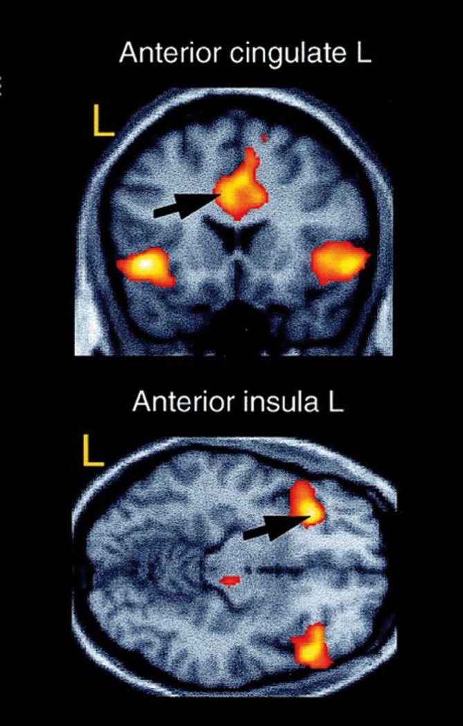
Over the last few years, neuroimaging techniques have contributed greatly to the identification of the structural and functional neuroanatomy of anxiety disorders. The amygdala seems to be a crucial structure for fear and anxiety, and has consistently been found to be activated in anxiety-provoking situations. Apart from the amygdala, the insula and anterior cinguiate cortex seem to be critical, and ail three have been referred to as the "fear network." In the present article, we review the main findings from three major lines of research. First, we examine human models of anxiety disorders, including fear conditioning studies and investigations of experimentally induced panic attacks. Then we turn to research in patients with anxiety disorders and take a dose look at post-traumatic stress disorder and obsessive-compulsive disorder. Finally, we review neuroimaging studies investigating neural correlates of successful treatment of anxiety, focusing on exposure-based therapy and several pharmacological treatment options, as well as combinations of both.
Overview
The claims and statistics of this paper haven’t been checked yet.
Run the check — free, no account needed. It reads the open-access text and checks the sources it cites.
Integrity signals
Registry
Cites
Statistics
Figures
Verification
Claims the paper makes
The claims and statistics of this paper haven’t been checked yet.
Run the check — free, no account needed. It reads the open-access text and checks the sources it cites.
Claims other papers make about this one
Each is checked against this paper’s own text: does it say what the citing paper says it does?
The cited source says what the claim attributes to it.
In the citing paper
“and occurs during anxiety symptom provocation (Holzschneider and Mulert, 2011).”
From Neuroimaging and psychophysiological investigation of the link between anxiety, enhanced affective reactivity and interoception in people with joint hypermobility (2014) · cites this paper as Holzschneider and Mulert, 2011
Self-check
Percentages, sums and reported differences re-computed from the paper’s own numbers.
The self-check needs the paper’s full text.
Citations
Cites
How do you feel? Interoception: The sense of the physiological condition of the body
Craig, A. D. (2002) · Nature Reviews Neuroscience · doi:10.1038/nrn894 · Cited by 6,006
Functional neuroanatomy of emotion: A meta-analysis of emotion activation studies in PET and fMRI
Phan, K. L., Wager, T., Taylor, S. F., & Liberzon, I. (2002) · NeuroImage · doi:10.1006/nimg.2002.1087 · Cited by 3,502
Neural systems supporting interoceptive awareness
Critchley, H. D., Wiens, S., Rotshtein, P., Öhman, A., & Dolan, R. J. (2004) · Nature Neuroscience · doi:10.1038/nn1176 · Cited by 3,406
Etkin, A., & Wager, T. D. (2007) · American Journal of Psychiatry · doi:10.1176/appi.ajp.2007.07030504 · Cited by 3,391 · cited 3×
“logical characteristics specific to each disorder.(4) However, the number of neuroimaging studies conducted on each anxiety disorder varies greatly.”
InfluentialStress, memory and the amygdala
Roozendaal, B., McEwen, B. S., & Chattarji, S. (2009) · Nature Reviews Neuroscience · doi:10.1038/nrn2651 · Cited by 1,760
Phelps, E. A., Delgado, M. R., Nearing, K. I., & LeDoux, J. E. (2004) · Neuron · doi:10.1016/j.neuron.2004.08.042 · Cited by 1,753
Oxytocin, vasopressin, and the neurogenetics of sociality
Donaldson, Z. R., & Young, L. J. (2008) · Science · doi:10.1126/science.1158668 · Cited by 1,645
Smaller hippocampal volume predicts pathologic vulnerability to psychological trauma
Gilbertson, M. W., Shenton, M. E., Ciszewski, A., Kasai, K., Lasko, N. B., Orr, S. P., & Pitman, R. K. (2002) · Nature Neuroscience · doi:10.1038/nn958 · Cited by 1,635
Goldin, P. R., & Gross, J. J. (2010) · Emotion · doi:10.1037/a0018441 · Cited by 1,242 · cited 1×
“For example, behavioral changes in patients with social anxiety disorder after mindfulnessbased stress reduction (MBSR) therapy seem to be reflected by distinct patterns of neural activity.(66) With regard to psychotherapy research, neuroimaging techniques offer the opportunity to monitor structural and functional neu…”
Amygdala, medial prefrontal cortex, and hippocampal function in PTSD
SHIN, L. M., RAUCH, S. L., & PITMAN, R. K. (2006) · Annals of the New York Academy of Sciences · doi:10.1196/annals.1364.007 · Cited by 1,203
Research works printed but not linked: 4
- American Psychiatric Association., 2000, Diagnostic and Statistical Manual of Mental Disorders. 4th ed. Text Revision. Washington, DC: American Psychiatric Association;
- Vermetten E., 2007, Psychopharmacol Bull., 40, 8
- Simon D., 2010, Psychophysiology., 47, 728
- Bryant RA., 2008, J Psychiatry Neurosci., 33, 142
Cited by
Sorted by citations
Ma, Z., Zhao, J., Li, Y., Chen, D., Wang, T., Zhang, Z., Chen, Z., Yu, Q., Jiang, J., Fan, F., et al. (2020) · Epidemiology and Psychiatric Sciences · doi:10.1017/s2045796020000931 · Cited by 437
Neuropathologic Correlates of Psychiatric Symptoms in Alzheimer’s Disease
Ehrenberg, A. J., Suemoto, C. K., Resende, E. D. P. F., Petersen, C., Leite, R. E. P., Rodriguez, R. D., Ferretti‐Rebustini, R. E. D. L., You, M. Y., Oh, J. Y., Nitríni, R., et al. (2018) · Journal of Alzheimer s Disease · doi:10.3233/jad-180688 · Cited by 233
Moylan, S., Jacka, F. N., Pasco, J. A., & Berk, M. (2013) · Brain and Behavior · doi:10.1002/brb3.137 · Cited by 186
Sex differences in mood disorders: Perspectives from humans and rodent models
Seney, M. L., & Sibille, E. (2014) · Biology of Sex Differences · doi:10.1186/s13293-014-0017-3 · Cited by 181
The vicious cycle of itch and anxiety
Sanders, K. M., & Akiyama, T. (2018) · Neuroscience & Biobehavioral Reviews · doi:10.1016/j.neubiorev.2018.01.009 · Cited by 187
Anxiety in Patients with Schizophrenia: Epidemiology and Management
Temmingh, H., & Stein, D. J. (2015) · CNS Drugs · doi:10.1007/s40263-015-0282-7 · Cited by 173
Almeida-Suhett, C. P., Graham, A. M., Chen, Y., & Deuster, P. A. (2016) · Physiology & Behavior · doi:10.1016/j.physbeh.2016.11.016 · Cited by 146
A systematic review of the neural bases of psychotherapy for anxiety and related disorders
Brooks, S. J., & Stein, D. J. (2015) · Dialogues in Clinical Neuroscience · doi:10.31887/dcns.2015.17.3/sbrooks · Cited by 134
The Physiology of Fear: Reconceptualizing the Role of the Central Amygdala in Fear Learning
Keifer, O. P., Hurt, R. C., Ressler, K. J., & Marvar, P. J. (2015) · Physiology · doi:10.1152/physiol.00058.2014 · Cited by 124
Hattingh, C. J., Ipser, J., Tromp, S. A., Syal, S., Lochner, C., Brooks, S. J., & Stein, D. J. (2013) · Frontiers in Human Neuroscience · doi:10.3389/fnhum.2012.00347 · Cited by 129
Almeida-Suhett, C. P., Prager, E. M., Pidoplichko, V. I., Figueiredo, T. H., Marini, A. M., Li, Z., Eiden, L. E., & Braga, M. F. M. (2014) · PLoS ONE · doi:10.1371/journal.pone.0102627 · Cited by 123
Streit, F., Haddad, L., Paul, T., Frank, J., Schäfer, A., Nikitopoulos, J. A., Akdeniz, C., Lederbogen, F., Treutlein, J., Witt, S. H., et al. (2014) · Stress · doi:10.3109/10253890.2014.921903 · Cited by 119
Measuring behavior in the home cage: study design, applications, challenges, and perspectives
Grieco, F., Bernstein, B. J., Biemans, B., Bikovski, L., Burnett, C. J., Cushman, J. D., van Dam, E. A., Fry, S. A., Richmond-Hacham, B., Homberg, J. R., et al. (2021) · Frontiers in Behavioral Neuroscience · doi:10.3389/fnbeh.2021.735387 · Cited by 109
Decreased saliency processing as a neural measure of Barratt impulsivity in healthy adults
Farr, O. M., Hu, S., Zhang, S., & Li, C. R. (2012) · NeuroImage · doi:10.1016/j.neuroimage.2012.07.049 · Cited by 109
Naik, R. R., Sotnikov, S., Diepold, R. P., Iurato, S., Markt, P. O., Bultmann, A., Brehm, N., Mattheus, T., Lutz, B., Erhardt, A., et al. (2017) · Translational Psychiatry · doi:10.1038/s41398-017-0025-2 · Cited by 111
MallorquÃ-Bagué, N., Garfinkel, S. N., Engels, M., Eccles, J. A., Pailhez, G., Bulbena, A., & Critchley, H. D. (2014) · Frontiers in Psychology · doi:10.3389/fpsyg.2014.01162 · Cited by 97
Functional neuroanatomy in panic disorder: Status quo of the research
Sobanski, T., & Wagner, G. (2017) · World Journal of Psychiatry · doi:10.5498/wjp.v7.i1.12 · Cited by 99
Heritability of subcortical brain measures: A perspective for future genome-wide association studies
den Braber, A., Bohlken, M. M., Brouwer, R. M., Ent, D. V. ‘., Ryota Kanai, C., Kahn, R. S., de Geus, E. J. C., Pol, H. E. H., & Boomsma, D. I. (2013) · NeuroImage · doi:10.1016/j.neuroimage.2013.06.027 · Cited by 93
The Interplay of Environmental Exposures and Mental Health: Setting an Agenda
Reuben, A., Manczak, E. M., Cabrera, L. Y., Alegria, M. M., Bucher, M. L., Freeman, E. C., Miller, G. W., Solomon, G. M., & Perry, M. J. (2022) · Environmental Health Perspectives · doi:10.1289/ehp9889 · Cited by 111
Gender Differences in the Neurobiology of Anxiety: Focus on Adult Hippocampal Neurogenesis
Marques, A. A., Bevilaqua, M. C. D. N., da Fonseca, A. M. P., Nardi, A. E., Thuret, S., & Dias, G. P. (2016) · Neural Plasticity · doi:10.1155/2016/5026713 · Cited by 78
Automated volumetry of the mesiotemporal structures in antibody-associated limbic encephalitis
Wagner, J., Witt, J., Helmstaedter, C., Malter, M. P., Weber, B., & Elger, C. E. (2014) · Journal of Neurology Neurosurgery & Psychiatry · doi:10.1136/jnnp-2014-307875 · Cited by 70
Neural correlates of a computerized attention modification program in anxious subjects
Taylor, C. T., Aupperle, R. L., Flagan, T., Simmons, A. N., Amir, N., Stein, M. B., & Paulus, M. P. (2013) · Social Cognitive and Affective Neuroscience · doi:10.1093/scan/nst128 · Cited by 65
Paul, S., Simon, D., Endrass, T., & Kathmann, N. (2016) · Psychological Medicine · doi:10.1017/s0033291715001610 · Cited by 60
Golpanian, R. S., Kim, H. S., & Yosipovitch, G. (2020) · Clinical Therapeutics · doi:10.1016/j.clinthera.2020.01.025 · Cited by 61
Olfaction in affective and anxiety disorders: A review of the literature
Burón, E., & Bulbena, A. (2013) · Psychopathology · doi:10.1159/000338717 · Cited by 52
Imaging the neural effects of cognitive bias modification training
Wiers, C. E., & Wiers, R. W. (2017) · NeuroImage · doi:10.1016/j.neuroimage.2016.07.041 · Cited by 52
Vergallito, A., Gallucci, A., Pisoni, A., Punzi, M., Caselli, G., Ruggiero, G. M., Sassaroli, S., & Lauro, L. J. R. (2021) · Journal of Psychiatry and Neuroscience · doi:10.1503/jpn.210050 · Cited by 53
Neuroimaging and Psychiatry: The Long Road from Bench to Bedside
Mayberg, H. S. (2014) · The Hastings Center Report · doi:10.1002/hast.296 · Cited by 49
Visuospatial Memory and Neuroimaging Correlates in Mild Cognitive Impairment
Mitolo, M., Gardini, S., Fasano, F., Crisi, G., Pelosi, A., Pazzaglia, F., & Caffarra, P. (2013) · Journal of Alzheimer s Disease · doi:10.3233/jad-121288 · Cited by 46
Altered Uncinate Fasciculus Microstructure in Childhood Anxiety Disorders in Boys But Not Girls
Tromp, D., Williams, L. E., Fox, A. S., Oler, J. A., Roseboom, P. H., Rogers, G. M., Benson, B. E., Alexander, A. L., Pine, D. S., & Kalin, N. H. (2019) · American Journal of Psychiatry · doi:10.1176/appi.ajp.2018.18040425 · Cited by 49
Poletti, S., Radaelli, D., Cucchi, M., Ricci, L., Vai, B., Smeraldi, E., & Benedetti, F. (2015) · Psychiatry Research Neuroimaging · doi:10.1016/j.pscychresns.2015.05.013 · Cited by 46
Liu, P., Taber‐Thomas, B. C., Fu, X., & Pérez‐Edgar, K. E. (2017) · Journal of the American Academy of Child & Adolescent Psychiatry · doi:10.1016/j.jaac.2017.11.016 · Cited by 45
Wearable Artificial Intelligence for Detecting Anxiety: Systematic Review and Meta-Analysis
Abd-Alrazaq, A. A., AlSaad, R., Harfouche, M., Aziz, S., Ahmed, A., Damseh, R., & Sheikh, J. I. (2023) · Journal of Medical Internet Research · doi:10.2196/48754 · Cited by 50
Transdiagnostic brain responses to disorder-related threat across four psychiatric disorders
Feldker, K., Heitmann, C. Y., Neumeister, P., Tupak, S. V., Schrammen, E., Moeck, R., Zwitserlood, P., Bruchmann, M., & Straube, T. (2016) · Psychological Medicine · doi:10.1017/s0033291716002634 · Cited by 45
Anticipating agoraphobic situations: the neural correlates of panic disorder with agoraphobia
Wittmann, A., Schlagenhauf, F., Guhn, A., Lueken, U., Gaehlsdorf, C., Stoy, M., Bermpohl, F., Fydrich, T., Pfleiderer, B., Bruhn, H., et al. (2014) · Psychological Medicine · doi:10.1017/s0033291713003085 · Cited by 38
Johnson, S. L. M., Wang, L., Alpert, K. I., GREENSTEIN, D. K., CLASEN, L. S., Lalonde, F. M., Miller, R., Rapoport, J. L., & Gogtay, N. (2013) · Journal of the American Academy of Child & Adolescent Psychiatry · doi:10.1016/j.jaac.2013.02.003 · Cited by 36
Functional MRI in the Investigation of Blast-Related Traumatic Brain Injury
Graner, J. L., Oakes, T. R., French, L. M., & Riedy, G. P. (2013) · Frontiers in Neurology · doi:10.3389/fneur.2013.00016 · Cited by 41
Brain responses to disorder‐related visual threat in panic disorder
Feldker, K., Heitmann, C. Y., Neumeister, P., Bruchmann, M., Vibrans, L., Zwitserlood, P., & Straube, T. (2016) · Human Brain Mapping · doi:10.1002/hbm.23320 · Cited by 37
van der Wee, N. J. A., Wen, M., Kandiah, N., Chander, R. J., Ng, A. Y. T., Au, W. L., & Tan, L. C. S. (2016) · Journal of the Neurological Sciences · doi:10.1016/j.jns.2016.10.021 · Cited by 35
Castelhano, J., Lima, G., Teixeira, M., Soares, C., Pais, M. L., & Castelo-Branco, M. S. (2021) · Frontiers in Pharmacology · doi:10.3389/fphar.2021.739053 · Cited by 41
Weidt, S., Lutz, J., Rufer, M., Delsignore, A., Jakob, N. J., Herwig, U., & Bruehl, A. B. (2016) · Psychological Medicine · doi:10.1017/s0033291715002998 · Cited by 30
Directed threat imagery in generalized anxiety disorder
Buff, C., Schmidt, C. C., Brinkmann, L., Gathmann, B., Tupak, S. V., & Straube, T. (2017) · Psychological Medicine · doi:10.1017/s0033291717001957 · Cited by 32
Functional connectivity density abnormalities and anxiety in primary insomnia patients
Zhu, Y., Zhao, X., Yin, H., & Zhang, M. (2020) · Brain Imaging and Behavior · doi:10.1007/s11682-019-00238-w · Cited by 29
Temporal profile of brain response to alprazolam in patients with generalized anxiety disorder
Brown, G. G., Ostrowitzki, S., Stein, M. B., von Kienlin, M., Liu, T. T., Simmons, A. N., Wierenga, C. E., Stein, O. Y., Bruns, A., Bischoff‐Grethe, A., et al. (2015) · Psychiatry Research Neuroimaging · doi:10.1016/j.pscychresns.2015.06.016 · Cited by 26
Acute and repetitive fronto-cerebellar tDCS stimulation improves mood in non-depressed participants
Newstead, S., Young, H. A., Benton, D., Jiga‐Boy, G. M., Sienz, M. L. A., Clement, R. M., & Boy, F. (2017) · Experimental Brain Research · doi:10.1007/s00221-017-5109-y · Cited by 27
Legge, A. W. (2015) · Music Therapy Perspectives · doi:10.1093/mtp/miv025 · Cited by 31
Roy, B., CHOI, S. E., Freeby, M., & Kumar, R. (2023) · Scientific Reports · doi:10.1038/s41598-023-35522-9 · Cited by 28
Anglin, R., Rosebush, P. I., Noseworthy, M. D., Tarnopolsky, M. A., & Mazurek, M. F. (2012) · Translational Psychiatry · doi:10.1038/tp.2012.107 · Cited by 26
Williams, M., Honan, C., Skromanis, S., Sanderson, B., & Matthews, A. J. (2024) · Current Psychology · doi:10.1007/s12144-023-04695-x · Cited by 25
Icenhour, A., Petrakova, L., Hazzan, N., Theysohn, N., Merz, C. J., & Elsenbruch, S. (2021) · NeuroImage · doi:10.1016/j.neuroimage.2021.118229 · Cited by 23
Casarrubea, M., Pierucci, M., Aiello, S., Cassar, D., Deidda, G., Crescimanno, G., & Giovanni, G. D. (2019) · Progress in Neuro-Psychopharmacology and Biological Psychiatry · doi:10.1016/j.pnpbp.2019.109731 · Cited by 22
Mallorquí‐Bagué, N., Bulbena, A., Roé-Vellvé, N., Hoekzema, E. A., Carmona, S., Barba‐Müller, E., Fauquet, J., Pailhez, G., & Vilarroya, Ó. (2015) · European Psychiatry · doi:10.1016/j.eurpsy.2015.01.004 · Cited by 23
Streit, F., Akdeniz, C., Haddad, L., Kumsta, R., Entringer, S., Frank, J., Yim, I. S., Zänkert, S., Witt, S. H., Kirsch, P., et al. (2016) · Psychoneuroendocrinology · doi:10.1016/j.psyneuen.2016.10.027 · Cited by 23
Tromp, D., Fox, A. S., Oler, J. A., Alexander, A. L., & Kalin, N. H. (2019) · Biological Psychiatry · doi:10.1016/j.biopsych.2019.07.022 · Cited by 24
Neuroimaging the Microbiome-Gut–Brain Axis
Tillisch, K., & Labus, J. S. (2014) · Advances in experimental medicine and biology · doi:10.1007/978-1-4939-0897-4_18 · Cited by 23
Mental health and urban design – zoning in on PTSD
M., M. A. G. A., Golembiewski, J. A., & Moustafa, A. A. (2017) · Current Psychology · doi:10.1007/s12144-017-9746-x · Cited by 26
Neuroanatomy of Anxiety: A Brief Review
Schmidt, C. K., Khalid, S., Loukas, M., & Tubbs, R. S. (2018) · Cureus · doi:10.7759/cureus.2055 · Cited by 21
Clark, U. S., Sweet, L. H., Morgello, S., Philip, N. S., & Cohen, R. A. (2016) · Brain Imaging and Behavior · doi:10.1007/s11682-016-9542-5 · Cited by 18
Rozenkrantz, L., Weissgross, R., Weiss, T., Ravreby, I., Frumin, I., Shushan, S., Gorodisky, L., Reshef, N., Holzman, Y., Pinchover, L., et al. (2020) · eLife · doi:10.7554/elife.55305 · Cited by 16
Hinze, J., Röder, A., Menzie, N., Müller, U., Domschke, K., Riemenschneider, M. J., & Noll‐Hussong, M. (2021) · Frontiers in Psychiatry · doi:10.3389/fpsyt.2021.704174 · Cited by 24
Anesthesia and the neurobiology of fear and posttraumatic stress disorder
Vogt, K. M., & Pryor, K. O. (2022) · Current Opinion in Anaesthesiology · doi:10.1097/aco.0000000000001176 · Cited by 17
Brain Mechanisms Underlying Panic Attack and Panic Disorder
Guan, X., & Cao, P. (2023) · Neuroscience Bulletin · doi:10.1007/s12264-023-01088-9 · Cited by 22
Figueiredo, T. H., Harbert, C. L., Pidoplichko, V. I., Almeida-Suhett, C. P., Pan, H., Rossetti, K., Braga, M. F., & Marini, A. (2017) · Molecular Neurobiology · doi:10.1007/s12035-017-0732-y · Cited by 16
Sympathetic Neural Control in Humans with Anxiety‐Related Disorders
Bigalke, J. A., & Carter, J. R. (2021) · Comprehensive physiology · doi:10.1002/cphy.c210027 · Cited by 29
Divergent encoding of active avoidance behavior in corticostriatal and corticolimbic projections
Kajs, B. L., Loewke, A. C., Dorsch, J. M., Vinson, L. T., & Gunaydin, L. A. (2022) · Scientific Reports · doi:10.1038/s41598-022-14930-3 · Cited by 17
Steinhäuser, J. L., Teed, A. R., Zoubi, O. A., Hurlemann, R., Chen, G., & Khalsa, S. S. (2023) · Scientific Reports · doi:10.1038/s41598-023-35939-2 · Cited by 18
Opmeer, E. M., Kortekaas, R., van Tol, M., van der Wee, N. J. A., Woudstra, S., van Buchem, M. A., Penninx, B. W. J. H., Veltman, D. J., & Aleman, A. (2013) · Social Cognitive and Affective Neuroscience · doi:10.1093/scan/nst025 · Cited by 20
Mirante, O., Brandalise, F., Bohacek, J., & Mansuy, I. M. (2014) · Frontiers in Molecular Neuroscience · doi:10.3389/fnmol.2014.00062 · Cited by 18
Mueller, S. A. L., Oler, J. A., Roseboom, P. H., Aggarwal, N., Kenwood, M. M., Riedel, M. K., Elam, V. R., Olsen, M. E., DiFilippo, A. H., Christian, B. T., et al. (2023) · Current Research in Neurobiology · doi:10.1016/j.crneur.2023.100111 · Cited by 14
Lin, E. R.-H., Veenker, F., Manza, P., Yonga, M.-V., Abey, S. K., Wang, G., & Volkow, N. D. (2024) · Brain Sciences · doi:10.3390/brainsci14121285 · Cited by 15
Measures of mindfulness and anxiety in OM meditators and non-meditators: A cross-sectional study
Singh, D., Suhas, A., Visweswaraiah, N. K., & Nagendra, H. (2014) · International Journal of Medicine and Public Health · doi:10.4103/2230-8598.127170 · Cited by 13
Anxiety symptoms bias memory assessment in older adults
Williams, M., Kueider‐Paisley, A., Dmitrieva, N., Manly, J. J., Pieper, C. F., Verney, S. P., & Gibbons, L. E. (2016) · International Journal of Geriatric Psychiatry · doi:10.1002/gps.4557 · Cited by 13
The Origin of Anxiety Disorders - An Evolutionary Approach
Willers, L. E. W. G., Vulink, N. C. C., Denys, D. A. J. P., & Stein, D. J. (2013) · Modern trends in psychiatry · doi:10.1159/000351919 · Cited by 37
Wittmann, A., Schlagenhauf, F., Guhn, A., Lueken, U., Elle, M., Stoy, M., Liebscher, C., Bermpohl, F., Fydrich, T., Pfleiderer, B., et al. (2018) · Psychotherapy and Psychosomatics · doi:10.1159/000493146 · Cited by 10
Anxiety in old age and dementia - implications for clinical and research practice
Tales, A., & Basoudan, N. (2016) · Neuropsychiatry · doi:10.4172/neuropsychiatry.1000133 · Cited by 11
Oliva, A., Torre, S., Taranto, P., Delvecchio, G., & Brambilla, P. (2020) · Journal of Affective Disorders · doi:10.1016/j.jad.2020.12.085 · Cited by 11
Kim, S.-W., Kim, M.-K., Kim, B., Choi, T., & Lee, S.-H. (2020) · Journal of Affective Disorders · doi:10.1016/j.jad.2019.09.032 · Cited by 9
Transcranial sonography in obsessive-compulsive disorder.
Mavrogiorgou, P., Nalato, F., Meves, S., Luksnat, S., Norra, C., Gold, R., Juckel, G., & Krogias, C. (2013) · Journal of Psychiatric Research · doi:10.1016/j.jpsychires.2013.07.020 · Cited by 11
Effect of diazepam and yohimbine on neuronal activity in sham and hemiparkinsonian rats
O'Connor, K. A., Mahoney, E. C., Ramirez‐Zamora, A., Molho, E., Pilitsis, J. G., & Shin, D. S.-H. (2017) · Neuroscience · doi:10.1016/j.neuroscience.2017.03.039 · Cited by 10
Neural responses to acute stress predict chronic stress perception in daily life over 13 months
Giglberger, M., Peter, H. L., Henze, G., Kraus, E., Bärtl, C., Konzok, J., Kreuzpointner, L., Kirsch, P., Kudielka, B. M., & Wüst, S. (2023) · Scientific Reports · doi:10.1038/s41598-023-46631-w · Cited by 14
Brain activation during disorder-related script-driven imagery in panic disorder: a pilot study
Burkhardt, A., Buff, C., Brinkmann, L., Feldker, K., Gathmann, B., Hofmann, D., & Straube, T. (2019) · Scientific Reports · doi:10.1038/s41598-019-38990-0 · Cited by 11
Yamada, D., Miyajima, M., Ishibashi, H., Wada, K., Seki, K., & Sekiguchi, M. (2012) · The Journal of Physiology · doi:10.1113/jphysiol.2012.235010 · Cited by 10
Amygdala and Emotional Learning in Vertebrates – A Comparative Perspective
Pedro, J., Carlos, J., & Portavell, M. (2012) · InTech eBooks · doi:10.5772/51552 · Cited by 9
Influence of the inflammasome complex on psychiatric disorders: clinical and preclinical studies
Kwon, S., & Cheon, S. Y. (2021) · Expert Opinion on Therapeutic Targets · doi:10.1080/14728222.2021.2005027 · Cited by 9
Neurological Research on Music Therapy for Mental Health: A Summary of Imaging and Research Methods
Hunt, A. M., & Legge, A. W. (2015) · Music Therapy Perspectives · doi:10.1093/mtp/miv024 · Cited by 8
Structural connectome alterations in anxious dogs: a DTI-based study
Chen, Q., Xu, Y., Christiaen, E., Wu, G., Witte, S. D., Vanhove, C., Saunders, J. H., Peremans, K. Y., & Baeken, C. (2023) · Scientific Reports · doi:10.1038/s41598-023-37121-0 · Cited by 8
Shan, Y., Sun, G., Ji, J., Li, Z., Chen, X., Zhang, X., Ma, Y., Zhang, Y., Zhang, T., & Zhang, Y. (2024) · Frontiers in Psychiatry · doi:10.3389/fpsyt.2024.1336233 · Cited by 9
Braga, M. F. M., Juranek, J. J., Eiden, L. E., Li, Z., Figueiredo, T. H., Furtado, M. D. A., & Marini, A. M. (2022) · Amino Acids · doi:10.1007/s00726-022-03184-y · Cited by 8
A network analysis of obsessive-compulsive symptoms and their comorbidity with other disorders
Brakoulias, V., Elhindi, J., & Starčević, V. (2025) · Journal of Psychiatric Research · doi:10.1016/j.jpsychires.2025.02.010 · Cited by 9
Sympathetic Neural Control in Humans with Anxiety‐Related Disorders
Bigalke, J. A., & Carter, J. R. (2021) · Comprehensive physiology · doi:10.1002/j.2040-4603.2022.tb00206.x · Cited by 6
Shirokova, O. M., Kuzmina, D. M., Zaborskaya, O. G., Щелчкова, Н. А., Kozliaeva, E. V., Александровна, К. С., Pershin, V. I., Vasilchikov, P. I., & Mukhina, I. V. (2025) · International Journal of Molecular Sciences · doi:10.3390/ijms26031251 · Cited by 9
To BD or not to BD: functional neuroimaging and the boundaries of bipolarity
Kuiper, S., McLean, L., & Malhi, G. S. (2012) · Expert Review of Neurotherapeutics · doi:10.1586/ern.12.126 · Cited by 6
Sliding Scale Theory of Attention and Consciousness/Unconsciousness
Bowins, B. E. (2022) · Behavioral Sciences · doi:10.3390/bs12020043 · Cited by 6
Lakhawat, S. S., Mech, P., Kumar, A., Malik, N., Kumar, V., Sharma, V., Bhatti, J. S., Jaswal, S. K., Kumar, S., & Sharma, P. K. (2024) · Metabolic Brain Disease · doi:10.1007/s11011-024-01453-1 · Cited by 6
Plag, J., Petzold, M. B., Gechter, J., Liebscher, C., & Ströhle, A. (2018) · Nordic Journal of Psychiatry · doi:10.1080/08039488.2018.1457178 · Cited by 5
Woody, M. L., Yang, J. O., Cummings, L. R., Gilchrist, D., Graur, S., Siegle, G. J., & Price, R. B. (2019) · Translational Psychiatry · doi:10.1038/s41398-019-0458-x · Cited by 6
SpiDa-MRI: behavioral and (f)MRI data of adults with fear of spiders
Zhang, M., Karner, A., Kostorz, K., Shea, S., Steyrl, D., Melinščak, F., Sladky, R., Lor, C. S., & Scharnowski, F. (2025) · Scientific Data · doi:10.1038/s41597-025-04569-w · Cited by 9
Neuroimaging in Post-Traumatic Stress Disorder (PTSD): a Narrative Review
Stokłosa, I., Marwaha, R., Stokłosa, M., Zacharzewska-Gondek, A., Piegza, M. J., Gorczyca, P. W., & Więckiewicz, G. (2024) · Archives of Medical Science · doi:10.5114/aoms/188377 · Cited by 8
Complex Sympathetic Regulation in Adolescent Mitral Valve Prolapse
Olexova, L. B., Višňovcová, Z., Ferencová, N., Jurko, A., & Tonhajzerová, I. (2021) · Physiological Research · doi:10.33549/physiolres.934830 · Cited by 4
Yan, Y., Li, M., Jia, H., Fu, L., Qiu, J., & Yang, W. (2023) · Brain and Cognition · doi:10.1016/j.bandc.2023.105976 · Cited by 4
Metadata
- Authors
- Holzschneider, K., & Mulert, C.
- Affiliations
- University Medical Center Hamburg-Eppendorf (DE)
- Corresponding author
- Mulert, Christoph
- ORCID
- Mulert, Christoph
- Venue
- Dialogues in Clinical Neuroscience
- Publisher
- Informa UK Limited
- Published
- 2011-12-31
- Volume
- 13
- Issue
- 4
- Pages
- 453-461
- ISSN
- 1294-8322 (ISSN-L)
- 1958-5969
- PMC
- PMC3263392
- OpenAlex
- W4298412747
- Cited by
- 146
- References
- 72
- Citations by year
- 2026: 2 · 2025: 8 · 2024: 7 · 2023: 13 · 2022: 9 · 2021: 12 · 2020: 7 · 2019: 8 · 2018: 6 · 2017: 10 · 2016: 14 · 2015: 14 · 2014: 14 · 2013: 12 · 2012: 9
- FWCI
- 14.43
- Type
- journal article
- Language
- en
- Open access
- diamond
- doi.org
- Licence
- Locations
- Dialogues in Clinical Neuroscience (publishedVersion, cc-by-nc-nd)
- PubMed Central (publishedVersion, cc-by-nc-nd)
- PubMed (publishedVersion)
- PubMed
- Topics
- Social Sciences › Psychology › Experimental and Cognitive Psychology › Anxiety, Depression, Psychometrics, Treatment, Cognitive Processes
- Life Sciences › Neuroscience › Cognitive Neuroscience › Memory and Neural Mechanisms
- Life Sciences › Neuroscience › Cognitive Neuroscience › Neural and Behavioral Psychology Studies
- Keywords
- anxiety disorders, amygdala, fear network, insula, anterior cingulate cortex, fear conditioning, panic attacks, post-traumatic stress disorder, obsessive-compulsive disorder, exposure therapy, pharmacological treatment of anxiety
- MeSH terms
- Amygdala, Anxiety Disorders, Humans, Magnetic Resonance Imaging, Tomography, Emission-Computed, Single-Photon, Positron-Emission Tomography
About these checks
Registry, retraction, Cites and statistics results are computed, not judged, with one exception: a printed reference that no index lists is read once by a model before it counts as a citation error. Claims are extracted by a model and kept only when their quote appears verbatim in the text; each cited source is then checked, with a model’s help, for whether it says what the claim relies on. Those verdicts can be wrong — read the claim’s quote and the cited source before relying on one.
Statistics and arithmetic are recomputed, not judged.
Confidence bands are shown only for fields calibrated on our annotated test set. None is calibrated yet, so no band is shown.
What counts as a claim ›How we measure confidence ›How we check this ›
Are you an author, or do you see a problem with this page? Request a review
