

The DOI matches its Crossref record; 218 of the 220 references it cites were found in a registry or an index.
The pathophysiology of osteoporosis in obesity and type 2 diabetes in aging women and men: The mechanisms and roles of increased bone marrow adiposity
Open accessAbstract
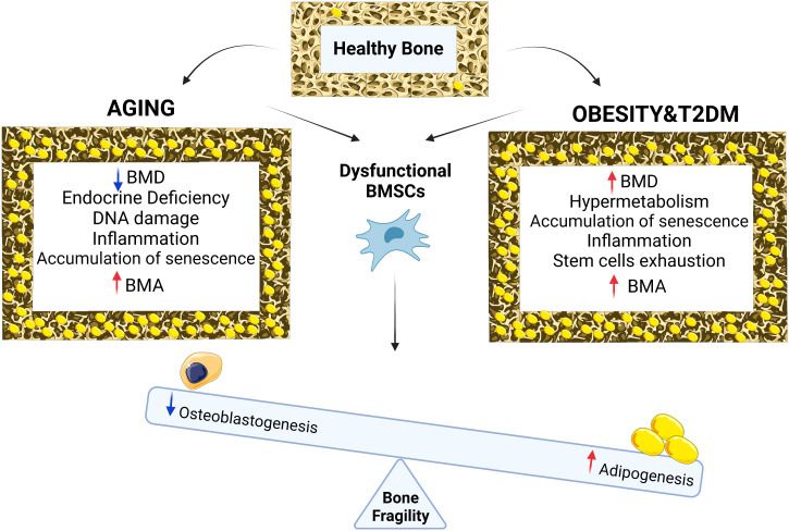
Osteoporosis is defined as a systemic skeletal disease characterized by decreased bone mass and micro-architectural deterioration leading to increased fracture risk. Osteoporosis incidence increases with age in both post-menopausal women and aging men. Among other important contributing factors to bone fragility observed in osteoporosis, that also affect the elderly population, are metabolic disturbances observed in obesity and Type 2 Diabetes (T2D). These metabolic complications are associated with impaired bone homeostasis and a higher fracture risk. Expansion of the Bone Marrow Adipose Tissue (BMAT), at the expense of decreased bone formation, is thought to be one of the key pathogenic mechanisms underlying osteoporosis and bone fragility in obesity and T2D. Our review provides a summary of mechanisms behind increased Bone Marrow Adiposity (BMA) during aging and highlights the pre-clinical and clinical studies connecting obesity and T2D, to BMA and bone fragility in aging osteoporotic women and men.
Overview
The claims and statistics of this paper haven’t been checked yet.
Run the check — free, no account needed. It reads the open-access text and checks the sources it cites.
Integrity signals
Registry
Cites
Statistics
Figures
Verification
Claims the paper makes
The claims and statistics of this paper haven’t been checked yet.
Run the check — free, no account needed. It reads the open-access text and checks the sources it cites.
Claims other papers make about this one
Each is checked against this paper’s own text: does it say what the citing paper says it does?
The cited source says what the claim attributes to it.
In the citing paper
“As aging-related osteoporosis typically features low bone mass, microarchitectural alterations and bone marrow adipose tissue expansion40–42, our findings suggest that AD mice at a younger age recapitulate an aged skeletal phenotype.”
From Serum amyloid P secreted by bone marrow adipocytes drives skeletal amyloidosis (2025) · cites this paper as [40]
Self-check
Percentages, sums and reported differences re-computed from the paper’s own numbers.
The self-check needs the paper’s full text.
Citations
Cites
López-Otín, C., Blasco, M. A., Partridge, L., Serrano, M., & Kroemer, G. (2013) · Cell · doi:10.1016/j.cell.2013.05.039 · Cited by 14,982 · cited 1×
“Aging is defined as a gradual loss of normal tissue homeostasis and progressive deterioration of the organ functions, due to accumulation of cellular/DNA damage and senescence throughout aging (11).”
BackgroundThe senescence-associated secretory phenotype: the dark side of tumor suppression
Coppé, J.-P., Desprez, P.-Y., Krtolica, A., & Campisi, J. (2010) · Annual Review of Pathology: Mechanisms of Disease · doi:10.1146/annurev-pathol-121808-102144 · Cited by 5,206
An estimate of the worldwide prevalence and disability associated with osteoporotic fractures
Johnell, O., & Kanis, J. (2006) · Osteoporosis International · doi:10.1007/s00198-006-0172-4 · Cited by 4,770 · cited 1×
“It is the most common bone disease worldwide, affecting more than 200 million people and causing more than 9 million fractures in the year 2000 (2).”
BackgroundJohnston, C., & Slemenda, C. (1992) · Southern medical journal (Birmingham, Ala. Print) · doi:10.1097/00007611-199208001-00002 · Cited by 3,785
Adipose tissue, adipokines, and inflammation
Fantuzzi, G. (2005) · Journal of Allergy and Clinical Immunology · doi:10.1016/j.jaci.2005.02.023 · Cited by 2,606
Kanis, J. А., & Kanis, J. А. (1994) · Osteoporosis International · doi:10.1007/bf01622200 · Cited by 2,532
Cellular senescence in aging and age-related disease: from mechanisms to therapy
Childs, B. G., Durik, M., Baker, D. J., & van Deursen, J. M. (2015) · Nature Medicine · doi:10.1038/nm.4000 · Cited by 2,363
Lips, P. (2001) · Endocrine reviews · doi:10.1210/edrv.22.4.0437 · Cited by 2,203
Obesity-induced inflammatory changes in adipose tissue
Wellen, K. E., & Hotamisligil, G. S. (2003) · Journal of Clinical Investigation · doi:10.1172/jci200320514 · Cited by 1,823
Cellular senescence and the senescent secretory phenotype: therapeutic opportunities
Tchkonia, T., Zhu, Y., van Deursen, J., Campisi, J., & Kirkland, J. L. (2013) · Journal of Clinical Investigation · doi:10.1172/jci64098 · Cited by 1,774
Tang, B., Eslick, G., Nowson, C., Smith, C. A., & Bensoussan, A. (2007) · The Lancet · doi:10.1097/01.ogx.0000298339.68546.3c · Cited by 1,365
Tang, B. M., Eslick, G. D., Nowson, C., Smith, C., & Bensoussan, A. (2007) · The Lancet · doi:10.1016/s0140-6736(07)61342-7 · Cited by 1,248
Systematic Review of Type 1 and Type 2 Diabetes Mellitus and Risk of Fracture
Janghorbani, M., van Dam, R. M., Willett, W. C., & Hu, F. B. (2007) · American Journal of Epidemiology · doi:10.1093/aje/kwm106 · Cited by 1,223
Targeting cellular senescence prevents age-related bone loss in mice
Farr, J. N., Xu, M., Weivoda, M. M., Monroe, D. G., Fraser, D. G., Onken, J. L., Negley, B. A., Sfeir, J. G., Ogrodnik, M. B., Hachfeld, C. M., et al. (2017) · Nature Medicine · doi:10.1038/nm.4385 · Cited by 1,182 · cited 1×
“Furthermore genetic depletion of senescent cells, using the inducible INKATTAC ‘suicide’ transgene that eliminates the p16 expressing cells, in a murine pre-clinical model of age-related osteoporosis, exhibited anti-resorptive and anabolic impact on bone, leading to decreased BMAT and increased bone mass (32).”
BackgroundBone-marrow adipocytes as negative regulators of the haematopoietic microenvironment
Naveiras, O., Nardi, V., Wenzel, P. L., Hauschka, P. V., Fahey, F., & Daley, G. Q. (2009) · Nature · doi:10.1038/nature08099 · Cited by 1,129
Mechanisms of diabetes mellitus-induced bone fragility
On behalf of the IOF Bone and Diabetes Working Group, Napoli, N., Chandran, M., Pierroz, D. D., Abrahamsen, B., Schwartz, A. V., & Ferrari, S. L. (2017) · Nature Reviews Endocrinology · doi:10.1038/nrendo.2016.153 · Cited by 1,107 · cited 1×
“Changes in BMA during aging in males and females 145 females, 114 males; age range (62-90) years.”
BackgroundPathogenesis of bone fragility in women and men
Seeman, E. (2002) · The Lancet · doi:10.1016/s0140-6736(02)08706-8 · Cited by 909 · cited 1×
“Aging is associated with significant bone loss and structural bone damage, due to accelerated trabecular thinning and disconnection, cortical thinning, and porosity (12), leading to increased fracture risk.”
BackgroundBianco, P., & Robey, P. G. (2000) · Journal of Clinical Investigation · doi:10.1172/jci10413 · Cited by 908
LONG-TERM PREVENTION OF POSTMENOPAUSAL OSTEOPOROSIS BY ŒSTROGEN
Lindsay, R., Aitken, J. M., Anderson, L., Hart, D., Macdonald, E., & Clarke, A. (1976) · The Lancet · doi:10.1016/s0140-6736(76)92217-0 · Cited by 875
Ambrosi, T. H., Scialdone, A., Graja, A., Gohlke, S., Jank, A.-M., Bocian, C., Woelk, L., Fan, H., Logan, D. W., Schürmann, A., et al. (2017) · Cell Stem Cell · doi:10.1016/j.stem.2017.02.009 · Cited by 856
Adipocyte tissue volume in bone marrow is increased with aging and in patients with osteoporosis
Justesen, J., Stenderup, K., Ebbesen, E., Mosekilde, L., Steiniche, T., & Kassem, M. (2004) · Biogerontology (Dordrecht) · doi:10.1023/a:1011513223894 · Cited by 847
Self-citationMoerman, E., Teng, K., Lipschitz, D., & Lecka-Czernik, B. (2004) · Aging Cell · doi:10.1111/j.1474-9728.2004.00127.x · Cited by 840 · cited 2×
“BMSCs obtained from old mice also exhibited increased expression of SASP markers, leading to increased osteoclast formation, as well as increased expression of the adipogenic transcription factor PPAR-g (45).”
BackgroundChanges in Bone Mineral Density of the Proximal Femur and Spine with Aging
Riggs, B. L., Wahner, H. W., Seeman, E., Offord, K. P., Dunn, W. L., Mazess, R. B., Johnson, K. A., & Melton, L. J. (1982) · Journal of Clinical Investigation · doi:10.1172/jci110667 · Cited by 816 · cited 1×
“Changes in BMA during aging in males and females 145 females, 114 males; age range (62-90) years.”
BackgroundOlder Women with Diabetes Have an Increased Risk of Fracture: A Prospective Study
Schwartz, A. V., Sellmeyer, D. E., Ensrud, K. E., Cauley, J. A., Tabor, H. K., Schreiner, P. J., Jamal, S. A., Black, D. M., & Cummings, S. R. (2001) · The Journal of Clinical Endocrinology & Metabolism · doi:10.1210/jcem.86.1.7139 · Cited by 788
An atlas of genetic influences on osteoporosis in humans and mice
23andMe Research Team, Morris, J. A., Kemp, J. P., Youlten, S. E., Laurent, L., Logan, J. G., Chai, R. C., Vulpescu, N. A., Forgetta, V., Kleinman, A., et al. (2019) · Nature Genetics · doi:10.1038/s41588-018-0302-x · Cited by 773 · cited 1×
“Changes in BMA during aging in males and females 145 females, 114 males; age range (62-90) years.”
BackgroundFalahati-Nini, A., Riggs, B. L., Atkinson, E. J., O’Fallon, W. M., Eastell, R., & Khosla, S. (2000) · Journal of Clinical Investigation · doi:10.1172/jci10942 · Cited by 757 · cited 2×
“Another human study investigated the impact of endogenous estrogen and testosterone production on bone, using 59 elderly men and demonstrated that estrogen is the dominant sex-steroid hormone protecting men from age-related bone resorption, whereas both hormones of estrogen and testosterone are critical for bone forma…”
BackgroundVanderschueren, D., Vandenput, L., Boonen, S., Lindberg, M. K., Bouillon, R., & Ohlsson, C. (2004) · Endocrine Reviews · doi:10.1210/er.2003-0003 · Cited by 717
Association of BMD and FRAX Score with Risk of Fracture in Older Adults with Type 2 Diabetes
Schwartz, A., Vittinghoff, E., Bauer, D. C., Hillier, T., Strotmeyer, E., Ensrud, K., Donaldson, M., Cauley, J., Harris, T., Koster, A., et al. (2011) · Journal of the American Medical Association (JAMA) · doi:10.1001/jama.2011.715 · Cited by 700 · cited 1×
“Interestingly in humans, obesity is associated with increased BMD in children and adults (89, 101, 102).”
BackgroundAtashi, F., Modarressi, A., & Pepper, M. S. (2015) · Stem Cells and Development · doi:10.1089/scd.2014.0484 · Cited by 660 · cited 1×
“Aging is also accompanied with increased levels of Reactive Oxygen Species (ROS) and oxidative stress in the bone microenvironment , leading to impaired osteoblast differentiation and function, and enhanced adipocyte differentiation (33).”
BackgroundRisk of fracture in women with type 2 diabetes: the Women's Health Initiative Observational Study.
Bonds, D., Larson, J. C., Schwartz, A., Strotmeyer, E., Robbins, J. A., Rodríguez, B., Johnson, K. C., & Margolis, K. (2006) · Journal of Clinical Endocrinology and Metabolism · doi:10.1210/jc.2006-0614 · Cited by 602 · cited 1×
“contributed to the delayed bone healing in monocortical fracture model (100).”
BackgroundVestergaard, P., Rejnmark, L., & Mosekilde, L. (2005) · Diabetologia · doi:10.1007/s00125-005-1786-3 · Cited by 568
A crucial role for thiol antioxidants in estrogen-deficiency bone loss.
Lean, J., Davies, J., Fuller, K., Jagger, C., Kirstein, B., Partington, G., Urry, Z., & Chambers, T. (2003) · Journal of Clinical Investigation · doi:10.1172/jci18859 · Cited by 567 · cited 2×
“Estrogen deficiency also leads to decreased production of endogenous antioxidants (68, 69), which together with the elevated ROS production and the senescence microenvironment observed during aging or in obese mice and human, can lead to significantly increased levels of oxidative stress in the bone microenvironment (…”
BackgroundChapuy, M., Pamphile, R., Paris, E., Kempf, C., Schlichting, M., Arnaud, S., Garnero, P., & Meunier, P. (2002) · Osteoporosis International · doi:10.1007/s001980200023 · Cited by 567
Identification of senescent cells in the bone microenvironment
Farr, J. N., Fraser, D. G., Wang, H., Jaehn, K., Ogrodnik, M. B., Weivoda, M. M., Drake, M. T., Tchkonia, T., LeBrasseur, N. K., Kirkland, J. L., et al. (2016) · Journal of Bone and Mineral Research · doi:10.1002/jbmr.2892 · Cited by 562 · cited 1×
“Analysis of human bone s p e c im en s h a d s h own p r e s e n c e o f s e n e s c e n c e microenvironment in the bone tissue obtained from old individuals compared to young control subjects, evidenced by increased expression of p16 and p21 (31).”
BackgroundObesity Is Not Protective against Fracture in Postmenopausal Women: GLOW
Compston, J. E., Watts, N. B., Chapurlat, R., Cooper, C., Boonen, S., Greenspan, S., Pfeilschifter, J., Silverman, S., Díez-Pérez, A., Lindsay, R., et al. (2011) · The American Journal of Medicine · doi:10.1016/j.amjmed.2011.06.013 · Cited by 552
Rosiglitazone-Associated Fractures in Type 2 Diabetes
Kahn, S. E., Zinman, B., Lachin, J. M., Haffner, S. M., Herman, W. H., Holman, R. R., Kravitz, B. G., Yu, D., Heise, M. A., Aftring, R. P., et al. (2008) · Diabetes Care · doi:10.2337/dc07-2270 · Cited by 537
Boonen, S., Lips, P. T. A. M., Bouillon, R., Bischoff-Ferrari, H. A., Vanderschueren, D., & Haentjens, P. (2007) · The Journal of Clinical Endocrinology & Metabolism · doi:10.1210/jc.2006-1404 · Cited by 536
Reid, I., Ames, R., Evans, M., Sharpe, S., Gamble, G., France, J., Lim, T., & Cundy, T. (1992) · Journal of Clinical Endocrinology and Metabolism · doi:10.1097/00006254-199301000-00025 · Cited by 531
Oxidative stress in type 2 diabetes: the role of fasting and postprandial glycaemia
Wright, E., Scism-Bacon, J. L., & Glass, L. C. (2006) · International Journal of Clinical Practice · doi:10.1111/j.1368-5031.2006.00825.x · Cited by 517
In Vivo Assessment of Bone Quality in Postmenopausal Women With Type 2 Diabetes
Farr, J. N., Drake, M. T., Amin, S., Melton, L. J., McCready, L. K., & Khosla, S. (2014) · Journal of Bone and Mineral Research · doi:10.1002/jbmr.2106 · Cited by 499 · cited 1×
“Interestingly in humans, obesity is associated with increased BMD in children and adults (89, 101, 102).”
BackgroundYue, R., Zhou, B. O., Shimada, I. S., Zhao, Z., & Morrison, S. J. (2016) · Cell stem cell · doi:10.1016/j.stem.2016.02.015 · Cited by 495
Kudo, O., Sabokbar, A., Pocock, A., Itonaga, I., Fujikawa, Y., & Athanasou, N. (2002) · Bone · doi:10.1016/s8756-3282(02)00915-8 · Cited by 487
Mundy, G. R. (2008) · Nutrition Reviews · doi:10.1111/j.1753-4887.2007.tb00353.x · Cited by 485
Pathophysiology of Osteoporosis
Armas, L. A. G., & Recker, R. R. (2012) · Endocrinology and Metabolism Clinics of North America · doi:10.1016/j.ecl.2012.04.006 · Cited by 466 · cited 1×
“Osteoporosis is a chronic-skeletal disorder characterized by low bone mass, and deterioration in the microarchitecture of the bone tissue that results in bone fragility and increased fracture risk (1).”
BackgroundBone marrow adipocytes promote the regeneration of stem cells and hematopoiesis by secreting SCF
Zhou, B., Yu, H., Yue, R., Zhao, Z.-Y., Rios, J., Naveiras, O., & Morrison, S. (2017) · Nature Cell Biology · doi:10.1038/ncb3570 · Cited by 466
Khosla, S. (1998) · Journal of Clinical Endocrinology & Metabolism · doi:10.1210/jc.83.7.2266 · Cited by 447 · cited 2×
“This study also indicated that age-related bone loss in men is associated with decline in testosterone as well as estrogen (63).”
BackgroundA role of miR‐27 in the regulation of adipogenesis
Lin, Q., Gao, Z., Alarcon, R. M., Ye, J., & Yun, Z. (2009) · FEBS Journal · doi:10.1111/j.1742-4658.2009.06967.x · Cited by 439 · cited 1×
“This study also indicated a direct correlation between the increased senescence burden in the bone tissue and up-regulation of mir-27a, which is known to be involved in obesity and regulation of adipose-tissue related transcription factors (40).”
BackgroundGriffith, J., Yeung, D., Antonio, G., Lee, F. H., Hong, A., Wong, S., Lau, E., & Leung, P. (2005) · Radiology · doi:10.1148/radiol.2363041425 · Cited by 435 · cited 2×
“In addition, human studies have shown that aging and loss of steroid hormones make women more vulnerable to the negative impacts of BMAT on loss of trabecular bone at the spine and femoral neck, and greater loss of spine strength (75), while in men vertebral BMAT is significantly increased with osteoporosis (76).”
BackgroundZhu, X., Chen, Z., Shen, W., Huang, G., Sedivy, J. M., Wang, H., & Ju, Z. (2021) · Signal Transduction and Targeted Therapy · doi:10.1038/s41392-021-00646-9 · Cited by 425 · cited 1×
“increased expression of cell cycle regulators/tumor suppressors such as p16 and p53), mitochondrial dysfunction, and resistance to apoptosis, reviewed in (21, 22).”
BackgroundHuman cell senescence as a DNA damage response
Zglinicki, T. V., Saretzki, G., Ladhoff, J., Fagagna, F. D. D., & Jackson, S. P. (2005) · Mechanisms of Ageing and Development · doi:10.1016/j.mad.2004.09.034 · Cited by 419
Fracture Risk in Type 2 Diabetes: Update of a Population-Based Study
Melton, L. J., Leibson, C. L., Achenbach, S. J., Therneau, T. M., & Khosla, S. (2008) · Journal of Bone and Mineral Research · doi:10.1359/jbmr.080323 · Cited by 411
Dimri, G. P., Itahana, K., Acosta, M., & Campisi, J. (2000) · Molecular and Cellular Biology · doi:10.1128/mcb.20.1.273-285.2000 · Cited by 401 · cited 1×
“Few studies have reported that senescence can be regulated via transcription factors (36, 37).”
BackgroundRelation between body size and bone mineral density in elderly men and women.
Edelstein, S. L., & Barrett-Connor, E. (1993) · American Journal of Epidemiology · doi:10.1093/oxfordjournals.aje.a116842 · Cited by 400
Rios, H., Koushik, S. V., Wang, H., Wang, J., Zhou, H.-M., Lindsley, A. W., Rogers, R., Chen, Z., Maeda, M., Kruzynska-Frejtag, A., et al. (2005) · Molecular and Cellular Biology · doi:10.1128/mcb.25.24.11131-11144.2005 · Cited by 398
Mechanisms and Regulation of Cellular Senescence
Roger, L., Tomas, F., & Gire, V. (2021) · International Journal of Molecular Sciences · doi:10.3390/ijms222313173 · Cited by 395 · cited 1×
“increased expression of cell cycle regulators/tumor suppressors such as p16 and p53), mitochondrial dysfunction, and resistance to apoptosis, reviewed in (21, 22).”
BackgroundPathophysiology of Age-Related Bone Loss and Osteoporosis
Khosla, S., & Riggs, B. L. (2005) · Endocrinology and Metabolism Clinics of North America · doi:10.1016/j.ecl.2005.07.009 · Cited by 376 · cited 2×
“Thus, osteoporosis is an exaggerated expression of aging process in the bone tissue (13).”
BackgroundMitochondrial effectors of cellular senescence: beyond the free radical theory of aging
Ziegler, D. V., Wiley, C. D., & Velarde, M. C. (2014) · Aging Cell · doi:10.1111/acel.12287 · Cited by 366
Reactive oxygen species mediate RANK signaling in osteoclasts
Ha, H., Bok Kwak, H., Woong Lee, S., Mi Jin, H., Kim, H.-M., Kim, H.-H., & Hee Lee, Z. (2004) · Experimental Cell Research · doi:10.1016/j.yexcr.2004.07.035 · Cited by 347 · cited 2×
“Estrogen deficiency also leads to decreased production of endogenous antioxidants (68, 69), which together with the elevated ROS production and the senescence microenvironment observed during aging or in obese mice and human, can lead to significantly increased levels of oxidative stress in the bone microenvironment (…”
BackgroundBredella, M. A., Torriani, M., Ghomi, R. H., Thomas, B. J., Brick, D. J., Gerweck, A. V., Rosen, C. J., Klibanski, A., & Miller, K. K. (2010) · Obesity · doi:10.1038/oby.2010.106 · Cited by 346 · cited 3×
“Aging, osteoporosis and metabolic diseases such as obesity and diabetes robustly affect microstructural changes in the BM and contribute to impairment of bone homeostasis, which leads to higher risk of bone fractures (88, 89).”
BackgroundEffect of GH/IGF-1 on Bone Metabolism and Osteoporsosis
Locatelli, V., & Bianchi, V. E. (2014) · International Journal of Endocrinology · doi:10.1155/2014/235060 · Cited by 332 · cited 1×
“A number of underlying pathogenic mechanisms of osteoporosis have been proposed, such as estrogen deficiency-associated Bone Marrow (BM) inflammation (8) or decreased levels of anabolic hormones (9, 10).”
BackgroundO'Brien, C. A., Gubrij, I., Lin, S.-C., Saylors, R., & Manolagas, S. (1999) · Journal of Biological Chemistry · doi:10.1074/jbc.274.27.19301 · Cited by 326
Adipogenic Differentiation of Human Adult Stem Cells From Bone Marrow Stroma (MSCs)
Sekiya, I., Larson, B., Vuoristo, J. T., Cui, J.-G., & Prockop, D. (2003) · Journal of Bone and Mineral Research · doi:10.1359/jbmr.0301220 · Cited by 320
Molecular mechanism of 1,25-dihydroxyvitamin D 3 inhibition of adipogenesis in 3T3-L1 cells
Kong, J., & Li, Y. C. (2005) · American Journal of Physiology-Endocrinology and Metabolism · doi:10.1152/ajpendo.00410.2005 · Cited by 318
Stress, cell senescence and organismal ageing
de Magalhães, J. P., & Passos, J. F. (2018) · Mechanisms of Ageing and Development · doi:10.1016/j.mad.2017.07.001 · Cited by 316
RANKL inhibition improves muscle strength and insulin sensitivity and restores bone mass
Bonnet, N., Bourgoin, L., Biver, E., Douni, E., & Ferrari, S. (2019) · Journal of Clinical Investigation · doi:10.1172/jci125915 · Cited by 314
Estrogen and the Male Skeleton
Khosla, S., Melton, L. J., & Riggs, B. L. (2002) · The Journal of Clinical Endocrinology & Metabolism · doi:10.1210/jcem.87.4.8417 · Cited by 308 · cited 1×
“Bone growth and maintenance of the skeleton is regulated via different endocrine factors (hormones) such as estrogens and androgens (49, 50).”
BackgroundCoppé, J.-P., Patil, C. K., Rodier, F., Sun, Y., Muñoz, D. P., Goldstein, J., Nelson, P. S., Desprez, P.-Y., & Campisi, J. (2008) · PLoS Biology · doi:10.1371/journal.pbio.0060301 · Cited by 302
Dietary Protein: An Essential Nutrient For Bone Health
Bonjour, J. (2005) · Journal of the American College of Nutrition · doi:10.1080/07315724.2005.10719501 · Cited by 295
Recker, R. R., Benson, C., Matsumoto, T., Bolognese, M. A., Robins, D. A., Alam, J., Chiang, A. Y., Hu, L., Krege, J. H., Sowa, H., et al. (2014) · Journal of Bone and Mineral Research · doi:10.1002/jbmr.2351 · Cited by 295
Cellular machineries for chromosomal DNA repairPreprint
Peterson, C. L., & Côté, J. (2004) · Genes & Development · doi:10.1101/gad.1182704 · Cited by 294 · cited 1×
“DNA damage can be caused by exogenous factors such as Ionizing Radiation (IR), chemotherapeutic agents, and Ultraviolet (UV) light exposure, as well as endogenous factors such as ROS that are generated by mitochondria in the process of ATP production (41).”
BackgroundReinisch, A., Etchart, N., Thomas, D., Hofmann, N., Frühwirth, M., Sinha, S., Chan, C. K. F., Senarath-Yapa, K., Seo, E. Y., Wearda, T. L., et al. (2014) · Blood · doi:10.1182/blood-2014-04-572255 · Cited by 270 · cited 1×
“The expansion of BMAT in aging can lead to space limitation for other cells that are required for normal skeletal homeostasis, such as BMSCs, osteoblastic, or hematopoietic cells (16).”
BackgroundSahota, O., Mundey, M., San, P., Godber, I., Lawson, N., & Hosking, D. (2004) · Bone · doi:10.1016/j.bone.2004.02.003 · Cited by 264
Update on the pathogenesis and treatment of skeletal fragility in type 2 diabetes mellitus
Khosla, S., Samakkarnthai, P., Monroe, D. G., & Farr, J. N. (2021) · Nature Reviews Endocrinology · doi:10.1038/s41574-021-00555-5 · Cited by 260 · cited 1×
“Changes in BMA during aging in males and females 145 females, 114 males; age range (62-90) years.”
BackgroundSwanson, E. C., Manning, B., Zhang, H., & Lawrence, J. B. (2013) · Journal of Cell Biology · doi:10.1083/jcb.201306073 · Cited by 251
Schwartz, A. V., Sigurdsson, S. O., Hue, T. F., Lang, T., Harris, T. B., Rosen, C. J., Vittinghoff, E., Siggeirsdóttir, K., Sigurdsson, G. T., Oskarsdóttir, D., et al. (2013) · The Journal of Clinical Endocrinology & Metabolism · doi:10.1210/jc.2012-3949 · Cited by 249
Rauch, A., Haakonsson, A. K., Madsen, J. G. S., Larsen, M., Forss, I., Madsen, M. R., Hauwaert, E. L. V., Wiwie, C., Jespersen, N. Z., Tencerová, M., et al. (2019) · Nature Genetics · doi:10.1038/s41588-019-0359-1 · Cited by 236 · cited 1×
“Comprehensive characterization of the epigenomic and transcriptional changes associated with osteoblast and adipocyte differentiation of BMSCs has shown that the inverse correlation between fate specification of BMSCs into osteoblast versus adipocyte lineages is regulated by a large and diverse network of transcriptio…”
BackgroundSelf-citationCharatcharoenwitthaya, N., Khosla, S., Atkinson, E. J., McCready, L. K., & Riggs, B. L. (2007) · Journal of Bone and Mineral Research · doi:10.1359/jbmr.070207 · Cited by 235 · cited 1×
“A number of underlying pathogenic mechanisms of osteoporosis have been proposed, such as estrogen deficiency-associated Bone Marrow (BM) inflammation (8) or decreased levels of anabolic hormones (9, 10).”
BackgroundOsteoblasts in osteoporosis: Past, emerging, and future anabolic targets
Marie, P. J., & Kassem, M. (2011) · European Journal of Endocrinology · doi:10.1530/eje-11-0132 · Cited by 234 · cited 1×
“Osteoporosis is primarily attributed to aging and sex-steroid deficiency, which at the cellular level leads to increased bone resorption by osteoclasts and decreased bone formation by osteoblasts (6, 7).”
BackgroundSelf-citationWeivoda, M. M., Chew, C. K., Monroe, D. G., Farr, J. N., Atkinson, E. J., Geske, J. R., Eckhardt, B., Thicke, B., Ruan, M., Tweed, A. J., et al. (2020) · Nature Communications · doi:10.1038/s41467-019-14003-6 · Cited by 230
Self-citationObesity, Type 2 Diabetes and Bone in Adults
Walsh, J., & Vilaca, T. (2017) · Calcified Tissue International · doi:10.1007/s00223-016-0229-0 · Cited by 228 · cited 1×
“Frontiers in Endocrinology 05 BMAT formation evaluated by measurement of BMAT volume, BMAd size and number (70, 92–98), which was correlated with increased, unchanged, or decreased BMD in mice depending on the employed diabetic model (T1D or T2D) (70, 71, 94, 95, 99), but associated with a higher risk of fractures.”
MethodSenescence‐associated intrinsic mechanisms of osteoblast dysfunctions
Kassem, M., & Marie, P. (2011) · Aging Cell · doi:10.1111/j.1474-9726.2011.00669.x · Cited by 228 · cited 1×
“Osteoporosis is primarily attributed to aging and sex-steroid deficiency, which at the cellular level leads to increased bone resorption by osteoclasts and decreased bone formation by osteoblasts (6, 7).”
BackgroundSelf-citationGreco, E., Fornari, R., Rossi, F., Santiemma, V., Prossomariti, G., Annoscia, C., Aversa, A., Brama, M., Marini, M., Donini, L., et al. (2010) · International journal of clinical practice · doi:10.1111/j.1742-1241.2009.02301.x · Cited by 228
Baum, T. H., Yap, S. P., Karampinos, D. C., Nardo, L., Kuo, D. Z., Burghardt, A. J., Masharani, U. B., Schwartz, A. V., Li, X., & Link, T. M. (2011) · Journal of Magnetic Resonance Imaging · doi:10.1002/jmri.22757 · Cited by 227
Kajkenova, O., Lecka‐Czernik, B., Gubrij, I. B., Hauser, S. P., Takahashi, K., PARFITT, A. M., Jilka, R. L., Manolagas, S. C., & Lipschitz, D. A. (1997) · Journal of Bone and Mineral Research · doi:10.1359/jbmr.1997.12.11.1772 · Cited by 226 · cited 1×
“microenvironment, leading to compromised bone formation and decreased bone mass (30).”
BackgroundBone marrow fat content in the elderly: A reversal of sex difference seen in younger subjects
Griffith, J., Yeung, D., Ma, H. T., Leung, J., Kwok, T., & Leung, P. (2012) · Journal of Magnetic Resonance Imaging · doi:10.1002/jmri.23619 · Cited by 226 · cited 2×
“Frontiers in Endocrinology 04 An interesting question that has been raised is whether the biological sex has an impact on the expansion of BMAT during aging? Griffith et al, investigated BMD and BMAT in the lumbar spine of 259 healthy subjects (145 females, 114 males; age range: 62-90 years) using MR spectroscopy of L…”
BackgroundMethodGoulding, A., & Taylor, R. (1998) · Calcified Tissue International · doi:10.1007/s002239900557 · Cited by 220
Tencerova, M., Figeac, F., Ditzel, N., Taipaleenmäki, H., Nielsen, T., & Kassem, M. (2018) · Journal of Bone and Mineral Research · doi:10.1002/jbmr.3408 · Cited by 218 · cited 4×
“Frontiers in Endocrinology 05 BMAT formation evaluated by measurement of BMAT volume, BMAd size and number (70, 92–98), which was correlated with increased, unchanged, or decreased BMD in mice depending on the employed diabetic model (T1D or T2D) (70, 71, 94, 95, 99), but associated with a higher risk of fractures.”
BackgroundMethodInfluentialSelf-citationAntiresorptive therapies for osteoporosis: a clinical overview
Chen, J. S., & Sambrook, P. N. (2011) · Nature Reviews Endocrinology · doi:10.1038/nrendo.2011.146 · Cited by 213
Baxter-Jones, A., Kontulainen, S., Faulkner, R., & Bailey, D. (2008) · Bone · doi:10.1016/j.bone.2008.07.245 · Cited by 213
Harrison, S. A., Alkhouri, N., Davison, B. A., Sanyal, A., Edwards, C., Colca, J. R., Lee, B. H., Loomba, R., Cusi, K., Kolterman, O., et al. (2020) · Journal of Hepatology · doi:10.1016/j.jhep.2019.10.023 · Cited by 211
Effects of estrogen therapy on bone marrow adipocytes in postmenopausal osteoporotic women
Syed, F., Oursler, M., Hefferanm, T. E., Peterson, J., Riggs, B., & Khosla, S. (2008) · Osteoporosis International · doi:10.1007/s00198-008-0574-6 · Cited by 207
Cohen, A., Dempster, D. W., Recker, R. R., Lappe, J. M., Zhou, H., Zwahlen, A., Müller, R., Zhao, B., Guo, X., Lang, T., et al. (2013) · The Journal of Clinical Endocrinology & Metabolism · doi:10.1210/jc.2013-1047 · Cited by 206 · cited 1×
“Along with this finding, several clinical studies including ours have noted reduced bone turnover in patients with metabolic diseases (71, 103).”
ResultThe Role and Mechanism of SIRT1 in Resveratrol-regulated Osteoblast Autophagy in Osteoporosis Rats
Yang, X., Jiang, T., Wang, Y., & Guo, L. (2019) · Scientific Reports · doi:10.1038/s41598-019-44766-3 · Cited by 201
State of the art in osteoporosis risk assessment and treatment
Liu, J., Curtis, E. M., Cooper, C. C., & Harvey, N. C. (2019) · Journal of Endocrinological Investigation · doi:10.1007/s40618-019-01041-6 · Cited by 198 · cited 1×
“Fractures, due to osteoporosis, require hospitalization and increase risk of mortality by 20%, and in 50% of the cases, result in chronic disability (3).”
BackgroundBone Fractures and Hypoglycemic Treatment in Type 2 Diabetic Patients
Monami, M., Cresci, B., Colombini, A., Pala, L., Balzi, D., Gori, F., Chiasserini, V., Marchionni, N., Rotella, C. M., & Mannucci, E. (2008) · Diabetes Care · doi:10.2337/dc07-1736 · Cited by 194
DNA damage and senescence in osteoprogenitors expressing Osx1 may cause their decrease with age
Kim, H., Chang, J., Shao, L., Han, L., Iyer, S., Manolagas, S. C., O'Brien, C. A., Jilka, R. L., Zhou, D., & Almeida, M. (2017) · Aging Cell · doi:10.1111/acel.12597 · Cited by 192 · cited 3×
“BMSCs obtained from old mice also exhibited increased expression of SASP markers, leading to increased osteoclast formation, as well as increased expression of the adipogenic transcription factor PPAR-g (45).”
BackgroundObesity in menopause - our negligence or an unfortunate inevitability
Kozakowski, J., Gietka-Czernel, M., Leszczyńska, D., & Majos, A. (2017) · Menopausal Review · doi:10.5114/pm.2017.68594 · Cited by 189
The Role of Vitamin D in the Aging Adult
Meehan, M., & Penckofer, S. (2014) · Journal of Aging and Gerontology · doi:10.12974/2309-6128.2014.02.02.1 · Cited by 189
High Fat Diet-Induced Animal Model of Age-associated Obesity and Osteoporosis
Halade, G., Rahman, M. M., Williams, P., & Fernandes, G. (2010) · Journal of Nutritional Biochemistry · doi:10.1016/j.jnutbio.2009.10.002 · Cited by 186 · cited 2×
“Another question that has been raised recently is whether gender-affirming interventions have an impact on bone microstructure and BMA? Gender-affirming interventions using hormone therapy or surgery aim to align the physical characteristics with an individual’s gender identity (85).”
BackgroundRibot, C. A., Trémollières, F., Pouillès, J. M., Bonneu, M., Germain, F., & Louvet, J. (1987) · Bone · doi:10.1016/8756-3282(87)90062-7 · Cited by 183 · cited 1×
“In addition, it is shown that lumbar spine BMD is inversely associated with the lumbar adiposity in adults with morbid obesity, and that morbid obesity and T2D exhibits a higher BMA than nondiabetic controls of similar weight (98).”
BackgroundFOXP1 controls mesenchymal stem cell commitment and senescence during skeletal aging
Li, H., Liu, P., Xu, S., Li, Y., Dekker, J. D., Li, B., Fan, Y., Zhang, Z., Hong, Y., Yang, G., et al. (2017) · Journal of Clinical Investigation · doi:10.1172/jci89511 · Cited by 180 · cited 1×
“Mouse genetic studies had shown that conditional depletion of Foxp1 in BMSCs leads to premature aging, bone loss, and increased BMA via Foxp1 interactions with different members of C/EBP family proteins, such as C/EBP b/d, which are key modulators of adipogenesis (38).”
BackgroundZinman, B., Haffner, S. M., Herman, W. H., Holman, R. R., Lachin, J. M., Kravitz, B. G., Paul, G., Jones, N. C., Aftring, R. P., Viberti, G. C., et al. (2009) · The Journal of Clinical Endocrinology & Metabolism · doi:10.1210/jc.2009-0572 · Cited by 180
Dipeptidyl Peptidase-4 Inhibitors and Bone Fractures
Monami, M., Dicembrini, I., Antenore, A., & Mannucci, E. (2011) · Diabetes Care · doi:10.2337/dc11-1099 · Cited by 178
McColm, J., Hu, L.-J., Womack, T., Tang, C.-C., & Chiang, A. Y. (2014) · Journal of Bone and Mineral Research · doi:10.1002/jbmr.2092 · Cited by 173
Editorial: Cytokines, Estrogen, and Postmenopausal Osteoporosis-The Second Decade.
Pacifici, R. (1998) · Endocrinology · doi:10.1210/endo.139.6.6087 · Cited by 173 · cited 1×
“Changes in BMA during aging in males and females 145 females, 114 males; age range (62-90) years.”
BackgroundBone quality changes associated with aging and disease: a review
Boskey, A., & Imbert, L. (2017) · Annals of the New York Academy of Sciences · doi:10.1111/nyas.13572 · Cited by 163
Doucette, C. R., Horowitz, M., Berry, R., MacDougald, O., Anunciado-Koza, R., Koza, R. A., & Rosen, C. J. (2015) · Journal of Cellular Physiology · doi:10.1002/jcp.24954 · Cited by 163 · cited 2×
“Changes in BMA during aging in males and females 145 females, 114 males; age range (62-90) years.”
BackgroundMethodAlendronate Has an Anabolic Effect on Bone Through the Differentiation of Mesenchymal Stem Cells
Duque, G., & Rivas, D. (2007) · Journal of Bone and Mineral Research · doi:10.1359/jbmr.070701 · Cited by 156
Calcium Supplementation Suppresses Bone Turnover During Weight Reduction in Postmenopausal Women
Ricci, T. A., Chowdhury, H., Heymsfield, S., Stahl, T. J., Pierson, R. N., & Shapses, S. (1998) · Journal of Bone and Mineral Research · doi:10.1359/jbmr.1998.13.6.1045 · Cited by 155
The Bones of Children With Obesity
Fintini, D., Cianfarani, S., Cofini, M., Andreoletti, A., Ubertini, G., Cappa, M., & Manco, M. (2020) · Frontiers in Endocrinology · doi:10.3389/fendo.2020.00200 · Cited by 154
Cigarette smoking, obesity, and bone mass
Slemenda, C. W., Hui, S. L., Longcope, C., & Johnston, C. C. (1989) · Journal of Bone and Mineral Research · doi:10.1002/jbmr.5650040513 · Cited by 153
Direct and Indirect Estrogen Actions on Osteoblasts and Osteoclasts
Zallone, A. (2006) · Annals of the New York Academy of Sciences · doi:10.1196/annals.1346.019 · Cited by 150 · cited 1×
“Changes in BMA during aging in males and females 145 females, 114 males; age range (62-90) years.”
BackgroundYamaguchi, T., Kanazawa, I., Yamamoto, M., Kurioka, S., Yamauchi, M., Yano, S., & Sugimoto, T. (2009) · Bone · doi:10.1016/j.bone.2009.05.003 · Cited by 145
Is obesity in women protective against osteoporosis?
Migliaccio, S., Greco, E. A., Fornari, F., Donini, L. M., & Lenzi (2011) · Diabetes Metabolic Syndrome and Obesity · doi:10.2147/dmso.s11920 · Cited by 143
Lecka-Czernik, B., Stechschulte, L. A., Czernik, P., & Dowling, A. R. (2015) · Molecular and Cellular Endocrinology · doi:10.1016/j.mce.2015.01.001 · Cited by 141 · cited 1×
“Changes in BMA during aging in males and females 145 females, 114 males; age range (62-90) years.”
BackgroundThe skeletal cell‐derived molecule sclerostin drives bone marrow adipogenesis
Fairfield, H. E., Falank, C. R., Harris, E., DeMambro, V., McDonald, M. M., Pettitt, J. A., Mohanty, S. T., Croucher, P. I., Krämer, I., Kneissel, M., et al. (2017) · Journal of Cellular Physiology · doi:10.1002/jcp.25976 · Cited by 140
Vanderschueren, D., & Bouillon, R. (1995) · Calcified Tissue International · doi:10.1007/bf00301598 · Cited by 137 · cited 1×
“Bone growth and maintenance of the skeleton is regulated via different endocrine factors (hormones) such as estrogens and androgens (49, 50).”
BackgroundChanges in Skeletal Integrity and Marrow Adiposity during High-Fat Diet and after Weight Loss
Scheller, E. L., Khoury, B. M., Moller, K. L., Wee, N. K. Y., Khandaker, S., Kozloff, K. M., Abrishami, S., Zamarron, B. F., & Singer, K. (2016) · Frontiers in Endocrinology · doi:10.3389/fendo.2016.00102 · Cited by 137 · cited 2×
“showed lower bone mass acquisition and greater increases in BMAT indices in Transgender and Gender Non-Conforming (TGNC) youth after 12 months of pubertal suppression with gonadotropin-releasing hormone agonists (87).”
BackgroundEbeling, P. R. (2013) · The biomedical & life sciences collection. · doi:10.1097/bor.0b013e328362164d · Cited by 131 · cited 1×
“Gradual reduction in the testosterone levels in aging men are associated with reduction in bone mass (60, 61), and osteoblast dysfunction (62).”
BackgroundAlmeida, M., Martín-Millán, M., Ambrogini, E., Bradsher, R., Han, L., Chen, X.-D., Roberson, P., Weinstein, R., O'Brien, C. A., Jilka, R., et al. (2009) · Journal of Bone and Mineral Research · doi:10.1359/jbmr.091017 · Cited by 126 · cited 1×
“Estrogen is involved in molecular signaling in bone and regulation of BMAds (53–55) as estrogen is a positive regulator of osteogenesis through activation of estrogen receptor (ER)-dependent cytoplasmic kinases, BMPs and Wnt signaling (56, 57).”
BackgroundKelly, K. A., & Gimble, J. (1998) · Endocrinology · doi:10.1210/endo.139.5.5970 · Cited by 126
Tencerova, M., Frost, M., Figeac, F., Nielsen, T., Ali, D., Lauterlein, J., Andersen, T. L., Haakonsson, A., Rauch, A., Madsen, J. S., et al. (2019) · Cell Reports · doi:10.1016/j.celrep.2019.04.066 · Cited by 125 · cited 7×
“Along with this finding, several clinical studies including ours have noted reduced bone turnover in patients with metabolic diseases (71, 103).”
BackgroundMethodResultInfluentialSelf-citationHamann, C., Rauner, M., Höhna, Y., Bernhardt, R., Mettelsiefen, J., Goettsch, C., Günther, K., Stolina, M., Han, C.-Y. E., Asuncion, F., et al. (2013) · Journal of Bone and Mineral Research · doi:10.1002/jbmr.1803 · Cited by 120
The interrelationship between bone and fat: from cellular see-saw to endocrine reciprocity
Gijsen, H. S., Crowther, N. J., Hough, F. S., & Ferris, W. F. (2012) · Cellular and Molecular Life Sciences · doi:10.1007/s00018-012-1211-2 · Cited by 115
Boonen, S., Mohan, S., Dequeker, J., Aerssens, J., Vanderschueren, D., Verbeke, G., Broos, P., Bouillon, R., & Baylink, D. (1999) · Journal of Bone and Mineral Research · doi:10.1359/jbmr.1999.14.12.2150 · Cited by 114 · cited 1×
“A number of underlying pathogenic mechanisms of osteoporosis have been proposed, such as estrogen deficiency-associated Bone Marrow (BM) inflammation (8) or decreased levels of anabolic hormones (9, 10).”
BackgroundEffect of estrogens on bone marrow adipogenesis and Sirt1 in aging C57BL/6J mice
Elbaz, A., Rivas, D., & Duque, G. (2009) · Biogerontology (Dordrecht) · doi:10.1007/s10522-009-9221-7 · Cited by 111
Suen, P. K., & Qin, L. (2015) · Journal of Orthopaedic Translation · doi:10.1016/j.jot.2015.08.004 · Cited by 111
Chandra, A., Lin, T., Young, T. J., Tong, W., Ma, X., Tseng, W., Krämer, I., Kneissel, M., Levine, M. A., Zhang, Y., et al. (2016) · Journal of Bone and Mineral Research · doi:10.1002/jbmr.2996 · Cited by 110
The Role of Oxidative Stress in the Development of Diabetes Mellitus and Its Complications
dos Santos, J. D., Tewari, S., & Mendes, R. (2019) · Journal of Diabetes Research · doi:10.1155/2019/4189813 · Cited by 110
Factors affecting bone mineral density in postmenopausal women
Heidari, B., Hosseini, R., Javadian, Y., Bijani, A., Sateri, M. H., & Nouroddini, H. G. (2015) · Archives of Osteoporosis · doi:10.1007/s11657-015-0217-4 · Cited by 104
Association between Obesity and Bone Mineral Density by Gender and Menopausal Status
Salamat, M. R., Salamat, A. H., & Janghorbani, M. (2016) · Endocrinology and Metabolism · doi:10.3803/enm.2016.31.4.547 · Cited by 103
Exercise Decreases Marrow Adipose Tissue Through ß-Oxidation in Obese Running Mice
Styner, M., Pagnotti, G. M., McGrath, C., Wu, X., Sen, B., Uzer, G., Xie, Z., Zong, X., Styner, M. A., Rubin, C. T., et al. (2017) · Journal of Bone and Mineral Research · doi:10.1002/jbmr.3159 · Cited by 103
The role of insulin-like growth factor I components in the regulation of vitamin D
Manuel Gomez, J. (2006) · Current Pharmaceutical Biotechnology · doi:10.2174/138920106776597621 · Cited by 96
Prakasam, G., Yeh, J. K., Chen, M. M., Castro-Magaña, M., Liang, C. T., & Aloia, J. F. (1999) · Bone · doi:10.1016/s8756-3282(99)00018-6 · Cited by 96 · cited 1×
“Testosterone exhibits an anabolic impact on bone formation in the periosteal surface in mice (58), and that oral administration of testosterone reduces the accelerated bone loss in orchiectomized mice (59).”
BackgroundMarrow adipose tissue composition in adults with morbid obesity
Yu, E. W., Greenblatt, L. B., Eajazi, A., Torriani, M., & Bredella, M. (2016) · Bone · doi:10.1016/j.bone.2016.12.018 · Cited by 96 · cited 2×
“Osteoporosis and aging are characterized with decreased BMD, along with increased BMAT and accompanied by increased resorption activity in the BM in mice and humans (90, 91).”
BackgroundMaddalozzo, G., Turner, R. T., Edwards, C., Howe, K., Widrick, J., Rosen, C. J., & Iwaniec, U. (2009) · Osteoporosis International · doi:10.1007/s00198-009-0836-y · Cited by 96
Ameri, P., Giusti, A., Boschetti, M., Bovio, M., Teti, C., Leoncini, G., Ferone, D., Murialdo, G., & Minuto, F. (2013) · European Journal of Endocrinology · doi:10.1530/eje-13-0510 · Cited by 95
Marie, P., Sabbagh, A., de Vernejoul, M., & Lomri, A. (1989) · Journal of Clinical Endocrinology and Metabolism · doi:10.1210/jcem-69-2-272 · Cited by 93 · cited 2×
“increased bone resorption and bone formation, manifested by increased number of osteoblast precursors, osteoblast proliferation and increased osteoblast number (72, 73).”
BackgroundWhitfield, G. K., Hsieh, J., Nakajima, S., MacDonald, P. N., Thompson, P. D., Jurutka, P. W., Haussler, C. A., & Haussler, M. R. (1995) · Molecular Endocrinology · doi:10.1210/mend.9.9.7491109 · Cited by 90
Fretz, J. A., Zella, L. A., Kim, S., Shevde, N. K., & Pike, J. W. (2006) · Molecular Endocrinology · doi:10.1210/me.2006-0102 · Cited by 89
Bolan, P., Arentsen, L., Sueblinvong, T., Zhang, Y., Moeller, S., Carter, J. S., Downs, L. S., Ghebre, R., Yee, D., Froelich, J., et al. (2013) · Journal of Magnetic Resonance Imaging · doi:10.1002/jmri.24071 · Cited by 88
Marie, P. J., de Vernejoul, M., Connes, D., & Hott, M. (1991) · Journal of Clinical Investigation · doi:10.1172/jci115418 · Cited by 87 · cited 2×
“Gradual reduction in the testosterone levels in aging men are associated with reduction in bone mass (60, 61), and osteoblast dysfunction (62).”
BackgroundLi, Y., Lu, L., Xie, Y., Chen, X., Tian, L., Liang, Y., Li, H., Zhang, J., Liu, Y., & Yu, X. (2021) · Frontiers in Endocrinology · doi:10.3389/fendo.2020.622950 · Cited by 87
Kousteni, S., Almeida, M., Han, L., Bellido, T., Jilka, R., & Manolagas, S. (2006) · Molecular and Cellular Biology · doi:10.1128/mcb.01550-06 · Cited by 86 · cited 1×
“Estrogen is involved in molecular signaling in bone and regulation of BMAds (53–55) as estrogen is a positive regulator of osteogenesis through activation of estrogen receptor (ER)-dependent cytoplasmic kinases, BMPs and Wnt signaling (56, 57).”
BackgroundPPARG Post-translational Modifications Regulate Bone Formation and Bone Resorption
Stechschulte, L. A., Czernik, P., Rotter, Z. C., Tausif, F. N., Corzo, C. A., Marciano, D., Asteian, A., Zheng, J., Bruning, J., Kamenecka, T., et al. (2016) · EBioMedicine · doi:10.1016/j.ebiom.2016.06.040 · Cited by 85
Martin, R. B., Chow, B. D., & Lucas, P. A. (1990) · Calcified Tissue International · doi:10.1007/bf02555043 · Cited by 85
Cianferotti, L., & Demay, M. (2007) · Journal of Cellular Biochemistry · doi:10.1002/jcb.21151 · Cited by 84
Early-onset type 2 diabetes impairs skeletal acquisition in the male TALLYHO/JngJ mouse.
Devlin, M. J., van Vliet, M., Motyl, K., Karim, L., Brooks, D. J., Louis, L., Conlon, C., Rosen, C., & Bouxsein, M. (2014) · Endocrinology · doi:10.1210/en.2014-1041 · Cited by 82
Bone Health in the Transgender Population
Rothman, M. S., & Iwamoto, S. J. (2019) · Clinical Reviews in Bone and Mineral Metabolism · doi:10.1007/s12018-019-09261-3 · Cited by 82
Vitamin D endocrine system and osteoblasts.
van Driel, M., & van Leeuwen, J. (2014) · BoneKEy Reports · doi:10.1038/bonekey.2013.227 · Cited by 81
Bone Turnover Is Suppressed in Insulin Resistance, Independent of Adiposity
Tonks, K., White, C. P., J. Center, Samocha-Bonet, D., & Greenfield, J. (2017) · Journal of Clinical Endocrinology and Metabolism · doi:10.1210/jc.2016-3282 · Cited by 80 · cited 1×
“Frontiers in Endocrinology 05 BMAT formation evaluated by measurement of BMAT volume, BMAd size and number (70, 92–98), which was correlated with increased, unchanged, or decreased BMD in mice depending on the employed diabetic model (T1D or T2D) (70, 71, 94, 95, 99), but associated with a higher risk of fractures.”
MethodShort‐Term Effect of Estrogen on Human Bone Marrow Fat
Limonard, E., Veldhuis-Vlug, A., van Dussen, L., Runge, J., Tanck, M. W., Endert, E., Heijboer, A., Fliers, E., Hollak, C., Akkerman, E., et al. (2015) · Journal of Bone and Mineral Research · doi:10.1002/jbmr.2557 · Cited by 80
Osteoporosis Treatment with Anti-Sclerostin Antibodies—Mechanisms of Action and Clinical Application
Rauner, M., Taipaleenmäki, H., Tsourdi, E. A., & Winter, E. M. (2021) · Journal of Clinical Medicine · doi:10.3390/jcm10040787 · Cited by 79
Sclerostin monoclonal antibody enhanced bone fracture healing in an open osteotomy model in rats
Suen, P.-K., He, Y.-X., Chow, D., Huang, L., Li, C., Ke, H., Ominsky, M., & Qin, L. (2014) · Journal of Orthopaedic Research · doi:10.1002/jor.22636 · Cited by 78
Kyle, K., Willett, T., Baggio, L., Drucker, D., & Grynpas, M. (2011) · Endocrinology · doi:10.1210/en.2010-1098 · Cited by 78
Extrinsic Mechanisms Involved in Age-Related Defective Bone Formation
Marie, P. J., & Kassem, M. S. (2011) · The Journal of Clinical Endocrinology & Metabolism · doi:10.1210/jc.2010-2113 · Cited by 78 · cited 1×
“leading to inhibited bone formation and enhanced bone resorption (48).”
BackgroundSelf-citationRecknor, C., Recker, R., Benson, C. T., Robins, D., Chiang, A. Y., Alam, J., Hu, L.-J., Matsumoto, T., Sowa, H., Sloan, J. H., et al. (2015) · Journal of Bone and Mineral Research · doi:10.1002/jbmr.2489 · Cited by 77
The Effects of Adiponectin and Leptin on Changes in Bone Mineral Density
Barbour, K. E., Zmuda, J., Boudreau, R., Strotmeyer, E., Horwitz, M., Evans, R., Kanaya, A., Harris, T. B., & Cauley, J. (2011) · Osteoporosis International · doi:10.1007/s00198-011-1768-x · Cited by 77
Sheu, Y., Amati, F., Schwartz, A., Danielson, M., Li, X.-J., Boudreau, R., & Cauley, J. (2017) · Bone · doi:10.1016/j.bone.2017.02.001 · Cited by 75
Sclerostin Antibody Treatment Improves Fracture Outcomes in a Type I Diabetic Mouse Model
Yee, C. S., Xie, L.-Q., Hatsell, S., Hum, N., Murugesh, D., Economides, A., Loots, G., & Collette, N. (2015) · Bone · doi:10.1016/j.bone.2015.04.048 · Cited by 74
The implications of bone marrow adipose tissue on inflammaging
Aaron, N., Costa, S., Rosen, C. J., & Qiang, L. (2022) · Frontiers in Endocrinology · doi:10.3389/fendo.2022.853765 · Cited by 74
Jafari, A., Qanie, D., Andersen, T. L., Zhang, Y., Chen, L., Postert, B., Parsons, S., Ditzel, N., Khosla, S., Johansen, H., et al. (2017) · Stem Cell Reports · doi:10.1016/j.stemcr.2017.01.003 · Cited by 73
Self-citationAli, D., Chen, L., Kowal, J. M., Okla, M., Manikandan, M., Alshehri, M. K., Mana, Y. A., AlObaidan, R. H., AlOtaibi, N., Hamam, R., et al. (2020) · Bone · doi:10.1016/j.bone.2020.115252 · Cited by 73 · cited 10×
“IL-6 is one of the cytokines that is secreted by BMAds and increases osteoclast formation and bone resorption, thereby leading to decreased BMD in men and women (150, 151).”
BackgroundResultInfluentialSelf-citationBredella, M. A., Gill, C. M., Keating, L., Torriani, M., Anderson, E. J., Punyanitya, M., Wilson, K. E., Kelly, T. L., & Miller, K. K. (2013) · Obesity · doi:10.1002/oby.20424 · Cited by 72 · cited 2×
“Aging, osteoporosis and metabolic diseases such as obesity and diabetes robustly affect microstructural changes in the BM and contribute to impairment of bone homeostasis, which leads to higher risk of bone fractures (88, 89).”
BackgroundGreater Bone Marrow Adiposity Predicts Bone Loss in Older Women
Woods, G. N., Ewing, S. K., Sigurdsson, S. O., Kado, D. M., Eiríksdóttir, G., Gudnason, V. G., Hue, T. F., Lang, T., Vittinghoff, E., Harris, T. B., et al. (2019) · Journal of Bone and Mineral Research · doi:10.1002/jbmr.3895 · Cited by 72 · cited 3×
“2) years BMA is associated with higher loss of trabecular bone at the spine area and femoral neck, and greater loss of spine strength (75)”
BackgroundDLK1 is a novel regulator of bone mass that mediates estrogen deficiency–induced bone loss in mice
Abdallah, B. M., Ditzel, N., Mahmood, A., Isa, A., Traustadóttir, G. Á., Schilling, A. F., Ruiz‐Hidalgo, M. J., Laborda, J., Amling, M., & Kassem, M. S. (2011) · Journal of Bone and Mineral Research · doi:10.1002/jbmr.346 · Cited by 71
Self-citationInfluence of obesity on bone density in postmenopausal women.
da Silva, H. G. V., Mendonça, L. M. C., Conceição, F., Zahar, S., & Farias, M. (2007) · Arquivos brasileiros de endocrinologia e metabologia · doi:10.1590/s0004-27302007000600008 · Cited by 71
Effects of risedronate on bone marrow adipocytes in postmenopausal women
Duque, G., Li, W., Adams, M., Xu, S., & Phipps, R. (2009) · Osteoporosis International · doi:10.1007/s00198-010-1353-8 · Cited by 70
Jankowska, E., Rogucka, E., & Mędraś, M. (2001) · Andrologia · doi:10.1046/j.1439-0272.2001.00469.x · Cited by 70
Bone damage in type 2 diabetes mellitus.
Carnevale, V., Romagnoli, E., D'Erasmo, L., & d'Erasmo, E. (2014) · NMCD. Nutrition Metabolism and Cardiovascular Diseases · doi:10.1016/j.numecd.2014.06.013 · Cited by 69
Dombrowski, S., Kostev, K., & Jacob, L. (2017) · Osteoporosis International · doi:10.1007/s00198-017-4051-y · Cited by 68
Aging and lineage allocation changes of bone marrow skeletal (stromal) stem cells
Nehlin, J. O., Jafari, A., Tencerová, M., & Kassem, M. S. (2019) · Bone · doi:10.1016/j.bone.2019.03.041 · Cited by 68
Self-citationBone marrow fat changes after gastric bypass surgery are associated with loss of bone mass
Kim, T. Y., Schwartz, A., Li, X.-J., Xu, K.-P., Black, D. M., Petrenko, D., Stewart, L., Rogers, S. J., Posselt, A., Carter, J. T., et al. (2017) · Journal of Bone and Mineral Research · doi:10.1002/jbmr.3212 · Cited by 67
Wiren, K. M., Semirale, A. A., Zhang, X.-W., Woo, A., Tommasini, S. M., Price, C., Schaffler, M. B., & Jepsen, K. J. (2008) · Bone · doi:10.1016/j.bone.2008.04.026 · Cited by 65 · cited 1×
“Testosterone exhibits an anabolic impact on bone formation in the periosteal surface in mice (58), and that oral administration of testosterone reduces the accelerated bone loss in orchiectomized mice (59).”
BackgroundAdiponectin is associated with low bone mineral density in elderly men.
Basurto, L., Galván, R., Córdova, N., Saucedo, R., Vargas, C., Campos, S., Halley, E., Avelar, F., & Zarate, A. (2008) · European Journal of Endocrinology · doi:10.1530/eje-08-0569 · Cited by 65
Lee, R., Sloane, R., Pieper, C., Lyles, K., Adler, R., Houtven, C. V., LaFleur, J., & Colón-Emeric, C. (2019) · Journal of Bone and Mineral Research · doi:10.1002/jbmr.3826 · Cited by 64
Insulin-Dependent Adipogenesis in Stromal ST2 Cells Derived from Murine Bone Marrow
Ding, J., Nagai, K., & Woo, J. (2003) · Bioscience, biotechnology and biochemistry · doi:10.1271/bbb.67.314 · Cited by 64
Figeac, F., Tencerová, M., Ali, D., Andersen, T. L., Appadoo, D. R. C., Kerckhofs, G., Ditzel, N., Kowal, J. M., Rauch, A., & Kassem, M. S. (2022) · Stem Cells · doi:10.1093/stmcls/sxab011 · Cited by 63
Self-citationMetformin prevents anti-osteogenic in vivo and ex vivo effects of rosiglitazone in rats.
Sedlinsky, C., Molinuevo, M. S., Cortizo, A., Tolosa, M. J., Felice, J. I., Sbaraglini, M. L., Schurman, L., & McCarthy, A. D. (2011) · European Journal of Pharmacology · doi:10.1016/j.ejphar.2011.07.033 · Cited by 62
McDonald, M., Morse, A., Mikulec, K., Peacock, L., Yu, N., Baldock, P., Birke, O., Liu, M., Ke, H., & Little, D. (2012) · Journal of Orthopaedic Research · doi:10.1002/jor.22109 · Cited by 59
Targeting the Androgen Receptor
Friedlander, T. W., & Ryan, C. J. (2012) · Urologic Clinics of North America · doi:10.1016/j.ucl.2012.07.003 · Cited by 55
Andreoli, A., Bazzocchi, A., Celi, M., Lauro, D., Sorge, R., Tarantino, U., & Guglielmi, G. (2011) · La radiologia medica · doi:10.1007/s11547-011-0689-2 · Cited by 55
Chandra, A., Lagnado, A. B., Farr, J. N., Schleusner, M., Monroe, D. G., Saul, D., Passos, J. F., Khosla, S., & Pignolo, R. J. (2020) · Journal of Bone and Mineral Research · doi:10.1002/jbmr.4537 · Cited by 52 · cited 3×
“In addition, in vivo murine studies using irradiation or physiological aging models, in which increased levels of ROS within the bone microenvironment are associated with expansion of BMAT and compromised skeletal homeostasis, indicated that antioxidant agents Dasatinib and Quercetin reduced BMAT and enhanced bone for…”
BackgroundInfluentialThe dynamics of human bone marrow adipose tissue in response to feeding and fasting
Fazeli, P. K., Bredella, M., Pachón-Peña, G., Zhao, W.-X., Zhang, X., Faje, A., Resulaj, M., Polineni, S., Holmes, T. M., Lee, H., et al. (2021) · JCI Insight · doi:10.1172/jci.insight.138636 · Cited by 49
Effect of duration of diabetes on bone mineral density: a population study on East Asian males
Jang, M., Kim, H.-K., Lea, S., Oh, S., Kim, J. S., & Oh, B. (2018) · BMC Endocrine Disorders · doi:10.1186/s12902-018-0290-y · Cited by 49 · cited 1×
“Frontiers in Endocrinology 05 BMAT formation evaluated by measurement of BMAT volume, BMAd size and number (70, 92–98), which was correlated with increased, unchanged, or decreased BMD in mice depending on the employed diabetic model (T1D or T2D) (70, 71, 94, 95, 99), but associated with a higher risk of fractures.”
MethodMasamoto, Y., Arai, S., Sato, T., Kubota, N., Takamoto, I., Kadowaki, T., & Kurokawa, M. (2017) · Stem Cells · doi:10.1002/stem.2640 · Cited by 49
Androgens as anabolic agents for bone
ORWOLL, E. S. (1996) · Trends in Endocrinology and Metabolism · doi:10.1016/1043-2760(96)00024-0 · Cited by 47 · cited 1×
“Gradual reduction in the testosterone levels in aging men are associated with reduction in bone mass (60, 61), and osteoblast dysfunction (62).”
BackgroundKin, K.-K., Kushida, K., Yamazaki, K., Okamoto, S., & Inoue, T. (1991) · Calcified Tissue International · doi:10.1007/bf02565129 · Cited by 47 · cited 1×
“Human studies had shown that expansion of visceral fat is associated with enhanced adipocyte formation in the BM microenvironment in osteoporotic obese women (97).”
BackgroundKerckhofs, G., Durand, M., Vangoitsenhoven, R., Marín, C., der Schueren, B. V., Carmeliet, G., Luyten, F., Geris, L., & Vandamme, K. (2016) · Scientific Reports · doi:10.1038/srep35517 · Cited by 46
An Insulin-Sensitizing Thiazolidinedione, Which Minimally Activates PPARγ, Does Not Cause Bone Loss
Fukunaga, T., Zou, W., Rohatgi, N., Colca, J. R., & Teitelbaum, S. L. (2014) · Journal of Bone and Mineral Research · doi:10.1002/jbmr.2364 · Cited by 45
The reproducibility of dietary data from a self-administered questionnaire. The Tromsø Study.
Jacobsen, B. K., & Bønaa, K. (1990) · International Journal of Epidemiology · doi:10.1093/ije/19.2.349 · Cited by 41
Bone Microarchitecture in Transgender Adults: A Cross-Sectional Study
Bretherton, I., Ghasem-Zadeh, A., Leemaqz, S., Seeman, E., Wang, X., McFarlane, T., Spanos, C., Grossmann, M., Zajac, J. D., & Cheung, A. S. (2020) · Journal of Bone and Mineral Research · doi:10.1002/jbmr.4497 · Cited by 41
Exercise Regulation of Marrow Adipose Tissue
Pagnotti, G. M., & Styner, M. (2016) · Frontiers in Endocrinology · doi:10.3389/fendo.2016.00094 · Cited by 40
Effects of Resveratrol on Bone Mineral Density in Ovarectomized Rats
Lin, Q., Huang, Y.-M., Xiao, B., & Ren, G.-F. (2005) · International Journal of Biomedical Science · doi:10.59566/ijbs.2005.1076 · Cited by 39
Oxidative Aging and Insulin Receptor Signaling
Dröge, W. (2005) · The Journals of Gerontology Series A · doi:10.1093/gerona/60.11.1378 · Cited by 36
Marrow Fat-Secreted Factors as Biomarkers for Osteoporosis
Herrmann, M. (2019) · Current Osteoporosis Reports · doi:10.1007/s11914-019-00550-w · Cited by 36
Influence of obesity on vertebral fracture prevalence and vitamin D status in postmenopausal women
Maghraoui, A. E., Sadni, S., Maataoui, A. E., Majjad, A., Rezqi, A., Ouzzif, Z., & Mounach, A. (2015) · Nutrition and Metabolism · doi:10.1186/s12986-015-0041-2 · Cited by 35
Increased proliferation of osteoblast precursor cells in estrogen-deficient rats.
Modrowski, D., Miravet, L., Feuga, M., & Marie, P. J. (1993) · American Journal of Physiology · doi:10.1152/ajpendo.1993.264.2.e190 · Cited by 35 · cited 2×
“increased bone resorption and bone formation, manifested by increased number of osteoblast precursors, osteoblast proliferation and increased osteoblast number (72, 73).”
BackgroundLoss of chemerin triggers bone remodeling in vivo and in vitro
Han, L., Zhang, Y., Wan, S., Wei, Q., Shang, W.-B., Huang, G., Fang, P., & Min, W. (2021) · Molecular Metabolism · doi:10.1016/j.molmet.2021.101322 · Cited by 33
Research works printed but not linked: 4
- Rozman, 1989, Age-related variations of fat tissue fraction in normal human bone marrow depend both on size and number of adipocytes: a stereological study, Exp Hematol, 17
- Kearns, 2008, Receptor activator of nuclear factor kappaB ligand and osteoprotegerin regulation of bone remodeling in health and disease, Endocr Rev, 29, 10.1210/er.2007-0014 · DOI · Paper page
- Carter, 2014, Physical activity and bone health, Mo Med, 111, 59
- Karimifar, 2012, Evaluation of bone loss in diabetic postmenopausal women, J Res Med Sci, 17
Cited by
Sorted by citations
Links among Obesity, Type 2 Diabetes Mellitus, and Osteoporosis: Bone as a Target
Martiniaková, M., Bíro, R., Pénzes, N., Šarocká, A., Kováčová, V., Mondočková, V., & Omelka, R. (2024) · International Journal of Molecular Sciences · doi:10.3390/ijms25094827 · Cited by 106
“More than 30 million people in Europe are expected to be affected by osteoporosis by 2050 [7–9].”
BackgroundInfluentialThe multi-faceted nature of age-associated osteoporosis
Smit, A. E., Meijer, O. C., & Winter, E. M. (2024) · Bone Reports · doi:10.1016/j.bonr.2024.101750 · Cited by 78
“In addition, several disorders that are associated with age, such as diabetes mellitus (DM) and obesity, both induce bone marrow adipogenesis and reduce bone formation (Ali et al., 2022).”
BackgroundObesity and lipid metabolism in the development of osteoporosis (Review)
Wang, X., Zhang, C., Zhao, G.-T., Yang, K., & Tao, L. (2024) · International Journal of Molecular Medicine · doi:10.3892/ijmm.2024.5385 · Cited by 70
“Based on the balance of osteo‑ genesis and adipogenesis, the expansion of bone marrow adipose tissue is common in populations at a high risk for osteoporosis, leading to decreased bone formation (24).”
BackgroundZhang, Y.-S., He, X., Wang, K., Xue, Y., Hu, S.-H., Jin, Y.-S., Zhu, G., Rui, Y.-J., & Shi, Q. (2024) · Journal of Advanced Research · doi:10.1016/j.jare.2024.04.013 · Cited by 49
“Accumulating evidence affirms that obesity is deeply involved in osteoporosis [35]; however, its relevant mechanism should be strongly emphasized.”
Endocrinal metabolic regulation on the skeletal system in post-menopausal women
Thapa, S., Nandy, A., & Rendina-Ruedy, E. (2022) · Frontiers in Physiology · doi:10.3389/fphys.2022.1052429 · Cited by 53
“, 2009), as well as during obesity (Elia, 2012; Rinonapoli et al., 2021; Ali et al., 2022).”
Background
Metadata
- Authors
- Ali, D., Tencerova, M., Figeac, F., Kassem, M., & Jafari, A.
- Affiliations
- University of Southern Denmark (DK)
- University of Copenhagen (DK)
- Corresponding author
- Ali, Dalia
- Jafari, Abbas
- Venue
- Frontiers in Endocrinology
- Publisher
- Frontiers Media SA
- Published
- 2022-09-15
- Volume
- 13
- Pages
- 981487-981487
- ISSN
- 1664-2392 (ISSN-L)
- PMC
- PMC9520254
- OpenAlex
- W4295942629
- Cited by
- 122
- References
- 220
- Citations by year
- 2026: 25 · 2025: 42 · 2024: 43 · 2023: 10 · 2022: 2
- FWCI
- 5.32
- Type
- journal article
- Language
- en
- Open access
- gold
- doi.org
- Licence
- Locations
- Frontiers in Endocrinology (publishedVersion, cc-by)
- PubMed Central (publishedVersion, cc-by)
- PubMed (publishedVersion)
- Research at the University of Copenhagen (University of Copenhagen) (publishedVersion, cc-by)
- DOAJ (DOAJ: Directory of Open Access Journals) (submittedVersion, cc-by-sa)
- University of Southern Denmark Research Portal (University of Southern Denmark) (submittedVersion, cc-by)
- Funders
- Olav Thon Stiftelsen
- Grantová Agentura České Republiky
- Novo Nordisk Fonden
- Topics
- Health Sciences › Medicine › Orthopedics and Sports Medicine › Bone health and osteoporosis research
- Life Sciences › Biochemistry, Genetics and Molecular Biology › Molecular Biology › Bone Metabolism and Diseases
- Health Sciences › Medicine › Cardiology and Cardiovascular Medicine › Cardiovascular Disease and Adiposity
- Keywords
- osteoporosis, bone marrow adipose tissue, bone marrow adiposity, obesity, type 2 diabetes, aging, bone fragility, bone homeostasis, bone formation, fracture risk, postmenopausal women, older men
- MeSH terms
- Aged, Aging, Bone Marrow, Diabetes Mellitus, Type 2, Female, Humans, Male, Obesity, Osteoporosis, Adiposity, Fractures, Bone
About these checks
Registry, retraction, Cites and statistics results are computed, not judged, with one exception: a printed reference that no index lists is read once by a model before it counts as a citation error. Claims are extracted by a model and kept only when their quote appears verbatim in the text; each cited source is then checked, with a model’s help, for whether it says what the claim relies on. Those verdicts can be wrong — read the claim’s quote and the cited source before relying on one.
Statistics and arithmetic are recomputed, not judged.
Confidence bands are shown only for fields calibrated on our annotated test set. None is calibrated yet, so no band is shown.
What counts as a claim ›How we measure confidence ›How we check this ›
Are you an author, or do you see a problem with this page? Request a review
