

The DOI matches its Crossref record; 82 of the 82 references it cites were found in a registry or an index; 22 of the 62 sources its claims cite were checked for support.
Superior Longitudinal Fasciculus: A Review of the Anatomical Descriptions With Functional Correlates
Open accessAbstract
The superior longitudinal fasciculus (SLF) is part of the longitudinal association fiber system, which lays connections between the frontal lobe and other areas of the ipsilateral hemisphere. As a dominant association fiber bundle, it should correspond to a well-defined structure with a clear anatomical definition. However, this is not the case, and a lot of confusion and overlap surrounds this entity. In this review/opinion study, we survey relevant current literature on the topic and try to clarify the definition of SLF in each hemisphere. After a comparison of postmortem dissections and data obtained from diffusion MRI studies, we discuss the specifics of this bundle regarding its anatomical landmarks, differences in lateralization, as well as individual variability. We also discuss the confusion regarding the arcuate fasciculus in relation to the SLF. Finally, we recommend a nomenclature based on the findings exposed in this review and finalize with a discussion on relevant functional correlates of the structure.
Overview
AI snapshot
Written from the paper’s full text.
TL;DR
This review/opinion argues that the superior longitudinal fasciculus (SLF) remains anatomically and nomenclaturally controversial, despite being a major association fiber system. It recommends distinguishing the SLF from the arcuate fasciculus, questioning SLF I as separate from the cingulum, and recognizing asymmetric SLF II/III functional roles.
Key findings
- The SLF is best defined as a fronto-parietal tract, whereas the arcuate fasciculus (AF) is a fronto-temporal tract; classifications that include the AF as part of the SLF are described as misleading.
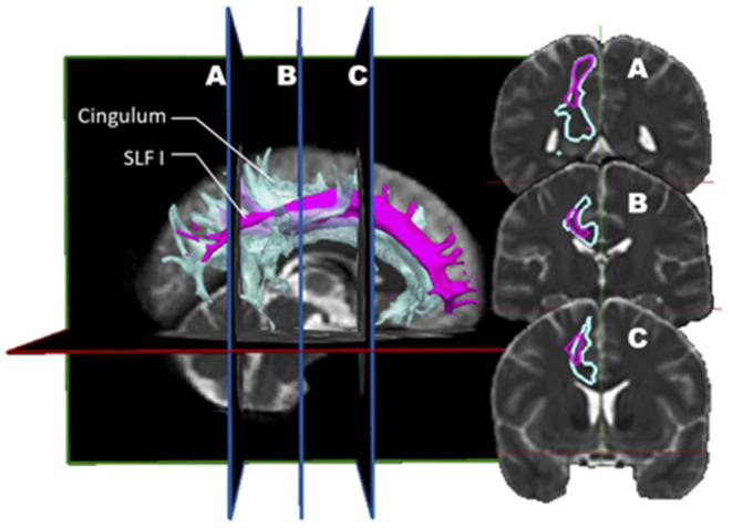
- SLF I, connecting parietal precuneus to superior frontal gyrus, appears assimilable to the cingulum, so its existence and role are unclear; SLF II and SLF III are described as distinct and asymmetric, with left-sided SLF II/III implicated in speech processing and right-sided SLF II/III in visuospatial functioning.
- Wang et al.’s combined dMRI (\(n=10\)) and dissection (\(n=5\)) study found SLF II represented about 66% of total SLF volume and SLF III about 33%; left SLF II connectivity was linked to motor planning of language and/or syntax, right SLF II to attention regulation in spatial orientation.
- The review highlights dMRI validation problems: 63–90% of white matter volume is estimated to contain crossing fibers, and in an international tractography competition most tractograms contained more invalid than valid bundles.
Methods
- Narrative review/opinion based on a literature survey of SLF anatomy, nomenclature, lateralization, individual variability, AF relationship, and functional correlates.
- The authors compare postmortem dissection and diffusion MRI/tractography studies and summarize studies reporting 2, 3, 3 plus AF, and 5 SLF divisions in a table.
- No original cohort is analyzed; illustrative figures use open-source MRI images and the Imeka test tractogram, with segmentation methods derived from Wakana et al. and Fitzgerald et al.
Limitations
- The authors state that inter- and intra-individual variability is greater than previously suspected and recommend larger cohorts; they also note that cited studies often have low subject numbers.
- The authors discuss dMRI pitfalls including false positives, poor inter-protocol segmentation agreement, and orientation errors up to 90° at gray-white matter transitions; postmortem dissection cannot resolve crossing fibers or distal terminations and is biased toward elderly brains and fixation effects.
- Evident from the text: as a review/opinion without a systematic search or quantitative synthesis, its nomenclature recommendations are expert opinion rather than empirically tested consensus.
Model-written summary. Check it against the paper before relying on it.
Integrity signals
Registry
Cites
Statistics
Figures
Verification
Claims the paper makes
28 Own interpretation · 18 No full text of the sources · 6 Supported so far · 5 Partly supported · 4 Supported · 3 Couldn’t confirm · 2 Own result · 2 No basis given · 1 Mixed support · 1 Not supported · accuracy not yet measured
At least one cited source doesn’t support this claim or points to a different paper, and none confirmed it — see the verdicts below.
In the paper
“However, there is one domain where histological postmortem studies have shown a consistent failure of dMRI, which is in regions of transition between white and gray matter, where orientation errors of as much as 90° have been observed (49, 50).”
§ Superior Longitudinal Fasciculus: A Review of the Anatomical Descriptions With Functional Correlates > Introduction > Anatomical Description Derived From Diffusion MRI Studies
What the sources say
- Not checked — no full text of the sourcecites [49] Dyrby, 2007, Validation of in vitro probabilistic tractography, Neuroimage source
- Disputedcites [50] Seehaus, 2015, Histological validation of high-resolution DTI in human post mortem tissue, Front Neuroanat sourceExperimental — accuracy not yet measured
The Results (Orientation differences) say that at the white-to-gray-matter transition, orientation differences often remained small despite reduced FA. The near-90° differences were instead found mostly in bands within gray matter, at intermediate cortical depths (Discussion, Orientations). Thus the paper does not describe a consistent failure at the transition. (LLM, conf=0.88)
Checked by a reasoning model — may be wrong.
plaindirect- Asserted: passes — The authors put it forward as true.
- Checkable: passes — It can be checked against evidence.
- Load-bearing: unclear — It is unclear whether the paper’s conclusions depend on it.
- Based: passes — The paper gives a reason why it holds.
The sources with a verdict support this claim; the rest weren’t checked yet, have no full text we could read, or their verdicts aren’t rated yet.
In the paper
“Postmortem factors, such as the interval between death and fixation, as well as the known effects of the fixative on the tissue can also alter results (18–21).”
§ Superior Longitudinal Fasciculus: A Review of the Anatomical Descriptions With Functional Correlates > Introduction > Anatomical Description Derived From Postmortem Dissections
What the sources say
- Not checked — no full text of the sourcecites [18] Chu, 2005, Ultrasound-accelerated formalin fixation of tissue improves morphology, antigen and mRNA preservation, Mod Pathol source
- Supportedcites [19] Schmierer, 2008, Quantitative magnetic resonance of postmortem multiple sclerosis brain before and after fixation, Magnetic Resonance in Medicine source
The Introduction says short postmortem delays are desirable to avoid structural damage from autolysis. The Discussion reports significant changes in quantitative MR measures following fixation. The Results found no confounding from the measured death-to-retrieval or death-to-MRI intervals in this sample, but that does not contradict the claim that postmortem factors can alter results. (LLM, conf=0.85)
- Not checked — no full text of the sourcecites [20] Stüber, 2014, Myelin and iron concentration in the human brain: a quantitative study of MRI contrast, NeuroImage source
- Not checked — no full text of the sourcecites [21] van Duijn, 2011, artifacts in human brain tissue after prolonged formalin storage, Magnetic Resonance in Medicine source
plaindirect- Asserted: passes — The authors put it forward as true.
- Checkable: passes — It can be checked against evidence.
- Load-bearing: passes — The paper’s conclusions depend on it.
- Based: passes — The paper gives a reason why it holds.
Every source the paper cites for this claim was checked and supports it.
In the paper
“Indeed, they found that most tractograms produced in this competition were made up of more invalid than valid bundles (52).”
§ Superior Longitudinal Fasciculus: A Review of the Anatomical Descriptions With Functional Correlates > Introduction > Anatomical Description Derived From Diffusion MRI Studies
What the sources say
- Supportedcites [52] Maier-Hein, 2017, The challenge of mapping the human connectome based on diffusion tractography, Nat Commun source
The Results section, “Tractograms contained more invalid than valid bundles,” reports an average of 88 invalid bundles per submission, more than four times the average number of valid bundles. It also reports that 69 submissions with at least 23 valid bundles had no fewer than 37 invalid bundles, supporting the claim about most competition tractograms. (LLM, conf=0.94)
plain- Asserted: passes — The authors put it forward as true.
- Checkable: passes — It can be checked against evidence.
- Load-bearing: passes — The paper’s conclusions depend on it.
- Based: passes — The paper gives a reason why it holds.
- Supported
The sources with a verdict support this claim; the rest weren’t checked yet, have no full text we could read, or their verdicts aren’t rated yet.
In the paper
“overall, the right-sided SLF as a whole appears more voluminous than on the left (3, 55).”
§ Superior Longitudinal Fasciculus: A Review of the Anatomical Descriptions With Functional Correlates > Lateralization
What the sources say
- Not checked — no full text of the sourcecites [3] Martino, 2013, Analysis of the subcomponents and cortical terminations of the perisylvian superior longitudinal fasciculus: a fiber dissection and DTI tractography study, Brain Struct Funct source
- Supportedcites [55] Hecht, 2015, Virtual dissection and comparative connectivity of the superior longitudinal fasciculus in chimpanzees and humans, Neuroimage source
The Results (§3.4) report that the SLF was right-lateralized as a whole across the chimpanzees and humans studied, supporting the statement that the right-sided SLF appears more voluminous overall. (LLM, conf=0.94)
hedgedcorrelational- Asserted: passes — The authors put it forward as true.
- Checkable: passes — It can be checked against evidence.
- Load-bearing: unclear — It is unclear whether the paper’s conclusions depend on it.
- Based: passes — The paper gives a reason why it holds.
Every source the paper cites for this claim was checked and supports it.
In the paper
“One of them was that current state-of-the-art tractography algorithms do not control for false positives.”
§ Superior Longitudinal Fasciculus: A Review of the Anatomical Descriptions With Functional Correlates > Introduction > Anatomical Description Derived From Diffusion MRI Studies
What the sources say
- Supportedcites [52] Maier-Hein, 2017, The challenge of mapping the human connectome based on diffusion tractography, Nat Commun source
The Introduction reports that most of the 96 evaluated tractography pipelines produced large numbers of false-positive bundles. The Discussion adds that current pipelines produced substantial false-positive bundles even when tracking ground-truth fiber orientations, supporting the claim about their inability to control false positives in this evaluation. (LLM, conf=0.91)
plain- Asserted: passes — The authors put it forward as true.
- Checkable: passes — It can be checked against evidence.
- Load-bearing: passes — The paper’s conclusions depend on it.
- Based: passes — The paper gives a reason why it holds.
- Supported
The cited sources support only part of this claim — read each source’s verdict below.
In the paper
“Even when given the same set of underlying streamlines, variability across protocols for bundle segmentation was found to be greater than all other sources of potential unevenness.”
§ Superior Longitudinal Fasciculus: A Review of the Anatomical Descriptions With Functional Correlates > Introduction > Anatomical Description Derived From Diffusion MRI Studies
What the sources say
- Partly supportedcites [69] Schilling, 2021, Tractography dissection variability: what happens when 42 groups dissect 14 white matter bundles on the same dataset?, Neuroimage sourceExperimental — accuracy not yet measured
The Discussion (page unavailable) says that, using the same underlying streamlines, variability across segmentation protocols exceeded variability within protocols, including acquisition, registration, reconstruction, and streamline-generation effects. But it describes inter-protocol variability as “similar to (or greater than)” inter-subject variability, not greater than every other source. The Results for the corticospinal tract also show that the comparison depends on the measure used. (LLM, conf=0.88)
plain- Asserted: passes — The authors put it forward as true.
- Checkable: passes — It can be checked against evidence.
- Load-bearing: passes — The paper’s conclusions depend on it.
- Based: passes — The paper gives a reason why it holds.
- Partly supported
Every source the paper cites for this claim was checked and supports it.
In the paper
“In terms of validation, ex vivo dMRI in cadaveric samples appears ideally suited to the study of normal anatomy as it can be obtained with maximal quality.”
§ Superior Longitudinal Fasciculus: A Review of the Anatomical Descriptions With Functional Correlates > Introduction > Anatomical Description Derived From Diffusion MRI Studies
What the sources say
- Supportedcites [53] Takahashi, 2013, Detection of postmortem human cerebellar cortex and white matter pathways using high angular resolution diffusion tractography: a feasibility study, Neuroimage source
The Introduction says ex vivo imaging avoids motion and permits very high resolution and adequate signal-to-noise for HARDI. The Discussion describes high-resolution, high-signal-to-noise postmortem imaging as a way to reveal cerebellar pathways, while the validation section connects the findings to understanding cerebellar organization in healthy individuals. The claim is a reasonable paraphrase of those advantages, though the study specifically examines the cerebellum. (LLM, conf=0.88)
hedgeddirect- Asserted: passes — The authors put it forward as true.
- Checkable: passes — It can be checked against evidence.
- Load-bearing: unclear — It is unclear whether the paper’s conclusions depend on it.
- Based: passes — The paper gives a reason why it holds.
- Supported
The cited sources support only part of this claim — read each source’s verdict below.
In the paper
“This variability can be observed at different levels such as in cortical morphology, cytoarchitecture, task-evoked activation, or in dMRI connectivity pattern (65–68).”
§ Superior Longitudinal Fasciculus: A Review of the Anatomical Descriptions With Functional Correlates > Individual Variability
What the sources say
- Not checked — no full text of the sourcecites [65] Amunts, 2004, Analysis of neural mechanisms underlying verbal fluency in cytoarchitectonically defined stereotaxic space - The roles of Brodmann areas 44 and 45, Neuroimage source
- Partly supportedcites [66] Baldassarre, 2012, Individual variability in functional connectivity predicts performance of a perceptual task, Proc Natl Acad Sci U S A sourceExperimental — accuracy not yet measured
The Discussion (page unavailable) proposes individual variability in structural connectivity as a possible explanation for its functional-connectivity findings. It does not establish the claimed anatomical variability across cortical morphology, cytoarchitecture, task-evoked activation, and dMRI connectivity patterns; the study measured resting-state functional connectivity, not dMRI connectivity. (LLM, conf=0.79)
- Not checked — no full text of the sourcecites [67] Good, 2001, voxel-based morphometric study of ageing in 465 normal adult human brains, Neuroimage source
- Partly supportedcites [68] Hill, 2010, Similar patterns of cortical expansion during human development and evolution, PNAS sourceExperimental — accuracy not yet measured
The Results (page not available) show differences in cortical surface area among individuals within each age group, supporting variability in cortical morphology. The Abstract discusses regional differences in cellular structure, not cytoarchitectural differences between individuals. The cited passages do not establish individual variability in task-evoked activation or dMRI connectivity patterns. (LLM, conf=0.85)
plain- Asserted: passes — The authors put it forward as true.
- Checkable: passes — It can be checked against evidence.
- Load-bearing: unclear — It is unclear whether the paper’s conclusions depend on it.
- Based: passes — The paper gives a reason why it holds.
Every source the paper cites for this claim was checked and supports it.
In the paper
“If we are to still consider the SLF I as part of the SLF, then this trunk appears to be distributed symmetrically between the right and left hemispheres (9).”
§ Superior Longitudinal Fasciculus: A Review of the Anatomical Descriptions With Functional Correlates > Lateralization
What the sources say
- Supportedcites [9] Thiebaut de Schotten, 2011, A lateralized brain network for visuospatial attention, Nat Neurosci source
The 'Asymmetry of Parieto-Frontal Spatial Networks' section treats SLF I as one of the three branches of the SLF and reports a symmetrical distribution between the left and right hemispheres. This directly supports the attributed claim. (LLM, conf=0.94)
hedged- Asserted: passes — The authors put it forward as true.
- Checkable: passes — It can be checked against evidence.
- Load-bearing: unclear — It is unclear whether the paper’s conclusions depend on it.
- Based: passes — The paper gives a reason why it holds.
- Supported
The cited sources that could be checked couldn’t confirm or dispute this claim.
In the paper
“from one individual to another, the volume of the SLF appears different.”
§ Superior Longitudinal Fasciculus: A Review of the Anatomical Descriptions With Functional Correlates > Individual Variability
What the sources say
- Not checked — no full text of the sourcecites [67] Good, 2001, voxel-based morphometric study of ageing in 465 normal adult human brains, Neuroimage source
- Could not verifycites [68] Hill, 2010, Similar patterns of cortical expansion during human development and evolution, PNAS sourceExperimental — accuracy not yet measured
The Results describe differences in cortical surface area among individuals, not differences in superior longitudinal fasciculus (SLF) volume. A search for SLF and superior longitudinal fasciculus found no passage in our copy addressing its volume across individuals; the Discussion's reference to white-matter volume concerns postnatal maturation instead. (LLM, conf=0.76)
hedged- Asserted: passes — The authors put it forward as true.
- Checkable: passes — It can be checked against evidence.
- Load-bearing: passes — The paper’s conclusions depend on it.
- Based: passes — The paper gives a reason why it holds.
- Probably not a substantial claim. 24
- It may state two separate claims as one. 19
Some cited sources support this claim and some don’t — read each source’s verdict below.
In the paper
“Indeed, some studies even describe a fifth division of the SLF comprised of a distinct fasciculus (61–63) that would connect the superior temporal gyrus to the superior parietal.”
§ Superior Longitudinal Fasciculus: A Review of the Anatomical Descriptions With Functional Correlates > Introduction > Anatomical Description Derived From Diffusion MRI Studies
What the sources say
- Not checked — no full text of the sourcecites [61] Frey, 2008, Dissociating the human language pathways with high angular resolution diffusion fiber tractography, J Neurosci source
- Disputedcites [62] Makris, 2009, Delineation of the middle longitudinal fascicle in humans: A quantitative, in vivo, DT-MRI study, Cerebral Cortex sourceExperimental — accuracy not yet measured
The Abstract and Discussion identify the tract as the middle longitudinal fascicle, connecting the superior temporal gyrus to the angular gyrus of the inferior parietal lobule—not the superior parietal region. The Discussion also explicitly describes it as distinct from the SLF, rather than a fifth division of it. (LLM, conf=0.89)
Checked by a reasoning model — may be wrong.
- Partly supportedcites [63] Zhang, 2010, Atlas-guided tract reconstruction for automated and comprehensive examination of the white matter anatomy, Neuroimage sourceExperimental — accuracy not yet measured
The Results (Comparison between the automated and manual methods; page unavailable) describe a separately reconstructed SLF segment connecting the parietal and temporal lobes. They do not call it a fifth division or identify its endpoints as the superior temporal gyrus and superior parietal region; the parietal regions specified are the angular and supramarginal gyri. (LLM, conf=0.85)
hedgedcorrelational- Asserted: passes — The authors put it forward as true.
- Checkable: passes — It can be checked against evidence.
- Load-bearing: fails — The paper’s conclusions may not depend on it.
- Based: passes — The paper gives a reason why it holds.
Probably not a substantial claim. The paper’s conclusions may not depend on it.
The cited sources support only part of this claim — read each source’s verdict below.
In the paper
“Classical DTI postmortem histological validation studies have shown that fiber orientation is correctly represented in large unidirectional fiber bundles, but fails in complex regions with fiber crossings and low anisotropy (44–46).”
§ Superior Longitudinal Fasciculus: A Review of the Anatomical Descriptions With Functional Correlates > Introduction > Anatomical Description Derived From Diffusion MRI Studies
What the sources say
- Not checked — no full text of the sourcecites [44] Dauguet, 2007, Comparison of fiber tracts derived from in-vivo DTI tractography with 3D histological neural tract tracer reconstruction on a macaque brain, Neuroimage source
- Partly supportedcites [45] Kaufman, 2005, Anatomical analysis of an aye-aye brain (Daubentonia madagascariensis, primates: prosimii) combining histology, structural magnetic resonance imaging, and diffusion-tensor imaging, Anat Rec A Discov Mol Cell Evol Biol sourceExperimental — accuracy not yet measured
The Diffusion-Tensor Imaging section (page unavailable) reports good correspondence between DTI and histological measurements in the cingulum bundle, supporting the claim for coherent fibers. It also finds broader fiber-orientation distributions where anisotropy is low, but does not show that DTI fails there or test regions with crossing fibers. (LLM, conf=0.88)
- Not checked — no full text of the sourcecites [46] Fernandez-Miranda, 2008, Three-dimensional microsurgical and tractographic anatomy of the white matter of the human brain, Neurosurgery source
boosteddirect- Asserted: passes — The authors put it forward as true.
- Checkable: passes — It can be checked against evidence.
- Load-bearing: passes — The paper’s conclusions depend on it.
- Based: passes — The paper gives a reason why it holds.
It may state two separate claims as one.
The cited sources support only part of this claim — read each source’s verdict below.
In the paper
“A great many studies clearly distinguish the 2 bundles as distinct entities, both anatomically and functionally (56, 59, 70, 71, 75, 76).”
§ Superior Longitudinal Fasciculus: A Review of the Anatomical Descriptions With Functional Correlates > Is The AF Part of the SLF?
What the sources say
- Not checked — no full text of the sourcecites [56] Thiebaut de Schotten, 2011, Monkey to human comparative anatomy of the frontal lobe association tracts, Cortex source
- Partly supportedcites [59] Cabeen, 2016, Kernel regression estimation of fiber orientation mixtures in diffusion MRI, Neuroimage sourceExperimental — accuracy not yet measured
The Results (§5.5; page unavailable) describe the arcuate fasciculus and three portions of the superior longitudinal fasciculus separately, supporting an anatomical distinction. The paper does not address whether they are functionally distinct or establish that a great many studies distinguish them on both grounds. (LLM, conf=0.84)
- Not checked — no full text of the sourcecites [70] Catani, 2012, Atlas of Human Brain Connections source
boosted- Asserted: passes — The authors put it forward as true.
- Checkable: passes — It can be checked against evidence.
- Load-bearing: passes — The paper’s conclusions depend on it.
- Based: passes — The paper gives a reason why it holds.
It may state two separate claims as one.
The cited sources that could be checked couldn’t confirm or dispute this claim.
In the paper
“This variability is a correlate of the phenotypic variability in general, determined by the genetic substrates and the environmental exposure that shapes this diversity for any trait, including the brain's anatomy, (68)”
§ Superior Longitudinal Fasciculus: A Review of the Anatomical Descriptions With Functional Correlates > Individual Variability
What the sources say
- Could not verifycites [68] Hill, 2010, Similar patterns of cortical expansion during human development and evolution, PNAS sourceExperimental — accuracy not yet measured
The Discussion considers intrinsic and extrinsic signals in the maturation of cortical areas and suggests that postnatal experience may contribute to development in some regions. It does not address whether genetic substrates and environmental exposure determine variability among individuals or phenotypic variability across traits (searched: individual variability, genetics, environment, and phenotype). (LLM, conf=0.72)
plaincorrelational- Asserted: passes — The authors put it forward as true.
- Checkable: passes — It can be checked against evidence.
- Load-bearing: fails — The paper’s conclusions may not depend on it.
- Based: passes — The paper gives a reason why it holds.
Probably not a substantial claim. The paper’s conclusions may not depend on it.
- Could not verify
The sources with a verdict support this claim; the rest weren’t checked yet, have no full text we could read, or their verdicts aren’t rated yet.
In the paper
“the SLF is in its essence a fronto-parietal tract, hence connecting the frontal and parietal cortices”
- Also stated in: § Functional Correlates of SLF · plain
“The SLF is a massive bundle connecting widespread areas of the frontal and the parietal lobes.”
§ Superior Longitudinal Fasciculus: A Review of the Anatomical Descriptions With Functional Correlates > Is The AF Part of the SLF?
What the sources say
- Supportedcites [55] Hecht, 2015, Virtual dissection and comparative connectivity of the superior longitudinal fasciculus in chimpanzees and humans, Neuroimage source
The Introduction defines the SLF as direct fronto-parietal connections. The Results (§3.2) describe SLF tracts connecting frontal and parietal cortical regions, supporting the verbatim citing sentence. (LLM, conf=0.94)
- Not checked — no full text of the sourcecites [56] Thiebaut de Schotten, 2011, Monkey to human comparative anatomy of the frontal lobe association tracts, Cortex source
plaincorrelational- Asserted: passes — The authors put it forward as true.
- Checkable: passes — It can be checked against evidence.
- Load-bearing: passes — The paper’s conclusions depend on it.
- Based: passes — The paper gives a reason why it holds.
It may state two separate claims as one.
- Also stated in: § Functional Correlates of SLF · plain
The sources with a verdict support this claim; the rest weren’t checked yet, have no full text we could read, or their verdicts aren’t rated yet.
In the paper
“the AF is recognized as a distinctive bundle with connection areas and a trajectory different from that of the SLF”
§ Superior Longitudinal Fasciculus: A Review of the Anatomical Descriptions With Functional Correlates > Is The AF Part of the SLF?
What the sources say
- Supportedcites [55] Hecht, 2015, Virtual dissection and comparative connectivity of the superior longitudinal fasciculus in chimpanzees and humans, Neuroimage source
The Introduction distinguishes the arcuate fasciculus (AF) as a fronto-temporal bundle from the fronto-parietal SLF. Discussion §4.1 adds that the AF travels beneath the SLF without terminating in parietal cortex, supporting the claimed differences in connections and trajectory. The paper notes that naming conventions vary, but adopts this distinction. (LLM, conf=0.92)
- Not checked — no full text of the sourcecites [56] Thiebaut de Schotten, 2011, Monkey to human comparative anatomy of the frontal lobe association tracts, Cortex source
plain- Asserted: passes — The authors put it forward as true.
- Checkable: passes — It can be checked against evidence.
- Load-bearing: passes — The paper’s conclusions depend on it.
- Based: passes — The paper gives a reason why it holds.
It may state two separate claims as one.
- Supported
The cited sources that could be checked couldn’t confirm or dispute this claim.
In the paper
“Fractional anisotropy of SLF also varies between individuals, but less importantly, than volume (67, 68).”
§ Superior Longitudinal Fasciculus: A Review of the Anatomical Descriptions With Functional Correlates > Individual Variability
What the sources say
- Not checked — no full text of the sourcecites [67] Good, 2001, voxel-based morphometric study of ageing in 465 normal adult human brains, Neuroimage source
- Could not verifycites [68] Hill, 2010, Similar patterns of cortical expansion during human development and evolution, PNAS sourceExperimental — accuracy not yet measured
The Results and Materials and Methods passages discuss individual differences in cortical surface area and patterns of cortical expansion, not the superior longitudinal fasciculus (SLF). Searches for SLF fractional anisotropy and its variability relative to volume found no passage addressing the claim. (LLM, conf=0.76)
plaincorrelational- Asserted: passes — The authors put it forward as true.
- Checkable: passes — It can be checked against evidence.
- Load-bearing: unclear — It is unclear whether the paper’s conclusions depend on it.
- Based: passes — The paper gives a reason why it holds.
It may state two separate claims as one.
The sources with a verdict support this claim; the rest weren’t checked yet, have no full text we could read, or their verdicts aren’t rated yet.
In the paper
“whereas the AF is a fronto-temporal tract that passes through the parietal white matter beneath the SLF (55, 56).”
§ Superior Longitudinal Fasciculus: A Review of the Anatomical Descriptions With Functional Correlates > Is The AF Part of the SLF?
What the sources say
- Supportedcites [55] Hecht, 2015, Virtual dissection and comparative connectivity of the superior longitudinal fasciculus in chimpanzees and humans, Neuroimage source
Section 4.1 (page not provided) describes the arcuate fasciculus as a fronto-temporal tract that travels through parietal white matter beneath the SLF. The authors explicitly adopt this anatomical distinction, although they did not track the arcuate fasciculus in this study. (LLM, conf=0.95)
- Not checked — no full text of the sourcecites [56] Thiebaut de Schotten, 2011, Monkey to human comparative anatomy of the frontal lobe association tracts, Cortex source
plain- Asserted: passes — The authors put it forward as true.
- Checkable: passes — It can be checked against evidence.
- Load-bearing: passes — The paper’s conclusions depend on it.
- Based: passes — The paper gives a reason why it holds.
It may state two separate claims as one.
- Supported
The sources with a verdict support this claim; the rest weren’t checked yet, have no full text we could read, or their verdicts aren’t rated yet.
In the paper
“a lot of the prior work which has made its way in the literature and anatomy textbooks is based on non-human primates, which present a rather different connectivity anatomy, thus steering the nomenclature toward a biased pattern (5, 56).”
§ Superior Longitudinal Fasciculus: A Review of the Anatomical Descriptions With Functional Correlates > Nomenclature
What the sources say
- Supportedcites [5] Mandonnet, 2018, The nomenclature of human white matter association pathways: proposal for a systematic taxonomic anatomical classification, Front Neuroanat source
In the Introduction, under “The Current Status of WM Terminology,” the paper says that extensive monkey tracing work was used as a basis for classifying human white-matter pathways, calling this a “drift.” The same section explains that macaque pathways cannot be treated as the ground truth of human neuroanatomy and that interspecies analogy has made bundle names confusing. (LLM, conf=0.91)
- Not checked — no full text of the sourcecites [56] Thiebaut de Schotten, 2011, Monkey to human comparative anatomy of the frontal lobe association tracts, Cortex source
plaindirect- Asserted: passes — The authors put it forward as true.
- Checkable: passes — It can be checked against evidence.
- Load-bearing: passes — The paper’s conclusions depend on it.
- Based: passes — The paper gives a reason why it holds.
It may state two separate claims as one.
- Supported
A conclusion the paper draws from its own results — it cites no other source for it, so there is none to check. The quote shows it is in the paper.
In the paper
“As such, this fiber bundle is likely to be one of the most affected whenever we undertake a surgery for an intrinsic brain tumor.”
§ Superior Longitudinal Fasciculus: A Review of the Anatomical Descriptions With Functional Correlates > Introduction
hedgedconditional- Asserted: passes — The authors put it forward as true.
- Checkable: passes — It can be checked against evidence.
- Load-bearing: fails — The paper’s conclusions may not depend on it.
- Based: fails — The paper may give no reason why it holds.
Probably not a substantial claim. The paper’s conclusions may not depend on it. The paper may give no reason why it holds.
- Rests on an assumption: The SLF is part of the longitudinal association fiber system, which lays connections between the frontal lobe and other areas of the ipsilateral hemisphere.
- Rests on an assumption: The SLF connects the perisylvian areas in the hemisphere (frontal, temporal, and parietal).
A conclusion the paper draws from its own results — it cites no other source for it, so there is none to check. The quote shows it is in the paper.
In the paper
“However, this is not the case, and a lot of confusion and overlap surrounds this entity.”
§ Superior Longitudinal Fasciculus: A Review of the Anatomical Descriptions With Functional Correlates > Abstract
plain- Asserted: passes — The authors put it forward as true.
- Checkable: passes — It can be checked against evidence.
- Load-bearing: passes — The paper’s conclusions depend on it.
- Based: passes — The paper gives a reason why it holds.
Probably not a substantial claim. It may state two separate claims as one.
A conclusion the paper draws from its own results — it cites no other source for it, so there is none to check. The quote shows it is in the paper.
In the paper
“It stands to reason that there would be asymmetric findings between both SLF.”
§ Superior Longitudinal Fasciculus: A Review of the Anatomical Descriptions With Functional Correlates > Lateralization
hedgedcorrelational- Asserted: passes — The authors put it forward as true.
- Checkable: passes — It can be checked against evidence.
- Load-bearing: passes — The paper’s conclusions depend on it.
- Based: passes — The paper gives a reason why it holds.
Probably not a substantial claim.
- Rests on an assumption: There is a clear asymmetry in speech function (left-sided characteristically) and visuospatial functioning (right-sided characteristically).
In the paper
“In this diffusion tensor imaging study, the SLF of 12 normal right-handed participants was assessed to find that projection to Broca's area was minimal or absent in the majority of cases.”
§ Superior Longitudinal Fasciculus: A Review of the Anatomical Descriptions With Functional Correlates > Introduction > Anatomical Description Derived From Diffusion MRI Studies
What the sources say
- Not checked — no full text of the sourcecites [57] Bernal, 2010, The connectivity of the superior longitudinal fasciculus: A tractography DTI study, Magn Reson Imaging source
plainn = 12- Asserted: passes — The authors put it forward as true.
- Checkable: passes — It can be checked against evidence.
- Load-bearing: fails — The paper’s conclusions may not depend on it.
- Based: passes — The paper gives a reason why it holds.
Probably not a substantial claim. The paper’s conclusions may not depend on it.
In the paper
“Hence, they consider the SLF as only composed of a dorsal (SLF II) and a ventral (SLF III) segment.”
§ Superior Longitudinal Fasciculus: A Review of the Anatomical Descriptions With Functional Correlates > Introduction > Anatomical Description Derived From Diffusion MRI Studies
What the sources say
- Not checked — no full text of the sourcecites [7] Wang, 2016, Subcomponents and connectivity of the superior longitudinal fasciculus in the human brain, Brain Struct Funct source
plain- Asserted: passes — The authors put it forward as true.
- Checkable: passes — It can be checked against evidence.
- Load-bearing: fails — The paper’s conclusions may not depend on it.
- Based: passes — The paper gives a reason why it holds.
Probably not a substantial claim. The paper’s conclusions may not depend on it.
In the paper
“Wang et al., on the other hand, found a slight asymmetric difference in all SLF sub-component with a tendency toward right-hemispheric dominance (7).”
§ Superior Longitudinal Fasciculus: A Review of the Anatomical Descriptions With Functional Correlates > Lateralization
What the sources say
- Not checked — no full text of the sourcecites [7] Wang, 2016, Subcomponents and connectivity of the superior longitudinal fasciculus in the human brain, Brain Struct Funct source
plaincorrelational- Asserted: passes — The authors put it forward as true.
- Checkable: passes — It can be checked against evidence.
- Load-bearing: fails — The paper’s conclusions may not depend on it.
- Based: passes — The paper gives a reason why it holds.
Probably not a substantial claim. The paper’s conclusions may not depend on it. It may state two separate claims as one.
In the paper
“In this construct, the SLF II represents 66% of the total volume, whereas the SLF III about 33%.”
§ Superior Longitudinal Fasciculus: A Review of the Anatomical Descriptions With Functional Correlates > Introduction > Anatomical Description Derived From Diffusion MRI Studies
What the sources say
- Not checked — no full text of the sourcecites [7] Wang, 2016, Subcomponents and connectivity of the superior longitudinal fasciculus in the human brain, Brain Struct Funct source
plain- Asserted: passes — The authors put it forward as true.
- Checkable: passes — It can be checked against evidence.
- Load-bearing: fails — The paper’s conclusions may not depend on it.
- Based: passes — The paper gives a reason why it holds.
Probably not a substantial claim. The paper’s conclusions may not depend on it. It may state two separate claims as one.
In the paper
“The first conclusion is that there is a high discrepancy between the simian and human anatomy; hence, all conclusions derived from the study of the former cannot be applied to the latter.”
§ Superior Longitudinal Fasciculus: A Review of the Anatomical Descriptions With Functional Correlates > Introduction > Anatomical Description Derived From Diffusion MRI Studies
What the sources say
- Not checked — no full text of the sourcecites [7] Wang, 2016, Subcomponents and connectivity of the superior longitudinal fasciculus in the human brain, Brain Struct Funct source
plaindirect- Asserted: passes — The authors put it forward as true.
- Checkable: passes — It can be checked against evidence.
- Load-bearing: passes — The paper’s conclusions depend on it.
- Based: passes — The paper gives a reason why it holds.
Probably not a substantial claim. It may state two separate claims as one.
A conclusion the paper draws from its own results — it cites no other source for it, so there is none to check. The quote shows it is in the paper.
In the paper
“There is no such thing as a singular brain blueprint!”
§ Superior Longitudinal Fasciculus: A Review of the Anatomical Descriptions With Functional Correlates > Individual Variability
plain- Asserted: passes — The authors put it forward as true.
- Checkable: fails — It may be too vague to check against evidence.
- Load-bearing: passes — The paper’s conclusions depend on it.
- Based: passes — The paper gives a reason why it holds.
Probably not a substantial claim. It may be too vague to check against evidence.
- Rests on an assumption: Some areas and connectivity routes are highly conserved amongst individuals (such as primary areas and large projection bundles), while others are more variable.
- Rests on an assumption: The brain connectivity pattern of an individual is constantly changing, and the density of white matter changes during the development of the individual.
A conclusion the paper draws from its own results — it cites no other source for it, so there is none to check. The quote shows it is in the paper.
In the paper
“However, the inclusion of the AF as the 4th component of the SLF is far from unanimous and remains a subject of controversy in the nomenclature that is addressed in a later section of this study.”
§ Superior Longitudinal Fasciculus: A Review of the Anatomical Descriptions With Functional Correlates > Introduction > Anatomical Description Derived From Diffusion MRI Studies
plain- Asserted: passes — The authors put it forward as true.
- Checkable: passes — It can be checked against evidence.
- Load-bearing: fails — The paper’s conclusions may not depend on it.
- Based: passes — The paper gives a reason why it holds.
Probably not a substantial claim. The paper’s conclusions may not depend on it.
A claim is checked only against the cited paper’s full text, never its abstract, and none is available for its sources.
In the paper
“other researchers found the SLF II subcomponents' lateralization to be dominant in the left hemisphere (7, 56, 72).”
§ Superior Longitudinal Fasciculus: A Review of the Anatomical Descriptions With Functional Correlates > Lateralization
What the sources say
- Not checked — no full text of the sourcecites [7] Wang, 2016, Subcomponents and connectivity of the superior longitudinal fasciculus in the human brain, Brain Struct Funct source
- Not checked — no full text of the sourcecites [56] Thiebaut de Schotten, 2011, Monkey to human comparative anatomy of the frontal lobe association tracts, Cortex source
- Not checked — no full text of the sourcecites [72] Vernooij, 2007, Fiber density asymmetry of the arcuate fasciculus in relation to functional hemispheric language lateralization in both right- and left-handed healthy subjects: a combined fMRI and DTI study, Neuroimage source
plaincorrelational- Asserted: passes — The authors put it forward as true.
- Checkable: passes — It can be checked against evidence.
- Load-bearing: fails — The paper’s conclusions may not depend on it.
- Based: passes — The paper gives a reason why it holds.
Probably not a substantial claim. The paper’s conclusions may not depend on it.
A conclusion the paper draws from its own results — it cites no other source for it, so there is none to check. The quote shows it is in the paper.
In the paper
“This is an illustration of the poor consensus on the precise anatomical definition of white matter bundles.”
§ Superior Longitudinal Fasciculus: A Review of the Anatomical Descriptions With Functional Correlates > Introduction > Anatomical Description Derived From Diffusion MRI Studies
plain- Asserted: passes — The authors put it forward as true.
- Checkable: fails — It may be too vague to check against evidence.
- Load-bearing: passes — The paper’s conclusions depend on it.
- Based: passes — The paper gives a reason why it holds.
Probably not a substantial claim. It may be too vague to check against evidence.
A conclusion the paper draws from its own results — it cites no other source for it, so there is none to check. The quote shows it is in the paper.
In the paper
“some difference in connectivity is expected.”
§ Superior Longitudinal Fasciculus: A Review of the Anatomical Descriptions With Functional Correlates > Lateralization
hedged- Asserted: passes — The authors put it forward as true.
- Checkable: fails — It may be too vague to check against evidence.
- Load-bearing: fails — The paper’s conclusions may not depend on it.
- Based: passes — The paper gives a reason why it holds.
Probably not a substantial claim. It may be too vague to check against evidence. The paper’s conclusions may not depend on it.
- Rests on an assumption: There is a clear asymmetry in speech function (left-sided characteristically) and visuospatial functioning (right-sided characteristically).
A conclusion the paper draws from its own results — it cites no other source for it, so there is none to check. The quote shows it is in the paper.
In the paper
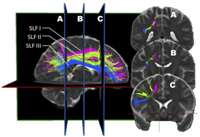
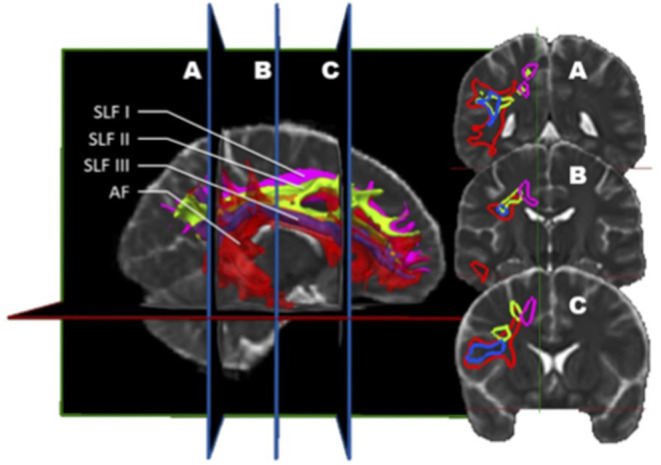
“The first three SLF subdivisions are usually reported alike in most studies, except for a few minor differences (Figure 2).”
§ Superior Longitudinal Fasciculus: A Review of the Anatomical Descriptions With Functional Correlates > Introduction > Anatomical Description Derived From Diffusion MRI Studies
plain- Asserted: passes — The authors put it forward as true.
- Checkable: passes — It can be checked against evidence.
- Load-bearing: fails — The paper’s conclusions may not depend on it.
- Based: passes — The paper gives a reason why it holds.
Probably not a substantial claim. The paper’s conclusions may not depend on it.
A conclusion the paper draws from its own results — it cites no other source for it, so there is none to check. The quote shows it is in the paper.
In the paper
“Their findings are rather provocative, differing from the rest of the literature and challenging the current knowledge on the SLF.”
§ Superior Longitudinal Fasciculus: A Review of the Anatomical Descriptions With Functional Correlates > Introduction > Anatomical Description Derived From Diffusion MRI Studies
plain- Asserted: passes — The authors put it forward as true.
- Checkable: passes — It can be checked against evidence.
- Load-bearing: fails — The paper’s conclusions may not depend on it.
- Based: passes — The paper gives a reason why it holds.
Probably not a substantial claim. The paper’s conclusions may not depend on it. It may state two separate claims as one.
A conclusion the paper draws from its own results — it cites no other source for it, so there is none to check. The quote shows it is in the paper.
In the paper
“This would functionally be related to its role in language.”
§ Superior Longitudinal Fasciculus: A Review of the Anatomical Descriptions With Functional Correlates > Lateralization
hedgedcorrelational- Asserted: passes — The authors put it forward as true.
- Checkable: passes — It can be checked against evidence.
- Load-bearing: fails — The paper’s conclusions may not depend on it.
- Based: passes — The paper gives a reason why it holds.
Probably not a substantial claim. The paper’s conclusions may not depend on it.
A conclusion the paper draws from its own results — it cites no other source for it, so there is none to check. The quote shows it is in the paper.
In the paper
“This finding contradicts the preconceived SLF structure and role, and does not represent the dominant consensus.”
§ Superior Longitudinal Fasciculus: A Review of the Anatomical Descriptions With Functional Correlates > Introduction > Anatomical Description Derived From Diffusion MRI Studies
plainn = 12- Asserted: passes — The authors put it forward as true.
- Checkable: passes — It can be checked against evidence.
- Load-bearing: fails — The paper’s conclusions may not depend on it.
- Based: passes — The paper gives a reason why it holds.
Probably not a substantial claim. The paper’s conclusions may not depend on it. It may state two separate claims as one.
A claim is checked only against the cited paper’s full text, never its abstract, and none is available for its sources.
In the paper
“Martino et al. used dMRI and cortex-sparing fiber dissection and came to the conclusion that there were two segments to the SLF, an anterior and a posterior segment, much like Martino et al. and Wang et al. (3, 7).”
§ Superior Longitudinal Fasciculus: A Review of the Anatomical Descriptions With Functional Correlates > Introduction > Anatomical Description Derived From Diffusion MRI Studies
What the sources say
- Not checked — no full text of the sourcecites [3] Martino, 2013, Analysis of the subcomponents and cortical terminations of the perisylvian superior longitudinal fasciculus: a fiber dissection and DTI tractography study, Brain Struct Funct source
- Not checked — no full text of the sourcecites [7] Wang, 2016, Subcomponents and connectivity of the superior longitudinal fasciculus in the human brain, Brain Struct Funct source
plain- Asserted: passes — The authors put it forward as true.
- Checkable: passes — It can be checked against evidence.
- Load-bearing: unclear — It is unclear whether the paper’s conclusions depend on it.
- Based: passes — The paper gives a reason why it holds.
It may state two separate claims as one.
In the paper
“these long associative axons comprise only 2% of the total intrahemispheric cortico-cortical fibers and that 98% of the fibers are short U-fibers (74).”
§ Superior Longitudinal Fasciculus: A Review of the Anatomical Descriptions With Functional Correlates > Individual Variability
What the sources say
- Not checked — no full text of the sourcecites [74] Duffau, 2012 source
plain- Asserted: passes — The authors put it forward as true.
- Checkable: passes — It can be checked against evidence.
- Load-bearing: unclear — It is unclear whether the paper’s conclusions depend on it.
- Based: passes — The paper gives a reason why it holds.
It may state two separate claims as one.
A conclusion the paper draws from its own results — it cites no other source for it, so there is none to check. The quote shows it is in the paper.
In the paper
“Other authors also support the idea that the SLF is indeed comprised of two main divisions.”
§ Superior Longitudinal Fasciculus: A Review of the Anatomical Descriptions With Functional Correlates > Introduction > Anatomical Description Derived From Diffusion MRI Studies
plain- Asserted: passes — The authors put it forward as true.
- Checkable: passes — It can be checked against evidence.
- Load-bearing: fails — The paper’s conclusions may not depend on it.
- Based: passes — The paper gives a reason why it holds.
Probably not a substantial claim. The paper’s conclusions may not depend on it.
A conclusion the paper draws from its own results — it cites no other source for it, so there is none to check. The quote shows it is in the paper.
In the paper
“This would be in keeping with a role for the left SLF II in motor planning of speech and/or syntax processing.”
§ Superior Longitudinal Fasciculus: A Review of the Anatomical Descriptions With Functional Correlates > Functional Correlates of SLF
hedgedcorrelational- Asserted: passes — The authors put it forward as true.
- Checkable: passes — It can be checked against evidence.
- Load-bearing: passes — The paper’s conclusions depend on it.
- Based: passes — The paper gives a reason why it holds.
It may state two separate claims as one.
A conclusion the paper draws from its own results — it cites no other source for it, so there is none to check. The quote shows it is in the paper.
In the paper
“As the bundle depicts clear asymmetry between both hemispheres, this must translate into obvious functional implications.”
§ Superior Longitudinal Fasciculus: A Review of the Anatomical Descriptions With Functional Correlates > Functional Correlates of SLF
boosteddirect- Asserted: passes — The authors put it forward as true.
- Checkable: passes — It can be checked against evidence.
- Load-bearing: passes — The paper’s conclusions depend on it.
- Based: fails — The paper may give no reason why it holds.
Probably not a substantial claim. The paper may give no reason why it holds.
- Rests on an assumption: There is a clear asymmetry in speech function (left-sided characteristically) and visuospatial functioning (right-sided characteristically).
In the paper
“the anterior segment of the AF appears to somehow correspond to the SLF III, and the two terms are used interchangeably.”
§ Superior Longitudinal Fasciculus: A Review of the Anatomical Descriptions With Functional Correlates > Is The AF Part of the SLF?
What the sources say
- Not checked — no full text of the sourcecites [70] Catani, 2012, Atlas of Human Brain Connections source
hedgedcorrelational- Asserted: passes — The authors put it forward as true.
- Checkable: passes — It can be checked against evidence.
- Load-bearing: unclear — It is unclear whether the paper’s conclusions depend on it.
- Based: passes — The paper gives a reason why it holds.
It may state two separate claims as one.
A conclusion the paper draws from its own results — it cites no other source for it, so there is none to check. The quote shows it is in the paper.
In the paper
“The SLF II and III are clearly defined as distinct entities that are asymmetrical in their function as well as presumably in their role”
§ Superior Longitudinal Fasciculus: A Review of the Anatomical Descriptions With Functional Correlates > Conclusion
boostedcorrelational- Asserted: passes — The authors put it forward as true.
- Checkable: passes — It can be checked against evidence.
- Load-bearing: passes — The paper’s conclusions depend on it.
- Based: passes — The paper gives a reason why it holds.
It may state two separate claims as one.
In the paper
“According to some authors, this connectivity pattern is probably involved in spatial awareness functioning (7).”
§ Superior Longitudinal Fasciculus: A Review of the Anatomical Descriptions With Functional Correlates > Functional Correlates of SLF
What the sources say
- Not checked — no full text of the sourcecites [7] Wang, 2016, Subcomponents and connectivity of the superior longitudinal fasciculus in the human brain, Brain Struct Funct source
hedgedconditional- Asserted: passes — The authors put it forward as true.
- Checkable: passes — It can be checked against evidence.
- Load-bearing: fails — The paper’s conclusions may not depend on it.
- Based: passes — The paper gives a reason why it holds.
Probably not a substantial claim. The paper’s conclusions may not depend on it.
A conclusion the paper draws from its own results — it cites no other source for it, so there is none to check. The quote shows it is in the paper.
In the paper
“the SLF itself in its current definition is not a consensual entity.”
§ Superior Longitudinal Fasciculus: A Review of the Anatomical Descriptions With Functional Correlates > Conclusion
plain- Asserted: passes — The authors put it forward as true.
- Checkable: passes — It can be checked against evidence.
- Load-bearing: passes — The paper’s conclusions depend on it.
- Based: passes — The paper gives a reason why it holds.
Probably not a substantial claim.
- Rests on an assumption: Individual variability had been underestimated in the past.
A conclusion the paper draws from its own results — it cites no other source for it, so there is none to check. The quote shows it is in the paper.
In the paper
“the very definition of the SLF, connecting the frontal to the parietal lobe, allows a clear distinction with the AF, connecting the frontal lobe to the temporal lobe.”
§ Superior Longitudinal Fasciculus: A Review of the Anatomical Descriptions With Functional Correlates > Conclusion
boosteddirect- Asserted: passes — The authors put it forward as true.
- Checkable: passes — It can be checked against evidence.
- Load-bearing: passes — The paper’s conclusions depend on it.
- Based: passes — The paper gives a reason why it holds.
Probably not a substantial claim.
A conclusion the paper draws from its own results — it cites no other source for it, so there is none to check. The quote shows it is in the paper.
In the paper
“its existence, as well as its role is unclear at this time.”
§ Superior Longitudinal Fasciculus: A Review of the Anatomical Descriptions With Functional Correlates > Conclusion
plain- Asserted: passes — The authors put it forward as true.
- Checkable: passes — It can be checked against evidence.
- Load-bearing: passes — The paper’s conclusions depend on it.
- Based: passes — The paper gives a reason why it holds.
It may state two separate claims as one.
The paper relies on this without support.
The SLF is part of the longitudinal association fiber system, which lays connections between the frontal lobe and other areas of the ipsilateral hemisphere.
“The superior longitudinal fasciculus (SLF) is part of the longitudinal association fiber system, which lays connections between the frontal lobe and other areas of the ipsilateral hemisphere.”
The paper relies on this without support.
The SLF connects the perisylvian areas in the hemisphere (frontal, temporal, and parietal).
“To put it simply, it connects the perisylvian areas in the hemisphere (frontal, temporal, and parietal).”
The paper relies on this without support.
There is a clear asymmetry in speech function (left-sided characteristically) and visuospatial functioning (right-sided characteristically).
“As there is a clear asymmetry in speech function (left-sided characteristically) and visuospatial functioning (right-sided characteristically)”
The paper relies on this without support.
Some areas and connectivity routes are highly conserved amongst individuals (such as primary areas and large projection bundles), while others are more variable.
“Although some areas and connectivity routes are highly conserved amongst individuals (such as primary areas and large projection bundles), others are more variable.”
The paper relies on this without support.
The brain connectivity pattern of an individual is constantly changing, and the density of white matter changes during the development of the individual.
“the brain connectivity pattern of an individual is constantly changing, and the density of white matter has been shown to change during the development of the individual.”
The paper relies on this without support.
Individual variability had been underestimated in the past.
“the individual variability had been underestimated in the past.”
Self-check
Percentages, sums and reported differences re-computed from the paper’s own numbers.
Each contradiction between two passages listed here was confirmed by an independent check.
Rated major: it would touch a result — a value a result rests on, reported and recomputed p-values on opposite sides of the significance level, two passages that conflict with no sign which is right, or a cited work retracted before this paper was published.
In the paper
§ Superior Longitudinal Fasciculus: A Review of the Anatomical Descriptions With Functional Correlates > Introduction > Anatomical Description Derived From Diffusion MRI Studies
“In the left hemisphere, the connectivity pattern was in keeping with a predominant functional role in speech (connections between the supramarginal gyrus with the dorsal precentral gyrus and caudal middle frontal gyrus).”
§ Superior Longitudinal Fasciculus: A Review of the Anatomical Descriptions With Functional Correlates > Introduction > Anatomical Description Derived From Diffusion MRI Studies
“Dorsal segment (SLF II) in the left hemisphere | Angular gyrus, Brodmann Areas (BA) 39 | Caudal middle frontal gyrus and dorsal precentral gyrus”
Rated minor: it would not touch a result — a value no result rests on, a p-value that stays on the same side of the significance level, or a conflict that is clerical, resolved elsewhere in the paper, or outside its own findings.
In the paper
§ Superior Longitudinal Fasciculus: A Review of the Anatomical Descriptions With Functional Correlates > Lateralization
“other researchers found the SLF II subcomponents' lateralization to be dominant in the left hemisphere (7, 56, 72).”
§ Superior Longitudinal Fasciculus: A Review of the Anatomical Descriptions With Functional Correlates > Lateralization
“Wang et al., on the other hand, found a slight asymmetric difference in all SLF sub-component with a tendency toward right-hemispheric dominance (7).”
Citations
Cites
Spin Diffusion Measurements: Spin Echoes in the Presence of a Time-Dependent Field Gradient
Stejskal, E. O., & Tanner, J. E. (1965) · The Journal of Chemical Physics · doi:10.1063/1.1695690 · Cited by 8,239
The basis of anisotropic water diffusion in the nervous system—A technical review.
Beaulieu, C. (2002) · NMR in Biomedicine · doi:10.1002/nbm.782 · Cited by 4,628
A Voxel-Based Morphometric Study of Ageing in 465 Normal Adult Human Brains
Good, C. D., Johnsrude, I. S., Ashburner, J., Henson, R. N. A., Friston, K. J., & Frackowiak, R. S. J. (2001) · NeuroImage · doi:10.1006/nimg.2001.0786 · Cited by 4,561
Three-dimensional tracking of axonal projections in the brain by magnetic resonance imaging
Mori, S., Crain, B. J., Chacko, V. P., & van Zijl, P. C. M. (1999) · Annals of Neurology · doi:10.1002/1531-8249(199902)45:2 · Cited by 3,513
Diffusion tensor imaging of the brain
Alexander, A. L., Lee, J. E., Lazar, M., & Field, A. S. (2007) · Neurotherapeutics · doi:10.1016/j.nurt.2007.05.011 · Cited by 2,747
Diffusion tensor MR imaging of the human brain
Pierpaoli, C., Jezzard, P., Basser, P. J., Barnett, A., & Di Chiro, G. (1996) · Radiology · doi:10.1148/radiology.201.3.8939209 · Cited by 2,664
Jensen, J. H., Helpern, J. A., Ramani, A., Lu, H., & Kaczynski, K. (2005) · Magnetic Resonance in Medicine · doi:10.1002/mrm.20508 · Cited by 2,488
Tuch, D. S. (2004) · Magnetic Resonance in Medicine · doi:10.1002/mrm.20279 · Cited by 1,880
Network structure of cerebral cortex shapes functional connectivity on multiple time scales
Honey, C. J., Kötter, R., Breakspear, M. J., & Sporns, O. (2007) · Proceedings of the National Academy of Sciences · doi:10.1073/pnas.0701519104 · Cited by 1,727
Reproducibility of quantitative tractography methods applied to cerebral white matter
Wakana, S., Caprihan, A., Panzenboeck, M. M., Fallon, J. H., Perry, M., Gollub, R. L., Hua, K., Zhang, J., Jiang, H., Dubey, P., et al. (2007) · NeuroImage · doi:10.1016/j.neuroimage.2007.02.049 · Cited by 1,686
Research works printed but not linked: 2
- Descoteaux, 2015, Wiley Encyclopedia of Electrical and Electronics Engineering (EEEE), High Angular Resolution Diffusion Imaging (HARDI)
- Rheault, 2016, MI-Brain, a Software to Handle Tractograms and Perform Interactive Virtual Dissection
Cited by
Sorted by citations
Items 101–188 of 188
Lesion‐Based Disconnectome Explains Cognitive Outcomes in Multiple Sclerosis
Silva, J. D. S., Vasconcelos, C. C. F., Spedo, C. T., Azevedo, C. A., Lopes, L. C. S., Rosa, F. L., Moura, C. B., Nascimento, O. J. M., & Lopes, F. C. R. (2025) · Journal of Neuroimaging · doi:10.1111/jon.70081 · Cited by 1
“The right SLF is the primary structural pathway supporting visuospatial attention and working memory, and its disconnection would directly impair the ability to encode and mentally manipulate the complex figures of the BVMT-R [58].”
Fixel-Based White Matter Correlates of Sentence Comprehension in Post-Stroke Aphasia
Fang, D.-X., Ji, X.-T., Li, H.-Z., Xu, S., Yang, Y.-L., Zhan, J.-Y., Kong, A. P.-H., & Hu, R. (2025) · Brain Science · doi:10.3390/brainsci15101039 · Cited by 1
“The left SLF II is thought to support the integration of information necessary for understanding complex sentence structures, likely by facilitat‑ ing communication between regions responsible for higher‑level syntactic operations and working memory [83].”
Chen, Z., Tang, W., Shi, Y., Hu, S. H., Chen, B., Petersen, J. D., Huang, B., Liu, J., & You, Y. (2025) · Scientific Reports · doi:10.1038/s41598-025-30478-4 · Cited by 1
“As the largest associative fiber bundle in the brain, the superior longitudinal fasciculus forms critical connections between the frontal lobe and other ipsilateral regions 29,30 .”
Schwendner, M. J., Kram, L., Lackner, J., Zhang, H., Krieg, S. M., & Ille, S. (2025) · Cancers · doi:10.3390/cancers17244007 · Cited by 1
“This aligns with the observed impairments in task performance of the TMT-A, BT-t, and DSST for processing speed and the BT-AC for visuospatial attention [33].”
Lindsey, H. M., Rath, J. F., Bushnik, T. L., Flanagan, S. R., Lazar, M., & Voelbel, G. T. (2026) · Journal of Neurotrauma · doi:10.1177/08977151251414085 · Cited by 1
Ye, L., Zhao, W., Song, W.-Q., Yang, H.-Q., Xie, H.-X., & Cao, L. (2026) · Brain Imaging and Behavior · doi:10.1007/s11682-026-01106-0
“The SLF is a massive fibre connecting the ipsilateral areas of the frontal and parietal lobes and has been associated with both functional communication and integration within frontoparietal attention networks(Janelle et al., 2022 ).”
Guo, Y., Wen, S., Tao, T., et al. (2026) · Scientific Reports · doi:10.1038/s41598-026-39900-x · Cited by 1
Zhu, R., Cao, P., Tang, Y., Lin, Y., Wang, Y., Li, Y., Tao, R., Jin, Z., & Sui, Y. (2026) · CNS Neuroscience & Therapeutics · doi:10.1002/cns.70880 · Cited by 1
“The superior longitudinal fasciculus is a major fronto-parietal association tract involved in executive function, attention, and language-related processing [55, 56].”
Anatomy and Structural Connectivity of the Opercula
Charbonneau, L., Mrichi, S., Champagne, P., Theaud, G., Bojanowski, M. W., & Obaïd, S. (2026) · Journal of Clinical Neurophysiology · doi:10.1097/wnp.0000000000001276 · Cited by 1
Surgical Aspects of Opercular Epilepsies
Hsu, J., Bingaman, W. E., & Serletis, D. (2026) · Journal of Clinical Neurophysiology · doi:10.1097/wnp.0000000000001247 · Cited by 1
Gholami, M., Faramarzi, M., & Pakravan, M. (2026) · Biomedical Signal Processing and Control · doi:10.1016/j.bspc.2026.110910 · Cited by 1
Superficial white matter and gray matter jointly support cognition among older adults in India
Liu, Y., Lynch, K. M., Rentería, M. A., Nichols, E., Gross, A. L., Kobayashi, L. C., Jahanshad, N., John, J. P., Vishwakarma, H., Khobragade, P. Y., et al. (2026) · Alzheimer s & Dementia · doi:10.1002/alz.71697 · Cited by 2
“These SWM regions also lie near the cortical endpoints of two major deep WM pathways, namely the ILF (major component of sagittal stratum) and SLF. 48,49 Both pathways are well-established markers for language processing and development in early life.”
Hausmann, A. C., Querbach, S. K., Rubbert, C., Schnitzler, A., Caspers, J., & Hartmann, C. (2026) · Scientific Reports · doi:10.1038/s41598-026-65470-z · Cited by 1
“…dimensions of attention in healthy individuals 47 , while the superior longitudinal fasciculus plays a critical role in integrating information underlying complex cognitive functions, including language processing, visuospatial attention, verbal working memory, and global executive function 46,48 .”
Xie, Y., Wu, S., Su, H., Yao, Y., Zhu, H., Zhang, Y., & Zhu, W. (2024) · Multiple Sclerosis and Related Disorders · doi:10.1016/j.msard.2024.106147 · Cited by 2
Li, Y., Sun, Q., Zhu, S., Chu, C., & Wang, J. (2024) · eLife · doi:10.7554/elife.96020 · Cited by 1
Munoz, M. J., Arora, R., Rivera, Y. M., et al. (2024) · Frontiers in Neurology · doi:10.3389/fneur.2024.1463970 · Cited by 1
Bjork, J., Kenley, J. K., Gardner, C., Latham, A. R., Smyser, T. A., Miller, J. P., Shimony, J. S., Neil, J. J., Warner, B. B., Luby, J. L., et al. (2025) · The Journal of Pediatrics · doi:10.1016/j.jpeds.2025.114676 · Cited by 2
“The SLF and AF are more difficult to track in the neonate compared with the larger and established CC. 34,35,91 In addition, PSD and postnatal Social Disadvantage were stable over time, potentially reflecting the lack of social mobility in the region of recruitment.”
Neuronale Netzwerke der Sprachperzeption
Sidiropoulos, K. (2025) · Springer eBooks · doi:10.1007/978-3-662-70454-7_6 · Cited by 1
Zhang, W., Hou, Y., Wang, X., Sun, Y., Shao, J., Yan, R., Kang, X., Yao, Z., & Lü, Q. (2025) · Brain Imaging and Behavior · doi:10.1007/s11682-025-01049-y · Cited by 1
“SLF provides a strong structural connection foundation for the integration and regulation of brain networks, connecting the parietal, motor cortex (which belongs to SMN), and regions in the DMN, including the ACC, PCC, and prefrontal cortex (Janelle et al., 2022).”
Brandão, R. A. (2023) · Digital Library of Theses and Dissertations (Universidade de São Paulo) · doi:10.11606/t.17.2023.tde-11102023-095141
Aphasia: Etiology, symptomology, and treatment approaches
Harvey, D. Y., & Hamilton, R. H. (2024) · Elsevier eBooks · doi:10.1016/b978-0-12-820480-1.00079-6
Brain white matter pathways of resilience to chronic back pain: a multisite validationPreprint
Mišić, M., Lee, N., Zidda, F., Sohn, K., Usai, K., Löffler, M., Uddin, M. N., Farooqi, A., Schifitto, G., Zhang, Z., et al. (2024) · eLife · doi:10.7554/elife.96312.1
Li, S., Zhu, Y., Lai, H., Da, X., Liao, T., Liu, X., Deng, F., & Chen, L. (2024) · Neuroreport · doi:10.1097/wnr.0000000000002046
“In addition, superior longitudinal fasciculus presented increased MD, partly increase in AD or RD in PD patients with VBD, which may be correlated with freezing gait episodes in PD patients by impairing visuospatial coordination of movement [40,41], possibly due to restoration or demyelination [32].”
BackgroundNusbaum, F., Hannoun, S., Barile, B., Suprano, I., Mouchet-Mages, S., & Sappey‐Marinier, D. (2024) · Brain Connectivity · doi:10.1089/brain.2023.0077
“These connections facilitate the integration of cognitive processes such as decision-making, planning, problem-solving, and emotional regulation (Janelle et al., 2022, Rolls, 2019).”
Sex Differences in White Matter Diffusivity in Children with Developmental Dyslexia
Gupta, G., Arrington, C. N., & Morris, R. D. (2024) · Children · doi:10.3390/children11060721
“Based on DWI, other authors and researchers advocate for the existence of two main SLF bundles: anterior/dorsal and posterior/ventral [19].”
BackgroundBjork, J., Kenley, J. K., Gardner, C., Latham, A. R., Smyser, T. A., Miller, J. P., Shimony, J., Neil, J. J., Warner, B. B., Luby, J. L., et al. (2024) · medRxiv · doi:10.1101/2024.08.02.24311434
Brain white matter pathways of resilience to chronic back pain: a multisite validationPreprint
Mišić, M., Lee, N., Zidda, F., Sohn, K., Usai, K., Löffler, M., Uddin, M. N., Farooqi, A., Schifitto, G., Zhang, Z., et al. (2024) · eLife · doi:10.7554/elife.96312.2
White matter organization abnormalities in adults with 47,XXX: A 7 Tesla MRI study
Serrarens, C., Kashyap, S., Otter, M., et al. (2024) · Psychiatry Research Neuroimaging · doi:10.1016/j.pscychresns.2024.111915
Anatomy and White Matter Dissection for Surgery of Low-grade Gliomas
Güngör, A., & Doğruel, Y. (2024) · Springer eBooks · doi:10.1007/978-3-031-59838-8_60
Bonaudo, C., Castaldi, E., Pedone, A., et al. (2024) · Cancers · doi:10.3390/cancers16244250
White fibers anatomy through dissection - Klingler Method and its clinical correlation
Silva, G. B. C., Martini, C. S., Carvalho, R. P., da Cunha, J. M., Centeno, R. S., & de Aguiar, P. H. P. (2024) · Italian Journal of Anatomy and Embryology · doi:10.36253/ijae-15400
Tractography validation part 3: Lessons learned through validation studies
Schilling, K. G., Rheault, F., & Dyrby, T. B. (2025) · Elsevier eBooks · doi:10.1016/b978-0-12-818894-1.00004-5
MARRA, M., Reitano, M. R., Cirella, L., Pozzoli, S. M., & Videtta, G. (2025) · Minerva Psychiatry · doi:10.23736/s2724-6612.24.02557-0
Tracing human trait evolution through integrative genomics and temporal annotations
Zeng, J. (2025) · Cell Genomics · doi:10.1016/j.xgen.2025.100767
Ramírez-Guerrero, J. J., Narganes-Pineda, C., Martín-Signes, M., & Chica, A. B. (2025) · NeuroImage · doi:10.1016/j.neuroimage.2025.121137
Diffusion neuroimaging of speech acquisition in infants
Liu, F., Liu, J., Gu, J., Wang, Y., Cai, X., Wang, Z., Wu, H., Feng, J., Zhang, H., & Shen, D. G. (2026) · Communications Biology · doi:10.1038/s42003-026-10644-8
Townend, S., Staginnus, M., Rogers, J., Smaragdi, A., Martinelli, A., Bernhard, A., Raschle, N. M., Kohls, G., Konrad, K., Stadler, C., et al. (2025) · Development and Psychopathology · doi:10.1017/s0954579425000367
Konusu, S., Gupta, U., Talwar, T., et al. (2025) · Annals of Neurosciences · doi:10.1177/09727531251347122
“7 Unlike prior studies of Incekara et al. 2019, 9 Nakajima et al. 2018, 11 Vidorreta et al. 2011, 18 involvement of SLF was not correlated to the Picture Naming Test, as Sanvito et al. 2020 19 reported that involvement of SLF alone cannot be attributed to diminished performance in Picture Naming…”
Kim, J., Park, H., Park, I., Jang, J., Kim, M., & Kwon, J. S. (2025) · Psychiatry Investigation · doi:10.30773/pi.2025.0116
Rodrigues, M. A. S., Rodrigues, T. P., Martins-Bach, A. B., Paulo, A. J. M., Naslavsky, M. S., Duarte, Y. A. O., Zatz, M., & Amaro, E. (2025) · Einstein (São Paulo) · doi:10.31744/einstein_journal/2024ao1467
Liu, J., Yang, W., Wen, X., Wang, J., Du, Z., Yang, L., Zhang, J., & Yuan, K. (2026) · Psychological Medicine · doi:10.21203/rs.3.rs-7980274/v1
Yu, Q., Sun, Y., Ye, T., & Liu, K. (2025) · NeuroImage Clinical · doi:10.1016/j.nicl.2025.103909 · Cited by 1
“Anatomically, the SLF is proposed to comprise three to four subdivisions, with only two branches — the SLF_II and SLF_III — directly implicated in language organization (Schurr et al. 2020; Janelle et al. 2022; Zong et al. 2023).”
Traditional Methods of Neurovisualization
Patra, A., Pushpa, N. B., & Ravi, K. S. (2025) · Biomedical visualization · doi:10.1007/978-3-032-07380-8_2
No Evidence for Stronger Brain–Behavior Associations in the Bimanual Motor Network of Older AdultsPreprint
Novén, M., Lundbye‐Jensen, J., Lundell, H., Siebner, H. R., & Karabanov, A. N. (2025) · bioRxiv (Cold Spring Harbor Laboratory) · doi:10.1101/2025.11.15.688248
Liu, P., Sun, J., Hu, L., Chen, X., Han, J., Yu, L., Fan, S., Ye, R., Wang, K., Ji, G., et al. (2025) · iNew Medicine · doi:10.1002/inm3.70010
Wardana, N. D., Enriquez‐Geppert, S., Deyn, P. P. D., Marsman, J.-B. C., Reesink, F. E., Aleman, A., & Ćurĉić‐Blake, B. (2026) · Journal of Alzheimer s Disease · doi:10.1177/13872877251411478
“16 This broad connection is important for attention and executive function.”
Kamali, A., Narayana, N. P., Loiko, A., Gandhi, A., Schulz, P. E., Tandon, N., Shah, M. N., Kumar, V. A., Kramer, L. A., Zhu, J., et al. (2026) · Brain Sciences · doi:10.3390/brainsci16020142
“There is also direct connectivity of Broca’s area to the SPL and occipital lobe via the SFOF (Figure 7a) [79].”
Age and Reading Ability Shape White Matter Development Across the Lifespan: A Cross-Sectional StudyPreprint
Hengyu, M., Liu, X., Liu, J., Cheng, Q., Cai, M., Qi, T., & Liu, L. (2026) · SSRN Electronic Journal · doi:10.2139/ssrn.6154575
Popoviciu, A., Rajaram, S., Brackins, T., & Richmond, L. L. (2026) · Neuropsychologia · doi:10.1016/j.neuropsychologia.2026.109394
Shahir, A. A. A., Gharib, M. H., Ramsheh, M. S., Zahedpasha, R., Razman, A., Omidi, A., Kharazm, P., Amir, G., Ghorbani, S., & Hashemian, S.-S. (2026) · BMC Medical Imaging · doi:10.1186/s12880-026-02211-8
Fan, Y., & Lei, X. (2026) · Brain Structure and Function · doi:10.1007/s00429-025-03037-w
“This fasciculus has been implicated in spatial vision, attentional processes, and language functions (Janelle et al. 2022).”
Daniel, E., Young, J. R., Deng, F., Patel, S. K., Sedrak, M. S., Kim, H., Razavi, M., Sun, C.-L., Root, J. C., Ahles, T. A., et al. (2026) · Brain Sciences · doi:10.3390/brainsci16030266
“This white matter fiber tract is known to support language processing, complex motor planning, and visuospatial attention [41].”
Onuma, A., Sato, R., Hashimoto, Y., et al. (2026) · Brain Imaging and Behavior · doi:10.1007/s11682-026-01116-y
“Supporting these results, SVR-LSM in the present study revealed that SLF II and SLF III, which were associated with VI, overlapped with areas of high probability for tract disconnection.”
InfluentialJahanshahi, A., Mohammadi, S., Salehi, M. A., Dolatshahi, M., Mirakhori, S., Frounchi, N., Zakavi, S. S., Harandi, H., Ghasempour, H., & Raji, C. A. (2026) · Brain Imaging and Behavior · doi:10.1007/s11682-026-01084-3 · Cited by 1
Redžepi, S., Avdagić, E., Šahinović, A., & Pojskić, M. (2026) · Brain Sciences · doi:10.3390/brainsci16040345
“The SLF supports parieto-frontal control relevant to spatial manipulation; disruption may manifest as impaired transformation efficiency in spatial imagery tasks [13].”
InfluentialYu, Q., Sun, Y., Ju, X., Liu, K., Feng, Q., & Xu, X. (2026) · Brain Research Bulletin · doi:10.1016/j.brainresbull.2026.111851
Hausmann, A. C., Querbach, S. K., Rubbert, C., Schnitzler, A., Caspers, J., & Hartmann, C. J. (2026) · medRxiv · doi:10.64898/2026.03.27.26349503
Individualized structure–function coupling reveals behavioral signatures in the adolescent brain
Jafrasteh, B., Wang, Y., Hu, Q., Jamison, K. W., Kuceyeski, A. F., & Zhao, Q. (2026) · Imaging Neuroscience · doi:10.1162/imag.a.1223
“For example, the superior longitudinal and Arcuate Fasciculi are known to course through the Infe-rior Parietal Lobule to connect temporal and frontal regions ( Basile et al., 2024; Janelle et al., 2022; Kamali et al., 2013), both pathways reflected in their associated FC patterns.”
da Rocha, J. L. D., Stinnett, P. Z., McAfee, S., Rojas, C. T., Scoggins, M., Canabarro, A., Pandey, P., Merchant, T. E., Robinson, G. W., Gajjar, A. J., et al. (2026) · Cancers · doi:10.3390/cancers18172795
“…implicated in phonological storage [38] and sublexical grapheme–phoneme coding [39], with projections via superior longitudinal fasciculus [40] and arcuate fasciculus [41] to the premotor cortex https://doi.org/10.3390/cancers18172795 and inferior frontal gyrus supporting articulatory…”
InfluentialKokubun, K., Nemoto, K., Okamoto, M., et al. (2026) · Brain and Behavior · doi:10.1002/brb3.71446
“In particular, the superior longitudinal fasciculus (SLF), the brain’s largest associative tract (Janelle et al. 2022 ), interconnects frontal, temporal, and parietal structures through fibers passing through the inferior parietal, premotor, prefrontal, medial and inferior frontal, postcentral, and…”
Ozzoude, M., Gorbet, D. J., Macpherson, A., & Sergio, L. E. (2026) · Frontiers in Human Neuroscience · doi:10.3389/fnhum.2026.1772037
“…network and the posterior parietal lobule anatomically, and the integrity of the SLF-2 is related to visuospatial and visuomotor control processes (Janelle et al., 2022; Nakajima et al., 2020), whereas the CC splenium and CC-body premotor serve as connections between the parieto-occipital and…”
Sharma, D., Gupta, N., Agrawal, D., Kaul, R., & Sharma, A. (2026) · Indian Journal of Neurotrauma · doi:10.1055/s-0046-1819637
“In 10 – 12 The present study fi ndings provide a structural explanation for these observations and highlights the relevance of stopping resection at functional white matter limits rather than anatomical borders.”
A radiomics-based method for studying seed-based voxel-wise morphological connectivity
Jiang, C., Wang, X., Pan, N., Lin, L., Li, J., & Wang, J. (2026) · NeuroImage · doi:10.1016/j.neuroimage.2026.121999
Wasolua, T. T., Bommersbach, T. J., McIntyre, R. S., & Rhee, T. G. (2026) · Research Square · doi:10.21203/rs.3.rs-9827580/v1
“117”
Svaldi, C., Vos, K., Reinders, A., Ćurĉić‐Blake, B., Tseng, J., Jonkers, R., & de Aguiar, V. (2026) · Cortex · doi:10.1016/j.cortex.2026.05.012
The Left Ventral Premotor Cortex: Powerful Roles in Reading
Nakamura, K. (2026) · The Neuroscientist · doi:10.1177/10738584261450880
“…agreed that its most ventral component arises from the inferior parietal cortex (including the angular/supramarginal gyri), terminates in the posterior inferior frontal cortex (including the PMv), and thereby contributes to visuomotor integration (Dick and Tremblay 2012; Janelle et al 2022).”
Akyüz, M. E., Sahin, M. H., Temtek, U., Gok, E., Keleş, A., Bozdag, S., Erginoğlu, U., Kadıoğlu, H. H., Ture, U., & Başkaya, M. K. (2026) · Acta Neurochirurgica · doi:10.1007/s00701-026-06931-2
“However, its distinct anatomical identity remains debated, as some recent studies suggest that its close spatial relationship to the cingulum may render it difficult to distinguish as a separate tract [19].”
Barreto, B., Ge, H., Nafeli, M., Guzman, V., Levin, B., Assuras, S., Rosa, J. D., Wright, C., Elkind, M. S. V., Rundek, T., et al. (2026) · Research Square · doi:10.21203/rs.3.rs-9634268/v1
Mundorf, A., Merklein, S. A., Reinke, P., Peterburs, J., Getzmann, S., Gajewski, P. D., Larra, M. F., Wascher, E., & Ocklenburg, S. (2026) · Frontiers in Human Neuroscience · doi:10.3389/fnhum.2026.1796350
“…contributions of frontoparietal circuits linked by the superior longitudinal fasciculus, which connects regions involved in motor execution, spatial attention, and perceptual decision-making and has been strongly implicated in neglect-related deficits (Lunven et al., 2015; Janelle et al., 2022).”
Novén, M., Lundbye‐Jensen, J., Lundell, H., Siebner, H. R., & Karabanov, A. N. (2026) · European Journal of Neuroscience · doi:10.1111/ejn.70619
“Structurally, the SLF III connects the inferior parietal cortex with the inferior frontal cortex (Janelle et al. 2022).”
Luo, L., Zhang, X., & Lu, L. (2026) · Cognition · doi:10.1016/j.cognition.2026.106666
Martínez, S. S. (2026) · Psiquiatría Biológica · doi:10.1016/j.psiq.2026.100950
Bolte, L., Gifford, G., Serpa, M. H., Cottaar, M., Dazzan, P., Fusar‐Poli, P., Tognin, S., Kempton, M. J., Rossum, I. W.-V., Slot, M. I. E., et al. (2026) · bioRxiv (Cold Spring Harbor Laboratory) · doi:10.64898/2026.07.20.739516
“These tracts are thought to support cogniZve processes that are disrupted in psychosis, such as execuZve funcZoning, language, memory and self-monitoring, underscoring their clinical relevance 17–19 .”
Obersnel, M., Probst, K. X., Angelini, C., & Rubio, R. R. (2026) · Cureus · doi:10.7759/cureus.113416
Age and reading ability shape white matter development across the lifespan: A cross-sectional study
Mao, H., Liu, X., Liu, J., Cheng, Q., Cai, M., Qi, T., & Liu, L. (2026) · Brain and Language · doi:10.1016/j.bandl.2026.105817
Vallar, G. (2026) · Elsevier eBooks · doi:10.1016/b978-0-443-29258-3.00069-0
White matter regional homogeneity alterations in internet gaming disorder
Zhou, Z., Su, C., Huang, Y., Xu, W., Ye, H., 王艳丽, Wang, S., Xu, Y., Liu, K., Chen, J., et al. (2026) · Computers in Human Behavior Reports · doi:10.1016/j.chbr.2026.101234
Lam, A. K. F., Kong, S. D., Palmer, J. R., Calamante, F., Almgren, H., D'Rozario, A. L., & Naismith, S. L. (2026) · Brain Communications · doi:10.1093/braincomms/fcag303
“62 Furthermore, our exploratory analyses demonstrated that for individuals with MCI, greater SLF-II connectivity was associated with higher spindle amplitude, a relationship that was not observed in the SCD group.”
Rosinvil, T., Postuma, R. B., Lina, J., et al. (2026) · Journal of Sleep Research · doi:10.1111/jsr.70408
“…studies have reported microstructural white matter changes in PD with EDS, including alterations in the superior and inferior longitudinal fasciculi and superior corona radiata (Ashraf-Ganjouei et al. 2019; Chondrogiorgi et al. 2016), tracts that project to occipital regions (Janelle et al. 2022).”
Podwalski, P., Dawidowski, B., Franczak, Ł., Tyburski, E. M., Misiak, B., Sagan, L. M., & Samochowiec, J. (2026) · Science Progress · doi:10.1177/00368504261484783
“60,61 Across the reviewed studies, these tracts frequently showed reduced FA and/or increased diffusivity, particularly within the SLF and ILF, which may reflect alterations in fronto-parietal and fronto-temporal connectivity.”
Kovacs, B. Z., Poortman, S. R., Clement, E. A. V. D. P., van Haren, N. E. M., & van Houtum, L. A. E. M. (2026) · JAACAP Open · doi:10.1016/j.jaacop.2026.09.001
Henderson, D., Moxham, B. J., Plaisant, O., & Guédon, A. (2026) · Anatomical Science International · doi:10.1007/s12565-026-00970-6
Bondarenko, Y. D., Kauk, O. I., Hovbakh, I. O., Cherkashyna, L., & Різниченко, О. К. (2026) · Ukrainian Neurosurgical Journal · doi:10.25305/unj.340921
“At the white matter level, the key trajectories are pulvinar-cortical loop fibers, arcuate and superior longitudinal fasciculi (connectivity STS–IPS– PFC), as well as tectothalamic and tectobulbar pathways that provide subcortical routing [99-105].”
Kokubun, K., Nemoto, K., & Yamakawa, Y. (2026) · Brain Structure and Function · doi:10.1007/s00429-026-03205-6
Boons, J. H. C., Luca, A. D., Dingemanse, G., Vinke, E. J., Kremer, B., Vernooij, M. W., & Goedegebure, A. (2026) · GeroScience · doi:10.1007/s11357-026-02589-x
Metadata
- Authors
- Janelle, F., Iorio-Morin, C., D'amour, S., & Fortin, D.
- Affiliations
- Université de Sherbrooke (CA)
- Corresponding author
- Fortin, David
- ORCID
- Fortin, David
- Venue
- Frontiers in Neurology
- Publisher
- Frontiers Media SA
- Published
- 2022-04-27
- Volume
- 13
- Pages
- 794618-794618
- ISSN
- 1664-2295 (ISSN-L)
- PMC
- PMC9093186
- OpenAlex
- W4224927688
- Semantic Scholar
- 24b650219ef5424d102e7ef0e0af039754115939
- Cited by
- 189
- References
- 81
- Citations by year
- 2026: 57 · 2025: 61 · 2024: 45 · 2023: 25 · 2022: 1
- FWCI
- 16.98
- Type
- journal article
- Language
- en
- Open access
- gold
- doi.org
- Licence
- Locations
- Frontiers in Neurology (publishedVersion, cc-by)
- PubMed (publishedVersion)
- PubMed Central (submittedVersion, cc-by)
- DOAJ (DOAJ: Directory of Open Access Journals) (submittedVersion, cc-by-sa)
- Topics
- Health Sciences › Medicine › Radiology, Nuclear Medicine and Imaging › Advanced Neuroimaging Techniques and Applications
- Health Sciences › Medicine › Anatomy › Classical Anatomy and Histology
- Life Sciences › Neuroscience › Cognitive Neuroscience › Functional Brain Connectivity Studies
- Keywords
- superior longitudinal fasciculus, arcuate fasciculus, frontal lobe, anatomical landmarks, hemispheric lateralization, interindividual variability, diffusion MRI, postmortem dissection, functional correlates, white matter tracts, nomenclature
About these checks
Registry, retraction, Cites and statistics results are computed, not judged, with one exception: a printed reference that no index lists is read once by a model before it counts as a citation error. Claims are extracted by a model and kept only when their quote appears verbatim in the text; each cited source is then checked, with a model’s help, for whether it says what the claim relies on. Those verdicts can be wrong — read the claim’s quote and the cited source before relying on one.
A language model drew the claims from the paper on 2026-10-18, under version 3 of the claim definition (model deepseek-v4.1-flash, prompt extract-v3).
A second model labelled each claim on 2026-10-18 (model jev-1.13-20260917, definition revision gold-guideline-v0.2).
Cited sources are checked with a language model’s help (gpt-6-sol).
Statistics and arithmetic are recomputed, not judged.
Confidence bands are shown only for fields calibrated on our annotated test set. None is calibrated yet, so no band is shown.
What counts as a claim ›How we measure confidence ›How we check this ›
Are you an author, or do you see a problem with this page? Request a review
