

The DOI matches its Crossref record; 98 of the 100 references it cites were found in a registry or an index, and 2 are not checked yet.
Dipy, a library for the analysis of diffusion MRI data
Open accessAbstract
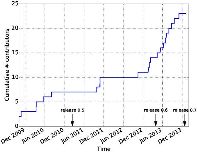
Diffusion Imaging in Python (Dipy) is a free and open source software project for the analysis of data from diffusion magnetic resonance imaging (dMRI) experiments. dMRI is an application of MRI that can be used to measure structural features of brain white matter. Many methods have been developed to use dMRI data to model the local configuration of white matter nerve fiber bundles and infer the trajectory of bundles connecting different parts of the brain. Dipy gathers implementations of many different methods in dMRI, including: diffusion signal pre-processing; reconstruction of diffusion distributions in individual voxels; fiber tractography and fiber track post-processing, analysis and visualization. Dipy aims to provide transparent implementations for all the different steps of dMRI analysis with a uniform programming interface. We have implemented classical signal reconstruction techniques, such as the diffusion tensor model and deterministic fiber tractography. In addition, cutting edge novel reconstruction techniques are implemented, such as constrained spherical deconvolution and diffusion spectrum imaging (DSI) with deconvolution, as well as methods for probabilistic tracking and original methods for tractography clustering. Many additional utility functions are provided to calculate various statistics, informative visualizations, as well as file-handling routines to assist in the development and use of novel techniques. In contrast to many other scientific software projects, Dipy is not being developed by a single research group. Rather, it is an open project that encourages contributions from any scientist/developer through GitHub and open discussions on the project mailing list. Consequently, Dipy today has an international team of contributors, spanning seven different academic institutions in five countries and three continents, which is still growing.
Overview
The claims and statistics of this paper haven’t been checked yet.
Run the check — free, no account needed. It reads the open-access text and checks the sources it cites.
Integrity signals
Registry
Cites
Statistics
Figures
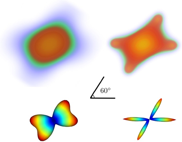
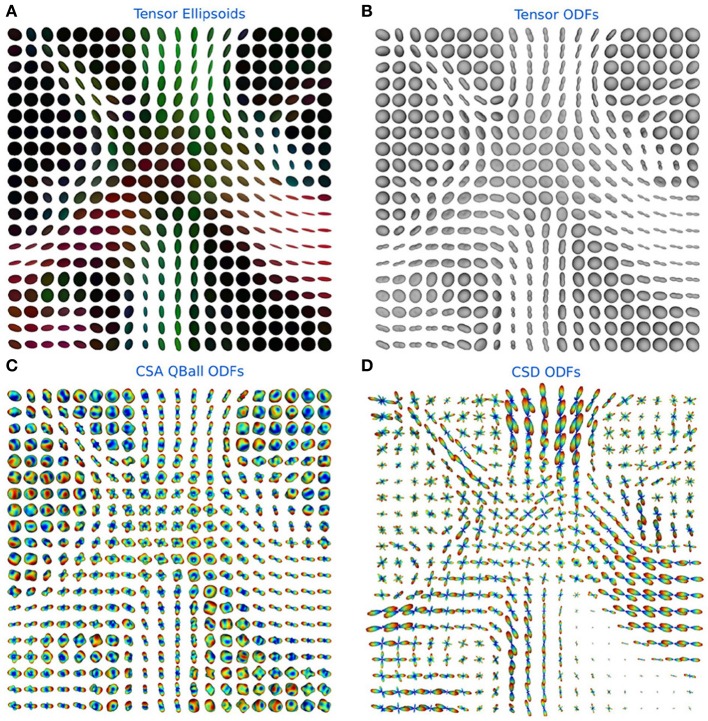
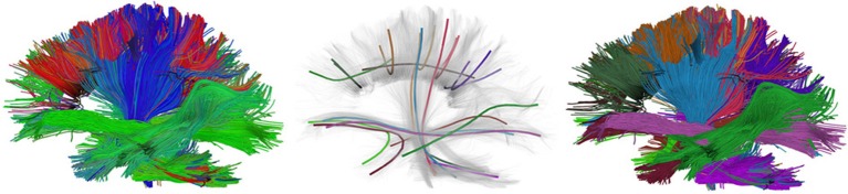
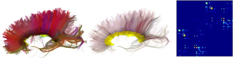
Verification
Claims the paper makes
The claims and statistics of this paper haven’t been checked yet.
Run the check — free, no account needed. It reads the open-access text and checks the sources it cites.
Claims other papers make about this one
Each is checked against this paper’s own text: does it say what the citing paper says it does?
The cited source says what the claim attributes to it.
In the citing paper
“In contrast to projects such as DiPy (Garyfallidis et al., 2014) , this library does not provide functionality to manipulate raw image data within Python itself, but provides functionality to invoke and manage external high-performance executables to perform the actual computations.”
From MRtrix3: A fast, flexible and open software framework for medical image processing and visualisation (2019) · cites this paper as Garyfallidis et al., 2014
Self-check
Percentages, sums and reported differences re-computed from the paper’s own numbers.
The self-check needs the paper’s full text.
Citations
Cites
A Threshold Selection Method from Gray-Level Histograms
Otsu, N. (1979) · IEEE Transactions on Systems, Man and Cybernetics · doi:10.1109/tsmc.1979.4310076 · Cited by 44,708 · cited 1×
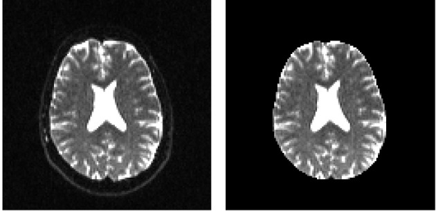
“…median_otsu (see Figure 3 ). from dipy.segment.mask import median_otsu mask, S0_mask = median_otsu(data [:, :, :, 0]) median_otsu uses first a median filter to smooth the S0 and then Otsu’s method, an automated histogram method (Otsu, 1979) to separate the brain (foreground) from its background.”
MethodAdvances in functional and structural MR image analysis and implementation as FSL
Smith, S. M., Jenkinson, M., Woolrich, M. W., Beckmann, C. F., Behrens, T. E. J., Johansen-Berg, H., Bannister, P. R., De Luca, M., Drobnjak, I., Flitney, D. E., et al. (2004) · NeuroImage · doi:10.1016/j.neuroimage.2004.07.051 · Cited by 13,630
Fischl, B. (2012) · NeuroImage · doi:10.1016/j.neuroimage.2012.01.021 · Cited by 9,526 · cited 4×
“…al., 2006), AFNI (Cox, 2012), MITK (Fritzsche et al., 2012), BrainVoyager QX (Goebel, 2012), DTI-Query/Quench (Sherbondy et al., 2005), FreeSurfer (Fischl, 2012), FSL-FDT (Smith et al., 2004), MedInria (Toussaint et al., 2007), MRtrix (Tournier et al., 2012), Diffusion Toolkit/Trackvis (Wang et…”
MethodInfluentialThe NumPy Array: A Structure for Efficient Numerical Computation
van der Walt, S., Colbert, S. C., & Varoquaux, G. (2011) · Computing in Science & Engineering · doi:10.1109/mcse.2011.37 · Cited by 9,381
MR diffusion tensor spectroscopy and imaging
Basser, P. J., Mattiello, J., & LeBihan, D. (1994) · Biophysical Journal · doi:10.1016/s0006-3495(94)80775-1 · Cited by 5,970 · cited 2×
“Another alternative is to make assumptions about the distribution of the PDF such as a Gaussian assumption, which leads to the popular diffusion tensor imaging (DTI) technique (Basser et al., 1994).”
BackgroundMethodThe WU-Minn Human Connectome Project: An overview
Van Essen, D. C., Smith, S. M., Barch, D. M., Behrens, T. E. J., Yacoub, E., & Ugurbil, K. (2013) · NeuroImage · doi:10.1016/j.neuroimage.2013.05.041 · Cited by 5,651
The basis of anisotropic water diffusion in the nervous system—A technical review.
Beaulieu, C. (2002) · NMR in Biomedicine · doi:10.1002/nbm.782 · Cited by 4,628
Basser, P. J., & Pierpaoli, C. (1996) · Journal of Magnetic Resonance, Series B · doi:10.1006/jmrb.1996.0086 · Cited by 4,306 · cited 3×
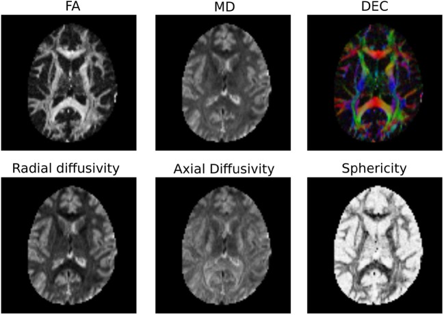
“…of the myelin wrapping the axons in the voxel may affect both MD and FA, changes in other factors, such as the distribution of directions of crossing of fiber populations through the voxel may also affect these measures (Basser and Pierpaoli, 1996; Jones et al., 2013; Wandell and Yeatman, 2013).”
BackgroundMethodMedical image analysis: progress over two decades and the challenges ahead
Duncan, J. S., & Ayache, N. (2000) · IEEE Transactions on Pattern Analysis and Machine Intelligence · doi:10.1109/34.824822 · Cited by 4,167
IPython: A System for Interactive Scientific Computing
Pérez, F., & Granger, B. (2007) · Computing in Science & Engineering · doi:10.1109/mcse.2007.53 · Cited by 4,129 · cited 1×
“There has been large growth in the number of Python users, there are many Python tools for scientific computing (Pérez and Granger, 2007; Pérez et al., 2011; McKinney, 2012), and there are complementary neuroimaging packages in Python1.”
Background
Research works printed but not linked: 9
- Chamberland, 2013, Explore the brain white matter networks in real-time: multi-sticks fiber tracking
- Garyfallidis, 2012, Towards an Accurate Brain Tractography
- Garyfallidis, 2011, Dipy - a novel software library for diffusion MR and tractography
- LeBihan, 1985, Imagerie de diffusion in vivo par résonance magnétique nucléaire, C. R. Acad. Sci. Paris, 301, 1109
- Pajevic, 1999, Color schemes to represent the orientation of anisotropic tissues from diffusion tensor data: application to white matter fiber tract mapping in the human brain, Magn. Reson. Med, 42, 526, 10.1002/(SICI)1522-2594(199909)42:3526::AID-MRM153.3.CO;2-A · DOI
- Schroeder, 2001, The Visualization Toolkit-User's Guide
- Vaillancourt, 2011, A fiber navigator for neurosurgical planning
- Wang, 2007, Diffusion toolkit: a software package for diffusion imaging data processing and tractography, Proc. Int. Soc. Magn. Reson. Med, 15, 3720
- Westin, 1997, Geometrical diffusion measures for MRI from tensor basis analysis, Proc. ISMRM, 97, 1742
Cited by
Sorted by citations
Items 901–1,000 of 1,000
Cho, D., & Wallraven, C. (2022) · Lecture notes in computer science · doi:10.1007/978-3-031-17976-1_2 · Cited by 3
“We then used dipy [7] to register the scans onto the standard MNI152 template including symmetric diffeomorphic registration, removing border voxels outside the brain.”
MethodJaatela, J., Aydogan, D. B., Nurmi, T., Vallinoja, J., Mäenpää, H., & Piitulainen, H. (2023) · Cerebral Cortex · doi:10.1093/cercor/bhad279 · Cited by 3
“2016) using Dipy (Garyfallidis et al. 2014) and visually verified.”
Advanced analysis of diffusion MRI data
Gu, X. (2019) · Linköping studies in science and technology. Dissertations · doi:10.3384/diss.diva-161288 · Cited by 4
MRI2MRI: A deep convolutional network that accurately transforms between brain MRI contrastsPreprint
Xiao, S., Wu, Y., Lee, A., & Rokem, A. (2018) · bioRxiv (Cold Spring Harbor Laboratory) · doi:10.1101/289926 · Cited by 3
“We used a state-of-the-art registration algorithm (12) , implemented in DIPY (13) to register DWI b=0 to corresponding T1w images, using a mutual information metric to find the best affine transformation for registration.”
MethodSelf-citationMitigating Gyral Bias in Cortical Tractography via Asymmetric Fiber Orientation DistributionsPreprint
Wu, Y., Hong, Y., Feng, Y., Shen, D. G., & Yap, P. (2019) · arXiv (Cornell University) · doi:10.48550/arxiv.1904.03328 · Cited by 3
Computational and informatics advances for reproducible data analysis in neuroimagingPreprint
Poldrack, R. A., Gorgolewski, K. J., & Varoquaux, G. P. (2018) · arXiv (Cornell University) · doi:10.48550/arxiv.1809.10024 · Cited by 3
“…for brain imaging is strong and diverse, with many libraries of various size and focus, such as –in addition to these mentioned previously– dipy (73) for processing of diffusion-weighted MRI data, or nibabel (74), which provides tools to read and operate on many of the file formats used in…”
MethodBrain connectivity and motor improvements after ballet intervention in multiple sclerosis: pilotPreprint
Camacho, P., Sutton, B. P., & López‐Ortiz, C. (2021) · medRxiv · doi:10.1101/2021.03.10.21252757 · Cited by 3
Poirier, C., & Descoteaux, M. (2022) · bioRxiv (Cold Spring Harbor Laboratory) · doi:10.1101/2022.12.18.520881 · Cited by 3
Self-citationZhang, J., Hu, Q., Li, F., & Wu, G. (2024) · Research Square · doi:10.21203/rs.3.rs-3921380/v1 · Cited by 3
Chandler, T., Guo, M. Y., Su, Y. P., Chen, J., Wu, Y., Liu, J., Agashe, A., Fischer, R. S., Mehta, S. B., Kumar, A., et al. (2024) · bioRxiv (Cold Spring Harbor Laboratory) · doi:10.1101/2024.03.09.584243 · Cited by 3
Magnetic Resonance Imaging Data Phenotypes for the Parkinson’s Progression Markers InitiativePreprint
Avants, B., Fonville, L., Hampton, O. L., Reardon, A., Stenger, V. A., Wang, X., Tustison, N. J., Stone, J. R., Cook, P. A., Marebwa, B., et al. (2024) · medRxiv · doi:10.1101/2024.09.23.24313179 · Cited by 3
Eggl, M. F., & Santis, S. D. (2024) · bioRxiv (Cold Spring Harbor Laboratory) · doi:10.1101/2024.11.11.622925 · Cited by 3
“All simulations and tensor-specific metric calculations were performed using Dipy functions [60], while we implemented an in-house Python implementation of the AxCaliber framework.”
A Python Toolbox for Representational Similarity AnalysisPreprint
van den Bosch, J. J. F., Golan, T., Peters, B., Taylor, J., Shahbazi, M., Lin, B., Charest, I., Diedrichsen, J., Kriegeskorte, N., Mur, M., et al. (2025) · eLife · doi:10.7554/elife.107828 · Cited by 3
Cortical connectome registration using spherical demons
Isaev, D., Gutman, B. A., Moyer, D., Faskowitz, J., & Thompson, P. M. (2017) · Proceedings of SPIE, the International Society for Optical Engineering/Proceedings of SPIE · doi:10.1117/12.2256975 · Cited by 2
Lei, W., Ding, M. L., Zhang, Y., & Wang, H. (2022) · NeuroImage · doi:10.1016/j.neuroimage.2022.119514 · Cited by 2
Deep Learning Framework using Sparse Diffusion MRI for Diagnosis of Frontotemporal Dementia
Tiwari, A. S., Singhal, A., Shigwan, S. J., & Singh, R. K. (2023) · IEEE International Conference on Computer Vision Workshops · doi:10.1109/iccvw60793.2023.00413 · Cited by 2
“Our neural network was trained on a dataset consisting of 20,000 tuples per image obtained from three different datasets: NIFD − 41 , NIFD − 21 , and NIFD − 5 [9, 25].”
MethodInfluentialXiao, X.-Y., Zeng, J.-Y., Cao, Y., Tang, Y., Zou, Z., Li, J., & Chen, H. (2024) · Quantitative Imaging in Medicine and Surgery · doi:10.21037/qims-24-236 · Cited by 2
Ma, X., Seres, P., Kinnaird, A. S., Fung, C. I., Feiweier, T., & Beaulieu, C. (2024) · Magnetic Resonance in Medicine · doi:10.1002/mrm.30408 · Cited by 2
“Diffusion image processing was performed with DIPY, 32 which included Marchenko-Pastur Principal Component Analysis denoising, 33 symmetric diffeomorphic registration, 34 and tensor analysis.”
Bertalan, G., Hainc, N., Dehn, F. D. V., Hortobágyi, T., Bink, A., Rhun, É. L., Weller, M., & Kulcsár, Z. (2025) · American Journal of Neuroradiology · doi:10.3174/ajnr.a8493 · Cited by 2
Sun, H., Yan, Z., Gao, J., Zheng, Y., Zheng, Y., Song, Y., Jin, F., Qu, H., Song, Y., Diao, Y., et al. (2025) · Neurosurgical Review · doi:10.1007/s10143-025-03495-0 · Cited by 2
Corpus Callosum Parcellation Methods: What Can Tractography Tell Us About Them?
Santana, C. P., Román, C., Appenzeller, S., Guevara, P., & Rittner, L. (2025) · Lecture notes in computer science · doi:10.1007/978-3-031-86920-4_19 · Cited by 2
Badea, A., Mahzarnia, A., Reddy, D., Dong, Z., Anderson, R. J., Moon, H. S., Stout, J. A., Williams, J., Hirschler, L., Barbier, E., et al. (2025) · Magnetic Resonance Imaging · doi:10.1016/j.mri.2025.110406 · Cited by 3
Toward diffusion MRI in the diagnosis and treatment of pancreatic cancer
Lee, J., Lin, T., He, Y., Wu, Y., & Qin, J. (2025) · Medical Oncology · doi:10.1007/s12032-025-02759-5 · Cited by 2
“Furthermore, open-source software platforms, such as the Diffusion Imaging in Python (Dipy) library, provide critical resources for clinicians and researchers operating in resource-limited settings, enabling dMRI data analysis without reliance on costly proprietary software [95].”
Little, G. E., Poirier, C., Boré, A., Parent, M., Petit, L., & Descoteaux, M. (2025) · Human Brain Mapping · doi:10.1002/hbm.70245 · Cited by 3
“For the 760 μm data and clinical test–retest data, and in order to avoid registration of the structural images to the diffusion data (distorted in the clinical data), initial tensor models were first fit on unprocessed diffusion data for each subject (Garyfallidis et al. 2014) (DIPY v1.”
Self-citationProgressive Remote Axonal Degeneration Following Spinal Cord Injury: A Histological and MRI Study
Dávid, G., Motovylyak, A., Schlegel, F., Kovacs‐Balint, Z., Kündig, C. W., Filous, A. R., Schwab, J. M., Budde, M. D., Klohs, J., & Freund, P. (2025) · Neurotrauma Reports · doi:10.1089/neur.2025.0011 · Cited by 2
Zhao, S., Ma, X., Li, L., Gao, E., Zhao, K., Wang, M., Yang, G., Zheng, H., Cheng, J., & Zhao, G. (2025) · Frontiers in Neurology · doi:10.3389/fneur.2025.1577811 · Cited by 2
Ricchi, M., Axford, A., McGing, J., Shinozaki, A., Yeung, K., Mills, R., Zaccagna, F., Tyler, D. J., Testa, C. M., & Grist, J. T. (2025) · Scientific Reports · doi:10.1038/s41598-025-12777-y · Cited by 2
“DKI and MSDKI models fit using the Diffusion Imaging in Python (DIPY) package 44 .”
Validation of structure tensor analysis for orientation estimation in brain tissue microscopy
Gray, B., Smith, A. W., MacKenzie‐Graham, A. J., Shattuck, D. W., & Tward, D. J. (2025) · Journal of Neuroscience Methods · doi:10.1016/j.jneumeth.2025.110539 · Cited by 2
“In three dimensions, we chose 100 angles distributed on a hemisphere using an electrostatic dispersion algorithm (Jones et al., 1999), implemented in Python as the disperse_charges method in Dipy (Garyfallidis et al., 2014), using 2000 iterations and a step size constant of 0.1.”
InfluentialLongitudinal diffusion tensor imaging correlates with amyloid burden in Down syndrome
LeMerise, L., Guerrero-Gonzalez, J. M., McVea, A. K., Zammit, M. D., Kecskemeti, S., Hartley, S. L., Head, E., Mapstone, M., Handen, B. L., Alexander, A. L., et al. (2025) · Alzheimer s & Dementia · doi:10.1002/alz.70572 · Cited by 3
“43 The co-registered DWIs were fit to a DTI model using the DiPy python package 44 for quantitative DTI maps, including FA, MD,RD,andAxD.”
Lillebostad, P. A. G., Njølstad, T. S., Hogstad, S., Riemer, F., Kverneng, S. U., Stige, K. E., Biermann, M., Jog, M., Buch, S., Haacke, E. M., et al. (2025) · Imaging Neuroscience · doi:10.1162/imag.a.158 · Cited by 2
“A diffusion tensor model was fitted onto the pre-processed data with the DiPy library (version 1.5.0) ( Garyfallidis et al., 2014).”
InfluentialDiffusion MRI of the prenatal fetal brain: a methodological scoping review
Stefano, M. D., Ciceri, T., Leemans, A., de Zwarte, S. M. C., Luca, A. D., & Peruzzo, D. (2025) · NeuroImage · doi:10.1016/j.neuroimage.2025.121453 · Cited by 2
Luo, D., Zeng, P., Zeng, B., B. L., Nie, L., Peng, Y., Li, Y., & Luo, T. (2025) · The Journals of Gerontology Series A · doi:10.1093/gerona/glaf225 · Cited by 2
Anatomy-to-tract mapping infers white matter pathways without diffusion streamline propagation
Tan, Y. F., Huynh, K. M., Liu, S., Phan, R. C. ‐., Ting, C., & Yap, P. (2025) · Nature Communications · doi:10.1038/s41467-025-66615-w · Cited by 2
“To learn the mapping from T1w images to white matter tracts, 3000 streamlines per bundle were randomly selected and resampled to 128 equidistant points along each streamline using Dipy 31 .”
Mak, E., Przybelski, S. A., Reid, R. I., Wiste, H. J., Fought, A. J., Schwarz, C. G., Senjem, M. L., Vemuri, P., Jack, C. R., Lowe, V. J., et al. (2026) · Alzheimer s Research & Therapy · doi:10.1186/s13195-026-01995-9 · Cited by 3
“Diffusion tensors were estimated using nonlinear least squares fitting to calculate FA and MD maps using the dipy software package [32].”
Wang, T., Wan, Z., Cao, S., Yu, J., He, Y., Xie, Y., Zhang, F., & Wu, Y. (2026) · Human Brain Mapping · doi:10.1002/hbm.70513 · Cited by 2
A geometry aware framework enhances noninvasive mapping of whole human brain dynamics
Wang, S., Lou, K., Wei, C., Sheng, Z., Tang, J., Peng, K., Shen, X., Mei, S., Chen, L., Gu, D., et al. (2026) · Nature Biomedical Engineering · doi:10.1038/s41551-026-01664-0 · Cited by 2
“Resting-state For MEG participants (HCP dataset, n = 80), ViEEG was estimated at standardized cortical contact coordinates from the MNI Open iEEG atlas [55], mapped to each participant’s native anatomy via affine registration (DIPY’s AffineRegistration with mutual-information objective [110]).”
Wei, Y., Huang, W.-H., Lin, C., Chen, C.-K., Hsu, C.-C. H., Ding, C.-H. M., Chen, Y.-L., & Lin, C.-P. (2026) · Journal of Alzheimer s Disease · doi:10.1177/13872877261447822 · Cited by 2
“20”
InfluentialZhou, X., Hu, Y., Yang, J., Zhang, J., Yong, T., Tang, L., Zheng, J., Lan, H., Yin, T., Liu, D., et al. (2026) · Brain Behavior and Immunity · doi:10.1016/j.bbi.2026.106819 · Cited by 2
Li, L., Pan, P., Li, J., Tang, M., Zhang, D., Lei, X., Zhang, X., & Gao, J. (2026) · Frontiers in Endocrinology · doi:10.3389/fendo.2026.1838132 · Cited by 2
“A regularized bi-tensor FW model from the Diffusion Imaging in Python (DIPY) package (https://dipy.org/) was employed to obtain FW fraction maps from the preprocessed diffusion-weighted images, separating the diffusion signal into tissue and isotropic FW components (29).”
Learning-Based Ensemble Average Propagator Estimation
Ye, C. (2017) · Lecture notes in computer science · doi:10.1007/978-3-319-66182-7_68 · Cited by 2
“Here, we selected the implementation of SHORE in the Dipy software [3] and used the default parameters (( N, ζ, λ N , λ L ) = (6 , 700 , 10 − 8 , 10 − 8 )) provided at http://nipy.org/dipy/examples_built/reconst_shore.html .”
MethodInformation-Theoretic Registration with Explicit Reorientation of Diffusion-Weighted Images
Jensen, H. G., Lauze, F., & Darkner, S. (2021) · Journal of Mathematical Imaging and Vision · doi:10.1007/s10851-021-01050-2 · Cited by 2
“To our knowledge, there are no open sources of simulated DWI data for comparing registration frameworks, although the DIPY project may be a good source for generating simulations [31].”
BackgroundMarcotte, K., Sanchez, E., Arbour, C., Brambati, S. M., Bedetti, C., Martineau, S., Descoteaux, M., & Gosselin, N. (2020) · Brain and Language · doi:10.1016/j.bandl.2020.104769 · Cited by 3
Self-citationManifold-aware synthesis of high-resolution diffusion from structural imaging
Anctil-Robitaille, B., Théberge, A., Jodoin, P., Descoteaux, M., Desrosiers, C., & Lombaert, H. (2022) · Frontiers in Neuroimaging · doi:10.3389/fnimg.2022.930496 · Cited by 2
Self-citationTanedo, J., Gajawelli, N., Guo, S., Nelson, M. B., & Leporé, N. (2022) · Frontiers in Neuroimaging · doi:10.3389/fnimg.2022.845609 · Cited by 2
“Thus, our future research will utilize methods which can distinguish multiple fibers (Garyfallidis et al., 2014).”
MethodNeural Registration and Segmentation of White Matter Tracts in Multi-modal Brain MRI
Barzilay, N., Nelkenbaum, I., Konen, E., Kiryati, N., & Mayer, A. (2023) · Lecture notes in computer science · doi:10.1007/978-3-031-25066-8_12 · Cited by 2
Kanakaraj, P., Cai, L. Y., Yao, T., Rheault, F., Rogers, B. P., Anderson, A. W., Schilling, K. G., & Landman, B. A. (2023) · Magnetic Resonance Imaging · doi:10.1016/j.mri.2023.03.014 · Cited by 2
Rauland, A., & Merhof, D. (2023) · IEEE International Symposium on Biomedical Imaging (ISBI) · doi:10.1109/isbi53787.2023.10230737 · Cited by 2
Little, G. E., Acosta‐Franco, J. A., & Beaulieu, C. (2026) · NMR in Biomedicine · doi:10.1002/nbm.70353 · Cited by 2
“To generate consistent brain masks that were inclusive of all brain tissue voxels, initial tensor models were first fit on unprocessed diffusion data for each subject (Garyfallidis et al., 2014) (DIPY v1.”
Mihailović, J. M., Sanganahalli, B. G., Hyder, F., Chitturi, J., Elkabes, S., Heary, R. F., & Kannurpatti, S. S. (2023) · Neuroscience Letters · doi:10.1016/j.neulet.2023.137607 · Cited by 2
BundleCleaner: Unsupervised Denoising and Subsampling of Diffusion MRI-Derived Tractography Data
Feng, Y., Chandio, B. Q., Villalón‐Reina, J. E., Thomopoulos, S. I., Joshi, H., Nair, G., Joshi, A. A., Venkatasubramanian, G., John, J. P., & Thompson, P. M. (2023) · Lecture notes in computer science · doi:10.1007/978-3-031-47292-3_14 · Cited by 2
Ciceri, T., Luca, A. D., Agarwal, N., Arrigoni, F., & Peruzzo, D. (2024) · NMR in Biomedicine · doi:10.1002/nbm.5141 · Cited by 2
“28,29 The mathematical formulation of the framework is included in Appendix A, the acquired data are freely available (10.5281/zenodo.10801619), and the Python code is available upon request.”
InfluentialMoon, H. S., Mahzarnia, A., Stout, J. A., Anderson, R. J., Han, Z. Y., Badea, C. T., & Badea, A. (2024) · SPIE Medical Imaging: Computer-Aided Diagnosis · doi:10.1117/12.3005695 · Cited by 2
Mazur, W., Spees, W. M., & Krzyżak, A. T. (2024) · NMR in Biomedicine · doi:10.1002/nbm.5259 · Cited by 2
“18 Secondly, denoised data were corrected for B1 oscillatory field inhomogeneity in MRTrix3 software 34 using the N4 algorithm as provided in ANTs.”
Ground-Truth Effects in Learning-Based Fiber Orientation Distribution Estimation in Neonatal Brains
Lin, R., Kebiri, H., Gholipour, A., Chen, Y., Thiran, J., Karimi, D., & Cuadra, M. B. (2025) · Lecture notes in computer science · doi:10.1007/978-3-031-86920-4_3 · Cited by 2
“…validation was performed based on the agreement rate (AR) [17] between the number of fibers estimated by the number of peaks extracted using Dipy [10] (with a mean separation angle of 45°, a maximum number of 3 peaks and relative peak threshold of 0.5), the angular error (AE) between those peaks…”
MethodInfluentialBertalan, G., Krepuska, M., Toth, D., Madjidyar, J., Thurner, P., Schubert, T. B., & Kulcsár, Z. (2024) · Sci · doi:10.3390/sci6040064 · Cited by 2
Raj, A., Sipes, B. S., Verma, P., Mathalon, D. H., Biswal, B. B., & Nagarajan, S. S. (2024) · Imaging Neuroscience · doi:10.1162/imag_a_00381 · Cited by 2
Chand, K., Fritsch, T., Oster, S., Ulbricht, A., Poka, K., & Bruno, G. (2025) · Journal of Nondestructive Evaluation · doi:10.1007/s10921-025-01174-0 · Cited by 2
“9usingtheDIPY3Dimageregistrationlibrary[38].”
Replace2Self: Self-Supervised Denoising Based on Voxel Replacing and Image Mixing for Diffusion MRI
Wu, L., Wang, L., Deng, Z., Zhu, Y., & Wei, H. (2025) · IEEE Transactions on Medical Imaging · doi:10.1109/tmi.2025.3552611 · Cited by 2
Sutkus, L. T., Sommer, K. M., Li, Z., Sutton, B. P., Donovan, S. M., & Dilger, R. N. (2025) · Frontiers in Neuroscience · doi:10.3389/fnins.2025.1484497 · Cited by 2
“Raw DWI images were first denoised using the Marchenko-Pastur Principal Component Analysis (MPPCA) method (Manjón et al., 2013; Neto Henriques, 2018; Veraart et al., 2016a; Veraart et al., 2016b) within the DIPY toolbox (Garyfallidis et al., 2014).”
Lan, Z., Chen, Y., Rushmore, J., Zekelman, L., Makris, N., Yogesh Rathi, A., Golby, A. J., Zhang, F., & O’Donnell, L. J. (2025) · Imaging Neuroscience · doi:10.1162/imag_a_00569 · Cited by 2
“The AFQ method ( Yeatman et al., 2012) implemented in the dipy software ( Garyfallidis et al., 2014) automatically computes the profile, which consists of mean FA values at sequential locations along a fiber tract.”
Zhang, H., Zhou, R., Lu, W., Su, Y., Cai, Y., Huang, J., Shen, H., Ding, L., Wang, Y., Zhang, M., et al. (2025) · Journal of Affective Disorders · doi:10.1016/j.jad.2025.04.064 · Cited by 2
Koryciński, M., Ciecierski, K. A., & Niewiadomska‐Szynkiewicz, E. (2025) · Neural Networks · doi:10.1016/j.neunet.2025.107624 · Cited by 2
Lasek, J., Stefańska, A. K., Kierońska-Siwak, S., Obuchowicz, R., & Krzyżak, A. T. (2025) · Computers in Biology and Medicine · doi:10.1016/j.compbiomed.2025.110503 · Cited by 2
Evaluating the Effect of Post-processing Steps When Analyzing Cardiac Diffusion Tensor Data
Cork, T. E., Hannum, A. J., Loecher, M., Perotti, L. E., & Ennis, D. B. (2025) · Lecture notes in computer science · doi:10.1007/978-3-031-94562-5_13 · Cited by 2
Ishida, S., Yamada, S., Oki, T., Otani, T., Hiratsuka, S., Wada, S., Mase, M., & Watanabe, Y. (2025) · Journal of Magnetic Resonance Imaging · doi:10.1002/jmri.29834 · Cited by 2
Evaluation of EPI‐Based Distortion Correction Techniques for Cardiac Diffusion Tensor Imaging
Cork, T. E., Middione, M. J., Loecher, M., Liao, C., Setsompop, K., & Ennis, D. B. (2025) · NMR in Biomedicine · doi:10.1002/nbm.70147 · Cited by 2
“The entire cDTI image processing pipeline was integrated using the Dipy Framework [39] with the exception of the shot-rejection module and the respiratory sorting module.”
Zheng, R., Zhang, W., Li, Y., Zhu, X., Lan, Z., Rushmore, J., Yogesh Rathi, A., Makris, N., O’Donnell, L. J., & Zhang, F. (2025) · Medical Image Analysis · doi:10.1016/j.media.2025.103892 · Cited by 2
Sound Source Localisation in Ambisonic Audio Using Peak Clustering
Green, M., & Murphy, D. T. (2019) · Detection and Classification of Acoustic Scenes and Events Workshop (DCASE) · doi:10.33682/05my-4306 · Cited by 2
“We used the peak-finding function from the dipy Python library [18].”
MethodStructural parcellation of the thalamus using shortest-path tractography
Kasenburg, N., Darkner, S., Hahn, U., Liptrot, M. G., & Feragen, A. (2016) · IEEE International Symposium on Biomedical Imaging (ISBI) · doi:10.1109/isbi.2016.7493330 · Cited by 2
“Fibre orientation distribution functions (fODFs) were computed for every voxel using constrained spherical deconvolution [17] with order 8 using the DiPy [18] package.”
MethodXu, T., Feng, Y., Wu, Y., Zeng, Q., Zhang, J., He, J., & Zhuge, Q. (2017) · PLoS ONE · doi:10.1371/journal.pone.0168864 · Cited by 2
Kim, H., Hong, S., Styner, M. A., Piven, J., Botteron, K. N., & Gerig, G. (2020) · Lecture notes in computer science · doi:10.1007/978-3-030-59728-3_31 · Cited by 2
Jordan, K., Lauricella, M., Licata, A. E., Sacco, S., Asteggiano, C., Wang, C., Sudarsan, S. P., Watson, C., Scheffler, A. W., Battistella, G., et al. (2021) · Journal of Neuroimaging · doi:10.1111/jon.12854 · Cited by 2
Denoising Diffusion MRI via Graph Total Variance in Spatioangular Domain
Wu, H., & Yan, S. (2021) · Computational and Mathematical Methods in Medicine · doi:10.1155/2021/4645544 · Cited by 2
“Both ANLM and MP-PCA are implemented by DIPY [33] (3) XQ nonlocal means (XQ-NLM): XQ-NLM [17] denoises the signal via weighted averaging of selfsimilar information, which is defined in the spatialangular domain.”
MethodAssessing the Reliability of Template-Based Clustering for Tractography in Healthy Human Adults
Kai, J., & KHAN, A. R. (2022) · Frontiers in Neuroinformatics · doi:10.3389/fninf.2022.777853 · Cited by 2
“, 2012) clustering, as implemented within the DIPY library (Garyfallidis et al., 2014), was also used as a secondary clustering technique.”
MethodRole of insular cortex lesions in determining the pathogenetic subtype of ischemic stroke
Kulesh, A. A., Kulikova, S. P., Евгеньевич, Д. В., Александрович, М. С., Bartuli, E., Владимирович, Б. А., Syromyatnikova, L. I., Sobyanin, K., & Karakulova, Y. V. (2022) · Neurology neuropsychiatry Psychosomatics · doi:10.14412/2074-2711-2022-2-11-17 · Cited by 2
High Resolution Ex Vivo Diffusion Tensor Distribution MRI of Neural Tissue
Magdoom, K. N., Komlosh, M. E., Saleem, K. S., Gasbarra, D., & Basser, P. J. (2022) · Frontiers in Physics · doi:10.3389/fphy.2022.807000 · Cited by 2
“The thermal noise in the images was filtered using a Marchenko-Pastur principal component analysis (PCA) algorithm [25] implemented in DIPY software [26], and the effect of eddy current induced image translation and shearing were reduced by registering the individual DWI volumes with b = 0 s/mm(2) acquisition using 3D…”
MethodGamal, A., Elattar, M., & Selim, S. (2022) · Communications in computer and information science · doi:10.1007/978-3-031-23119-3_1 · Cited by 2
Murray, C., Oladosu, O., Joshi, M., Kolind, S., Oh, J., & Zhang, Y. (2023) · Magnetic Resonance Imaging · doi:10.1016/j.mri.2023.03.023 · Cited by 2
Towards Frugal Unsupervised Detection of Subtle Abnormalities in Medical Imaging
Oudoumanessah, G., Lartizien, C., Dojat, M., & Forbes, F. (2023) · Lecture notes in computer science · doi:10.1007/978-3-031-43898-1_40 · Cited by 2
Comparing White Matter Fiber Bundle Segmentation Methods for Autism Prediction
Vidal, N., Navarrete, S., Román, C., Houenou, J., Mangin, J., Hernández, C., & Guevara, P. (2023) · International Symposium on Medical Information Processing and Analysis (SIPAIM) · doi:10.1109/sipaim56729.2023.10373470 · Cited by 2
A taxonomic guide to diffusion MRI tractography visualization tools
Laamoumi, M., Hendriks, T., & Chamberland, M. (2024) · NMR in Biomedicine · doi:10.1002/nbm.5267 · Cited by 2
Multi-Shell Diffusion MRI Measures of Brain Aging: A Preliminary Comparison From ADNI3
Nir, T. M., Thomopoulos, S. I., Villalón‐Reina, J. E., Zavaliangos‐Petropulu, A., Dennis, E. L., Reid, R. I., Bernstein, M. A., Borowski, B., Jack, C. R., Weiner, M. W., et al. (2019) · IEEE International Symposium on Biomedical Imaging (ISBI) · doi:10.1109/isbi.2019.8759486 · Cited by 2
“To compare performance with a measure derived from a HARDI technique non-parametric q-ball imaging (QBI) we also fit constant solid angle orientation distribution functions (CSA-ODFs) [18, 19] using the DiPy package [20] to generate generalized FA (GFA1000) maps.”
MethodTractography in Curvilinear Coordinates
Hussain, U., Baron, C. A., & KHAN, A. R. (2021) · Frontiers in Neuroscience · doi:10.3389/fnins.2021.716538 · Cited by 2
“Both of these algorithms are implemented in the dipy library (Garyfallidis et al., 2014).”
MethodJordan, K., Keshavan, A., Caverzasi, E., Osorio, J. A., Papinutto, N., Amirbekian, B., Berger, M. S., & Henry, R. G. (2017) · bioRxiv (Cold Spring Harbor Laboratory) · doi:10.1101/140137 · Cited by 2
“A tensor model was fit to the corrected HARDI data using the open-source package Diffusion Imaging in Python (Dipy) (Garyfallidis et al. 2014) and the resulting parameters used to calculate tensor metrics.”
MethodSelf-citationData-driven brain-types and their cognitive consequencesPreprint
Bathelt, J., Johnson, A., Zhang, M., the CALM team, & Astle, D. E. (2017) · bioRxiv (Cold Spring Harbor Laboratory) · doi:10.1101/237859 · Cited by 2
“8.0 implementation (Garyfallidis et al. 2014) of a non-local means de-noising algorithm (Coupe et al. 2008) to boost signal-to-noise ratio.”
MethodInfluentialPre-operarative Multivariate Connectome Analysis for Glioma PatientsPreprint
Crimi, A., Wiestler, B., Kirschke, J. S., & Krieg, S. M. (2018) · bioRxiv (Cold Spring Harbor Laboratory) · doi:10.1101/436220 · Cited by 2
“Initially tractographies for all subjects have been generated by processing DTI data with the Python library Dipy [9], stemming from 2,000,000 seed-points and stopping when the fractional anisotropy was smaller than < 0.”
MethodInfluentialCollegiate athlete brain data for white matter mapping and network neurosciencePreprint
Caron, B. A., Stuck, R., McPherson, B. C., Bullock, D. N., Kitchell, L. M., Faskowitz, J., Kellar, D., Cheng, H., Newman, S. D., Port, N., et al. (2020) · bioRxiv (Cold Spring Harbor Laboratory) · doi:10.1101/2020.08.25.267005 · Cited by 1
StimVis: A tool for interactive computation of the TMS-induced effects over tractography data
Kulikova, S. P. (2020) · SoftwareX · doi:10.1016/j.softx.2020.100594 · Cited by 2
Kiar, G., Chatelain, Y., Salari, A., Evans, A. C., & Tristan Glatard, B. (2020) · bioRxiv (Cold Spring Harbor Laboratory) · doi:10.1101/2020.12.16.423084 · Cited by 2
An edge-centric model for harmonizing multi-relational network datasetsPreprint
Faskowitz, J., Tanner, J., Mišić, B., & Betzel, R. F. (2021) · bioRxiv (Cold Spring Harbor Laboratory) · doi:10.1101/2021.01.07.425450 · Cited by 2
Unraveling micro-architectural modulations in neural tissue upon ischemia by Correlation Tensor MRIPreprint
Alves, R., Henriques, R. N., Kerkelä, L., Chavarrías, C., Jespersen, S. N., & Shemesh, N. (2021) · bioRxiv (Cold Spring Harbor Laboratory) · doi:10.1101/2021.02.20.432088 · Cited by 2
A low-resource reliable pipeline to democratize multi-modal connectome estimation and analysisPreprint
Chung, J., Lawrence, R., Loftus, A., Pisner, D., Kiar, G., Bridgeford, E., Roncal, W. G., Chandrashekhar, V., Mhembere, D., Ryman, S. G., et al. (2021) · bioRxiv (Cold Spring Harbor Laboratory) · doi:10.1101/2021.11.01.466686 · Cited by 2
“The dMRI file is then resliced to the desired voxel size (specified by the user, with a reasonable default of 2mm 3 if not specified) using Dipy’s reslice function with trilinear interpolation[2].”
MethodInfluentialCalibrated Diffusion Tensor EstimationPreprint
Karimi, D., Warfield, S. K., & Gholipour, A. (2021) · arXiv (Cornell University) · Cited by 2
Zhou, D., Kang, Y., Cosme, D., Jovanova, M., He, X., Mahadevan, A. S., Stanoi, O. A., Brynildsen, J. K., Cooper, N., Cornblath, E. J., et al. (2021) · doi:10.31234/osf.io/u83my · Cited by 2
Imaging Biomarkers for Alzheimer's Disease Using Magnetic Resonance Microscopy
Badea, A., Stout, J. A., Anderson, R. J., Cofer, G. P., Duan, L. L., & Vogelstein, J. T. (2022) · Wiley eBooks · doi:10.1002/9783527827244.ch13 · Cited by 2
Specific and non-linear effects of glaucoma on optic radiation tissue propertiesPreprint
Kruper, J., Richie-Halford, A., Benson, N. C., Caffarra, S., Owen, J. P., Wu, Y., Lee, A., Lee, C. S., Yeatman, J. D., & Rokem, A. (2023) · bioRxiv (Cold Spring Harbor Laboratory) · doi:10.1101/2023.01.17.524459 · Cited by 2
Self-citationSuper-resolution mapping of anisotropic tissue structure with diffusion MRI and deep learningPreprint
Abramian, D., Eklund, A., & Özarslan, E. (2023) · bioRxiv (Cold Spring Harbor Laboratory) · doi:10.1101/2023.04.04.535586 · Cited by 2
Longitudinal stability of brain and spinal cord quantitative MRI measuresPreprint
Boudreau, M., Karakuzu, A., Boré, A., Pinsard, B., Zelenkovski, K., Alonso‐Ortiz, E., Boyle, J., Bellec, P., & Cohen‐Adad, J. (2023) · doi:10.55458/neurolibre.00018 · Cited by 2
Shoby, A., Francis, J., Raju, J., Pillai, M. J., John, A., Valakkada, J., Amma, C. U., Rajan, G., & Rajaram, P. (2024) · SSRN Electronic Journal · doi:10.2139/ssrn.4777617 · Cited by 2
Mapping Along-Tract White Matter Microstructural Differences in AutismPreprint
Kim, G. S., Chandio, B. Q., Benavidez, S. M., Feng, Y., Thompson, P. M., & Lawrence, K. E. (2025) · bioRxiv (Cold Spring Harbor Laboratory) · doi:10.1101/2025.03.21.644498 · Cited by 2
Metadata
- Authors
- Garyfallidis, E., Brett, M., Amirbekian, B., Rokem, A., van der Walt, S., Descoteaux, M., Nimmo-Smith, I., & Contributors, D.
- Affiliations
- Université de Sherbrooke (CA)
- University of California, Berkeley (US)
- University of California, San Francisco (US)
- Stanford University (US)
- Corresponding author
- Garyfallidis, Eleftherios
- Venue
- Frontiers in Neuroinformatics
- Publisher
- Frontiers Media SA
- Published
- 2014-02-21
- Volume
- 8
- Pages
- 8-8
- ISSN
- 1662-5196 (ISSN-L)
- PMC
- PMC3931231
- OpenAlex
- W2062791478
- Cited by
- 1,371
- References
- 98
- Citations by year
- 2026: 147 · 2025: 192 · 2024: 177 · 2023: 162 · 2022: 151 · 2021: 147 · 2020: 100 · 2019: 89 · 2018: 54 · 2017: 79 · 2016: 34 · 2015: 26 · 2014: 12
- FWCI
- 39.38
- Type
- journal article
- Language
- en
- Open access
- gold
- doi.org
- Licence
- cc-by
- Locations
- Frontiers in Neuroinformatics (publishedVersion, cc-by)
- Europe PMC (PubMed Central) (publishedVersion, cc-by)
- PubMed Central (publishedVersion, cc-by)
- PubMed (publishedVersion)
- DOAJ (DOAJ: Directory of Open Access Journals) (submittedVersion)
- University of Birmingham Research Portal (University of Birmingham) (submittedVersion, cc-by)
- Funders
- Fonds Québécois de la Recherche sur la Nature et les Technologies
- Natural Sciences and Engineering Research Council of Canada
- National Eye Institute (F32EY022294)
- Topics
- Health Sciences › Medicine › Radiology, Nuclear Medicine and Imaging › Advanced Neuroimaging Techniques and Applications
- Life Sciences › Neuroscience › Cognitive Neuroscience › Functional Brain Connectivity Studies
- Health Sciences › Medicine › Radiology, Nuclear Medicine and Imaging › Advanced MRI Techniques and Applications
- Keywords
- diffusion MRI, DIPY, white matter, fiber tractography, diffusion tensor model, constrained spherical deconvolution, diffusion spectrum imaging, probabilistic tracking, open source software, Python library
About these checks
Registry, retraction, Cites and statistics results are computed, not judged, with one exception: a printed reference that no index lists is read once by a model before it counts as a citation error. Claims are extracted by a model and kept only when their quote appears verbatim in the text; each cited source is then checked, with a model’s help, for whether it says what the claim relies on. Those verdicts can be wrong — read the claim’s quote and the cited source before relying on one.
Statistics and arithmetic are recomputed, not judged.
Confidence bands are shown only for fields calibrated on our annotated test set. None is calibrated yet, so no band is shown.
What counts as a claim ›How we measure confidence ›How we check this ›
Are you an author, or do you see a problem with this page? Request a review
