

The DOI matches its Crossref record; 98 of the 100 references it cites were found in a registry or an index, and 2 are not checked yet.
Dipy, a library for the analysis of diffusion MRI data
Open accessAbstract
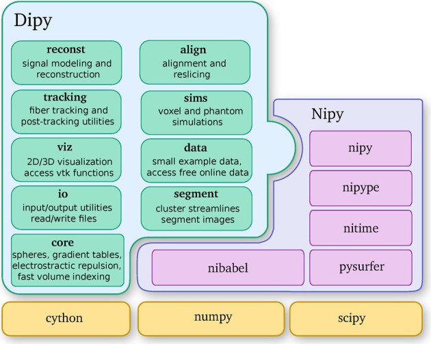
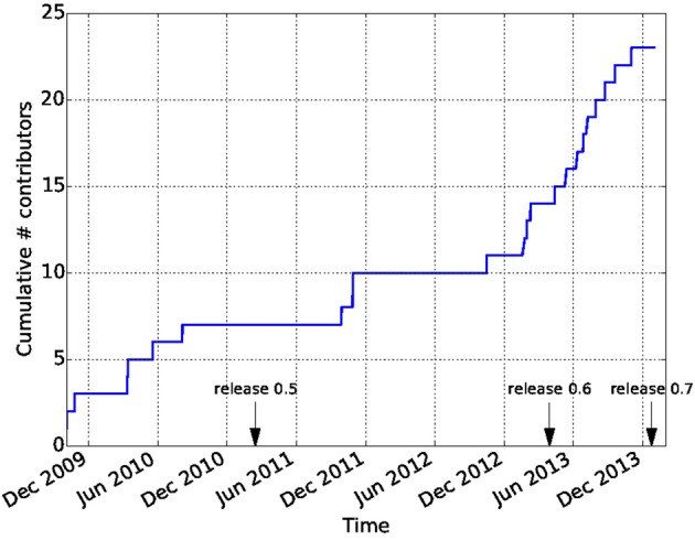
Diffusion Imaging in Python (Dipy) is a free and open source software project for the analysis of data from diffusion magnetic resonance imaging (dMRI) experiments. dMRI is an application of MRI that can be used to measure structural features of brain white matter. Many methods have been developed to use dMRI data to model the local configuration of white matter nerve fiber bundles and infer the trajectory of bundles connecting different parts of the brain. Dipy gathers implementations of many different methods in dMRI, including: diffusion signal pre-processing; reconstruction of diffusion distributions in individual voxels; fiber tractography and fiber track post-processing, analysis and visualization. Dipy aims to provide transparent implementations for all the different steps of dMRI analysis with a uniform programming interface. We have implemented classical signal reconstruction techniques, such as the diffusion tensor model and deterministic fiber tractography. In addition, cutting edge novel reconstruction techniques are implemented, such as constrained spherical deconvolution and diffusion spectrum imaging (DSI) with deconvolution, as well as methods for probabilistic tracking and original methods for tractography clustering. Many additional utility functions are provided to calculate various statistics, informative visualizations, as well as file-handling routines to assist in the development and use of novel techniques. In contrast to many other scientific software projects, Dipy is not being developed by a single research group. Rather, it is an open project that encourages contributions from any scientist/developer through GitHub and open discussions on the project mailing list. Consequently, Dipy today has an international team of contributors, spanning seven different academic institutions in five countries and three continents, which is still growing.
Overview
The claims and statistics of this paper haven’t been checked yet.
Run the check — free, no account needed. It reads the open-access text and checks the sources it cites.
Integrity signals
Registry
Cites
Statistics
Figures
Verification
Claims the paper makes
The claims and statistics of this paper haven’t been checked yet.
Run the check — free, no account needed. It reads the open-access text and checks the sources it cites.
Claims other papers make about this one
Each is checked against this paper’s own text: does it say what the citing paper says it does?
The cited source says what the claim attributes to it.
In the citing paper
“In contrast to projects such as DiPy (Garyfallidis et al., 2014) , this library does not provide functionality to manipulate raw image data within Python itself, but provides functionality to invoke and manage external high-performance executables to perform the actual computations.”
From MRtrix3: A fast, flexible and open software framework for medical image processing and visualisation (2019) · cites this paper as Garyfallidis et al., 2014
Self-check
Percentages, sums and reported differences re-computed from the paper’s own numbers.
The self-check needs the paper’s full text.
Citations
Cites
A Threshold Selection Method from Gray-Level Histograms
Otsu, N. (1979) · IEEE Transactions on Systems, Man and Cybernetics · doi:10.1109/tsmc.1979.4310076 · Cited by 44,708 · cited 1×
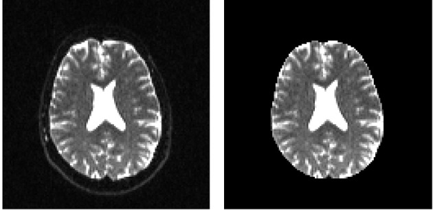
“…median_otsu (see Figure 3 ). from dipy.segment.mask import median_otsu mask, S0_mask = median_otsu(data [:, :, :, 0]) median_otsu uses first a median filter to smooth the S0 and then Otsu’s method, an automated histogram method (Otsu, 1979) to separate the brain (foreground) from its background.”
MethodAdvances in functional and structural MR image analysis and implementation as FSL
Smith, S. M., Jenkinson, M., Woolrich, M. W., Beckmann, C. F., Behrens, T. E. J., Johansen-Berg, H., Bannister, P. R., De Luca, M., Drobnjak, I., Flitney, D. E., et al. (2004) · NeuroImage · doi:10.1016/j.neuroimage.2004.07.051 · Cited by 13,630
Fischl, B. (2012) · NeuroImage · doi:10.1016/j.neuroimage.2012.01.021 · Cited by 9,526 · cited 4×
“…al., 2006), AFNI (Cox, 2012), MITK (Fritzsche et al., 2012), BrainVoyager QX (Goebel, 2012), DTI-Query/Quench (Sherbondy et al., 2005), FreeSurfer (Fischl, 2012), FSL-FDT (Smith et al., 2004), MedInria (Toussaint et al., 2007), MRtrix (Tournier et al., 2012), Diffusion Toolkit/Trackvis (Wang et…”
MethodInfluentialThe NumPy Array: A Structure for Efficient Numerical Computation
van der Walt, S., Colbert, S. C., & Varoquaux, G. (2011) · Computing in Science & Engineering · doi:10.1109/mcse.2011.37 · Cited by 9,381
MR diffusion tensor spectroscopy and imaging
Basser, P. J., Mattiello, J., & LeBihan, D. (1994) · Biophysical Journal · doi:10.1016/s0006-3495(94)80775-1 · Cited by 5,970 · cited 2×
“Another alternative is to make assumptions about the distribution of the PDF such as a Gaussian assumption, which leads to the popular diffusion tensor imaging (DTI) technique (Basser et al., 1994).”
BackgroundMethodThe WU-Minn Human Connectome Project: An overview
Van Essen, D. C., Smith, S. M., Barch, D. M., Behrens, T. E. J., Yacoub, E., & Ugurbil, K. (2013) · NeuroImage · doi:10.1016/j.neuroimage.2013.05.041 · Cited by 5,651
The basis of anisotropic water diffusion in the nervous system—A technical review.
Beaulieu, C. (2002) · NMR in Biomedicine · doi:10.1002/nbm.782 · Cited by 4,628
Basser, P. J., & Pierpaoli, C. (1996) · Journal of Magnetic Resonance, Series B · doi:10.1006/jmrb.1996.0086 · Cited by 4,306 · cited 3×
“…of the myelin wrapping the axons in the voxel may affect both MD and FA, changes in other factors, such as the distribution of directions of crossing of fiber populations through the voxel may also affect these measures (Basser and Pierpaoli, 1996; Jones et al., 2013; Wandell and Yeatman, 2013).”
BackgroundMethodMedical image analysis: progress over two decades and the challenges ahead
Duncan, J. S., & Ayache, N. (2000) · IEEE Transactions on Pattern Analysis and Machine Intelligence · doi:10.1109/34.824822 · Cited by 4,167
IPython: A System for Interactive Scientific Computing
Pérez, F., & Granger, B. (2007) · Computing in Science & Engineering · doi:10.1109/mcse.2007.53 · Cited by 4,129 · cited 1×
“There has been large growth in the number of Python users, there are many Python tools for scientific computing (Pérez and Granger, 2007; Pérez et al., 2011; McKinney, 2012), and there are complementary neuroimaging packages in Python1.”
Background
Research works printed but not linked: 9
- Chamberland, 2013, Explore the brain white matter networks in real-time: multi-sticks fiber tracking
- Garyfallidis, 2012, Towards an Accurate Brain Tractography
- Garyfallidis, 2011, Dipy - a novel software library for diffusion MR and tractography
- LeBihan, 1985, Imagerie de diffusion in vivo par résonance magnétique nucléaire, C. R. Acad. Sci. Paris, 301, 1109
- Pajevic, 1999, Color schemes to represent the orientation of anisotropic tissues from diffusion tensor data: application to white matter fiber tract mapping in the human brain, Magn. Reson. Med, 42, 526, 10.1002/(SICI)1522-2594(199909)42:3526::AID-MRM153.3.CO;2-A · DOI
- Schroeder, 2001, The Visualization Toolkit-User's Guide
- Vaillancourt, 2011, A fiber navigator for neurosurgical planning
- Wang, 2007, Diffusion toolkit: a software package for diffusion imaging data processing and tractography, Proc. Int. Soc. Magn. Reson. Med, 15, 3720
- Westin, 1997, Geometrical diffusion measures for MRI from tensor basis analysis, Proc. ISMRM, 97, 1742
Cited by
Sorted by citations
Items 301–400 of 1,000
Mitigating undersampling errors in MR fingerprinting by sequence optimization
Heesterbeek, D., Koolstra, K., van Osch, M. J. P., van Gijzen, M. B., Vos, F. M., & Nagtegaal, M. A. (2022) · Magnetic Resonance in Medicine · doi:10.1002/mrm.29554 · Cited by 20
Lin, C., Knoop, L., Frigerio, I., Bol, J. G. J. M., Rozemüller, A. J. M., Berendse, H. W., Pouwels, P. J. W., van de Berg, W. D. J., & Jonkman, L. E. (2023) · Movement Disorders · doi:10.1002/mds.29510 · Cited by 20
Henriques, R. N., Henson, R. N., Cam-CAN, & Correia, M. M. (2023) · Imaging Neuroscience · doi:10.1162/imag_a_00051 · Cited by 22
Kebiri, H., Gholipour, A., Lin, R., Vasung, L., Calixto, C. A., Krsnik, Ž., Karimi, D., & Cuadra, M. B. (2023) · Medical Image Analysis · doi:10.1016/j.media.2024.103186 · Cited by 20
The relation of structural integrity and task-related functional connectivity in the aging brain
Burianová, H., Marstaller, L., Choupan, J., Sepehrband, F., Ziaei, M., & Reutens, D. C. (2015) · Neurobiology of Aging · doi:10.1016/j.neurobiolaging.2015.07.006 · Cited by 20
“Then, after skull and non-brain tissue were removed from the image using median Otsu segmentation (Garyfallidis et al., 2014), the tensor model was fitted to each voxel using nonlinear regression, and, finally, fractional anisotropy (FA) maps were calculated (Basser et al.”
Tracking white matter degeneration in asymptomatic and symptomatic MAPT mutation carriers
Chen, Q., Boeve, B. F., Schwarz, C. G., Reid, R. I., Tosakulwong, N., Lesnick, T. G., Bove, J., Brannelly, P., Brushaber, D. E., Coppola, G., et al. (2019) · Neurobiology of Aging · doi:10.1016/j.neurobiolaging.2019.08.011 · Cited by 20
Becker, D., Scherer, M., Neher, P., Jungk, C., Jesser, J., Pflüger, I., Brinster, R., Bendszus, M., Bruckner, T. M., Maier-Hein, K. H., et al. (2019) · World Neurosurgery · doi:10.1016/j.wneu.2019.10.138 · Cited by 20
Muñoz‐Moreno, E., Tudela, R., López-Gil, X., & Sòria, G. (2020) · Network Neuroscience · doi:10.1162/netn_a_00126 · Cited by 18
Kawashima, T., Wei, Z., Haruvi, R., Shainer, I., Narayan, S., Baier, H., & Ahrens, M. B. (2025) · Neuron · doi:10.1016/j.neuron.2025.05.017 · Cited by 21
Surfice: visualizing neuroimaging meshes, tractography streamlines and connectomes
Rorden, C. (2025) · Nature Methods · doi:10.1038/s41592-025-02764-6 · Cited by 19
Guberman, G. I., Houde, J.-C., Ptito, A., Gagnon, I., & Descoteaux, M. (2019) · Brain Structure and Function · doi:10.1007/s00429-019-02002-8 · Cited by 17
Self-citationWang, P., He, J., Ma, X., Weng, L., Wu, Q., Zhao, P., Ban, C., Hao, X., Hao, Z., Yuan, P., et al. (2022) · Academic Radiology · doi:10.1016/j.acra.2022.10.009 · Cited by 19
“MAP-MRI parameters were calculated using NeuDiLab, a software developed in-house with Python based on the open-source tool DIPY (Diffusion Imaging in Python) (25).”
MethodMaruhashi, J., Grewe, V., Frömming, C., Jöckel, P., & Dedoussi, I. C. (2022) · Atmospheric chemistry and physics · doi:10.5194/acp-22-14253-2022 · Cited by 19
Fiber-specific structural properties relate to reading skills in children and adolescents
Meisler, S. L., & Gabrieli, J. D. E. (2022) · eLife · doi:10.7554/elife.82088 · Cited by 19
Jurek, J., Materka, A., Ludwisiak, K., Majos, A., Gorczewski, K., Cepuch, K., & Zawadzka, A. (2023) · Journal of Applied Biomedicine · doi:10.1016/j.bbe.2022.12.006 · Cited by 19
Gallagher, R. L., Koscik, R. L., Moody, J. F., Vogt, N. M., Adluru, N., Kecskemeti, S., Hulle, C. A. V., Chin, N. A., Asthana, S., Kollmorgen, G., et al. (2023) · Alzheimer s Research & Therapy · doi:10.1186/s13195-023-01281-y · Cited by 22
“Diffusion tensor fitting was performed using Diffusion Imaging in Python (DIPY) [32] to generate fractional anisotropy (FA) maps.”
MethodSingh, N. A., Goodrich, A. W., Graff‐Radford, J., Machulda, M. M., Sintini, I., Carlos, A. F., Robinson, C. G., Reid, R. I., Lowe, V. J., Jack, C. R., et al. (2024) · NeuroImage · doi:10.1016/j.neuroimage.2024.120564 · Cited by 19
“Diffusion tensors were estimated using nonlinear least squares fitting and used to calculate Fractional Anisotropy (FA) and Mean Diffusivity (MD) images in dipy (Garyfallidis et al., 2014).”
MethodMotion-resolved quantitative phase imaging
Kellman, M., Chen, M., Phillips, Z. F., Lustig, M. S., & Waller, L. (2018) · Biomedical Optics Express · doi:10.1364/boe.9.005456 · Cited by 19
Adaptive phase correction of diffusion-weighted images
Pizzolato, M., Gilbert, G., Thiran, J., Descoteaux, M., & Rachid Deriche, B. (2019) · NeuroImage · doi:10.1016/j.neuroimage.2019.116274 · Cited by 19
“In all cases, the reconstruction was performed using Dipy (Garyfallidis et al., 2014).”
MethodSelf-citationCharles, A. S., Falk, B., Turner, N. L., Pereira, T., Tward, D. J., Pedigo, B. D., Chung, J., Burns, R. C., Ghosh, S., Kebschull, J. M., et al. (2020) · Annual Review of Neuroscience · doi:10.1146/annurev-neuro-100119-110036 · Cited by 16
Karimi, D., Vasung, L., Jaimes, C. E., Machado‐Rivas, F., Khan, S., Warfield, S. K., & Gholipour, A. (2021) · Medical Image Analysis · doi:10.1016/j.media.2021.102129 · Cited by 19
Numerical uncertainty in analytical pipelines lead to impactful variability in brain networks
Kiar, G., Chatelain, Y., Castro, P. D. O., Petit, E., Rokem, A., Varoquaux, G. P., Mišić, B., Evans, A. C., & Glatard, T. (2021) · PLoS ONE · doi:10.1371/journal.pone.0250755 · Cited by 19
Self-citationGuerrero-Gonzalez, J. M., Yeske, B., Kirk, G. R., Bell, M. J., Ferrazzano, P. A., & Alexander, A. L. (2022) · NeuroImage · doi:10.1016/j.neuroimage.2022.119475 · Cited by 18
“Diffusion tensors were estimated for each voxel using the robust estimation of tensors by outlier rejection (RESTORE) algorithm as part of the diffusion imaging in python (DIPY) software package (Garyfallidis et al., 2014).”
Cervantes, B., Van, A. T., Weidlich, D., Kooijman, H., Hock, A., Rummeny, E. J., Gersing, A. S., Kirschke, J. S., & Karampinos, D. C. (2018) · Magnetic Resonance in Medicine · doi:10.1002/mrm.27072 · Cited by 19
“Axial b1⁄4 400 DW ss-EPI data were registered to axial b1⁄4 0 DW ss-EPI data using an affine transformation (45,46) to minimize geometric distortions produced by eddy currents.”
MethodDiffusion Weighted Image Co-registration: Investigation of Best PracticesPreprint
Chen, D. Q., Dell’Acqua, F., Rokem, A., Garyfallidis, E., Hayes, D. J., zhong, J., & Hodaie, M. (2019) · bioRxiv (Cold Spring Harbor Laboratory) · doi:10.1101/864108 · Cited by 19
“…and MDWI images were created using FSL (Smith et al., 2004); FA images were created in 3D Slicer by calculating the scalars following diffusion tensor estimation (Pieper et al., 2006); GFA and AP images were created with Dipy software library (Descoteaux et al., 2007; Garyfallidis et al., 2014).”
MethodSelf-citationQSIPrep: An integrative platform for preprocessing and reconstructing diffusion MRIPreprint
Cieslak, M., Cook, P. A., He, X., Yeh, F.-C. F., Dhollander, T., Adebimpe, A., Aguirre, G. K., Bassett, D. S., Betzel, R. F., Bourque, J., et al. (2020) · bioRxiv (Cold Spring Harbor Laboratory) · doi:10.1101/2020.09.04.282269 · Cited by 20
Self-citationBathelt, J., Barnes, J., Raymond, F. L., Baker, K., & Astle, D. (2017) · Cerebral Cortex · doi:10.1093/cercor/bhx027 · Cited by 15
“11 package (Garyfallidis et al. 2014) to boost signal to noise ratio.”
BackgroundKern, K. C., Zagzoug, M. S., Gottesman, R. F., Wright, C. B., & Leigh, R. J. (2023) · Frontiers in Neurology · doi:10.3389/fneur.2023.1172031 · Cited by 18
“org), (24) and the Quantitative Imaging Toolkit (cabeen.”
BackgroundLittle, G. E., & Beaulieu, C. (2021) · NeuroImage · doi:10.1016/j.neuroimage.2021.118105 · Cited by 18
Bagdasarian, F. A., Yuan, X., Athey, J., Bunnell, B. A., & Grant, S. C. (2021) · Magnetic Resonance in Medicine · doi:10.1002/mrm.28929 · Cited by 18
Jones, R. J., Maffei, C., Augustinack, J. C., Fischl, B. R., Wang, H., Bilgiç, B., & Yendiki, A. (2021) · NeuroImage · doi:10.1016/j.neuroimage.2021.118621 · Cited by 17
Dadario, N. B., Young, I. M., Zhang, X., Teo, C., Doyen, S., & Sughrue, M. E. (2022) · Brain Network and Modulation · doi:10.4103/2773-2398.340144 · Cited by 18
Zimmermann, J., Friedli, N., Bavato, F., Stämpfli, P., Coray, R. C., Baumgartner, M. R., Grandgirard, D., Leib, S. L., Opitz, A., Seifritz, E., et al. (2022) · NeuroImage Clinical · doi:10.1016/j.nicl.2022.103191 · Cited by 17
Renauld, E., Théberge, A., Petit, L., Houde, J., & Descoteaux, M. (2023) · Scientific Reports · doi:10.1038/s41598-023-28560-w · Cited by 18
Self-citationKyle, K., Maller, J. J., Barnett, Y., Jonker, B., Barnett, M. H., D’Souza, A., Calamante, F., Maamary, J., Peters, J., Wang, C., et al. (2023) · Frontiers in Neurology · doi:10.3389/fneur.2023.1129430 · Cited by 18
“For each of the 10 subjects, the DRTT in the template space was split into three clusters—anterior (aDRTT), middle (mDRTT) and posterior (pDRTT), based on the trajectory of the streamlines between the red nucleus and thalamus using QuickBundles (39) in DIPY, an automated algorithm that groups streamlines together base…”
MethodAlignment of Tractograms As Graph Matching
Olivetti, E., Sharmin, N., & Avesani, P. (2016) · Frontiers in Neuroscience · doi:10.3389/fnins.2016.00554 · Cited by 17
“Other choices can be made, for example using different clustering algorithms, see for example Garyfallidis et al. (2012), or recent specific methods for tractogram simplification, like Gori et al. (2016). The motivation of our choice, for adopting the algorithm described in Olivetti et al. (2013), is eminently computa…”
BackgroundMethodInfluentialVersatile, robust, and efficient tractography with constrained higher-order tensor fODFs
Ankele, M., Lim, L., Groeschel, S., & Schultz, T. (2017) · International Journal of Computer Assisted Radiology and Surgery · doi:10.1007/s11548-017-1593-6 · Cited by 18
Bayesian uncertainty quantification in linear models for diffusion MRI
Sjölund, J., Eklund, A., Özarslan, E., Herberthson, M., Bånkestad, M., & Knutsson, H. E. (2018) · NeuroImage · doi:10.1016/j.neuroimage.2018.03.059 · Cited by 17
“We implemented the core algorithmic ideas by extending the free and open source software project Dipy (Diffusion Imaging in Python) (Garyfallidis et al., 2014).”
MethodKong, J., Giridharagopal, R., Harrison, J. S., & Ginger, D. S. (2018) · The Journal of Physical Chemistry Letters · doi:10.1021/acs.jpclett.8b01003 · Cited by 18
Kerkelä, L., Henriques, R. N., Hall, M. G., Clark, C. A., & Shemesh, N. (2019) · Magnetic Resonance in Medicine · doi:10.1002/mrm.28048 · Cited by 18
Gersing, A. S., Cervantes, B., Knebel, C., Schwaiger, B. J., Kirschke, J. S., Weidlich, D., Claudi, C., Peeters, J., Pfeiffer, D., Rummeny, E. J., et al. (2020) · European Journal of Radiology · doi:10.1016/j.ejrad.2020.109110 · Cited by 18
Acquiring and Predicting Multidimensional Diffusion (MUDI) Data: An Open Challenge
Pizzolato, M., Palombo, M., Bonet-Carné, E., Tax, C. M. W., Grussu, F., Ianuș, A., Bogusz, F., Pieciak, T., Ning, L., Larochelle, H., et al. (2020) · Mathematics and visualization · doi:10.1007/978-3-030-52893-5_17 · Cited by 18
“Ad hoc affine volume registration was performed: collinear magnitude diffusion-weighted images (DWIs) acquired with different pairs (TI,TE) were first co-registered together using the highest TI and lowest TE volume as reference; the 106 reference volumes were then registered together based on a mutual information met…”
MethodSelf-citationJiang, R., Jiang, S., Song, S., Wei, X., Deng, K., Zhang, Z., & Xue, Y. (2020) · Korean Journal of Radiology · doi:10.3348/kjr.2020.0949 · Cited by 18
HashemizadehKolowri, S., Chen, R., Adluru, G., Dean, D., Wilde, E. A., Alexander, A. L., & DiBella, E. (2021) · Medical Image Analysis · doi:10.1016/j.media.2021.102000 · Cited by 18
Luca, A. D., Ianuș, A., Leemans, A., Palombo, M., Shemesh, N., Zhang, H. G., Alexander, D. C., Nilsson, M., Froeling, M., Biessels, G. J., et al. (2021) · NeuroImage · doi:10.1016/j.neuroimage.2021.118367 · Cited by 19
Self-citationOpen Environment for Multimodal Interactive Connectivity Visualization and Analysis
Taylor, P. A., Chen, G., Cox, R. W., & Saad, Z. S. (2015) · Brain Connectivity · doi:10.1089/brain.2015.0363 · Cited by 19
“…or Analysis of Functional NeuroImages (AFNI), but instead can easily be estimated using other packages, for example, DSI-Studio (http://dsi-studio.labsolver.org; an example processing script is included in the FATCAT Demo), Diffusion Toolkit (Wang et al., 2007), or Dipy (Garyfallidis et al., 2014).”
MethodXu, J., Devan, S. P., Shi, D., Pamulaparthi, A., Yan, N., Zu, Z., Smith, D. S., Harkins, K. D., Gore, J. C., & Jiang, X. (2025) · Magnetic Resonance Imaging · doi:10.1016/j.mri.2025.110428 · Cited by 17
Single‐shell NODDI using dictionary‐learner‐estimated isotropic volume fraction
Faiyaz, A., Doyley, M. M., Schifitto, G., Zhong, J., & Uddin, M. N. (2021) · NMR in Biomedicine · doi:10.1002/nbm.4628 · Cited by 19
Garic, D., Yeh, F.-C. F., Graziano, P. A., & Dick, A. S. (2021) · Brain Structure and Function · doi:10.1007/s00429-021-02364-y · Cited by 18
CTtrack: A CNN+Transformer-based framework for fiber orientation estimation & tractography
Hosseini, S. M., Hassanpour, M., Masoudnia, S., Iraji, S., Raminfard, S., & Nazem-Zadeh, M.-R. (2022) · Neuroscience Informatics · doi:10.1016/j.neuri.2022.100099 · Cited by 19
Führes, T., Saake, M., Lorenz, J., Seuß, H., Stemmer, A., Benkert, T., Uder, M., & Laun, F. B. (2022) · Magnetic Resonance in Medicine · doi:10.1002/mrm.29427 · Cited by 17
Sangüesa, G., Batlle, M., Muñoz‐Moreno, E., Sòria, G., Alcarraz, A., Rubies, C., Sitjà‐Roqueta, L., Solana, E., Martínez‐Heras, E., Meza‐Ramos, A., et al. (2022) · Annals of the New York Academy of Sciences · doi:10.1111/nyas.14912 · Cited by 16
Axial and radial axonal diffusivities and radii from single encoding strongly diffusion-weighted MRI
Pizzolato, M., Canales-Rodríguez, E. J., Andersson, M., & Dyrby, T. B. (2023) · Medical Image Analysis · doi:10.1016/j.media.2023.102767 · Cited by 18
Multi-planar 2.5D U-Net for image quality enhancement of dental cone-beam CT
Ryu, K., Lee, C., Han, Y., Pang, S., Kim, Y. H., Choi, C., Jang, I., & Han, S. (2023) · PLoS ONE · doi:10.1371/journal.pone.0285608 · Cited by 20
FD‐Net: An unsupervised deep forward‐distortion model for susceptibility artifact correction in EPI
Alkilani, A. Z., Çukur, T., & Sarıtaş, E. Ü. (2023) · Magnetic Resonance in Medicine · doi:10.1002/mrm.29851 · Cited by 17
“55 For all methods, hyperparameters were chosen empirically to maximize PSNR and SSIM over the four subjects reserved as validation data.”
MethodLebret, A., Lévy, S., Pfender, N., Farshad, M., Altorfer, F. C. S., Callot, V., Curt, A. E. P., Freund, P., & Seif, M. (2023) · Scientific Reports · doi:10.1038/s41598-023-49896-3 · Cited by 17
Kruper, J., Richie-Halford, A., Benson, N. C., Caffarra, S., Owen, J. P., Wu, Y., Egan, C. A., Lee, A., Lee, C. S., Yeatman, J. D., et al. (2024) · Communications Medicine · doi:10.1038/s43856-024-00496-w · Cited by 16
“Tissue properties were calculated using the diffusional kurtosis model implemented in DIPY 18,37 .”
Self-citationTutorial: a guide to diffusion MRI and structural connectomics
Shamir, I., & Assaf, Y. (2024) · Nature Protocols · doi:10.1038/s41596-024-01052-5 · Cited by 19
Merged Group Tractography Evaluation with Selective Automated Group Integrated Tractography
Chen, D. Q., zhong, J., Hayes, D. J., Behan, B., Walker, M., Hung, P. S.-P., & Hodaie, M. (2016) · Frontiers in Neuroanatomy · doi:10.3389/fnana.2016.00096 · Cited by 17
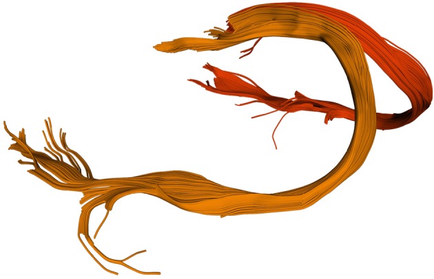
“We have previously studied fornix subregion anatomies extensively (Chen et al., 2015b), and it is a popular anatomy to showcase the ability of new tractography algorithms due to the curvature of the forniceal crura (Garyfallidis et al., 2014).”
BackgroundGallardo, G., Wells, W. M., Rachid Deriche, B., & Wassermann, D. (2017) · NeuroImage · doi:10.1016/j.neuroimage.2017.01.070 · Cited by 17
“For each subject, we fitted a CSD model (Tournier et al., 2004) to their diffusion data using Dipy (version 0.11) (Garyfallidis et al., 2014) and created 5000 streamlines per seed-voxel using the implementation of probabilistic tractography in Dipy.”
MethodInfluentialMultiuser virtual reality environment for visualising neuroimaging data
Shattuck, D. W. (2018) · Healthcare Technology Letters · doi:10.1049/htl.2018.5077 · Cited by 17
Effects of simvastatin on white matter integrity in healthy middle‐aged adults
Vogt, N. M., Hunt, J. F. V., Ma, Y., Hulle, C. A. V., Adluru, N., Chappell, R. J., Lazar, K. K., Jacobson, L. E., Austin, B. P., Asthana, S., et al. (2021) · Annals of Clinical and Translational Neurology · doi:10.1002/acn3.51421 · Cited by 17
Ren, M., Kim, H., Dey, N., & Gerig, G. (2021) · Lecture notes in computer science · doi:10.1007/978-3-030-87234-2_50 · Cited by 17
Semi-Automatic Segmentation of Optic Radiations and LGN, and Their Relationship to EEG Alpha Waves
Renauld, E., Descoteaux, M., Bernier, M., Garyfallidis, E., & Whittingstall, K. (2016) · PLoS ONE · doi:10.1371/journal.pone.0156436 · Cited by 15
“T1 images were denoised using non-local means (NLM) [60] (Dipy, [61]) and skull-stripped using FSL [62].”
MethodSelf-citationThe diffusion-simulated connectivity (DiSCo) dataset
Rafael‐Patiño, J., Girard, G., Truffet, R., Pizzolato, M., Caruyer, E., & Thiran, J. (2021) · Data in Brief · doi:10.1016/j.dib.2021.107429 · Cited by 17
Brain glymphatic fluid mapping in Alzheimer’s disease: a human MRI and PET study
Zhou, L., Nguyen, T. D., Chiang, G. C.-Y., Keil, S. A., Wang, X. H., Hu, T., Lan, H., Xi, K., Costa, A. P. F., Tanzi, E. B., et al. (2025) · Brain Communications · doi:10.1093/braincomms/fcaf200 · Cited by 17
“7,8,42 The DTI-FW was converted to FW mapping in the same way as described for pCSF mapping above and coregistered to FS T1w space using the transform between FA to FS T1w.”
HashemizadehKolowri, S., Chen, R., Adluru, G., & DiBella, E. (2022) · Magnetic Resonance in Medicine · doi:10.1002/mrm.29162 · Cited by 16
Hosseinpour, Z., Jonkman, L. E., Oladosu, O., Pridham, G., Pike, G. B., Inglese, M., Geurts, J. J. G., & Zhang, Y. (2022) · Journal of Neuroscience Methods · doi:10.1016/j.jneumeth.2022.109671 · Cited by 17
Prasse, G., Meyer, H., Scherlach, C., Maybaum, J., Hoffmann, A., Kasper, J., Fehrenbach, M. K., Wilhelmy, F., Meixensberger, J., Hoffmann, K., et al. (2022) · NeuroImage Clinical · doi:10.1016/j.nicl.2022.103310 · Cited by 16
Tractography passes the test: Results from the diffusion-simulated connectivity (disco) challenge
Girard, G., Rafael‐Patiño, J., Truffet, R., Aydogan, D. B., Adluru, N., Nair, V. A., Prabhakaran, V., Bendlin, B. B., Alexander, A. L., Bosticardo, S., et al. (2023) · NeuroImage · doi:10.1016/j.neuroimage.2023.120231 · Cited by 19
Self-citationLesh, T., Iosif, A.-M., Tanase, C., Vlasova, R., Ryan, A. M., Bennett, J. L., Hogrefe, C. E., Maddock, R., Geschwind, D., van de Water, J. A., et al. (2023) · Molecular Psychiatry · doi:10.1038/s41380-023-02213-w · Cited by 17
“The Dipy diffusion imaging library [57] and included free water elimination model [58] were used to calculate all diffusion metrics.”
Longitudinal evolution of diffusion metrics after left hemisphere ischaemic stroke
Boucher, J., Marcotte, K., Bedetti, C., Houzé, B., Descoteaux, M., Brisebois, A., García, A. O., Rochon, E. A., Léonard, C., Désautels, A., et al. (2023) · Brain Communications · doi:10.1093/braincomms/fcad313 · Cited by 21
“Second, diffusion weighted imaging (DWI)-weighted images were pre-processed through several processing steps, including noise correction with the Marchenko–Pastur principal component analysis method using Mrtrix3, 31 correction for subject movement, eddy-currents and susceptibility-induced distortions with AP-PA image…”
MethodSelf-citationSchilliger, Z., Alemán‐Gómez, Y., Smith, M. M., Celen, Z., Meuleman, B., Binz, P., Steullet, P., Kim, Q., Conus, P., Merglen, A., et al. (2024) · Translational Psychiatry · doi:10.1038/s41398-023-02728-4 · Cited by 20
“Dipy (v.1.5.0) [60] was applied over the corrected DWIs to fi t both second order tensors and intravoxel orientation distribution functions (ODF) via the Simple Harmonic Oscillator-based Reconstruction and Estimation method (SHORE) [61].”
MethodInfluentialBundleSeg: A Versatile, Reliable and Reproducible Approach to White Matter Bundle Segmentation
St‐Onge, E., Schilling, K. G., & Rheault, F. (2023) · Lecture notes in computer science · doi:10.1007/978-3-031-47292-3_5 · Cited by 17
Nazir, A., Assad, A., Hussain, A. B., & Singh, M. (2024) · International Journal of Systems Assurance Engineering and Management · doi:10.1007/s13198-024-02441-5 · Cited by 20
Sepehrband, F., O’Brien, K., & Barth, M. (2017) · Magnetic Resonance in Medicine · doi:10.1002/mrm.26608 · Cited by 16
“Dipy software (http://nipy.org/dipy/) version 0.8.0 was used to perform LiFE (68).”
MethodInfluentialModel‐based denoising in diffusion‐weighted imaging using generalized spherical deconvolution
Sperl, J. I., Sprenger, T., Tan, E. T., Menzel, M. I., Hardy, C. J., & Marinelli, L. (2017) · Magnetic Resonance in Medicine · doi:10.1002/mrm.26626 · Cited by 16
“TikReg was applied to SHORE using the Dipyimplementation (44), to SpherRidge using simple MoorePenrose inversion, and to genSD using quadratic programming to incorporate the nonnegativity constraints.”
MethodMeesters, S., Ossenblok, P., Wagner, L., Schijns, O. E. M. G., Boon, P., Florack, L. M. J., Vilanova, A., & Duits, R. (2017) · Journal of Neuroscience Methods · doi:10.1016/j.jneumeth.2017.05.029 · Cited by 16
Timmermans, C., Smeets, D., Verheyden, J., Terzopoulos, V., Anania, V., Parizel, P. M., & Maas, A. I. R. (2019) · Journal of Magnetic Resonance Imaging · doi:10.1002/jmri.26333 · Cited by 16
Muti-shell Diffusion MRI Harmonisation and Enhancement Challenge (MUSHAC): Progress and Results
Ning, L., Bonet-Carné, E., Grussu, F., Sepehrband, F., Kaden, E., Veraart, J., Blumberg, S. B., Khoo, C. S., Palombo, M., Coll‐Font, J., et al. (2019) · Mathematics and visualization · doi:10.1007/978-3-030-05831-9_18 · Cited by 16
White matter degeneration profile in the cognitive cortico‐subcortical tracts in Parkinson's disease
Hanganu, A., Houde, J., Fonov, V., Degroot, C., Mejia‐Constain, B., Lafontaine, A., Soland, V., Chouinard, S., Collins, D. L., Descoteaux, M., et al. (2018) · Movement Disorders · doi:10.1002/mds.27364 · Cited by 15
“Various statistics were computed along those bundles, including axial, radial, and mean diffusivity (AD, RD, MD), fractional anisotropy (FA), apparent fiber density (AFD), track volume (TV), and track count (number of reconstructed streamlines [RS]) using Dipy.60 AFD provides an estimate of the local voxel-wise fiber…”
MethodInfluentialSelf-citationProgressive axonopathy when oligodendrocytes lack the myelin protein CMTM5
Buscham, T. J., Eichel-Vogel, M. A., Steyer, A. M., Jahn, O., Strenzke, N., Dardawal, R., Memhave, T. R., Siems, S. B., Müller, C., Meschkat, M., et al. (2022) · eLife · doi:10.7554/elife.75523 · Cited by 18
Glymphatic dysfunction associated with cortisol dysregulation in major depressive disorder
Chen, S., Xu, Z., Guo, Z., Lin, S., Zhang, H., Liang, Q., Chen, Q., Zhang, Y., Peng, B., Zhao, Y., et al. (2025) · Translational Psychiatry · doi:10.1038/s41398-025-03486-1 · Cited by 15
Portable Learning Environments for Hands-On Computational Instruction
Holdgraf, C., Culich, A., Rokem, A., Deniz, F., Alegro, M., & Ushizima, D. (2017) · Practice and Experience in Advanced Research Computing · doi:10.1145/3093338.3093370 · Cited by 15
Self-citationJordan, K., Morin, O., Wahl, M. T., Amirbekian, B., Chapman, C. H., Owen, J. P., Mukherjee, P., Braunstein, S. E., & Henry, R. G. (2019) · Frontiers in Oncology · doi:10.3389/fonc.2019.00810 · Cited by 15
“The code to generate the WMPL maps is open-source and freely available on Github as part of the Diffusion Imaging in Python package (29).”
MethodInfluentialSelf-citationAutomatic Segmentation of White Matter Tracts Using Multiple Brain MRI Sequences
Nelkenbaum, I., Tsarfaty, G., Kiryati, N., Konen, E., & Mayer, A. (2020) · IEEE International Symposium on Biomedical Imaging (ISBI) · doi:10.1109/isbi45749.2020.9098454 · Cited by 15
“Raw DWI data was processed with Dipy [19] python module, and PDD [20], consisting of 3 channels per voxel, was generated.”
MethodPlanalp, E. M., Dowe, K. N., Alexander, A. L., Goldsmith, H. H., Davidson, R. J., & Dean, D. (2022) · Developmental Science · doi:10.1111/desc.13340 · Cited by 16
“DTI Diffusion tensors were estimated at each voxel using the robust estimation of tensors by an outlier rejection (RESTORE; Chang et al., 2005) algorithm as part of the Diffusion Imaging in Python open source software package (DIPY; (Garyfallidis et al., 2014).”
MethodThe importance of the dorsal branch of the arcuate fasciculus in phonological working memory
Barbeau, É. B., Kousaie, S., Brass, K., Descoteaux, M., Petrides, M., & Klein, D. (2023) · Cerebral Cortex · doi:10.1093/cercor/bhad226 · Cited by 15
Self-citationKirby, E., Andrushko, J., Rinat, S., D'Arcy, R. C. N., & Boyd, L. (2024) · Scientific Reports · doi:10.1038/s41598-024-56453-z · Cited by 15
Comparison of distances for supervised segmentation of white matter tractography
Olivetti, E., Bertò, G., Gori, P., Sharmin, N., & Avesani, P. (2017) · International Workshop on Pattern Recognition in NeuroImaging · doi:10.1109/prni.2017.7981502 · Cited by 15
“For each bundle and distance function, we observed a standard deviation 1 http://nipy.org/dipy, [22]. of DSC of approximately 0 .”
MethodClassifying Phenotypes Based on the Community Structure of Human Brain Networks
Kurmukov, A. G., Ananyeva, M., Dodonova, Y., Gutman, B. A., Faskowitz, J., Jahanshad, N., Thompson, P. M., & Евгеньевич, Ж. Л. (2017) · Lecture notes in computer science · doi:10.1007/978-3-319-67675-3_1 · Cited by 14
q-Space Learning with Synthesized Training Data
Ye, C., Cui, Y., & Li, X. (2019) · Mathematics and visualization · doi:10.1007/978-3-030-05831-9_10 · Cited by 15
FURY: advanced scientific visualization
Garyfallidis, E., Koudoro, S., Guaje, J., Côté, M.-A., Biswas, S., Reagan, D., Anousheh, N., Silva, F. N., Fox, G., & Contributors, F. (2021) · The Journal of Open Source Software · doi:10.21105/joss.03384 · Cited by 15
“…and allows to reprogram the low-level shaders for the creation of stunning effects (see Figure 2) not available in VTK. Historically, FURY had also a different path than these libraries as it was originally created for heavy-duty medical visualization purposes for DIPY (Garyfallidis et al., 2014).”
BackgroundSelf-citationLiesche-Starnecker, F., Prokop, G., Yakushev, I., Preibisch, C., Delbridge, C., Meyer, H. S., Aftahy, K., Barz, M., Meyer, B., Zimmer, C., et al. (2021) · EJNMMI Research · doi:10.1186/s13550-021-00817-3 · Cited by 15
“DTI data were analyzed using the open-source dipy framework [12].”
MethodGadolinium‐free assessment of synovitis using diffusion tensor imaging
Sandford, H. J. C., Mackay, J., Watkins, L., Gold, G. E., Kogan, F., & Mazzoli, V. (2021) · NMR in Biomedicine · doi:10.1002/nbm.4614 · Cited by 15
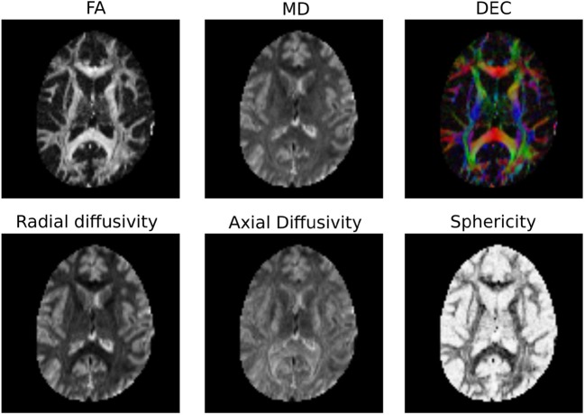
“Tensor fitting of the diffusion images was performed using the Dipy library.(30) Voxelwise maps of MD and FA were created (Figure 2C and 2D) by using the three eigen-values of the diffusion tensor (λ1, λ2, λ3) in the following formulas:”
MethodZhang, H., Ille, S., Sogerer, L., Schwendner, M. J., Schröder, A., Meyer, B., Wiestler, B., & Krieg, S. M. (2021) · Human Brain Mapping · doi:10.1002/hbm.25757 · Cited by 15
Brain‐age estimation accuracy is significantly increased using multishell free‐water reconstruction
Nemmi, F., Levardon, M., & Péran, P. (2022) · Human Brain Mapping · doi:10.1002/hbm.25792 · Cited by 15
Evaluating reproducibility and subject-specificity of microstructure-informed connectivity
Koch, P., Girard, G., Brügger, J., Cadic‐Melchior, A., Beanato, E., Park, C.-H., Morishita, T., Wessel, M. J., Pizzolato, M., Canales-Rodríguez, E. J., et al. (2022) · NeuroImage · doi:10.1016/j.neuroimage.2022.119356 · Cited by 16
“For each dataset, 5 million streamlines were generted, from which all streamlines with both endpoints in the individual ortical or subcortical brain masks were selected using the DIPY software ackage ( Garyfallidis et al., 2014 ).”
MethodIdentifying vulnerable brain networks associated with Alzheimer’s disease risk
Mahzarnia, A., Stout, J. A., Anderson, R. J., Moon, H. S., Han, Z. Y., Beck, K., Browndyke, J. N., Dunson, D. B., Johnson, K. G., O’Brien, R., et al. (2022) · Cerebral Cortex · doi:10.1093/cercor/bhac419 · Cited by 14
Blockmodels for connectome analysis
Moyer, D., Gutman, B. A., Prasad, G., Faskowitz, J., Steeg, G. V., & Thompson, P. M. (2015) · Proceedings of SPIE, the International Society for Optical Engineering/Proceedings of SPIE · doi:10.1117/12.2211519 · Cited by 15
Metadata
- Authors
- Garyfallidis, E., Brett, M., Amirbekian, B., Rokem, A., van der Walt, S., Descoteaux, M., Nimmo-Smith, I., & Contributors, D.
- Affiliations
- Université de Sherbrooke (CA)
- University of California, Berkeley (US)
- University of California, San Francisco (US)
- Stanford University (US)
- Corresponding author
- Garyfallidis, Eleftherios
- Venue
- Frontiers in Neuroinformatics
- Publisher
- Frontiers Media SA
- Published
- 2014-02-21
- Volume
- 8
- Pages
- 8-8
- ISSN
- 1662-5196 (ISSN-L)
- PMC
- PMC3931231
- OpenAlex
- W2062791478
- Cited by
- 1,371
- References
- 98
- Citations by year
- 2026: 147 · 2025: 192 · 2024: 177 · 2023: 162 · 2022: 151 · 2021: 147 · 2020: 100 · 2019: 89 · 2018: 54 · 2017: 79 · 2016: 34 · 2015: 26 · 2014: 12
- FWCI
- 39.38
- Type
- journal article
- Language
- en
- Open access
- gold
- doi.org
- Licence
- cc-by
- Locations
- Frontiers in Neuroinformatics (publishedVersion, cc-by)
- Europe PMC (PubMed Central) (publishedVersion, cc-by)
- PubMed Central (publishedVersion, cc-by)
- PubMed (publishedVersion)
- DOAJ (DOAJ: Directory of Open Access Journals) (submittedVersion)
- University of Birmingham Research Portal (University of Birmingham) (submittedVersion, cc-by)
- Funders
- Fonds Québécois de la Recherche sur la Nature et les Technologies
- Natural Sciences and Engineering Research Council of Canada
- National Eye Institute (F32EY022294)
- Topics
- Health Sciences › Medicine › Radiology, Nuclear Medicine and Imaging › Advanced Neuroimaging Techniques and Applications
- Life Sciences › Neuroscience › Cognitive Neuroscience › Functional Brain Connectivity Studies
- Health Sciences › Medicine › Radiology, Nuclear Medicine and Imaging › Advanced MRI Techniques and Applications
- Keywords
- diffusion MRI, DIPY, white matter, fiber tractography, diffusion tensor model, constrained spherical deconvolution, diffusion spectrum imaging, probabilistic tracking, open source software, Python library
About these checks
Registry, retraction, Cites and statistics results are computed, not judged, with one exception: a printed reference that no index lists is read once by a model before it counts as a citation error. Claims are extracted by a model and kept only when their quote appears verbatim in the text; each cited source is then checked, with a model’s help, for whether it says what the claim relies on. Those verdicts can be wrong — read the claim’s quote and the cited source before relying on one.
Statistics and arithmetic are recomputed, not judged.
Confidence bands are shown only for fields calibrated on our annotated test set. None is calibrated yet, so no band is shown.
What counts as a claim ›How we measure confidence ›How we check this ›
Are you an author, or do you see a problem with this page? Request a review
